PRO464TEO~1
Proteomic characterization of harvested pseudopodia with differential gel
electrophoresis and specific antibodies
Marie E Beckner 1,Xuan Chen 2,Jiyan An 2,Billy W Day 2,3and Ian F Pollack 4
1
Department of Pathology,University of Pittsburgh,Pittsburgh,PA,USA;2The Genomics and Proteomics Core Laboratories,University of Pittsburgh,Pittsburgh,PA,USA;3Departments of Pharmaceutical Sciences and Chemistry,University of Pittsburgh,Pittsburgh,PA,USA and 4Department of Neurological Surgery,University of Pittsburgh,Pittsburgh,PA,USA
Malignant gliomas (astrocytomas)are lethal tumors that invade the brain.Invasive cell migration is initiated by extension of pseudopodia into interstitial spaces.In this study,U87glioma cells formed pseudopodia in vitro as cells pushed through 3l m pores of polycarbonate membranes.Harvesting pseudopodia in a novel two-step method provided material for proteomic analysis.Differences in the protein profiles of pseudopodia and whole cells were found using differential gel electrophoresis (DIGE)and immunoblotting.Proteins from two-dimensional (2D)gels with M R ’s of 20–100kDa and p I ’s of 3.0–10.0were identified by peptide mass fingerprinting analysis using mass spectrometry.For DIGE,lysates of pseudopodia and whole cells were each labeled with electrophilic forms of fluorescent dyes,Cy3or Cy5,and analyzed as mixtures.Analysis was repeated with reciprocal labeling.Differences in protein distributions were detected by manual inspection and computer analysis.Topographical digital maps of the scanned gels were used for algorithmic spot matching,normalization of background,quantifying spot differences,and elimination of artifacts.Pseudopodial proteins in Coomassie-stained 2D gels included isoforms of glycolytic enzymes as the largest group,seven of 24proteins.Peptide mass fingerprint analysis of DIGE gels demonstrated increased isoforms of annexin (Anx)I,AnxII,enolase,pyruvate kinase,and aldolase,and decreased mitochondrial manganese superoxide dismutase and transketolase in pseudopodia.Specific antibodies showed restricted immunoreactivity of the hepatocyte growth factor (HGF)a chain to pseudopodia,indicating localization of its active form.Met (the HGF receptor),actin,and total AnxI were increased in pseudopodial lysates on immunoblots.Increased constituents of the pseudopodial proteome in glioma cells,identified in this study as actin,HGF,Met,and isoforms of AnxI,AnxII,and several glycolytic enzymes,represent therapeutic targets to consider for suppression of tumor https://www.360docs.net/doc/135809184.html,boratory Investigation (2005)85,316–327,advance online publication,17January 2005;doi:10.1038/labinvest.3700239Keywords:differential gel electrophoresis;HGF;mass spectrometry;migration;proteomics;pseudopodia
Tumor cells of diffuse astrocytomas (gliomas)are invasive,regardless of tumor grade.They migrate in response to certain cytokine growth factors,such as hepatocyte growth factor (HGF),also known as scatter factor.1–4Glioma cells preferentially move through interstitial spaces formed in the white matter by axons and glial processes,possibly widened by tumor-induced edema and proteolysis.Once embryonic development of the brain is
complete,most normal astrocytic cells maintain stable vascular contacts and do not migrate,with the exception of occasional,limited cell movement during some types of reactive gliosis.Normal astrocytes characteristically have a star shape due to slender cytoplasmic extensions stabilized by attachments with the vasculature and other cells.Some plasticity is retained in their fine,sheet-like perisynaptic processes that respond to neuronal activity.5In contrast,tumor cells produce dynamic pseudopodial cell extensions that mediate migration with only transitory vascular and neuronal contacts.Signal transduction,membrane formation and turn-over,cytoskeletal remodeling,adhesion,de-adhe-sion,and energy production occur within the pseudopodial leading edge during invasion.The proteins involved in mediating pseudopodial
Received 16November 2004;revised and accepted 7December 2004;published online 17January 2005
Correspondence:Dr ME Beckner,MD,Department of Pathology,200Lothrop St.,PUH,Rm.A-515,University of Pittsburgh,Pittsburgh,PA 15213,USA.E-mail:
becknerme@https://www.360docs.net/doc/135809184.html,
Laboratory Investigation (2005)85,316–327
&2005USCAP ,Inc All rights reserved 0023-6837/05$30.00
https://www.360docs.net/doc/135809184.html,
extension offer potential therapeutic targets to suppress tumor cell invasion.
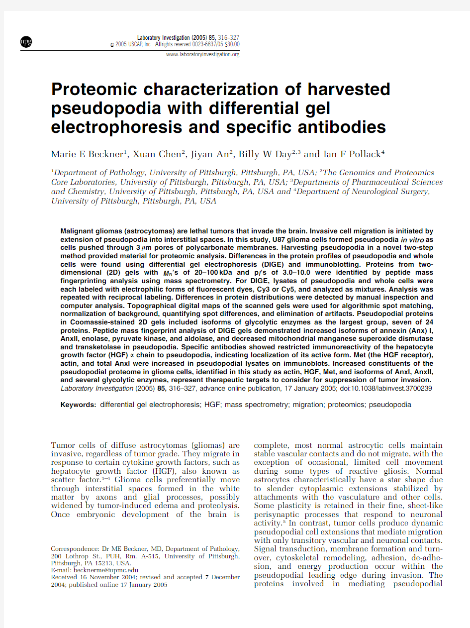
Proteomic investigations of migrated pseudopodia rely on obtaining sufficient lysate material for study. Obtaining migrated cellular material by scraping it from wafer-thin filters or chemically releasing it from chamber well surfaces in migration assays often results in low yields.To address this issue,we used a novel two-step method to harvest migrated pseudopodia from filters.Pseudopodia were trans-ferred from the filters onto glass slides and were then harvested in urea lysate buffer that crystallized so that all pseudopodial material could be retrieved by scraping.
A motile,human malignant glioma cell line,U87, was studied.Quantities of pseudopodial lysates were sufficient for two-dimensional(2D)gel electro-phoresis followed by peptide mass fingerprint (PMF)analysis.Numerous abundant proteins de-tected with Coomassie blue staining were identified. Some have been previously associated with tumor progression.On differential gel electrophoresis (DIGE)analysis of whole cells and pseudopodia, several of these proteins,or some of their isoforms, demonstrated increased pseudopodial distributions. Immunoreactivity for the a chain of activated HGF was detected only in pseudopodial lysates.The HGF receptor,Met,was present in both whole cells and pseudopodia and was increased in pseudopodia. Differential localization of certain proteins,includ-ing an activated signaling network for a motility cytokine in the pseudopodia,supports this metho-dology for defining the functional pseudopodial proteome.
Materials and methods
Cell Culture
Human U87MG astrocytoma cells(American Type Culture Collection,Rockville,MD,USA),were maintained in minimal essential media(MEM)Eagle (Cellgro,Mediatech,Herndon,VA,USA)with10% fetal bovine serum(FBS)(Invitrogen,Carlsbad,CA, USA).Half volumes of media were changed the day prior to each harvest of pseudopodia to normalize their metabolic state from batch to batch.
Harvesting Pseudopodia and Whole Cells
Confluent cells were trypsinized on the day of harvest and allowed to recover in10%FBS for2h at371C.Cells were centrifuged(500rpm,4.5min) and resuspended(1.5?106/ml)with minimal pipet-ting in motility media,which consisted of phos-phate-buffered saline(PBS)containing0.27% CaCl2á2H2O,0.02%MgCl2á6H2O,1%FBS,5mM sodium pyruvate,1mg/ml glucose,and0.1%bovine serum albumin(BSA),unless stated otherwise. Four-well Boyden separation chambers(Neuro Probe,Gaithersburg,MD,USA)were assembled
with9m m thick polycarbonate filters(Neuro Probe) containing3m m diameter pores,with and without a coating of0.01%porcine gelatin.The filters were inserted to separate upper and lower wells.The
lower wells contained motility media.Cell suspen-
sions were added to the upper wells and each chamber was placed in a large Petri dish on a moist Kimwipe and incubated at371C in a5%CO2tissue
culture incubator.To harvest pseudopodia,filters
were removed at4.5–5h.In the first step,each filter
was immersed in methanol for15s and placed with migrated cell materials down(attached to the undersurface of filters)on a5?7.5cm glass slide. Nonmigrated cell materials were completely wiped
from the top of each filter with large Kimwipes.
Fresh Kimwipes were used to press each filter firmly against a glass slide for a few seconds to promote adherence of migrated pseudopodia to the glass.The
filter was then gently peeled off the slide with forceps as shown(Figure1).If the pseudopodia did
not adhere on the first attempt,the filter was resuspended in methanol a second time and the process was repeated on a new slide.The glass
slides with attached pseudopodia were stored at
à801C in non-air tight plastic containers that allowed rapid freezing.During the second step, migrated materials on several glass slides were solubilized in lysate buffer consisting of6M urea,
4%3-[(3-chloamidopropyl)dimethylammonio]-1-pro-panesulfonate,2M thiourea,20mM dithiothreitol,
1.6mM
4-(2-hydroxyethyl)piperazine-1-ethanesul-
Figure1Method for harvesting pseudopodia.U87glioma cells
placed in the upper wells of four-well Boyden separation chambers pushed their cytoplasm through3m m pores of9m m
thick polycarbonate filters,during 4.5–5h incubations.The chambers were disassembled and filters were removed with forceps.Following a15s dip in methanol,each filter was placed
on a5?7.5cm glass slide with migrated materials trapped
between the filter and glass.Unmigrated cell materials were
wiped off the upper surface of the filter with large Kimwipes.
Fresh Kimwipes were used to press the filter firmly against the underlying glass slide.The filter was then gently peeled away,
leaving migrated pseudopodia attached to the slide.Urea lysis
buffer was used to harvest the pseudopodia as a liquid combined
with the dried residual material that was scraped off the slides
following crystallization.Pseudopodia from multiple slides were
batched.
Proteomic characterization of pseudopodia
ME Beckner et al
317
Laboratory Investigation(2005)85,316–327
fonic acid,pH 8.0,and removed by pipetting.After allowing the residual lysate to dry and crystallize,it was retrieved with razor blades to complete the harvesting process.Pseudopodia from approxi-mately 40–60million cells were combined to form each batch of lysate,1–3mg/ml total protein.Whole cells were allowed to adhere to the filters and then placed on the glass slides,as described for pseudo-podia,and harvested in a similar manner.The ratio of cell numbers used to obtain lysates of whole cells compared to pseudopodia was approximately 1:12.Most of the ‘whole’cells were not in contact with pores that would have permitted loss of their protrusions when harvested from the filters.Traditional 2D Gel Electrophoresis
The lysates were treated with a ReadyPrep 2D Cleanup Kit (BioRad,Hercules,CA,USA)to remove ions,DNA,RNA,etc.Their protein contents were measured using a 2D Quant kit (Amersham Bios-ciences,Piscataway,NJ,USA).After overnight rehydration in buffer consisting of 7M urea,2M thiourea,4%3-[(3-chloamidopropyl)dimethylam-monio]-1-propanesulfonate,25mM dithiothreitol,and 0.2%ampholyte,the protein samples,75m g,were loaded on a 17cm immobilized pH gradient (IPG)strip (BioRad),nonlinear pH 3–10.Focusing was accomplished with 250V for 15min,10000V for 3h,and a third step of 60000total V h,and then maintained at 500V,as needed.The resolved proteins were equilibrated in sodium dodecyl sulfate (SDS)buffer with reducing agents present and separated with SDS polyacrylamide gel electro-phoresis (PAGE)using 8–16%gradient Tris-HCl gels in a Protean II apparatus (BioRad)operated at 2,5,and 10W per gel for 8h.Coomassie blue stain was used to detect abundant proteins.Spots were manually retrieved with pipette tips,digested with trypsin,and submitted for PMF analysis.DIGE
Procedures described above were followed with the following modifications.The electrophilic fluores-cent dyes,PrCy3-N -hydroxysuccinimide ester and MeCy5-N -hydroxysuccinimide ester (Cy3and Cy5,respectively),were synthesized according to a previously published method 6and stored as single-use aliquots in anhydrous N,N -dimethylformamide at à801C.Pseudopodial and whole cell lysates were separately mixed with volumes of each dye solution to produce balanced covalent labeling of 1–5%of the available lysine side chains,without significant alteration of the protein isoelectric points (p I ’s).Quenching was accomplished with 40%aqueous methylamine,pH 8.6.Paired pseudopodial and whole cell lysates were mixed together for focusing and subsequent separation by SDS-PAGE.The gels were repeated with reciprocally labeled pseudopo-dial and whole cell lysates.The gels were viewed in a fluorescent gel imaging device built around a CCD camera (CH350model,16bit chip,Photometrics,Munchen,Germany)with an integral gel cutting tool.Imaging was performed at two excitation wavelengths,545710and 635715nm for Cy3and Cy5,respectively.Image manipulation and viewing was done with Image J (National Institutes of Health,Bethesda,MD,USA)and V ttPrecision Digital Imaging System software (Digital Optics,Auckland,New Zealand).Two-frame,‘flash’movies of the overlying Cy3and Cy5images were viewed in a continuous loop to visually detect differences in the protein signatures between the two samples sepa-rated in the same gels.The repeat gels of recipro-cally stained lysates revealed any differences due to dye uptake.Sample-dependent differences in pro-tein distributions were confirmed and quantified using an image analysis software package,DeCyder Differential Analysis Software,version 5.0(DeCy-der s ,Amersham Biosciences).DeCyder analyzed images as fragments,summed to generate a compo-site image,with proprietary algorithms that detected overlapping,differently colored images within the same gel to match spots and subtract background for normalization.DeCyder created measurement masks,each outlining an area of the gel containing a protein spot,that were applied to each of the matching fragments in the paired fluorescent images of each gel.The masks defined areas in which the pixel values were integrated to create three dimen-sional topographical maps with peaks representing each protein spot.An estimate of the local back-ground for each spot determined the base values across the masked area.Output from the image analysis,designated as fluorescence intensity,was the sum of pixel values in the mask area minus the background.Volumes of the peaks representing the relative strengths of fluorescent signals from matched spots were compared.Although a thresh-old was set at 1.5-fold to screen for differences,only proteins with Z 2.0-fold differences were reported.Visual inspection confirmed the differences indi-cated by the DeCyder software.Dust and artifacts were detected as peaks with slope values of less than one or as clusters of sharp spikes.Images of some protein spots were also scanned and digitized to compare total pixels,as described below for immunoblots.Protein spots that differed on DIGE between the labeled lysates were harvested from the gels using a robotic gel picker integrated with the high resolution CCD camera,digested with trypsin,and submitted for mass spectrometric PMF analysis.
Protein Identification
Following trypsin digestion,the peptides derived from each protein spot were analyzed with matrix assisted laser desorption ionization time of flight mass spectrometry (MALDI-TOF-MS)in a
4700
Proteomic characterization of pseudopodia
ME Beckner et al
318
Laboratory Investigation (2005)85,316–327
Proteomics Analyzer with TOF/TOF Optics(Ap-plied Biosystems,Foster City,CA,USA).Analysis of data with GPS Explorer Protein Analysis Software on a Remote Client workstation(Applied Biosys-tems)provided automated acquisition of optimized mass spectra and the derivation of monoisotopic PMF information.Searches of the NCBI nonredun-dant database,based on the peptide mass results using MASCOT(Matrix Science,Boston,MA,USA) via the GPS Explorer work station and MS-Fit (University of California,San Francisco MS Facility) via the internet identified proteins with matching PMFs.The MASCOT parameters were set to include all species(with the exception of spots from the gel region containing BSA),tolerance of one missed trypsin cleavage per protein,allowance of protein modification by methionine oxidation,peptide tolerance of50ppm(ie an example mass accuracy of100070.05Da),and restriction of peptides to 700–4000Da.The MS-Fit parameters included tol-erance of one missed trypsin cleavage per protein, cysteine modification by acrylamide,oxidation of methionine,and changes at the amino termini,such as conversion of glutamine to pyro-glutamate and acetylation.Protein identifications were accepted when the observed and predicted p I’s and M R’s were consistent and scores indicated nonrandom identi-fications at a significance level of P o0.05.
Densitometry of Immunoblots
Differential localization of proteins was also ana-lyzed with specific antibodies.Lysates of whole cells and pseudopodia,10m g each,were electro-phoresed in separate lanes of10%polyacrylamide 1D gels under reducing conditions.Gel contents were transferred onto polyvinylidene difluoride membranes(Invitrogen),blocked(Detector Block, Protein Detector Western Blot Kit LumiGLO System, Kirkegaard&Perry Laboratories,Gaithersburg,MD, USA),and reacted with anti-human HGF(2m g/ml) (Clone24612.11,Sigma,St.Louis,MO,USA),anti-Met(1:1000)(Clone25H2,Cell Signaling,Beverly, MA,USA),anti-actin(1:1000)(Sigma),and anti-AnxI,(0.2m g/ml)(Clone EH17a,Santa Cruz Biotech-nology,Inc.).The secondary antibodies,1:1000 (Kirkegaard&Perry Laboratories)were horseradish peroxidase-labeled anti-mouse for reactions with HGF,Met,and AnxI,and anti-rabbit for actin. Protein standards(MultiMark,Invitrogen)were loaded with8m g per lane.Immunoreactive bands were visualized with horseradish peroxidase con-version of a luminal-based solution to produce chemiluminescence,scanned(Epson Perfection 2450PHOTO transparency scanner,Epson America, Long Beach,CA,USA)and digitized(UN-SCAN-It gel,Silk Scientific,Orem,UT,USA).Band densities were corrected for adjacent background.Staining portions of the gels with Coomassie blue(Novex Colloidal Blue Stain Kit,Invitrogen)was used to confirm equal loading of pseudopodial and whole
cell lysates.
Results
Characterization of Pseudopodia in U87Cells
The cellular material that migrated through3m m diameter pores of filters,with and without a0.01%
gelatin(denatured collagen)coating,was stained
and examined microscopically.Nuclei were not
found after5h incubations at371C.Only a few
thick,fingerlike pseudopodia had extended through
some pores of the uncoated filters(Figure2a).With
gelatin present on the filters,migrated pseudopodia
at5h formed discontinuous,thin sheets of various
sizes that covered almost all the filters’surfaces, involving many(more than half)of the pores.After
the filters were removed,the slides and filters were separately stained with Diff Quik.Pseudopodia on
the glass slides retained their appearance as thin, discontinuous sheets(Figure2b)seen prior to filter removal.In separate assays,the nuclei also failed to migrate through the pores after6h.After overnight incubations(15h),however,some nuclei had mi-
grated through the filter pores,as shown in Figure
2c,thus verifying that pseudopodial formation at
4.5–5h included the leading edge of migrating cells.
Filters removed from the slides(Figure2b and c)did
not stain,indicating that no cell material remained
(not shown).
Pseudopodial Proteins Detected with Coomassie Blue Staining
Within ranges of20–100kD and p I’s of3–10, isoforms of24proteins were identified in pseudo-
podial lysates separated on2D gels and stained with Coomassie blue.The largest group of stained proteins included metabolic enzymes catalyzing
seven consecutive steps of the glycolytic pathway (aldolase,triose phosphate isomerase,glyceralde-
hyde-3-phosphate dehydrogenase(GAPDH),phos-phoglycerate kinase,phosphoglycerate mutase, enolase,and pyruvate kinase(PK)).A total of17 isoforms of these glycolytic enzymes represented
46%of the total number of protein isoforms(37) identified on Coomassie blue-stained gels.Proteins involved with the cytoskeleton(actin,tropomyosin, vimentin,tubulin and moesin),heat shock protein (HSP)s(27,60,70,and90kDa),and annexin(Anx)s
(I,II,and V)were also identified.Proteins that appeared as single isoforms on the gels,such as valosin-containing protein,HSP90,HSP60,calum-
enin precursor,tropomyosin,ubiquitin carboxyl-terminal esterase LI,HSP27,phosphoglycerate mutase and tropomyosin,had predicted p I’s of
5.14,5.3,5.7,4.4,4.67,5.33,5.98,
6.67and6.45, respectively,all of which approximately corre-sponded with the observed p I’s of the
migrated Proteomic characterization of pseudopodia
ME Beckner et al
319
Laboratory Investigation(2005)85,316–327
spots on the gel shown in Figure 3.These and other proteins are listed in Table 1with their database identification number,confidence scores,and other parameters.The protein expression patterns of whole cell lysates harvested at 2and 5h were similar.
Differential Distributions of Proteins Detected with DIGE
Image analysis of fluorescently labeled lysates loaded together on 2D gels identified different
distributions of protein isoforms in pseudopodia compared to whole cells.A representative DIGE gel is shown (Figure 4)with its DeCyder analysis portrayed topographically in Figure 5and the quantified differences listed in Table 2.The signals for two proteins,transketolase and mitochondrial manganese superoxide dismutase (MnSOD),were significantly decreased in pseudopodia compared to whole cells (Figure 5a).Abundant proteins that increased at least two-fold in pseudopodia and were identified on the DIGE gel shown (Figure 4)included isoforms of AnxI,enolase,and PK
(Figure
Figure 2Morphology of stained migrated U87glioma cell materials.(a )Pseudopodia that formed on an uncoated filter.Darkly stained cytoplasmic material formed a few fingerlike pseudopodia during 4.5–5h incubations without a matrix protein present.Pores,3m m in diameter,were evenly distributed in the overlying filter.No nuclei were present;250-fold magnification.(b )Pseudopodia formed on a filter coated with 0.01%gelatin (denatured-collagen).Under the same conditions described above,the morphology and quantity of migrated cell material changed markedly when the filters were coated with matrix material.The porous filter was removed as described in Figure 1,allowing an unobstructed view.No residual material remained on the removed filters that were stained and examined.Before and after filter removal,migrated cell material appeared as irregularly shaped,lightly stained sheets that varied in size and covered most of the glass surface.No nuclei were present;500-fold magnifica-tion.(c )Cell materials that migrated overnight.During overnight (15h)incubations,nuclei migrated through the 3m m pores of gelatin-coated filters,indicating that material found at 4.5–5h included the leading edge;500-fold magnification.Diff Quik stains for (a –c ).The migration media is described in the
text.
Figure 3Representative 2D gel of the relatively abundant proteins in U87pseudopodial lysates.Coomassie blue-stained gels,8–16%polyacrylamide,containing pseudopodial lysate showed the distribution of 22proteins identified with PMF analysis.A total of 17isoforms of glycolytic enzymes represented 46%of the total number of protein isoforms (37)seen.Table 1lists 24pseudopodial proteins with their parameters,identified by PMF analysis using MALDI-TOF-MS.Exogenous BSA,present in box 5,is not listed.The pH ranged from 3to 10,left and right margins of the gel,respectively.The expected p I ’s of proteins that migrated as single isoforms are stated in the Results
section.
Proteomic characterization of pseudopodia
ME Beckner et al
320
Laboratory Investigation (2005)85,316–327
5b).Inclusion of data from additional gels indicated that aldolase and AnxII were also elevated in pseudopodia.Although the DeCyder-determined ratio for aldolase was only1.94(almost significant) in the DIGE gel shown in Figure4,its ratio,2.09, was above the threshold of significance in another DIGE gel.A protein spot in Figure4’s gel that corresponded to the placement of AnxII isoforms in Figure3had a DeCyder ratio of2.48but did not yield satisfactory material for mass spectrometry.In another DIGE gel,a protein spot in the same location was identified as AnxII and demonstrated a DeCyder ratio of1.88.The left edge and lower mid region of the large,irregularly shaped spot identified as actin yielded DeCyder ratios greater than two-fold,but distinct isoforms of actin did not resolve for clearcut analysis.In a separate analysis using densitometric data from the scanned fluorescent signal,total actin increased by38%in the pseudopodial lysate on DIGE.Keratins were found preferentially in pseu-dopodia,but may have resulted from hair and skin contamination that accumulated in lysates when several slides were stored and batched during harvests of pseudopodia.Increased BSA,an exogen-ous protein in the media,was not considered biologically significant.BSA may have bound to glass surfaces not covered by pseudopodia or it may have preferentially adhered to the surfaces of https://www.360docs.net/doc/135809184.html,ck of successful identification with MALDI-TOF-MS for many of the fluorescent protein spots that yielded significant DeCyder ratios was mostly attributed to insufficient amounts of protein. None of the pseudopodial proteins with sufficient abundance to be detected and identified on gels stained with Coomassie blue(Table1)were found to be decreased compared to whole cells on DIGE gels.
Differential Distributions of Proteins on Immunoblots of1D Gels
Analysis of paired pseudopodial and whole cell lysates demonstrated immunoreactivity to the
Table1Pseudopodial proteins identified on Coomassie blue-stained gels
Name Location in
Figure3Accession
Number
M R(kDa)Score a Functional associations b
Actin12gi|1425040141137Cytoskeleton
Aldolase14gi|22967439.385Glycolysis
AnxI15gi|450210138.7141Tumor progression54–57,59
AnxII16gi|475775638.6138Proteolysis61–63
Actin bundling64
Glioma progression21,65
AnxV*gi|99992635.8148Apoptosis66
Calumenin precursor11gi|450255137.183Present in tumors55
Enolase10gi|450357147.1142Glycolysis
Glutathione-S-transferase*gi|220420723.497Drug resistance67
Glioma progression21
GAPDH17gi|316453693Glycolysis
HSP902gi|1312915083.3122Protein chaperones that stabilize
stressed cells68–71
HSP704gi|572987770.9176
gi|1650723772.399
HSP606gi|315429476179Glioma progression for HSP2721
HSP2720gi|66284122.376Glioma progression for HSP2721 Moesin3gi|1462582461.898Cytoskeleton
Phosphoglycerate kinase13gi|450576344.6(2.6e+06,MS Fit)Glycolysis
Phosphoglycerate mutase21gi|450575328.8123Glycolysis
Glioma progression21
Prolyl-4-hydroxylase,b*gi|2007012557.1126Collagen synthesis and regulation of
hypoxia inducible factor a72
PK7gi|12560457.9122Glycolysis
Triosephosphate
isomerase
22gi|450764526.7159Glycolysis
Tropomyosin18gi|450765128.5114Cytoskeleton
Tubulin-b-1chain9gi|13544849.7152Cytoskeleton
Ubiquitin carboxyl-
terminal esterase LI
19gi|2136109124.869Protein degradation73
Valosin containing
protein
1gi|600594289.369Tumor progression55,74–82
Vimentin8gi|211920453.6226Cytoskeleton
a Highest scores,à10log(P),where P was the probability that the observed match was a random event,obtained with MASCOT for all proteins listed,except phosphoglycerate kinase whose score(significant)was obtained with MS-Fit.MASCOT scores greater than63were considered to be significant(P o0.05).Exogenous BSA(box5in Figure3)was not included in the list of pseudopodial proteins.
b References for cytoskeletal and glycolyti
c associations were too numerous to list.
*Identified on another
gel.Proteomic characterization of pseudopodia
ME Beckner et al
321
Laboratory Investigation(2005)85,316–327
69kDa a -chain of activated HGF only in pseudo-podial lystates.The antibody was raised against recombinant human HGF.Met immunoreactivity,apparent as a 145kDa band,was present in both types of lysates and increased up to 79%in the pseudopodia.Increased immunoreactivity was pre-sent for total AnxI and actin,35and 66%,respectively,in pseudopodia (Figure 6a).Equalized loading of lysates was also demonstrated with Coomassie blue staining (Figure 6b).
Discussion
Malignancy of many tumors is defined by invasive and/or metastatic behavior.Diffuse astrocytomas invade widely through surrounding central nervous system tissue.These tumors migrate preferentially through white matter tracts formed by axons,glial processes,and the vasculature.Although white matter of the brain is a dense meshwork,it can be loosened by tumor edema and contains fewer blood vessels and neural processes than gray matter.The process of cell locomotion is often studied in single cells that crawl independently on flat surfaces in vitro .Their cytoplasmic protrusions have been named lamellipodia,filopodia,ruffles,spikes and blebs.Some of these structures have been character-ized by the proteins involved in their formation.7,8However,during brain invasion,tumor cells migrate mostly through the 3D,interstitial spaces of white matter.The term ‘pseudopodia’has been used to describe tumor cell protrusions into the 5–7m m openings of micropipette tips 9and protrusions through 1m m pores of filters.10In the current study,pseudopodia were formed by the tumor cell protru-sions through 3m m diameter pores of filters.
Microscopic examination of migrated pseudopo-dia at different time points confirmed inclusion of the leading edge in the harvested material by demonstrating nuclear migration at a later time point.The 2D analysis of pseudopodia formed by motile glioma cells provided a proteomic view,with some constituents noted for their association with high grade gliomas and progression in other types of tumors (Table 1).Immunoreactions with specific antibodies documented the potential for
localized
Figure 4Representative DIGE images of paired whole cell and pseudopodial lysates shown as inverse images of the fluorescent stains.(a )Whole cell lysate labeled with Cy5demonstrated relatively increased quantities of transketolase (arrow 1)and MnSOD (arrow 2)compared to the pseudopodial lysates.(b )
enolase,two isoforms for PK,and merged isoforms for actin (see text),as indicated with arrows,1and 2,3,4and 5,and 7,respectively.Increases in the isoforms for aldolase and AnxII (positions indicated on the Coomassie-stained 2D gel in Figure 3)are discussed in the text.Apparent increases in exogenous BSA,arrow 6,and keratin (probable contaminant)were not considered to be biologically relevant.Selected matching protein spots with digitized representations are topographically compared in Figure 5and the differences are quantified in Table 2.The background was normalized for both images.The 2D gel parameters were similar
to those described for Figure 3.
Figure 5The topographically rendered densitometry results for selected paired spots in the DIGE gel shown in Figure 4.Pairs of encircled (black)peaks,pseudopodial (left)and whole cells (right),represent the spots highlighted on the inserts of gel images.Arrows indicate the corresponding pseudopodial protein spots.Proteins were identified with MALDI-TOF-MS.(a )Two proteins,transketolase and MnSOD,were decreased in pseudo-podia compared to whole cells,indicated as 1and 2,respectively.(b )Selected proteins that were increased in pseudopodia compared to whole cells are indicated as follows,isoforms of AnxI as 1and 2,enolase as 3,isoforms of PK as 4and 5.These proteins are further described in Table 2.
Proteomic characterization of pseudopodia
ME Beckner et al
322
Laboratory Investigation (2005)85,316–327
Proteomic characterization of pseudopodia
ME Beckner et al
323
Laboratory Investigation(2005)85,316–327
signaling in pseudopodia by an activated,well-known glioma migration factor,HGF.DIGE detected changes in several constituents of the pseudopodial proteome compared to whole cells that potentially include important effectors of migration signaling.
Both HGF and Met are known to be produced by glial cells,in vivo and in vitro ,including the U87cell line.2,4,11–14Expression of HGF and Met are associated with tumor progression.3,4,15,16In other types of malignant cells,downstream events in-duced by HGF signaling have been shown to include increased glycolysis 17and phoshorylation of AnxI.18This study revealed the potential for HGF signaling to mediate similar changes within the leading edge of U87glioma cells.
Malignant astrocytic tumors are known to be glycolytic,possibly attributed to decreased numbers of functional mitochondria.19Several glycolytic enzymes,including GAPDH,PK,and phosphogly-cerate kinase,have been found on the cytoplasmic face of the plasma membrane in malignant astro-cytoma cells.20Phosphoglycerate mutase was pre-viously found to be increased in high-grade vs low-grade gliomas.21In the current study,the abundance of at least seven enzymes that catalyze consecutive steps in glycolysis (aldolase through PK reactions)in U87cells is consistent with the prominence of glycolytic metabolism in gliomas.In previous studies,glycolysis has been shown to provide energy for the migration of malignant cells when mitochondria were inhibited.With mitochon-drial inhibitors added to force reliance on glycolytic energy,we showed that A2058melanoma and U87glioma cells migrated at levels comparable to migration in normal conditions.22,23Others demon-strated the presence of GAPDH in pseudopodia and found that glycolysis was necessary for the forma-tion of protrusions in an invasive,transformed epithelial MDCK cell line.10
Site-directed actin polymerization in response to signaling is needed for the formation of cell protrusions.24Glycolytic enzymes are known to bind cytoskeletal proteins,including actin fila-ments,microtubules,and tropomyosin,in addi-tion to existing as unbound cytoplasmic proteins.Numerous studies have demonstrated interactions between the cytoskeleton and glycolytic enzymes,especially GAPDH,aldolase and PK.25–38In this study,quantities of these glycolytic enzymes,as well as actin,tubulin and tropomyosin,were sufficient in U87pseudopodia to be detected with
Table 2Ratios of differences in protein isoforms highlighted in Figures 4and 5
Protein isoforms a
Location in Figure 4
DeCyder ratios
Whole cells:Pseudopodia
Transketolase a1 2.01MnSOD a2 2.00
Pseudopodia:Whole Cells
AnxI b1-Upper spot 4.06b1-Lower spot 2.89AnxI b2-Upper spot 2.17b2-Lower spot
2.01Enolase b3 2.60PK b4 2.31PK b5 2.11
BSA b6N/A,Exogenous protein Actin
b7
3.05at left edge of large
spot
a
Differences for aldolase and AnxII (not listed)were established with additional
gels.
Figure 6Representative immunoblots comparing several proteins in lysates of pseudopodia (Ps)and whole cells (WC)with equalized gel loading.(a )Proteins were separated on 1D,10%polyacrylamide SDS gels under reducing conditions,blotted,and reacted with specific antibodies.An antibody raised against human recombinant HGF reacted with the 69kDa a -chain of activated HGF in Ps but no reactivity was found in lysates of WC.A specific antibody for Met,the receptor for HGF,reacted with 145kDa bands representing the b -subunit of Met in both lysates of Ps and WC with increased (79%)reactivity in Ps.Antibodies specific for total AnxI and actin reacted with 40and 42kDa bands,respectively,demonstrating 35and 66%increased reactivities,respectively,in Ps.(b )Equalized loading of Ps and WC lysates was demonstrated in a portion of the gel sliced from the one transferred and immunoblotted with anti-AnxI and anti-actin shown in (a ):Coomassie blue
stain.
Proteomic characterization of pseudopodia
ME Beckner et al
324
Laboratory Investigation (2005)85,316–327
Coomassie blue staining,thus establishing the potential for the energy-generating portion of the glycolytic pathway to energize cytoskeletal proteins for migration within U87pseudopodia.Polymeriza-tion of actin and tubulin shares a similar use of energy from nucleotide triphosphate hydrolysis.39–44 Increased actin in pseudopodia,as found in this study,has been previously documented by others.10,45Increased isoforms for enolase,aldolase, and PK detected with DIGE in U87pseudopodia may help to energize the leading edge.
Several types of annexins,I,II,and V,were found in the U87pseudopodia.The Anx family is comprised of numerous gene products,12in vertebrates,with various functions proposed for controlling membrane structure and transport, vesicle secretion,anti-inflammation,prostaglandin metabolism,apoptosis,and other functions.46AnxI is also known as lipocortin I,calpactin II,epidermal growth factor receptor substrate,and chromobindin 9.47Two isoforms of AnxI were increased in pseudopodia compared to whole cells as deter-mined by DIGE,and total AnxI was modestly increased when compared on immunoblots.Pre-vious studies have identified AnxI within CNS tumors,including astrocytomas,48and in the ante-rior pituitary gland.49Involvement of AnxI in cell surface changes induced by steroids has been demonstrated in C6rat astrocytoma cells.50,51AnxI is also involved in the regulation of prostaglandins and nitric oxide.52,53Previous associations of AnxI with tumor progression were found with2D electro-phoresis in studies that showed its differential expression in chemoresistant gastric carcinoma cell lines,54a cell line established from a metastasis compared to primary squamous carcinoma of the larynx,55and in tissue containing metastasized breast carcinoma cells compared to normal tissue.56 Immunohistochemical studies of breast and hepato-cellular carcinoma also indicated that AnxI is associated with tumor progression.57–59The contri-bution of AnxI to pseudopodial extension during migration may explain AnxI’s association with tumor progression and HGF signaling seen in other cells18and with activated HGF in U87pseudopodia. Also,AnxII was previously identified more fre-quently in high-grade than low-grade gliomas,50of 65and three of10tumors,respectively,following2D electrophoresis of tissue samples.21
Sensitivity for visualizing stained proteins in2D gels,approximately40ng for Coomassie blue,is increased by using fluorescent dyes and silver nitrate to5and0.5ng,respectively.However,our high success rate for identifying proteins detected with Coomassie blue fell to a much lower rate for spots demonstrated with the fluorescent dyes. Although the relatively low abundance of signaling proteins that control cell migration prevents their proteomic study by current2D gel technologies,60 specific antibodies on immunblots detected a strik-ing localization of active HGF to pseudopodia that also contained increased https://www.360docs.net/doc/135809184.html,bining DIGE with specific antibody reactivities on immunoblots de-monstrated significant increases in isoforms for
actin,enolase,PK,and AnxI,and probable increases
of aldolase and AnxII in the pseudopodia.Identify-
ing the functional proteome of tumor pseudopodia
will help to elucidate mechanisms of cell migration
in tissue and will aid in the selection of biomarkers
and molecular treatment targets for malignant gliomas.
Acknowledgements
We thank April E Engram,Fatima Burovic,and Beth
Arnold for technical assistance.We thank Sadie Aznavoorian Cheshire,PhD(Prism Medical Com-munications,Inc.,Morristown,NJ,USA)for helpful comments.We gratefully acknowledge financial support from The Nick Eric Wichman Foundation,
Ellicott City,MD,USA and The Walter L Copeland
Fund for Cranial Research of the Pittsburgh Founda-
tion,Pittsburgh,PA,USA.
References
1Brockmann MA,Ulbricht U,Gruner K,et al.Glioblas-
toma and cerebral microvascular endothelial cell
migration in response to tumor-associated growth
factors.Neurosurgery2003;52:1391–1399.
2Koochekpour S,Jeffers M,Rulong S,et al.Met and
hepatocyte growth factor/scatter factor expression in
human gliomas.Cancer Res1997;57:5391–5398.
3Lamszus K,Schmidt NO,Jin L,et al.Scatter factor
promotes motility of human glioma and neuromicro-
vascular endothelial cells.Int J Cancer1998;75:19–28.
4Lamszus K,Laterra J,Westphal M,et al.Scatter factor/
hepatocyte growth factor(SF/HGF)content and func-
tion in human gliomas.Int J Dev Neurosci1999;17:
517–530.
5Derouiche A,Frotscher M.Peripheral astrocyte pro-
cesses:monitoring by selective immunostaining for the
actin-binding ERM proteins.Glia2001;36:330–341.
6Unlu M,Morgan ME,Minden JS.Difference gel
electrophoresis:a single gel method for detecting
changes in cell extracts.Electrophoresis1997;18:
2071–2077.
7Horwitz AR,Parsons JT.Cell migration–movin’on.
Science1999;286:1102–1103.
8Ridley AJ,Schwartz MA,Burridge K,et al.Cell
migration:integrating signals from front to back.
Science2003;302:1704–1709.
9Dong C,Aznavoorian S,Liotta LA.Two phases of
pseudopod protrusion in tumor cells revealed by a
micropipette.Microvasc Res1994;47:55–67.
10Nguyen TN,Wang HJ,Zalzal S,et al.Purification and characterization of beta-actin-rich tumor cell pseudo-
podia:role of glycolysis.Exp Cell Res2000;258:
171–183.
11Abounader R,Ranganathan S,Lal B,et al.Reversion of human glioblastoma malignancy by U1small nuclear
RNA/ribozyme targeting of scatter
factor/hepatocyte Proteomic characterization of pseudopodia
ME Beckner et al
325
Laboratory Investigation(2005)85,316–327
growth factor and c-met expression.J Natl Cancer Inst 1999;91:1548–1556.
12Hollborn M,Krausse C,Iandiev I,et al.Glial cell expression of hepatocyte growth factor in vitreoretinal proliferative https://www.360docs.net/doc/135809184.html,b Invest 2004;84:963–972.13
Yamada T,Yoshiyama Y,Tsuboi Y,et al.Astroglial expression of hepatocyte growth factor and hepatocyte growth factor activator in human brain tissues.Brain Res 1997;762:251–255.
14
Welch WC,Kornblith PL,Michalopoulos GK,et al.Hepatocyte growth factor (HGF)and receptor (c-met)in normal and malignant astrocytic cells.Anticancer Res 1999;19:1635–1640.
15
Schmidt NO,Westphal M,Hagel C,et al.Levels of vascular endothelial growth factor,hepatocyte growth factor/scatter factor and basic fibroblast growth factor in human gliomas and their relation to angiogenesis.Int J Cancer 1999;84:10–18.
16
Yano H,Hara A,Murase S,et al.Expression of hepatocyte growth factor and matrix metalloprotei-nase-2in human glioma.Brain Tumor Pathol 2001;18:7–12.
17
Kaplan O,Firon M,Vivi A,et al.HGF/SF activates glycolysis and oxidative phosphorylation in DA3murine mammary cancer cells.Neoplasia 2000;2:365–377.
18
Skouteris GG,Schroder CH.The hepatocyte growth factor receptor kinase-mediated phosphorylation of lipocortin-1transduces the proliferating signal of the hepatocyte growth factor.J Biol Chem 1996;271:27266–27273.
19
Oudard S,Boitier E,Miccoli L,et al.Gliomas are driven by glycolysis:putative roles of hexokinase,oxidative phosphorylation and mitochondrial ultra-structure.Anticancer Res 1997;17:1903–1911.
20
Daum G,Keller K,Lange K.Association of glycolytic enzymes with the cytoplasmic side of the plasma membrane of glioma cells.Biochim Biophys Acta 1988;939:277–281.
21
Iwadate Y,Sakaida T,Hiwasa T,et al.Molecular classification and survival prediction in human glio-mas based on proteome analysis.Cancer Res 2004;64:2496–2501.
22Beckner ME,Stracke ML,Liotta LA,et al.Glycolysis as primary energy source in tumor cell chemotaxis.J Natl Cancer Inst 1990;82:1836–1840.
23Beckner ME,Pollack IF.Treatment of hypoxia-resistant astrocytoma cell migration with glucose inhibitors.Neuro-Oncology 2003;5:280.
24Carlier MF,Wiesner S,Le Clainche C,et al.Actin-based motility as a self-organized system:mechanism and reconstitution in vitro .C R Biol 2003;326:161–170.25Bronstein WW,Knull HR.Interaction of muscle glycolytic enzymes with thin filament proteins.Can J Biochem 1981;59:494–499.
26Durrieu C,Bernier-Valentin F,Rousset B.Binding of glyceraldehyde 3-phosphate dehydrogenase to micro-tubules.Mol Cell Biochem 1987;74:55–65.
27
Durrieu C,Bernier-Valentin F,Rousset B.Microtubules bind glyceraldehyde 3-phosphate dehydrogenase and modulate its enzyme activity and quaternary structure.Arch Biochem Biophys 1987;252:32–40.
28Masters C.Interactions between glycolytic enzymes and components of the cytomatrix.J Cell Biol 1984;99(1Part 2):222s–225s.
29
Karkhoff-Schweizer R,Knull HR.Demonstration of tubulin-glycolytic enzyme interactions using a novel
electrophoretic approach.Biochem Biophys Res Com-mun 1987;146:827–831.
30
Mejean C,Pons F,Benyamin Y,et al.Antigenic probes locate binding sites for the glycolytic enzymes glycer-aldehyde-3-phosphate dehydrogenase,aldolase and phosphofructokinase on the actin monomer in micro-filaments.Biochem J 1989;264:671–677.
31Minaschek G,Groschel-Stewart U,Blum S,et al.Microcompartmentation of glycolytic enzymes in cultured cells.Eur J Cell Biol 1992;58:418–428.
32
Somers M,Engelborghs Y,Baert J.Analysis of the binding of glyceraldehyde-3-phosphate dehydrogenase to microtubules,the mechanism of bundle formation and the linkage effect.Eur J Biochem 1990;193:437–444.33
Sirover MA.New insights into an old protein:the functional diversity of mammalian glyceraldehyde-3-phosphate dehydrogenase.Biochim Biophys Acta 1999;1432:159–184.
34
Walsh JL,Knull HR.Heteromerous interactions among glycolytic enzymes and of glycolytic enzymes with F-actin:effects of poly(ethylene glycol).Biochim Biophys Acta 1988;952:83–91.
35Walsh JL,Keith TJ,Knull HR.Glycolytic enzyme interactions with tubulin and microtubules.Biochim Biophys Acta 1989;999:64–70.
36Volker KW,Knull HR.Glycolytic enzyme–tubulin interactions:role of tubulin carboxy terminals.J Mol Recogn 1993;6:167–177.
37Volker KW,Reinitz CA,Knull HR.Glycolytic enzymes and assembly of microtubule https://www.360docs.net/doc/135809184.html,p Bio-chem Physiol B Biochem Mol Biol 1995;112:503–514.38Volker KW,Knull H.A glycolytic enzyme binding domain on tubulin.Arch Biochem Biophys 1997;338:237–243.
39Carlier MF,Pantaloni D,Evans JA,et al.The hydrolysis of ATP that accompanies actin polymerization is essentially irreversible.FEBS Lett 1988;235:211–214.40Carlier MF.Nucleotide hydrolysis regulates the dy-namics of actin filaments and microtubules.Philos Trans R Soc London B Biol Sci 1992;336:93–97.
41Korn ED,Carlier MF,Pantaloni D.Actin polymeriza-tion and ATP hydrolysis.Science 1987;238:638–644.42
Dufort PA,Lumsden CJ.How profilin/barbed-end synergy controls actin polymerization:a kinetic model of the ATP hydrolysis circuit.Cell Motil Cytoskeleton 1996;35:309–330.
43Loisel TP ,Boujemaa R,Pantaloni D,et al.Reconstitu-tion of actin-based motility of Listeria and Shigella using pure proteins.Nature 1999;401:613–616.
44Mitchison TJ.Evolution of a dynamic cytoskeleton.Philos Trans R Soc London B Biol Sci 1995;349:299–304.
45
Le PU,Nguyen TN,Drolet-Savoie P ,et al.Increased beta-actin expression in an invasive moloney sarcoma virus-transformed MDCK cell variant concentrates to the tips of multiple pseudopodia.Cancer Res 1998;58:1631–1635.
46Rescher U,Gerke V.Annexins—unique membrane binding proteins with diverse functions.J Cell Sci 2004;117(Part 13):2631–2639.
47Fernandez MP ,Morgan RO.Annexin Sequence Data-base,The New Nomenclature.European Annexin Homepage 2004;https://www.360docs.net/doc/135809184.html,.
48
Johnson MD,Kamso-Pratt J,Pepinsky RB;et al.Lipocortin-1immunoreactivity in central and periph-eral nervous system glial tumors.Hum Pathol 1989;20:
772–776.
Proteomic characterization of pseudopodia
ME Beckner et al
326
Laboratory Investigation (2005)85,316–327
49Turgeon JL,Cooper RH,Waring DW.Membrane-specific association of annexin I and annexin II in anterior pituitary cells.Endocrinology1991;128:96–102.
50McLeod JD,Goodall A,Jelic P,et al.Changes in the cellular distribution of lipocortin-1(Annexin-1)in C6 glioma cells after exposure to dexamethasone.Bio-chem Pharmacol1995;50:1103–1107.
51Mizuno H,Uemura K,Moriyama A,et al.Glucocorti-coid induced the expression of mRNA and the secretion of lipocortin1in rat astrocytoma cells.Brain Res1997;746:256–264.
52Cirino G,Flower RJ.Human recombinant lipocortin1 inhibits prostacyclin production by human umbilical artery in vitro.Prostaglandins1987;34:59–62.
53Minghetti L,Nicolini A,Polazzi E,et al.Down-regulation of microglial cyclooxygenase-2and induci-ble nitric oxide synthase expression by lipocortin1.
Br J Pharmacol1999;126:1307–1314.
54Sinha P,Hutter G,Kottgen E,et al.Increased expres-sion of annexin I and thioredoxin detected by two-dimensional gel electrophoresis of drug resistant human stomach cancer cells.J Biochem Biophys Methods1998;37:105–116.
55Wu W,Tang X,Hu W,et al.Identification and validation of metastasis-associated proteins in head and neck cancer cell lines by two-dimensional electro-phoresis and mass spectrometry.Clin Exp Metast 2002;19:319–326.
56Cicek M,Samant RS,Kinter M,et al.Identification of metastasis-associated proteins through protein analy-sis of metastatic MDA-MB-435and metastasis-sup-pressed BRMS1transfected-MDA-MB-435cells.Clin Exp Metast2004;21:149–157.
57Pencil SD,Toth M.Elevated levels of annexin I protein in vitro and in vivo in rat and human mammary adenocarcinoma.Clin Exp Metast1998;16:113–121. 58Masaki T,Tokuda M,Ohnishi M,et al.Enhanced expression of the protein kinase substrate annexin in human hepatocellular carcinoma.Hepatology 1996;24:72–81.
59Ahn SH,Sawada H,Ro JY,et al.Differential expression of annexin I in human mammary ductal epithelial cells in normal and benign and malignant breast tissues.
Clin Exp Metast1997;15:151–156.
60Lim MS,Elenitoba-Johnson KS.Proteomics in patho-logy https://www.360docs.net/doc/135809184.html,b Invest2004;84:1227–1244.
61Diaz VM,Hurtado M,Thomson TM,et al.Specific interaction of tissue-type plasminogen activator(t-PA) with annexin II on the membrane of pancreatic cancer cells activates plasminogen and promotes invasion in vitro.Gut2004;53:993–1000.
62Hajjar KA,Acharya SS.Annexin II and regulation of cell surface fibrinolysis.Ann NY Acad Sci2000;902: 265–271.
63Roshy S,Sloane BF,Moin K.Pericellular cathepsin B and malignant progression.Cancer Metast Rev2003;
22:271–286.
64Jones PG,Moore GJ,Waisman DM.A nonapeptide to the putative F-actin binding site of annexin-II tetramer inhibits its calcium-dependent activation of actin filament bundling.J Biol Chem1992;267: 13993–13997.
65Roseman BJ,Bollen A,Hsu J,et al.Annexin II marks astrocytic brain tumors of high histologic grade.Oncol Res1994;6:561–567.66Allen RT,Hunter Jr W,Agrawal DK.Morphological and biochemical characterization and analysis of
apoptosis.J Pharmacol Toxicol Methods1997;37:
215–228.
67Townsend DM,Tew KD.The role of glutathione-S-transferase in anti-cancer drug resistance.Oncogene
2003;22:7369–7375.
68Bagatell R,Beliakoff J,David CL,et al.Hsp90 inhibitors deplete key anti-apoptotic proteins in
pediatric solid tumor cells and demonstrate synergistic
anticancer activity with cisplatin.Int J Cancer2005;
113:179–188.
69Bagatell R,Whitesell L.Altered Hsp90function in cancer:a unique therapeutic opportunity.Mol Cancer
Ther2004;3:1021–1030.
70Beliakoff J,Bagatell R,Paine-Murrieta G,et al.
Hormone-refractory breast cancer remains sensitive to
the antitumor activity of heat shock protein90
inhibitors.Clin Cancer Res2003;9:4961–4971.
71Workman https://www.360docs.net/doc/135809184.html,binatorial attack on multistep onco-genesis by inhibiting the Hsp90molecular chaperone.
Cancer Lett2004;206:149–157.
72Myllyharju J.Prolyl4-hydroxylases,the key enzymes of collagen biosynthesis.Matrix Biol2003;22:15–24.
73Pickart CM,Rose IA.Ubiquitin carboxyl-terminal hydrolase acts on ubiquitin carboxyl-terminal amides.
J Biol Chem1985;260:7903–7910.
74Asai T,Tomita Y,Nakatsuka S,et al.VCP(p97) regulates NFkappaB signaling pathway,which is
important for metastasis of osteosarcoma cell line.
Jpn J Cancer Res2002;93:296–304.
75Tsujimoto Y,Tomita Y,Hoshida Y,et al.Elevated expression of valosin-containing protein(p97)is
associated with poor prognosis of prostate cancer.Clin
Cancer Res2004;10:3007–3012.
76Yamamoto S,Tomita Y,Hoshida Y,et al.Expression level of valosin-containing protein is strongly asso-
ciated with progression and prognosis of gastric
carcinoma.J Clin Oncol2003;21:2537–2544.
77Yamamoto S,Tomita Y,Nakamori S,et al.Elevated expression of valosin-containing protein(p97)in
hepatocellular carcinoma is correlated with increased
incidence of tumor recurrence.J Clin Oncol2003;21:
447–452.
78Yamamoto S,Tomita Y,Hoshida Y,et al.Expression of valosin-containing protein in colorectal carcinomas as
a predictor for disease recurrence and prognosis.Clin
Cancer Res2004;10:651–657.
79Yamamoto S,Tomita Y,Hoshida Y,et al.Increased expression of valosin-containing protein(p97)is
associated with lymph node metastasis and prognosis
of pancreatic ductal adenocarcinoma.Ann Surg Oncol
2004;11:165–172.
80Yamamoto S,Tomita Y,Hoshida Y,et al.Expression level of valosin-containing protein(p97)is correlated
with progression and prognosis of non-small-cell lung
carcinoma.Ann Surg Oncol2004;11:697–704.
81Yamamoto S,Tomita Y,Hoshida Y,et al.Expression level of valosin-containing protein(VCP)as a prog-
nostic marker for gingival squamous cell carcinoma.
Ann Oncol2004;15:1432–1438.
82Yamamoto S,Tomita Y,Hoshida Y,et al.Expression level of valosin-containing protein(p97)is associated
with prognosis of esophageal carcinoma.Clin Cancer
Res
2004;10:5558–5565.
Proteomic characterization of pseudopodia
ME Beckner et al
327
Laboratory Investigation(2005)85,316–327
JPEG图像的编解码实现
毕业论文论文题目(中文)JPEG图像的编解码实现 论文题目(外文)Encoding and decoding of JPEG image
摘要 JPEG是一种十分先进的图像压缩技术,它用有损压缩方式去除冗余的图像数据,在获得极高的压缩率的同时能展现十分丰富生动的图像。本文设计和实现一个JPEG图像编解码器来进行图像转换,利用离散余弦变换、熵编码、Huffman编码等图像压缩技术将BMP图像转换成JPEG图像,即进行图像的压缩。验证JPEG压缩编码算法的可行性。通过比对图像压缩前后实际效果,探讨压缩比,峰值信噪比等评价图像数据压缩程度及压缩质量的关键参数,对JPEG 压缩编码算法的实用性和优越性进行研究。 关键词:JPEG;编码;解码;图像压缩
Abstract JPEG is a very advanced image compression technology, it uses lossy compression to remove redundant image data, in obtaining a very high compression rate can show a very rich and vivid image. In this project, a JPEG image codec is designed and implemented to transform image, using discrete cosine transform, entropy coding, Huffman coding and other image compression techniques to convert BMP images into JPEG images. Verifies the feasibility of JPEG compression coding algorithm. Through the comparison of the actual effect of image compression, the key parameters of compression ratio, peak Snr, and the compression quality of image data are discussed, and the practicability and superiority of JPEG compression coding algorithm are researched. Key words: JPEG; encoding; decoding; image compression
医院健康教育与健康促进工作计划
一直以来,医院是健康教育与健康促进的重要场所,医院开展健康教育与健康促进是提高全体医务人员的健康知识知晓率,健康行为形成率,及住院病人相关知识知晓率的重要措施,是提高健康文明素质、提高病人生活质量的必须长期坚持不懈抓紧抓实的工作内容。为了做好我院今年的健康教育工作,特制订一下工作计划。 一、工作内容 1、做好年初健康教育宣传员的调整工作,做到院、科领导重视、积极参与,宣传员落实、各负其责。充分发挥健康教育网络的作用,层层深入,科室合作,推动健康促进工作的开展。 2、定期召开健康促进委员会及健康教育宣传员会议;年度内召开两次健康促进工作委员会会议;每季度召开健康教育宣传员工作例会。 3、开展健康教育知识培训。年度内计划举办两次全院性、纳入学分管理的健康教育讲座,以提高医务人员的卫生知识水平、健康意识,使医务人员的健康知识知晓率达80%以上,健康行为形成率达70%以上。 4、加强医院健康教育阵地建设。门诊设有的固定健康教育宣传栏,两月更换一次;候诊室设置的宣传架保持健教内容丰富;病区要设立白板报或宣传版块(以楼层计算),每季度更换一次;对上级下发的及
本院自制的健康教育资料及时张贴、分发。利用各种形式,积极传播健康信息。 5、大力开展健康教育活动。 职工健康教育:继续开展《中国公民健康素养》的学习,普及“健康素养66条”,针对职工中存在的不良生活方式,开展多种形式的行为干预活动,逐步提高医务人员健康促进观念,强化个人健康技能,充分发挥职工的潜力,以达到身心健康的目的。 门诊健康教育:门办医务人员应有针对性开展候诊教育与随诊教育;做好门诊35岁首诊患者测量血压工作,定期进行筛查;加大力度继续做好五种慢病门诊的健康干预和随访管理。 住院健康教育:做好入院教育与出院教育的同时,重点做好住院期教育。①医生在进行医疗活动时所运用的健康咨询、健康处方等对病人及其亲属开展健康教育;科室健教宣传员每月组织一次病人工休座谈会,使住院病人相关知识知晓率达≥80%。②健康处方:每位住院病人或家属至少持有一种健康教育处方;医务人员有针对性地对每位住院病人或家属开展健康教育2—3次。③病区的宣传专栏、宣传架为病人提供与本病区疾病相关的健康知识、健康行为等内容的宣教材料。护士、医生向病人传播健康知识,力求宣传内容通俗易懂,提高宣教质量,宣教覆盖率100%。④医院健康管理科每年对100名或以上病人进行相关知识知晓率调查。
医院健康教育的基本内容与形式
医院健康教育的基本内容与形式 1、医护人员健康教育 1)对专兼职健康教育人员的业务培训,系统学习健康教育基本理论和方法,提高业务技能。培养进行社区干预研究,健康教育计划设计、实施和评价的能力。 2)对全体医护人员进行培训,以业务学习、专题讲座等形式,结合本专业特点和工作需要,普及有关知识;进行人际沟通技巧培训,提高健康教育的知识和技能与工作热情。 3)开展医护人员健康促进活动。针对医护人员存在的问题,有计划有组织的实施干预活动,促使医护人员建立健康的生活方式、促进和增强自身的心身健康。 4)各医院每年对医护人员进行2次以上健康教育培训,培训覆盖率达到90%以上。培训资料(内容)、培训人员签到册、培训记录及照片、测评等资料完整。 5)开展无烟医院创建活动,创建活动有计划,有措施,有记录,有总结。 2、患者健康教育 患者健康教育可分为门诊教育、住院教育、出院教育、出院后(随访)教育。 1)门诊健康教育 门诊健康教育是指对病人在门诊治疗过程中进行的健康教育。门诊健康教育包括候诊健康教育、随诊健康教育、健康咨询等。 ①候诊健康教育:候诊健康教育是在病人候诊期间所进行的教育。主要采用的形式有在候诊厅放置健康知识资料、设置健康教育宣传栏、黑板报等。健康教育宣传栏内容要根据各类人群文化层次的特点精心设计,力求做到内容新颖、标题醒目、形式美观,注意科学性、针对性、通俗性和艺术性。 ②随诊健康教育:随诊健康教育是医生在给病人诊疗过程中,根据病人所患疾病的有关问题进行简短的讲解和指导。为解决门诊病人多,诊疗工作量大与开展随诊教育的矛盾,可使用健康教育处方来对口头教育进行补充完善,又便于病人保存阅读,指导病人进行自我保健和家庭保键是一种有效的辅助治疗手段。 ③健康咨询:健康咨询是医务人员对咨询者提出的有关疾病和健康问题进行的解答和医学指导,县级以上医院要设立咨询室或心理门诊,以满足各类人群不同需要。 指标要求:候诊厅设置一至两块健康教育宣传栏或健康教育橱窗,每块面积不小于3m2, 每两个月至少更换一次内容,每期内容原始稿、版面照片保存归档。各科室随时须有足量的针对该科各种常见病、多发病的健康教育处方,健康教育处方使用率达到80%以上。候诊厅要有“禁止吸烟”标志和宣传控烟的内容。 2)住院健康教育 住院健康教育是指医护人员对住院病人或病人家属进行的健康教育。住院健康教育可分
第五讲 领导与创业领导
第五讲领导与创业领导 一、如何定义领导 ?“领”者,颈也。整件衣服以领为纲,排扣对准领,依领而循行下来。 ?“袖”--- 可长可短可伸可缩。可短, 及时,出手以进;可长而覆掌,缩手可退。长袖善舞,视情境不同决定自己的行动的自由度与强弱度。 ?世界上存在着四种人: ☆第一种人促使事情发生 ☆第二种人看着事情发生 ☆第三种人不清楚所发生的事情 ☆第四种人完全不知所发生的任何事 领导的定义 ?领导是影响人们自动为实现团体目标而努力的一种行为。 ?领导是人们促使其部属充满信心,满怀热情来完成他们任务的艺术。 ?领导是对组织内群体或个人实行影响的活动过程。 领导是影响一个集体走向目标的能力。 ?火车跑得快,全靠车头带。 领导力是合力。 ?是领导者与追随者相互作用而迸发出的一种思想和行为能力。 ?用公式表达如下: 合力= 领导者的能力+ 追随者的能力–阻力 合力是一个团队显示出的整体能力。 ?在合力中,领导的个人能力所占的比例越小,整个团队越是成功;所占比例越大,团队整体效益越差。 电视剧“亮剑”的合力分析 李云龙的团队,其在装备、队伍数量弱于对手的情况下不断取得胜利,成功的秘诀?首先在于能力的恰当组织,化整体的弱为局部的强,从而取得进攻上的优势;其次在精神上有一种让士兵们嗷嗷叫的个人魅力和领导能力;再次有不墨守成规、常常出其不意的智慧和才能。 二、领导与管理的区别 人的四种成就 ①知道如何做好一项具体的工作是一个劳动者的基本成就。 ②传授知识和技能是一个教育者的基本成就。 ③激励他人提升业绩是一个管理者的成就。 ④而能完成上述三项则是一个领导者的成就。 360度反馈 领导与管理的区别 ?“领导人,像罗斯福、丘吉尔和里根等人,他们有办法激励一些有才干的人,让他们把事情做得更好。而管理者呢,总是在复杂事务的细节里打转,这些人在‘进行管理’的同时,‘把事情弄得复杂’。他们往往试图去控制和抑制,把大量的时间浪费在琐碎的细节上。” 领导者就是那些可以清楚地告诉人们如何做得更好,并且能够描绘出愿景构想来激发人们努力的人。 要做领导者,不要做管理者
医院健康促进医院工作总结与医院健康教育与健康促进工作总结汇编
---------------------------------------------------------------最新资料推荐------------------------------------------------------ 医院健康促进医院工作总结与医院健康教育与健康 促进工作总结汇编 医院健康促进医院工作总结与医院健康教育与健康促进工作总结汇编医院健康促进医院工作总结健康促进医院创建工作汇报为进一步加强医院健康促进与教育工作,我院在20**年初确定申报创建市级健康促进医院。 一年来,在市、区爱卫会的精心指导帮助下,对照《**市健康促进医院考核标准(试行)》,我院紧紧围绕健康促进医院创建五大具体目标任务,经过全院职工一年来的共同努力,已达到**市健康促进医院创建要求。 现将工作开展情况汇报如下。 一、基本情况**人民医院(原**第三人民医院)为省卫生厅核准的二级乙等综合性医院,创建于 1952 年,是一家集医疗、教学、科研、预防、保健为一体的非营利性县级医疗机构。 现为国际级爱婴医院,省级绿色医院,省级平安医院,市级文明单位,市级卫生先进单位,浙江省住院医师规范化临床培训基地。 医院内绿草如茵,环境优美。 医院在编职工 198 余人,其中高、中级职称医务人员 90 余人。 医院开放床位 140 张,设六个病区,门诊科室 22 个,医技辅助科室齐全。 无痛胃镜检查、肠镜检查,腹腔镜手术在全区处于领先水平。 1 / 20
年门诊量近 25 万人次,出院病人 9000 余人。 医院技术力量雄厚,设备先进,目前拥有日本日立全身螺旋 CT、日本富士 CR、500MA 遥控 X 线电视透视系统、C 型臂 X 光机、意大利百胜 MyLab60B 超、24 小时动态心电图、美国史赛克 1188 高清腹腔镜、奥林巴斯内窥镜、美国 ECKMANAU680 全自动生化分析仪、奥林巴斯 AU400 全自动生化仪、酶标仪、全自动血球计数仪、同光路手术显微镜、麻醉机、呼吸机、多功能电动手术床等价值千万余元的高级医疗仪器。 医院奉行以病人为中心,以质量为核心的办院宗旨。 近年来医院竭力加强转变职工思想,强化服务理念,改善服务态度和工作作风,不断提高医疗质量,塑造以人为本的医院形象,以崭新的面貌,满怀的激情,致力打造一所人民群众满意的公立医院。 二、主要做法医院以健康促进医院创建活动为契机,在全面提升医疗质量的同时,积极开展健康教育工作。 (一)建立机制,营造氛围,保障创建工作有序开展医院及时成立创建市级健康促进医院组织领导机构,进一步建立健全各科室相关工作,落实专兼职工作人员和工作经费,完善管理机制,逐步完善创建工作规划、计划、检查、考核、评比、总结等文字资料,使我院创建工作符合考核标准的有关要求。 创建领导小组及时研究解决创建工作中遇到的困难和问题,以强有力的组织领导保障创建工作顺利实施。 医院将创建工作任务分解到相关责任部门和科室,各科室按照医
医院健康教育促进工作自查报告
xxx医院健康教育促进工作自查报告 自xx年以来,紧紧围绕以改善就医环境和诊疗服务,提高医院职工、患者及其家属、社区居民的健康素养水平为工作重点,开展了一系列的健康教育促进活动,现将自查报告汇报如下: 一、开展医护人员健康教育 xx院调整充实了健康教育促进医院领导小组,由分管院长担任组长,并配备专职人员。各科室设健康教育领导小组组长及专职人员,负责实施健康教育各项工作。按照年初制定的健康教育工作计划,开展不同层次的健康教育和学术讲座 ,康教育专题讲座多次,主要内容包括《健康传播与技巧》,《健康素养66条》,妇产科围产期孕产妇保健指导,婴儿的特点及喂养,抚触训练等,年终组织健康教育专题考试一次,培训率和参考率达xxx%。为了保证学习效果,医院制定了学习制度,建立了学习考勤簿。通过开展医护人员健康教育活动,医务人员掌握了相关健康教育知识,在工作中充分发挥了健康教育主力军的作用,有针对性对住院病人开展入院、住院、出院教育,在住院过程中重视病人心理护理,促使病人树立信心,以最佳的心理状态接受并配合病人治疗,促进病人早日康复。上半年约有xxx多名病人在健康教育工作中受益,受到了病人的好评。 二、搞好健康教育宣传工作 由我院主办的《健康与生活》栏目在xxxx电视台开播,栏目包括医疗动态、健康知识、询医问药等,我院各科专家轮流做客,为百姓答疑解惑。院报坚持每两个月出版一期,发放给就诊群众,并寄发给各部门、各单位,其中“健康之窗”专版为群众提供健康
保健有关知识。设计了xx多种健康教育处方, 印刷了健康教育和家庭卫生手册x万多本,各种传染性疾病预防知识折页xxx多份,在医院侯诊区、诊室、咨询中心等处发放给病人。开展一期缺碘健康教育宣传活动,由内分泌科主任等相关宣传指导。住院病区每间病房放置一本相关疾病知识的健康教育宣传册,每个病区设有一个健康教育园地,内容定期更换补充。今年年共发放健康教育宣传资料xxx 余份,咨询者达xxx余人。全院设立宣传栏xx个,宣传内容包括高血压、糖尿病、冠心病、哮喘病、乳腺癌和宫颈癌、艾滋病、登革热、手足口病等重点疾病的健康教育,每年至少更换一期,公共场所根据需要随时更换。 三、开展义诊咨询活动 在各个节日如“五四”青年节、“世界爱牙日”、“高血压日”、“糖尿病日”等,深入社区开展“服务百姓、健康行动”的义诊活动,提高社区居民的整体健康水平。去年共开展义诊、下乡支农活动xx次,派出医务人员xx多人次,义诊人数xx多人次,受教育人数xx多人次,为当地百姓送医送药送器械近万元.帮扶卫生院xx元。此外,还下乡为xx多名留守儿童进行免费健康体检,为xx多名xx周岁以上农村计生二女户母亲免费进行常见妇女病检查,为xxx名农村妇女进行免费妇科两癌检查(宫颈癌和乳腺癌),把健康送到老百姓家门口。义诊活动中到场就诊及咨询的群众大多以中老年人为主,病种包含内、外、妇、儿科等常见疾病,多为老人病及慢性疾病,例如:高血压及其相关并发疾病、糖尿病、老慢支、冠心病、脑血管病变及各种疾病手术后康复等。根据老年人普遍存在血压偏高的现象,医院设有专人给到场咨询或就诊的群众免费测量血压,对测量结果进行记录并反馈到个人。对于血
jpeg编解码过程详解海王博客园
JPEG编解码过程详解- 海王- 博客园 JPEG(Joint Photographic Experts Group)是联合图像专家小组的英文缩写。它由国际电话与电报咨询委员会CCITT(The International Telegraph and Telephone Consultative Committee)与国际标准化组织ISO于1986年联合 成立的一个小组,负责制定静态数字图像的编码标准。 小组一直致力于标准化工作,开发研制出连续色调、多级灰度、静止图像的数字图像压缩编码方法,即JPEG 算法。JPEG算法被确定为国际通用标准,其适用范围广泛,除用于静态图像编码外,还推广到电视图像序列的帧 内图像压缩。而用JPEG算法压缩出来的静态图片文件称为JPEG文件,扩展名通常为*.jpg、*.jpe*.jpeg。 JPEG专家组开发了两种基本的压缩算法、两种数据编码方法、四种编码模式。具体如下: 压缩算法: l 有损的离散余弦变换(Discrete Cosine Transform,DCT);l 无损的预测技术压缩。 数据编码方法: l 哈夫曼编码; l 算术编码; 编码模式:
l 基于DCT顺序模式:编/解码通过一次扫描完成; l 基于DCT递进模式:编/解码需要多次扫描完成,扫描效果从粗糙到精细,逐级递进; l 无损模式:基于DPCM,保证解码后完全精确恢复到原图像采样值; l 层次模式:图像在多个空间多种分辨率进行编码,可以根据需要只对低分辨率数据作解码,放弃高分辨率信息。 在实际应用中,JPEG图像使用的是离散余弦变换、哈夫曼编码、顺序模式。 JPEG压缩编码算法的主要计算步骤如下: (0) 8*8分块。 (1) 正向离散余弦变换(FDCT)。 (2) 量化(quantization)。 (3) Z字形编码(zigzag scan)。 (4) 使用差分脉冲编码调制(DPCM)对直流系数(DC)进行编码。 (5) 使用行程长度编码(RLE)对交流系数(AC)进行编码。 (6) 熵编码。 笔者在实践过程中查阅了大量的资料,发现大多数书籍资料和网上资料都是从编码角度分析JPEG的编/解码方式,
第五讲 领导跟商议
组织权力(弱权力) 内在权力(强权力) 第五讲 领导与沟通 一、领导概述 领导含义: 指导和影响个人或组织,在一定条件下使组织成员为实现其共同的组织目标而作出努力和贡献的过程和艺术 理解: (1)领导活动是在组织中进行的; (2)领导者必须有权力和追随者; (3)领导者必须有影响追随者的能力或力量; (4)领导工作的目的是通过影响部下来达到组织目标; (5)领导的本质是一种影响力(影响力是一种追随、是一种自觉是一种认同、是非制度化的); (6)领导的作用在于指挥、激励、协调、造势。 ? 领导者:致力于实现这一过程的人 ? 决定领导者含义的是权力、责任、服务 权力 1.合法力:组织制定 2.奖赏力:利益引诱 3.威胁力:惩罚的威胁 4.专家力:专业技能 5.典范力:个人魅力 责任 责任是第一位的,权力是第二位的;权利是尽责的手段,责任是行使权力的目的。“责权一致,以责定权” 服务 是领导者地位所决定,是树立威信、巩固地位、取得支持的主要源泉 ● 管理者与领导者 管理者是被任命的,拥有合法的权力进行奖励和处罚,其影响力来自于他们所在职位所赋予的正式权力 领导者是可以是任命的,也可以是从一个群体中产生出来的,领导者可以不运用正式权力来影响他人的活动。
二、领导理论 1.关于领导者素质的研究 2.现代特质理论 3.情商论 20世纪末美国哈佛大学情商服务中心创始人格尔曼提出:成功的领导者必须具备高度的情商 高情商等于好表现 构成情商的五大因素 自我察觉(对自己的情绪、需要、优缺点的了解) 自我调节(调节自己的负面情绪) 动机(人行动的动力、韧性) 同情心(认知下属的感受和需要) 社交技巧(使自己思想被别人理解和支持) 特质理论的缺陷 该方法对领导者具备任何品质达到多大程度没有说明; 领导者的性格特征过于繁杂,且随不同情况而变化,难以寻求获得成功的真正因素; 忽略了被领导者的地位和影响作用; 实践证明并非所有领导人都具备这一切品质,并且许多非领导人也具有其中大部分或全部品质; 已完成的几十项研究对哪些品质是领导品质并不一致,大多数所谓品质实际是行为方式。 二、领导理论 ?(二)行为方式理论 1.三种领导方式理论(怀特和李皮特) 权威式领导(Authoritarian ),也称为专断独裁领导; 民主式领导(Democratic),也称为参与式领导; 放任式领导(Laissez-faine); 2.四种领导方式理论(利克特) 伦西斯·利克特教授及其同事,经过长期的领导方式研究,提出了领导的四种基本行为方式: (1)专制—权威式 (2)开明—权威式 (3)协商式 (4)集体参与式
医院健康教育与健康促进的基本内容
医院健康教育与健康促进的基本内容 医院健康教育的基本内容包括三个方面:常见病、多发病的预防控制知识教育、病人及家属的心理卫生教育、病人行为指导与行为矫正。 一、病人及家属的心理卫生教育 医学模式的转变,强调了社会、心理因素对健康影响的重要性。任何人都不能脱离社会而单独存在,其行为总是受社会制约的,各种社会因素作用于人脑后可激起人的心理变化。人的健康不仅与物质因素(如体质、遗传、生理、免疫等)有关,而且与本身的心理因素、心理状态有关。良好的心理状态有利于疾病的好转和病人的康复,不良的心理状态不仅可以致病,而且可以促使疾病的恶化。因此,对病人及家属的心理健康教育是促进健康的有效措施,不可忽视。 心理健康教育的着重点是:疾病是可以预防控制的,保持让病人及家属树立起防病治病的信心和决心,正确对待疾病;教育病人及时消除不良心理因素,调节情绪,维持心理平衡;保护病人心理状态,无论家属还是医护人员都要尽量避免对病人的恶性刺激,针对病人的心理特点和矛盾,解除其心理负担,促进康复或防止病人的病情恶化。 二、常见病、多发病的预防控制知识教育 有关常见病、多发病预防控制的基本卫生知识教育可概括为以下几个方面。 1.各种传染病预防控制知识 甲类传染病是指:鼠疫、霍乱。
乙类传染病是指:传染性非典型肺炎、艾滋病、病毒性肝炎、脊髓灰质炎、人感染高致病性禽流感、麻疹、流行性出血热、狂犬病、流行性乙型脑炎、登革热、炭疽、细菌性和阿米巴性痢疾、肺结核、伤寒和副伤寒、流行性脑脊髓膜炎、百日咳、白喉、新生儿破伤风、猩红热、布鲁氏菌病、淋病、梅毒、钩端螺旋体病、血吸虫病、疟疾。 丙类传染病是指:流行性感冒、流行性腮腺炎、风疹、急性出血性结膜炎、麻风病、流行性和地方性斑疹伤寒、黑热病、包虫病、丝虫病,除霍乱、细菌性和阿米巴性痢疾、伤寒和副伤寒以外的感染性腹泻病。 2.慢性非传染性疾病重点宣传心血管疾病(高血压、冠心病)、脑血管病(脑血管动脉硬化、脑栓塞、脑溢血)、肿瘤等的预防、治疗、康复。 3.各种常见外科疾病包括常见的外伤、感染、急性损伤(烧伤、烫伤、骨折等)、急腹症(急性阑尾炎、腹膜炎、肠梗阻等)等常见病、多发病、急症的防治知识和抢救措施。 4.小儿科疾病主要宣传小儿感冒、支气管炎、肺炎、小儿营养不良、佝偻病防治以及小儿科学喂养、小儿卫生保健常识等。 5.妇科主要介绍妇女常见病(月经不调、子宫脱垂、急慢性盆腔炎等)以及孕期卫生保健、优生优育、计划生育知识等。 6.皮肤科主要宣传荨麻疹、湿疹、痤疮、神经性皮炎、接触性皮炎、牛皮癣等疾病的防治知识。 7.五官科眼外伤、沙眼、急性结膜炎、曲光不正、青光眼、白内障、鼻炎、鼻窦炎、咽炎、化脓性中耳炎、扁桃体炎等疾病的防治知识。 8.各种仪器器械性治疗知识如放射线、红外线、激光、针灸、
JPEG编码过程详解
JPEG(Joint Photographic Experts Group)是联合图像专家小组的英文缩写。它由国际电话与电报咨询委员会 CCITT(The International Telegraph and Telephone Consultative Committee)与国际标准化组织ISO于1986年联合 成立的一个小组,负责制定静态数字图像的编码标准。 小组一直致力于标准化工作,开发研制出连续色调、多级灰度、静止图像的数字图像压缩编码方法,即JPEG 算法。JPEG算法被确定为国际通用标准,其适用范围广泛,除用于静态图像编码外,还推广到电视图像序列的帧 内图像压缩。而用JPEG算法压缩出来的静态图片文件称为JPEG文件,扩展名通常为*.jpg、*.jpe*.jpeg。 JPEG专家组开发了两种基本的压缩算法、两种数据编码方法、四种编码模式。具体如下: 压缩算法: ● 有损的离散余弦变换(Discrete Cosine Transform,DCT); ● 无损的预测技术压缩。 数据编码方法: ● 哈夫曼编码; ● 算术编码; 编码模式: ● 基于DCT顺序模式:编/解码通过一次扫描完成; ● 基于DCT递进模式:编/解码需要多次扫描完成,扫描效果从粗糙到精细, 逐级递进; ● 无损模式:基于DPCM,保证解码后完全精确恢复到原图像采样值; ● 层次模式:图像在多个空间多种分辨率进行编码,可以根据需要只对低 分辨率数据作解码,放弃高分辨率信息。 在实际应用中,JPEG图像使用的是离散余弦变换、哈夫曼编码、顺序模式。JPEG压缩编码算法的主要计算步骤如下: (0) 8*8分块。 (1) 正向离散余弦变换(FDCT)。 (2) 量化(quantization)。 (3) Z字形编码(zigzag scan)。 (4) 使用差分脉冲编码调制(DPCM)对直流系数(DC)进行编码。 (5) 使用行程长度编码(RLE)对交流系数(AC)进行编码。 (6) 熵编码。 笔者在实践过程中查阅了大量的资料,发现大多数书籍资料和网上资料都是从编码角度分析JPEG的编/解码方式, 并且都只是介绍编码过程中的主要方法。所以,本文从解码角度详细分析JPEG的编/解码过程,并且加入许多笔
医院健康教育与健康促进的形式
医院健康教育与健康促进的形式 医院健康教育的形式,按内容形式分有专题式、针对式、混合式。按载体形式分有板报式、电化式、信息储存式。按医院健康教育的内涵分有2个方面:一方面是以“病人”为中心,称“临床健康教育”或“病人健康教育”,是针对到医院接受医疗保健服务的患者及其家属所实施的健康教育活动,其目的是提高患者及家属的保健知识及保健技能,以促进康复;另一方面是以“健康”为中心,针对社区“健康群体”所实施的健康教育活动,其目的是预防疾病、维护与促进健康、提高人群的生活质量。 (一)医护人员教育 医护人员是健康教育的一支最重要、最基本、最权威的力量。他们不但具有专业的绝对优势,而且直接与病人打交道,最了解病人的心理和需求,最方便开展健康教育,也最受病人信赖。但我国医护人员大多缺少健康教育学科的正规教育,对健康促进与健康教育的内涵缺乏了解,他们苦于缺乏开展健康教育的技能、方法,因而首先对医护人员开展健康教育尤为重要,主要内容应侧重于掌握心理、社会、行为、健康教育学等新的科学知识,掌握健康促进计划的设计、组织实施及效果评价,学习健康教育的目的、意义,转变医护人员、行政领导的卫生观念,提高健康咨询的能力与技巧,使他们把对患者及家属进行健康教育、预防疾病、指导病人建立健康行为视为本职业务的一部分。 医护人员健康教育培训可分为两个层次: 1.专业健康教育骨干的业务培训以脱产办短训班或进修或在职自修、函授等方式,掌握健康促进基本理论和必要的传播手段和沟
通技巧。系统学习健康教育基本理论和方法,学习社会医学、行为科学、管理科学、心理学、美学等与健康教育相关的科学理论。 2.职前教育或在职教育将健康教育学纳入医护人员继续教育内容,以业务学习、专题讲座等形式,普及有关疾病健康教育的知识和技能,提高对健康教育的热情,帮助医护人员开展社区干预研究,培养对健康促进计划设计、执行和评价的能力。现在许多正规医学院校都已开设了健康教育学课程,有的院校还设置了健康教育学的硕士点或博士点,这为今后的健康教育培训提供了有利的条件。 (二)患者健康教育 1.门诊教育门诊教育是指对病人在门诊治疗过程中进行的教育。由于门诊病人流动性大,停留时间短,而且这些人的职业、年龄、性别、患病种类各异,他们的需求以及接受教育的能力差异也大。难以进行系统的、具体的健康教育。教育内容就以门诊病人普遍关心的问题为主,如夏季宣传肠道病的预防控制,秋天宣传流脑,冬天讲冻伤、感冒的预防等。另外对门诊病人,必须注意教育内容的精炼、新颖、通俗,以增进教育的吸引力。 有些健康教育的主要服务对象是门诊病人及家属。例如孕期妇女,一般不需要住院,但却需要有关劳动、休息、营养、用药对胎儿的影响、母体疾病及情绪与胎儿健康的关系,性生活注意事项、乳房卫生、妊娠并发症的发现及预防、定期产前检查、观察胎动、测听胎心音等方面的指导。另从缩短住院天数、降低医疗费用角度看,病人住院期间也常常没有足够的时间接受健康教育,所以在这类病人中,健康教育必须是门诊病人服务的主要项目,即通过咨询门诊服务或定期的门诊专题讲座或专题宣传栏完成健康教育计划。 门诊教育应伴随医疗活动开展,以稳定病人的情绪,维持良好的
JPEG图像压缩原理
JPEG编码 JPEG是联合图象专家组(Joint Picture Expert Group)的英文缩写,是国际标准化组织(ISO)和CCITT联合制定的静态图象的压缩编码标准。和相同图象质量的其它常用文件格式(如GIF,TIFF,PCX)相比,JPEG是目前静态图象中压缩比最高的。我们给出具体的数据来对比一下。例图采用Windows95目录下的Clouds.bmp,原图大小为640*480,256色。用工具SEA(version1.3)将其分别转成24位色BMP、24位色JPEG、GIF(只能转成256色)压缩格式、24位色TIFF压缩格式、24位色TGA压缩格式。得到的文件大小(以字节为单位)分别为:921,654,17,707,177,152,923,044,768,136。可见JPEG比其它几种压缩比要高得多,而图象质量都差不多(JPEG处理的颜色只有真彩和灰度图)。 正是由于JPEG的高压缩比,使得它广泛地应用于多媒体和网络程序中,例如HTML语法中选用的图象格式之一就是JPEG(另一种是GIF)。这是显然的,因为网络的带宽非常宝贵,选用一种高压缩比的文件格式是十分必要的。 JPEG有几种模式,其中最常用的是基于DCT变换的顺序型模式,又称为基线系统(Baseline),以下将针对这种格式进行讨论。 1.JPEG的压缩原理 JPEG的压缩原理其实上面介绍的那些原理的综合,博采众家之长,这也
正是JPEG有高压缩比的原因。其编码器的流程为: 图9.3 JPEG编码器流程 解码器基本上为上述过程的逆过程: 图9.4 解码器流程 DCT 下面对正向离散余弦变换(FDCT)变换作几点说明。 (1)对每个单独的彩色图像分量,把整个分量图像分成8×8的图像块,如图所示,并作为两维离散余弦变换DCT的输入。通过DCT变换,把能量集中在少数几个系数上。 (2)DCT变换使用下式计算: 它的逆变换使用下式计算:
JPEG文件编解码详解
JPEG文件编/解码详解 cat_ng 猫猫 JPEG(Joint Photographic Experts Group)是联合图像专家小组的英文缩写。它由国际电话与电报咨询委员会 CCITT(The International Telegraph and Telephone Consultative Committee)与国际标准化组织ISO 于1986年联合 成立的一个小组,负责制定静态数字图像的编码标准。 小组一直致力于标准化工作,开发研制出连续色调、多级灰度、静止图像的数字图像压缩编码方法,即JPEG 算法。JPEG算法被确定为国际通用标准,其适用范围广泛,除用于静态图像编码外,还推广到电视图像序列的帧 内图像压缩。而用JPEG算法压缩出来的静态图片文件称为JPEG文件,扩展名通常为*.jpg、*.jpe*.jpeg。 JPEG专家组开发了两种基本的压缩算法、两种数据编码方法、四种编码模式。具体如下: 压缩算法: ● 有损的离散余弦变换(Discrete Cosine Transform,DCT); ● 无损的预测技术压缩。 数据编码方法: ● 哈夫曼编码; ● 算术编码; 编码模式: ● 基于DCT顺序模式:编/解码通过一次扫描完成; ● 基于DCT递进模式:编/解码需要多次扫描完成,扫描效果从粗糙到精细, 逐级递进; ● 无损模式:基于DPCM,保证解码后完全精确恢复到原图像采样值;
层次模式:图像在多个空间多种分辨率进行编码,可以根据需要只对低分辨率数据作解码,放弃高分辨率信息。 在实际应用中,JPEG图像使用的是离散余弦变换、哈夫曼编码、顺序模式。 JPEG压缩编码算法的主要计算步骤如下: (0) 8*8分块。 (1) 正向离散余弦变换(FDCT)。 (2) 量化(quantization)。 (3) Z字形编码(zigzag scan)。 (4) 使用差分脉冲编码调制(DPCM)对直流系数(DC)进行编码。 (5) 使用行程长度编码(RLE)对交流系数(AC)进行编码。 (6) 熵编码。 笔者在实践过程中查阅了大量的资料,发现大多数书籍资料和网上资料都是从编码角度分析JPEG的编/解码方式, 并且都只是介绍编码过程中的主要方法。所以,本文从解码角度详细分析JPEG的编/解码过程,并且加入许多笔 者实践过程中遇到的问题和解决方法,希望从另一个角度说明问题,以更好帮助读者结合其他资料解决问题。 不过,介绍解码过程之前,首先要了解JPEG文件中数据的存储格式。 一、JPEG文件格式介绍 JPEG文件使用的数据存储方式有多种。最常用的格式称为JPEG文件交换格式(JPEG File Interchange Format,JFIF )。而JPEG文件大体上可以分成两个部分:标记码(Tag)和压缩数据。
医院健康教育与健康促进工作总结
医院健康教育与健康促进工作总结 篇一:XX年医院健康教育与健康促进工作总结 XX年电机厂医院健康教育与健康促进工作总结健康不仅仅是没有疾病,而是身体、心理和社会适应的状态,每个人都有维护自身和他人健康的责任。 一年来,我院按照上级卫生主管部门的指示精神,认真布置全面落实健康教育与健康促进工作,特别是医院领导班子非常重视由一名副院长亲自主抓,而且派一名专职人员负责此项工作,经过全院医务人员的共同努力,我院圆满地完成了一个工单程、二个项目、三个行动、四个络的健康教育与健康促进工作和任务。现总结如下: 一、健康教育整章建制 1、医院成立三级健康教育与健康促进工作组织机构和络小组。 2、制定各项有关健康教育与健康促进工作的制度及各类人员的职责。 3、制定健康教育与健康促进工作的长远规划,本年度计划、培训计划、宣传计划。 4、医院对全院职工进行了健康体检,建立职工健康档案。 二、健康教育宣传 1、利用厂闭路电视台定期制做健康宝典节目,向广大
辖区居民宣传卫生知识、疾病的预防、合理的用药等健康教育。 2、门诊部、住院部都安装了电子视屏健康教育联播系统,主题明确、形式多样、生动有趣、可看性强。 3、门诊部、住院部各科室结合本科特点每季度出宣传板报一期,图文并茂,版面清新活泼、温馨、美观、文字通俗易懂。 4、门诊设置导诊台、导诊员、健康教育处方健康教育宣传单、宣传手册等,方便就诊病人健康咨询和心理咨询。 5、医院院内设置健康教育橱窗,由专人负责每季度更新一次,收看率高,效果好。 6、卫生日宣传:我院除按上级卫生部门指定地点宣传外,还利用休息日分别在中央大街、乐松广场、厂文化宫门前、医院门前等地进行健康教育宣传和卫生日宣传。 7、为了把健康教育工作做到家喻户晓,我们经常深入到厂内、辖区居民委、学校、进行教育宣传和流行性疾病的预防的宣传,受到了赞扬和好评。 三、健康教育培训 1、通过不同形式、不同内容对门诊病人、住院病人进行健康教育培训,使病人接受了针对性的健康教育,了解了相关疾病防治知识和常识,同时缓解心理压力,使其配合治疗,促进早日康复。
年医院健康教育与健康促进工作计划
2017年医院健康教育与健康促进工作计划 一直以来,医院是健康教育与健康促进的重要场所,医院开展健康教育与健康促进是提高全体医务人员的健康知识知晓率,健康行为形成率,及住院病人相关知识知晓率的重要措施,是提高健康文明素质、提高病人生活质量的必须长期坚持不懈抓紧抓实的工作内容。为了做好我院今年的健康教育工作, 特制订一下工作计划。 一、工作内容 1、做好年初健康教育宣传员的调整工作,做到院、科领导重视、积极参与,宣传员落实、各负其责。充分发挥健康教育网络的作用,层层深入,科室合作,推动健康促进工作的开展。 2、定期召开健康促进委员会及健康教育宣传员会议;年度内召开两次健康促进工作委员会会议;每季度召开健康教育宣传员工作例会。 3、开展健康教育知识培训。年度内计划举办两次全院性、纳入学分管理的健康教育讲座,以提高医务人员的卫生知识水平、健康意识,使医务人员的健康知识知晓率达80%以上,健康行为形成率达70%以上。 4、加强医院健康教育阵地建设。门诊设有的固定健康教育宣传栏,两月更换一次;候诊室设置的宣传架保持健教内容丰富;病区要设立白板报或宣传版块(以楼层计算),每季度更换一次;对上级下发的及本院自制的健康教育资料及时张贴、分发。利用各种形式,积极传播健康信息。
5、大力开展健康教育活动。 职工健康教育:继续开展《中国公民健康素养》的学习,普及“健康素养66条”,针对职工中存在的不良生活方式,开展多种形式的行为干预活动,逐步提高医务人员健康促进观念,强化个人健康技能,充分发挥职工的潜力,以达到身心健康的目的。 门诊健康教育:门办医务人员应有针对性开展候诊教育与随诊教育;做好门诊35岁首诊患者测量血压工作,定期进行筛查;加大力度继续做好五种慢病门诊的健康干预和随访管理。 住院健康教育:做好入院教育与出院教育的同时,重点做好住院期教育。①医生在进行医疗活动时所运用的健康咨询、健康处方等对病人及其亲属开展健康教育;科室健教宣传员每月组织一次病人工休座谈会,使住院病人相关知识知晓率达≥80%。②健康处方:每位住院病人或家属至少持有一种健康教育处方;医务人员有针对性地对每位住院病人或家属开展健康教育2—3次。③病区的宣传专栏、宣传架为病人提供与本病区疾病相关的健康知识、健康行为等内容的宣教材料。护士、医生向病人传播健康知识,力求宣传内容通俗易懂,提高宣教质量,宣教覆盖率100%。④医院健康管理科每年对100名或以上病人进行相关知识知晓率调查。 社区健康教育:针对社区内的健康人群、亚健康人群、高危人群、重点保健人群等不同人群,结合社区卫生服务,对社区进行经常性指导。配合各种宣传日,深入社区开展咨询和宣传。利用预防接种、疾病普查等机会开展健康教育活动。 6、继续做好医务人员控烟工作,加强对患者及家属控烟知识的宣传,控制烟草危害与成瘾行为,保持无烟医院的称号。
JPEG图像格式详解
JPEG图像格式详解 JPEG 压缩简介 ------------- 1. 色彩模型 JPEG 的图片使用的是 YCrCb 颜色模型, 而不是计算机上最常用的 RGB. 关于色彩模型, 这里不多阐述. 只是说明, YCrCb 模型更适合图形压缩. 因为人眼对图片上的亮度 Y 的变化远比色度 C 的变化敏感. 我们完全可以每个点保存一个 8bit 的亮度值, 每 2x2 个点保存一个 Cr Cb 值, 而图象在肉眼中的感觉不会起太大的变化. 所以, 原来用 RGB 模型, 4 个点需要 4x3=12 字节. 而现在仅需要 4+2=6 字节; 平均每个点占 12bit. 当然 JPEG 格式里允许每个点的 C 值都记录下来; 不过 MPEG 里都是按 12bit 一个点来存放的, 我们简写为 YUV12. [R G B] -> [Y Cb Cr] 转换 ------------------------- (R,G,B 都是 8bit unsigned) | Y | | 0.299 0.587 0.114 | | R | | 0 | | Cb | = |- 0.1687 - 0.3313 0.5 | * | G | + |128| | Cr | | 0.5 - 0.4187 - 0.0813| | B | |128| Y = 0.299*R + 0.587*G + 0.114*B (亮度) Cb = - 0.1687*R - 0.3313*G + 0.5 *B + 128 Cr = 0.5 *R - 0.4187*G - 0.0813*B + 128 [Y,Cb,Cr] -> [R,G,B] 转换 ------------------------- R = Y + 1.402 *(Cr-128) G = Y - 0.34414*(Cb-128) - 0.71414*(Cr-128) B = Y + 1.772 *(Cb-128) 一般, C 值 (包括 Cb Cr) 应该是一个有符号的数字, 但这里被处理过了, 方法是加上了 128. JPEG 里的数据都是无符号 8bit 的. 2. DCT (离散余弦变换) JPEG 里, 要对数据压缩, 先要做一次 DCT 变换. DCT 变换的原理, 涉及到数学知识, 这里我们不必深究. 反正和傅立叶变换(学过高数的都知道) 是差不多了. 经过这个变换, 就把图片里点和点间的规律呈现出来了, 更方便压缩.JPEG 里是对每 8x8
第五讲人力资源管理与领导艺术
人力资源管理与领导艺术 第一讲战略性激励管理原理一、人力资源管理要义 (一)从摩托罗拉的人力资源管理看人力资源管理理念 (二)从TCL王牌公司的人力资源管理看人力资源管理的体系(三)从山东潍坊电业局的员工分线管理体系看人力资源管理的内容二、人力资源激励机制 (一)为什么士气低落 (二)激励分析 (三)激励菜谱 (四)激励的原则 (五)激励的策略 三、人力资源管理的战略境界 海尔集团人力资源管理战略的思考 第二讲有效的沟通 一、为什么沟而不通 (一)沟通不畅的16个原因 (二)沟通的三个环节 二、有效沟通的环节①——表达 (一)向谁表达——听众分析 (二)表达什么——内容分析 (三)不良表达 (四)有效表达的要点
三、有效沟通的环节②——倾听 (一)倾听的5个层次 (二)倾听的技巧 四、有效沟通的环节③——反馈 (一)什么是反馈? (二)JOHARI视窗 (二)JOHARI视窗 (三)如何给予反馈 (四)如何接受反馈 五、与上司沟通 六、水平沟通 七、与下属沟通 案例分析 武汉钢铁公司的“值班厂长”制度 “假戏真唱”,运用暂时的权力解决长期滞留的问题 上下支持,“值班厂长”取得良性效应 思考:武汉钢铁公司实行“值班厂长“制度的沟通意义何在? 第三讲合理的绩效评估一、绩效评估的难点 (一)绩效评估,难在哪里 (二)传统考核与绩效评估的区别 (三)、绩效期望的差异 (四)、管理者在绩效评估中的作用 二、建立绩效的标准 (一)绩效标准的特征
(二)谁来制定绩效标准 (三)如何为下属制定绩效标准 实例剖析 某集团的职位说明书与KPI设定 三、绩效观察 (一)绩效观察的障碍 (二)如何进行绩效观察 (三)绩效观察的方法 四、如何进行等第评定 (一)评估≠等第评定评估 (二)等地评定中常见的误区 (三)如何克服评定中的误区 五、绩效面谈 (一)面谈准备 (二)绩效面谈的过程 (三)负面反馈技术 案例分析 奥斯拉姆·塞尔维尼亚公司的绩效管理流程 公司绩效管理背景 创设一套新的绩效管理流程 新绩效管理流程的主要结果 第四讲团队建设与企业文化引导案例 微软公司的一次团队建设训练 一、团队的价值