Vascular endothelial growth factor-A (VEGFA)

REPRODUCTION
RESEARCH
Vascular endothelial growth factor regulates germ cell survival during establishment of spermatogenesis in the bovine testis Kyle C Caires,Jeanene de Avila and Derek J McLean
Department of Animal Sciences and Center for Reproductive Biology,Washington State University,Pullman, Washington99164,USA
Correspondence should be addressed to D J McLean;Email:dmclean@https://www.360docs.net/doc/26129204.html,
Abstract
Vascular endothelial growth factor-A(VEGFA)is a hypoxia-inducible peptide essential for angiogenesis and targets nonvascular cells in a variety of tissues and cell types.The objective of the current study was to determine the function of VEGF during testis development in bulls.We used an explant tissue culture and treatment approach to test the hypothesis that VEGFA-164could regulate the biological activity of bovine germ cells.We demonstrate that VEGFA,KDR,and FLT1proteins are expressed in germ and somatic cells in the bovine testis.Treatment of bovine testis tissue with VEGFA in vitro resulted in signi?cantly more germ cells following5days of culture when compared with controls.Quantitative real-time RT-PCR analysis determined that VEGF treatment stimulated an intracellular response that prevents germ cell death in bovine testis tissue explants,as indicated by increased expression of BCL2relative to BAX and decreased expression of BNIP3at3,6,and24h during culture.Blocking VEGF activity in vitro using antisera against KDR and VEGF signi?cantly reduced the number of germ cells in VEGF-treated testis tissue to control levels at120h.Testis grafting provided in vivo evidence that bovine testis tissue treated with VEGFA for5days in culture contained signi?cantly more differentiating germ cells compared with controls.These?ndings support the conclusion that VEGF supports germ cell survival and sperm production in bulls.
Reproduction(2009)138667–677
Introduction
Spermatogenesis includes the proliferation,meiotic division,and morphological differentiation of germ cells within the seminiferous tubules of the testis leading to the production of spermatozoa.The regulation of germ cell differentiation is regulated by interactions with Sertoli cells(Russell&Griswold1993)and extracellular signals including endocrine(Zirkin1998)and locally produced factors(Parvinen&Ventela1999).This process is initiated around puberty and results in sustained sperm production and potential fertility throughout the lifespan of an adult male.A key component of this process is the maintenance of the spermatogonial stem cell niche in the seminiferous tubule to provide differentiating spermatogonia(Oakberg1971,McLean2005). Vascular endothelial growth factor(VEGF)is critical for angiogenesis,and its role in promoting new blood vessel growth has been extensively investigated(Hicklin &Ellis2005).The VEGF family of proteins in mammals is encoded by?ve different genes:VEGFA,VEGFB, VEGFC,FIGF,and placenta growth factors(PGF-1and PGF-2),of which VEGFA has the most profound effects on stimulating endothelial cell proliferation,survival, and differentiation(Hicklin&Ellis2005).Alternative splicing of the VEGFA gene yields?ve different isoforms:VEGFA-120,VEGFA-144,VEGFA-164,VEGFA-188,and VEGFA-205;each differing in biological activity primarily associated with angiogenesis(Y amazaki& Morita2006).
VEGFA isoforms stimulate endothelial and hemato-poietic stem cells through two membrane-bound receptor tyrosine kinases,FLT1(also known as VEGFR1) and KDR(also known as VEGFR2and FLK1;Mac Gabhann&Popel2005),which bind VEGFA with high af?nity.Following the binding of VEGFA,each receptor elicits a distinct cellular response:FLT1regulates cell–cell interactions(Takahashi&Shibuya2005,Tammela et al. 2007),chemotaxis(Matsumoto et al.2002),and cell survival(LeCouter et al.2003),while KDR is responsible for the majority of signaling leading to proliferation, survival,and differentiation processes(Takahashi& Shibuya2005,Tammela et al.2007).
The importance of VEGFA,KDR,and FLT1in biological processes such as embryogenesis(Carmeliet et al.1996),folliculogenesis(Shimizu2006),and seminiferous cord formation in the embryonic testis (Bott et al.2006)has been demonstrated.An increasing number of reports suggest that VEGFA has direct effects on nonvascular cells including neuronal cells(Marti& Risau1998,Wada et al.2006,Nishijima et al.2007), muscle cells(Germani et al.2003,Bryan et al.2008),
q2009Society for Reproduction and Fertility DOI:10.1530/REP-09-0020 ISSN1470–1626(paper)1741–7899(online)Online version via https://www.360docs.net/doc/26129204.html,
and granulosa cells(Greenaway et al.2004).Data also suggest potential functional roles for VEGFA in the postnatal testis,as KDR and FLT1are expressed in testicular germ cells in humans(Ergun et al.1997),rats (Rudolfsson et al.2004),and mice(Nalbandian et al. 2003),while somatic Sertoli and Leydig cells produce VEGFA(Liu&Yang2004).Two studies have shown that overexpression of VEGFA-120and VEGFA-165causes detrimental effects on male fertility in transgenic mice (Korpelainen et al.1998,Huminiecki et al.2001). Mutant mice lacking KDR or FLT1have provided little insight into the role of VEGFA in the testis because embryonic death(E9–10)occurs before testis develop-ment(Ferrara et al.1996,Korpelainen et al.1998),and targeted inactivation of a single VEGF allele also results in embryonic death in utero(Ferrara et al.1996).As a result,the role of VEGF in extravascular cells is poorly understood.Microarray analysis identi?ed VEGFA as a candidate gene important for germ cell function in normal and grafted bovine testis in situ(Schmidt et al. 2007),and a study in our lab suggests that VEGFA may regulate germ cell proliferation in the bovine testis (Schmidt et al.2006).
We hypothesize that VEGF is important for germ cell proliferation and/or survival in the developing bovine testis.To test this hypothesis,the objective of the current study was to determine the role of VEGFA-164,and its receptors,in regulating the activity of germ cells during the initiation of spermatogenesis in the bovine testis.To accomplish this objective,we used the experimental approach of treating bovine testis tissue in culture with VEGFA with three endpoints.First,we evaluated the kinetics of cell proliferation of somatic and germ cells in the seminiferous tubule of the cultured tissue.Secondly, we used quantitative analysis of the expression of genes important for germ cell proliferation and differentiation along with somatic cell–testis development to determine the speci?c cell mechanism regulated by VEGF.Finally, we surgically grafted the testis tissue treated with VEGFA in vitro to determine whether VEGF actions on germ cell function resulted in increased germ cells in the tissue several months after the VEGFA treatment.Based on our results,we conclude that VEGFA-164is important for maintaining the survival of germ cells during the initiation of spermatogenesis in the bull testis.Thus,VEGFA-regulated germ cell survival is at least partially respon-sible for the eventual production of sperm in adult bulls. Results
Testicular expression of VEGFA and its receptors,KDR and FLT1
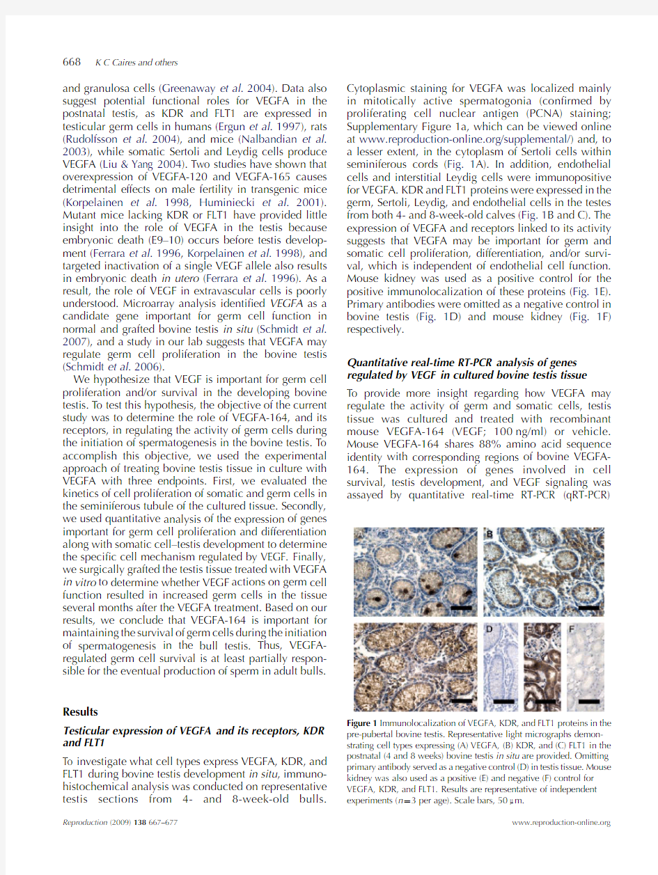
To investigate what cell types express VEGFA,KDR,and FLT1during bovine testis development in situ,immuno-histochemical analysis was conducted on representative testis sections from4-and8-week-old bulls.Cytoplasmic staining for VEGFA was localized mainly in mitotically active spermatogonia(con?rmed by proliferating cell nuclear antigen(PCNA)staining; Supplementary Figure1a,which can be viewed online at https://www.360docs.net/doc/26129204.html,/supplemental/)and,to a lesser extent,in the cytoplasm of Sertoli cells within seminiferous cords(Fig.1A).In addition,endothelial cells and interstitial Leydig cells were immunopositive for VEGFA.KDR and FLT1proteins were expressed in the germ,Sertoli,Leydig,and endothelial cells in the testes from both4-and8-week-old calves(Fig.1B and C).The expression of VEGFA and receptors linked to its activity suggests that VEGFA may be important for germ and somatic cell proliferation,differentiation,and/or survi-val,which is independent of endothelial cell function. Mouse kidney was used as a positive control for the positive immunolocalization of these proteins(Fig.1E). Primary antibodies were omitted as a negative control in bovine testis(Fig.1D)and mouse kidney(Fig.1F) respectively.
Quantitative real-time RT-PCR analysis of genes regulated by VEGF in cultured bovine testis tissue
To provide more insight regarding how VEGFA may regulate the activity of germ and somatic cells,testis tissue was cultured and treated with recombinant mouse VEGFA-164(VEGF;100ng/ml)or vehicle. Mouse VEGFA-164shares88%amino acid sequence identity with corresponding regions of bovine VEGFA-164.The expression of genes involved in cell survival,testis development,and VEGF signaling was assayed by quantitative real-time RT-PCR
(qRT-PCR)
Figure1Immunolocalization of VEGFA,KDR,and FLT1proteins in the pre-pubertal bovine testis.Representative light micrographs demon-strating cell types expressing(A)VEGFA,(B)KDR,and(C)FLT1in the postnatal(4and8weeks)bovine testis in situ are provided.Omitting primary antibody served as a negative control(D)in testis tissue.Mouse kidney was also used as a positive(E)and negative(F)control for VEGFA,KDR,and FLT1.Results are representative of independent experiments(n Z3per age).Scale bars,50m m.
668K C Caires and others
Reproduction(2009)138667–https://www.360docs.net/doc/26129204.html,
with gene-speci?c primer pairs (Table 1)at 0,3,6,and 24h during culture and treatment.Data are presented as fold change (mean normalized expression values)relative to the 0h (4-week-old donor)expression level.KDR and FLT1proteins are localized to germ and somatic cells in the bovine testis (Fig.1B and C);thus,we assayed whether VEGF treatment would affect the expression of these genes during explant culture.In comparison with baseline expression (0h),the expression of KDR and FLT1was signi?cantly induced at 3h,regardless of treatment.In addition,VEGF treatment signi?cantly increased (P !0.01)the expression of KDR and FLT1mRNA at 24h when compared with controls (Fig.2A and B).It is known that VEGF binding leads to downregulation of KDR and FLT1proteins at the cell surface as a means of receptor desensitization.However,it has been demonstrated that VEGF treatment upregulates the expression of KDR and FLT1mRNA and proteins in several cell types (Barleon
et al .1997,Herve
′et al .2006,Yao et al .2007),which is consistent with our observations in bovine testis tissue in vitro .
The BCL2family of proteins functions as regulators of cell survival and comprises both anti-apoptotic (BCL2and BCL2L1)and pro-apoptotic (BAX and BAD)members.Therefore,the ratio of mRNA expression between the pro-survival BCL2and pro-apoptotic BAX was measured in control-and VEGF-treated bovine testis tissue explants.In respect to the 0-h time point,VEGF treatment signi?cantly induced (P !0.001)the ratio of BCL2to BAX expression in bovine testis tissue at 3h (sixfold),6h (threefold),and 24h (threefold)when compared with controls (Fig.2C).VEGFA can protect extravascular cells (Lambrechts et al .2003,Jin et al .2006)from hypoxia-induced cell death;therefore,we investigated the mRNA expression of pro-apoptotic BCL2/adenovirus E1B 19kDa interacting protein (BNIP3),a hypoxia-responsive gene,following VEGF treatment.Regardless of treatment,BNIP3mRNA was signi?cantly higher at all time points during explant culture in comparison with the 0-h time point (Fig.2D).
However,BNIP3mRNA transcript levels were signi-?cantly suppressed at 3h (P !0.01),6h,and 24h (P !0.001)respectively after VEGF treatment compared with vehicle-treated testis tissue culture controls (Fig.2D).These data demonstrate that VEGFA stimulates an intracellular response that prevents germ cell death.BNIP3is a major downstream effector of hypoxia-inducible factor 1,a -subunit (HIF1A)protein (Althaus et al .2006);therefore,the expression of HIF1A was evaluated in bovine testis tissue during the explant culture period.Consistent with the qRT-PCR analysis for BNIP3expression,HIF1A transcript levels
were
Figure 2Expression of genes important for cell survival,testis
development,and VEGF signaling.Testis tissue explants from 4-week-old donors were cultured with VEGF (100ng/ml)or vehicle alone.Brie?y,samples obtained at 3,6,and 24h following treatment were assayed for gene expression by qRT-PCR for (A)KDR ,(B)FLT1,(C)ratio of BCL2:BAX ,(D)BNIP3,(E)HIF1A ,(F)PTGS2,(G)KIT ,and (H)GATA4.Results were normalized to RPS2expression,standardized to baseline expression at 0h,and are presented as the fold change of mean expression values G S .E .M .Asterisks indicate signi?cant differences between control and treatment means at a given time point.
***P !0.001;**P !0.01;and *P !0.05.Results are representative of independent experiments for each time point (n R 3),each performed on duplicate samples.
Table 1Bovine primer sequences used for quantitative real-time RT-PCR.Gene Forward
sequence (50–30)Reverse
sequence (50–30)Amplicon size BAX aacatggagctgcagaggat cagttgaagttgccgtcaga 104BCL2atgacttctctcggcgctac ctgaagagctcctccaccac 112BNIP3agaggtcgaaagcatcctga ggtgcttgaagagcaggaat 104FLT1accagatcatgttggactgc tacattggcttgaagcaggt 98GATA4gtgtagggccagtgttaccagat tttgagccgcggaagga 101HIF1A acctgcctctgaaactccaa tggggcatggtaaaagaaag 120KDR tcaatgtgtcactctgtgcaa actgaagcctttctggctgt 89KIT tggagtgcagggcttataacg gaacagggtgtgagcatgga 101PTGS2gaaatgatctacccgcctca tagccaaatggtggcataca 105RPS2
ggagcatccctgaaggatga
tccccgatagcaacaaacg
101
VEGFA-164regulates germ cell survival
669
https://www.360docs.net/doc/26129204.html,
Reproduction (2009)138667–677
signi?cantly reduced in VEGF-treated bovine testis tissue at3h(P!0.001)and6h(P!0.05)when compared with controls(Fig.2E).Prostaglandin-endoperoxide synthase 2(PTGS2)is critical for germ cell maturation and somatic–germ cell interactions in the mammalian gonad both in vivo and in vitro(Neal et al.1975, Takahashi et al.2006,Winnall et al.2007).Recent evidence suggests that this multifunctional enzyme is also responsible for protecting mouse embryonic stem cells and renal interstitial cells from oxidative stress and hypertonicity-induced apoptosis(Moeckel et al.2003, Liou et al.2007);thus,we evaluated the mRNA expression of PTGS2in response to VEGF treatment.In respect to the0-h time point,VEGF treatment signi-?cantly induced the expression of PTGS2in bovine testis tissue at3h,6h(P!0.001),and24h(P!0.01)when compared with controls(Fig.2F).Therefore,VEGF treatment promoted several independent means for cell survival in bovine testis tissue.
The mRNA expression of the testis germ cell-speci?c gene KIT and Sertoli and Leydig cells expressed gene GATA4were analyzed in explant-cultured bovine testis tissue treated with VEGF(100ng/ml)or vehicle. In comparison with the0-h time point,KIT expression was signi?cantly higher(P!0.05)in the VEGF treatment group at3and6h respectively,while control levels were not different from baseline levels at3,6,or24h. However,no differences in KIT were detected between control and treatment groups at3,6,and24h.GATA4 expression was twofold lower(P!0.05)in control-and VEGF-treated testis tissue at all time points when compared with the0-h baseline.Regardless of treatment, KIT and GATA4mRNA expression was not different between control-and VEGF-treated testis tissues at3,6, and24h during culture(Fig.2G and H).
Cell types affected by VEGF treatment in cultured bovine testis tissue
The speci?city of the VEGF treatment effect to stimulate cell survival pathways was investigated to determine the testicular cell type responding to the VEGF signal.We obtained testis tissue from4-week-old donors and treated the tissue with vehicle or100ng/ml VEGFA during explant culture.In addition,a negative control was added to investigate the speci?city of the VEGF signal during explant culture by supplementing media with neutralizing antibodies against human KDR (3mg/ml)and mouse VEGF(1.5mg/ml)at concen-trations necessary to yield one-half maximal inhibition (ND50)of VEGF activity from treatment(100ng/ml). Because KDR and FLT1receptors are present on germ and Sertoli cells(Fig.1),we investigated the population of these cells during(72h)and following(120h)the explant culture period by immunostaining for known markers of germ(DDX4/MVH)and Sertoli cells(GATA4). PCNA immunostaining indicated no differences in the rate of germ cell proliferation between control-and VEGF-treated bovine testis tissue grafts(data not shown). Germ cell numbers were30%greater(P!0.05)in bovine testis tissue treated with VEGF when compared with controls at120-h of culture(Fig.3A).Blocking VEGF activity using antisera against KDR and VEGF signi?cantly reduced(P!0.05)the number of germ cells in VEGF-treated testis tissue to control levels at120h. The effect of VEGF treatment on germ cell numbers was decreased by21%when neutralizing the activity of KDR.Directly mitigating the ligand activity in VEGF-treated testis tissue reduced germ cell numbers by52%. Immunostaining for DDX4to demonstrate the presence of germ cells in cultured tissues is shown in Fig.3B. In contrast,Sertoli cell number did not change during the explant culture period and was unaffected by
treatment Figure3Effect of VEGF treatment on survival of speci?c cell types in bovine testis tissue.(A)Analysis of germ and Sertoli cell populations in control-and VEGF-treated testis tissue following explant culture and blockade of VEGF activity.(B)Morphometric analysis of germ cell number was facilitated using DDX4/MVH immunostaining.An arrow indicates the presence of a DDX4-positive germ cell.Data are presented as the mean G S.E.M.Different letters indicate signi?cant differences between means(P!0.05).Scale bar,50m m.
670K C Caires and others
Reproduction(2009)138667–https://www.360docs.net/doc/26129204.html,
or blockage of VEGF biological activity(Fig.3A). Collectively,these data indicate that VEGF treatment promotes germ cell survival and results in increased germ cells following5days of explant culture. Testicular tissue explant culture followed by
xenografting bioassay
The testis tissue culture results demonstrated that VEGF supports germ cell survival in the bovine testis.The next step in the project was to determine whether increased germ cell survival following VEGF treatment in vitro resulted in long-term increased production of sperm in testis tissue.To complete this objective,treatment of testis tissue in vitro with VEGF was combined with testis tissue grafting to determine the extent of spermatid production that resulted from increased spermatogonia survival.Testicular tissue obtained from bovine donors was cultured and treated with VEGF at0(vehicle),100, or200ng/ml as tissue explants for5days then grafted onto nude mice.Bovine testis tissue grafts were removed from mice when testis tissue age equaled32weeks and categorized as functional based on evidence of germ cell differentiation in the seminiferous tubules of the testis graft.No differences existed between donor age(4weeks versus8weeks)and grafting period,so these datasets were pooled.Following explant culture,no gross morphological differences were observed due to treat-ment(Fig.4A–C).Immunostaining with von Willebrand factor(VWF)indicated no differences in blood vessel number or microvascular density(MVD)between control-and VEGF-treated testis tissue(Fig.4D). Treatment of testis tissue for5days in culture with VEGF or vehicle did not change the number of functional grafts recovered after grafting these tissues (Supplementary Figure1b).In addition,no differences were observed in graft weight or seminiferous tubule cross-section number in the recovered grafted tissue between the tissue groups treated in vitro with VEGF or vehicle(Fig.4E and F).The long-term ability of Leydig cells to produce testosterone was unaffected,as no difference in serum testosterone was detected by RIA in mice grafted with testis tissue treated with VEGF or vehicle during explant culture(Fig.4G).Similarly,the analysis of vesicular gland weights in recipient mice con?rmed that testosterone was biologically active (data not shown).
To determine whether treating bovine testis tissue with VEGF for5days in culture had a long-term effect on sperm production,we analyzed the germ cell popu-lations in functional testis grafts.Regardless of treatment, all testis xenografts contained spermatogonia and meiotic germ cells when removed from recipient mice. However,bovine testis tissue treated with VEGF in culture had signi?cantly more(P!0.005)differentiating germ cells after the grafting period compared with control tissue grafts(Fig.5A).VEGF treatment of cultured testis tissue for5days with100ng/ml resulted in36%of testis grafts supporting the completion of meiosis (determined by the presence of haploid spermatids) when compared with only4.8%of grafts in the control group.Similarly,40.9%of grafts in the200ng/ml VEGF treatment group contained spermatids.No dose effect (100vs200ng/ml)on germ cell differentiation was detected(P Z0.63).Thus,treatment of cultured testis tissue with VEGF at100and200ng/ml resulted in signi?cantly more spermatid production than controls.
A light micrograph demonstrating active spermato-genesis in a bovine testis tissue graft is shown in Fig.5B.Seminiferous tubule cross sections containing spermatogonia within grafts were classi?ed based on the existence of meiotic germ cells(primary and/or secondary spermatocytes)and haploid spermatids (round,elongating,and/or elongate spermatids).In addition to cell identi?cation based on morphological characteristics,immunostaining for synaptonemal complex protein3(SYCP3),a protein marker of meiosis (Parra et al.2004),con?rmed the presence
of
Figure4Effect of explant culture and VEGF
treatment on bovine testis tissue.Representative
light micrographs depicting morphology of
(A)donor testis tissue,explant-cultured testis
tissue treated with(B)vehicle or with(C)VEGF
prior to grafting.(D)Analysis of microvascular
density in control-and VEGF-treated testis tissue
following explant culture.Post-grafting analysis
of testis tissue growth including(E)graft weight
and(F)number of seminiferous tubule cross
sections per graft.(G)Serum testosterone from
recipient mice supporting control-and VEGF-
treated bovine testis grafts was evaluated by RIA.
Scale bars,50m m.
VEGFA-164regulates germ cell survival671 https://www.360docs.net/doc/26129204.html, Reproduction(2009)138667–677
spermatocytes in bovine testis tissue grafts (Supplemen-tary Figure 1c and d).When compared with controls,seminiferous tubules of bovine testis grafts cultured with VEGF contained signi?cantly more (P !0.05)meiotic germ cells and spermatids (Fig.5C and D).Thus,short-term treatment of bovine testis tissue with VEGF prior to grafting results in signi?cantly more spermatids present in the tissue 24and 28weeks later.
Seminiferous tubules of each testis graft were eval-uated for numbers of spermatogonia to determine the effect of VEGF treatment on long-term germ cell survival in cultured bovine testis tissues.Testis tissue cultured and treated with VEGF prior to grafting contained signi-?cantly more (P !0.001)spermatogonia than vehicle alone when removed from recipient mice (Fig.5E).VEGF treatment at 100and 200ng/ml resulted in more seminiferous tubules containing spermatogonia (78.8and 88.1%respectively)in testis grafts in comparison with controls (55.2%).The inverse relationship was observed for the proportion of seminiferous tubule cross sections containing only Sertoli cells (Fig.5F).Hence,germ cell loss was signi?cantly reduced (P !0.001)in
bovine testis tissue treated with VEGF in culture prior to ectopic grafting.These data provide in vivo evidence that VEGF support of spermatogonia survival leads to the production of more sperm.
Discussion
The purpose of this research was to investigate the role of VEGF in the regulation of germ cell survival and differentiation;two processes essential for sperm pro-duction.Work in our laboratory demonstrated that increased production of differentiating germ cells in bovine testis tissue ectopic grafts treated with a single dose of VEGF directly on the tissue at the time of grafting (Schmidt et al .2006).However,the speci?c target or effect of VEGF treatment was not determined.Owing to the complex nature of somatic and germ cells interaction in the testis required for germ cell differentiation,alteration of activity of several cell types may lead to changes in germ cell production.VEGFA,KDR,and FLT1protein expression was localized mainly to proliferating germ cells,Sertoli cells,and Leydig cells in the testis of 4-and 8-week-old bull calves.VEGF treatment signi-?cantly increased the population of germ cells in testis tissue cultures when compared with controls.No changes in the somatic cells were observed in these experiments leading us to conclude that VEGF acts to stimulate functional mechanisms in undifferentiated germ cells during postnatal bovine testis development to support the proliferation and/or survival of these cells.To test our hypothesis that VEGF promotes germ cell survival,we conducted qRT-PCR to evaluate expression of genes important for cellular survival,speci?cally the balance between anti-apoptotic and pro-apoptotic proteins that have been associated with VEGF action in other cell types.Therefore,we assayed the ratio of BCL2:BAX expression and found that VEGF treatment signi?cantly up-regulated the mRNA expression of anti-apoptotic BCL2relative to pro-apoptotic BAX when compared with controls at 3,6,and 24h of culture.Since VEGF has been implicated in protecting extra-vascular cells (Lambrechts et al .2003,Jin et al .2006)from hypoxia-induced cell death,we also investigated the mRNA expression of pro-apoptotic BNIP3,a hypoxia-responsive gene,during explant culture.VEGF treatment suppressed the expression of BNIP3mRNA at all time points during explant culture when compared with controls,and this correlates with HIF1A transcripts levels (Fig.2C).In contrast,VEGF treatment did not in?uence GATA4mRNA levels in the testis tissue explants culture,supporting our conclusion that VEGF does not regulate Sertoli and Leydig activity in the bovine testis.Similarly,VEGF treatment did not regulate the expression of KIT ,a gene associated with germ cell differentiation in the testis,when compared with controls.This indicates that VEGF regulates undiffer-entiated germ cells during explant
culture.
Figure 5Effect of VEGF treatment on the establishment of spermato-genesis in bovine testis tissue.(A)Number of grafts supporting complete germ cell differentiation in control-and VEGF-treated bovine testis tissue grafts.(B)Micrograph demonstrating active spermatogenesis in a bovine testis tissue graft.Seminiferous tubule cross sections were
evaluated for the presence of meiotic germ cells (C)and spermatids (D).Seminiferous tubule cross sections were evaluated for the presence of spermatogonia (E)or only Sertoli cells (F)in post-graft testis tissue.Data are presented as the mean G S .E .M .Different letters indicate signi?cant differences between means (P !0.05).Scale bar,50m m.
672
K C Caires and others
Reproduction (2009)138667–677
https://www.360docs.net/doc/26129204.html,
VEGF signaling is mediated through the trans-membrane receptor tyrosine kinases,KDR and FLT1. VEGF treatment signi?cantly induced the expression of its receptors KDR and FLT1at24h when compared with controls in bovine testis tissue cultures(Fig.2G and H). In normal and neoplastic cells,KDR activation rapidly induces several signaling cascades including phospha-tidylinositol3-kinase,phospholipase C-g,MAPK,and Src involved in promoting cellular proliferation,survival, and differentiation events(Takahashi&Shibuya2005, Yamazaki&Morita2006,Tammela et al.2007). Activation of FLT1elicits a poor mitogenic response(in contrast to KDR)and primarily functions to regulate chemotaxis,survival,and cell–cell interactions in a variety of cell types(Kaplan et al.2005,Takahashi& Shibuya2005,Fragoso et al.2006).It is also established that ligand binding to FLT1directly attenuates down-stream-signaling events mediated by KDR.The expression of VEGF,KDR,and FLT1in somatic and germ cells of the testis suggests this group of molecules may support spermatogenesis in several distinct ways. Indeed,signaling pathways stimulated by VEGF have been implicated in germ cell homeostasis(Dolci et al. 2001),and several studies have demonstrated the importance of VEGF for male fertility(Korpelainen et al.1998,Huminiecki et al.2001,Bott et al.2006). Gene expression of VEGF is regulated by a variety of stimuli such as hypoxia,growth factors,estrogen,and gonadotropins,and we demonstrate overlapping expression of VEGF and its receptors in germ and somatic cells in the pre-pubertal bovine testis,similar to the expression pattern found in the embryonic mouse testis(Bott et al.2006).Based on the data presented here, we hypothesize that proliferating undifferentiated germ cells may produce VEGF in response to local hypoxia and oxidative stress to prevent apoptosis in these cells preceding the onset of puberty.Accordingly,testicular germ cells of several species express KDR and FLT1 (Ergun et al.1997,Nalbandian et al.2003,Rudolfsson et al.2004),and although these receptors are expressed in endothelial cells,no active angiogenesis takes place within the postnatal testis.VEGF synthesis by Leydig cells is stimulated in response to hCG/LH(Rudolfsson et al.2004),and FSH triggers Sertoli cell production of VEGF in the adult testis(Marti&Risau1998,Liu&Y ang 2004,Reisinger et al.2007).Interestingly,FSH has been shown to stimulate proliferation of spermatogonia (De Rooij et al.1989),and type A spermatogonia express KDR in neonatal mice(Nalbandian et al.2003).Thus, autocrine and paracrine regulation of germ cell activity by VEGF in the mammalian testis during early develop-ment and adulthood may be important for the initiation and maintenance of sperm production.
Therefore,we sought to determine whether the changes induced by VEGF in vitro in germ cells lead to increased production of sperm in the tissue,on a long-term basis.To complete this objective,we grafted testis tissue explants after culture and treatment with VEGF or vehicle onto recipient nude mice and evaluated germ cell differentiation and sperm production in the grafts at recovery,6months later.While we have cultured and grafted bovine testis tissue successfully,no researchers have treated tissue in vitro prior to grafting to determine whether the treatment modi?es the germ cell population such that long-term sperm production is affected.Our testis tissue culture-grafting approach is a novel method to investigate factors important for germ cell biology because1)sperm production in testis tissue grafts is a direct means to determine whether a speci?c treatment manipulates the germ cell population and2)the specialized microenvironments(Honaramooz et al. 2002,McLean2005,Hess et al.2006,Y oshida et al. 2007)established between germ and Sertoli cells are maintained.
Treating bovine testis tissue explants with VEGF in culture prior to grafting did not affect growth,recovery rate,or testosterone synthesis by the grafted testis tissue when compared with controls.The rate of testis tissue graft growth in this study was consistent with previous explant culture experiments,and the number of seminiferous tubule cross sections within testis grafts treated with VEGF or vehicle alone did not differ (Schmidt et al.2006).Interestingly,a single VEGF treatment of bovine testis tissue at the time of grafting resulted in signi?cantly larger grafts32weeks later compared with controls,despite the fact that the number of seminiferous tubules was not different between groups (Schmidt et al.2006).Although the exact reason for the larger graft size has not been determined and an increase in blood vessel growth was not detected,it is possible that VEGF stimulated cell growth outside of the seminiferous tubules.Thus,our current approach aimed to minimize the effect of VEGF on neovascular-ization within the graft site of the recipient mouse. Regardless of treatment,all bovine testis tissue grafts contained spermatogonia and meiotic germ cells. However,culturing bovine testis explants with VEGF resulted in greater than sevenfold increase(P!0.05)in the extent of germ cell differentiation,as a greater number of testis grafts producing meiotic germ cells and spermatids.Morphometric evaluation of seminiferous tubule cross sections containing only Sertoli cells suggests that VEGF treatment signi?cantly increased (P!0.001)germ cell survival in testis tissue grafts. Abrogating the biological activity of VEGF during explant culture with neutralizing antibodies con?rmed these ?ndings in vitro.No differences in Sertoli cell number were observed in either study.Regardless of treatment, testis tissue grafts produced similar levels of testosterone, so we conclude Leydig cell function was not affected. Binding of VEGF to its cognate receptors initiates a variety of signaling cascades similar to those stimulated by GDNF and KIT in the postnatal testis,two factors of known importance for regulating germ cell proliferation,
VEGFA-164regulates germ cell survival673
https://www.360docs.net/doc/26129204.html, Reproduction(2009)138667–677
survival,and differentiation(Meng et al.2000,Ohta et al.2000,2003,Rossi et al.2000,Dolci et al.2001, Aponte et al.2006,Naughton et al.2006,Payne&Braun 2007).Rodent studies have shown that male fetal germ cells express VEGF protein until shortly after birth(Bott et al.2006),and Sertoli cells continue to produce VEGF throughout adult life(Marti&Risau1998,Nalbandian et al.2003).Interestingly,type A spermatogonia in close association with Sertoli cells also express KDR but do not produce VEGF in mice aged5days post partum and older.This developmental switch within the specialized microenvironment of the seminiferous tubule suggests that VEGF may be important in regulating both the onset and maintenance of spermatogenesis.Collectively,these data taken together with our recent observations in bovine testis tissue advocate the importance of VEGF for male fertility and support our hypothesis that VEGFA-164 maintains the survival of germ cells during the initiation of spermatogenesis in the developing bovine testis. Materials and Methods
Chemicals and reagents
All reagents were purchased from Sigma unless stated.
Donor bulls and tissue collection
The Washington State University Animal Care and Use Committee approved all animal procedures.Testis tissue samples were obtained from4-and8-week-old bull calves(ten bulls/age)at the Washington State University Beef Center using standard castration protocols.Testes were immediately processed for explant culture and histological examination,as previously described (Schmidt et al.2006).
Immunohistochemistry
Immunohistochemistry was performed,as previously described(Caires et al.2008),using the avidin–biotin complex method with af?nity-puri?ed primary antibodies(diluted in10%normal serum,pH7.4)as described in Table2.Immunoreactivity was detected following a5-min incubation in3,30-diaminobenzidine, and sections were counterstained with hematoxylin.As a negative control,serial sections were processed without primary antibody.
Testicular tissue explant culture
Testicular parenchyma(w3mg)obtained from4-and 8-week-old bovine donors were cultured as tissue explants on0.45-m m pore?oating?lter membranes (Millipore,Bedford,MA,USA)for5days prior to grafting.Eight tissue explants per donor(n Z13)were cultured at328C in DMEM containing10%fetal bovine serum,30mg/ml penicillin,and50mg/ml streptomycin, including recombinant mouse VEGFA-164(493-MV; R&D Systems,Minneapolis,MN,USA)at0(control), 100,or200ng/ml.Media were changed every2days including fresh addition of VEGF,and each experiment was conducted in duplicate.Following explant culture, tissues were processed for evaluation of angiogenesis using an established method(Vermeulen et al.1996). Brie?y,blood vessels were identi?ed by tubular morphology(single layer of endothelial cells and presence of intraluminal red blood cells)and positive immunostaining for VWF(1:400).MVD was then calculated by enumerating the mean number of blood vessels per40!?eld in ten randomly chosen areas within testis tissue from each treatment group(n Z5),as previously reported(Sinha et al.2004).
Quantitative real-time RT-PCR analysis
Bovine testis tissue(from4-week-old donors)explants treated with vehicle(0ng/ml)or VEGF(100ng/ml)were randomly selected at0,3,6,and24h during culture and homogenized in TRIzol(Invitrogen)according to the manufacturer’s protocol.Total RNA was puri?ed, dissolved in RNase-free water(Ambion,Foster City, CA,USA),and2m g total RNA was reverse transcribed into cDNA using an oligo(dT)12–18primer and M-MLV reverse transcriptase(Invitrogen).cDNA synthesis was con?rmed using primers and PCR for b-actin.Gene expression was evaluated in control-and VEGF-treated samples at each time point(n R3donors).Primers were
Table2Antibodies,suppliers,and dilutions used for immunohistochemical analysis.
Antigen Antibody description Source Dilution DDX4/VASA Rabbit anti-human DDX4/MVH polyclonal antibody Abcam,Cambridge,MA,USA1:500 GATA4Rabbit anti-mouse GATA4polyclonal antibody(C-20)SCBT,Santa Cruz,CA,USA1:200
PCNA Mouse monoclonal PCNA antibody(PC10)DAKO,Carpinteria,CA,USA1:200 SYCP3-C Rabbit anti-human SYCP3polyclonal antibody Novus,Littleton,CO,USA1:400 SYCP3-N Rabbit anti-mouse SYCP3polyclonal antibody Novus1:400 VEGF Rabbit anti-mouse VEGF polyclonal antibody(A-20)SCBT1:400 VEGFR1/FLT1Rabbit anti-mouse FLT1polyclonal antibody(C-17)SCBT1:200 VEGFR2/KDR Mouse anti-human FLK1monoclonal antibody(A-3)SCBT1:200 VWF Goat anti-human von Willebrand factor(C-20)SCBT1:400 ABCAM,Abcam,Inc.;DAKO,Dako Corporation;Novus,Novus Biologicals;SCBT,Santa Cruz Biotechnology.
674K C Caires and others
Reproduction(2009)138667–https://www.360docs.net/doc/26129204.html,
designed using the GenScript Real-Time PCR Primer Design tool(https://https://www.360docs.net/doc/26129204.html,/ssl-bin/app/primer) and are listed in Table1.Melt curve analyses were conducted to ensure speci?city of the PCR product for all target genes.The PCR reaction mixture contained 200ng cDNA,10pmol of each primer,12.5m l iQTM Supermix(Bio-Rad),SYBR Green I(Molecular Probes, Eugene,OR,USA)for PCR product detection,and water to a total volume of25m l.The PCR protocol was35 cycles of958C for30s,588C for30s,and728C for30s using an iCycler iQ Real-Time PCR Detection System (Bio-Rad),and samples were assayed in duplicate. Comparisons were made by normalizing the expression of the target gene to that of bovine ribosomal protein S2 (RPS2)using the Q-Gene method(Muller et al.2002). Data are presented as fold change(mean expression levels G S.E.M.)relative to the fresh tissue collected at0h from4-week-old donor calves.
Neutralization of VEGF bioactivity
All antisera were obtained from R&D Systems.Bovine testis tissue from4-week-old donors(n Z5)was cultured as explants(as described above)in the presence of VEGFA-164at0(control)and100ng/ml.Neutralizing antibodies including a goat anti-human KDR polyclonal antiserum(AF-357,3mg/ml)and a goat anti-mouse VEGF polyclonal antiserum(AF-493-NA, 1.5mg/ml) were added to media at concentrations to yield one-half maximal inhibition(ND50)of VEGFA-164activity due to treatment(100ng/ml).We evaluated germ cell and Sertoli cell populations in cultured explants by counting the mean number of each cell type found within ten randomly selected seminiferous tubule cross sections in testis tissue(n Z4;for each treatment group) at72and120h respectively.
Ectopic testis tissue xenografting bioassay
After5days in culture and in vitro treatment,testis tissues were ectopically grafted onto castrated immuno-de?cient NCr nude mice(Taconic,Hudson,NY,USA; CrTac:NCR-Fox1!nu O),as previously described (Oatley et al.2005).Recipient mice were killed when grafted bovine testis reached a tissue age of32weeks, serum was collected,and recovered tissues were processed for histological evaluation.The extent of germ cell survival and differentiation was based on the presence of spermatogonia,spermatocytes,and sperma-tids in testis grafts according to morphological charac-teristics,as previously described(Caires et al.2008). Statistical analysis
All datasets are presented as the mean G S.E.M.,and differences between treatments were considered signi-?cant at P!0.05.Parametric data were analyzed using ANOVA,and pairwise comparisons between groups were conducted using the Duncan’s multiple range test for signi?cance.Nonparametric datasets were evaluated using the Kruskal–Wallis test,and comparisons between groups were conducted using Mann–Whitney tests. Declaration of interest
There is no con?ict of interest prejudicing the impartiality of this research.
Funding
This work was supported in part by a graduate research fellowship provided by the Seattle Chapter of the Achievement Rewards for College Scientists(ARCS)Foundation on behalf of K C Caires.
Acknowledgements
The authors would like to thank Liang-Y u Chen and Nada Cummings for assisting with tissue collection and the grafting procedure.The mouse and human SYCP3antibodies were generous gifts from Dr Terry J Hassold(Washington State University,Pullman,WA,USA).
References
Althaus J,Bernaudin M,Petit E,Toutain J,Touzani O&Rami A2006 Expression of the gene encoding the pro-apoptotic BNIP3protein and stimulation of hypoxia-inducible factor-1alpha(HIF-1alpha)protein following focal cerebral ischemia in rats.Neurochemistry International 48687–695.
Aponte PM,Soda T,van de Kant HJ&de Rooij DG2006Basic features of bovine spermatogonial culture and effects of glial cell line-derived neurotrophic factor.Theriogenology651828–1847.
Barleon B,Siemeister G,Martiny-Baron G,Weindel K,Herzog C& Marme′D1997Vascular endothelial growth factor up-regulates its receptor fms-like tyrosine kinase1(FLT-1)and a soluble variant of FLT-1 in human vascular endothelial cells.Cancer Research575421–5425. Bott RC,McFee RM,Clopton DT,Toombs C&Cupp AS2006Vascular endothelial growth factor and kinase domain region receptor are involved in both seminiferous cord formation and vascular development during testis morphogenesis in the rat.Biology of Reproduction75 56–67.
Bryan BA,Walshe TE,Mitchell DC,Havumaki JS,Saint-Geniez M, Maharaj AS,Maldonado AE&D’Amore PA2008Coordinated vascular endothelial growth factor expression and signaling during skeletal myogenic differentiation.Molecular Biology of the Cell19994–1006. Caires KC,Schmidt JA,Oliver AP,de Avila J&McLean DJ2008Endocrine regulation of the establishment of spermatogenesis in pigs.Reproduction in Domestic Animals43280–287.
Carmeliet P,Ferreira V,Breier G,Pollefeyt S,Kieckens L,Gertsenstein M, Fahrig M,Vandenhoeck A,Harpal K,Eberhardt C et al.1996Abnormal blood vessel development and lethality in embryos lacking a single VEGF allele.Nature380435–439.
Dolci S,Pellegrini M,Di Agostino S,Geremia R&Rossi P2001Signaling through extracellular signal-regulated kinase is required for spermato-gonial proliferative response to stem cell factor.Journal of Biological Chemistry27640225–40233.
Ergun S,Kilie N,Fiedler W&Mukhopadhyay AK1997Vascular endothelial growth factor and its receptors in normal human testicular tissue.
Molecular and Cellular Endocrinology1319–20.
VEGFA-164regulates germ cell survival675
https://www.360docs.net/doc/26129204.html, Reproduction(2009)138667–677
Ferrara N,Carver-Moore K,Chen H,Dowd M,Lu L,O’Shea KS,Powell-Braxton L,Hillan KJ&Moore MW1996Heterozygous embryonic lethality induced by targeted inactivation of the VEGF gene.Nature380 439–442.
Fragoso R,Pereira T,Wu Y,Zhu Z,Cabecadas J&Dias S2006VEGFR-1 (FLT-1)activation modulates acute lymphoblastic leukemia localization and survival within the bone marrow,determining the onset of extramedullary disease.Blood1071608–1616.
Germani A,Di Carlo A,Mangoni A,Straino S,Giacinti C,Turrini P, Biglioli P&Capogrossi MC2003Vascular endothelial growth factor modulates skeletal myoblast function.American Journal of Pathology 1631417–1428.
Greenaway J,Connor K,Pedersen HG,Coomber BL,LaMarre J&Petrik J 2004Vascular endothelial growth factor and its receptor,Flk-1/KDR,are cytoprotective in the extravascular compartment of the ovarian follicle.
Endocrinology1452896–2905.
Herve′MA,Meduri G,Petit FG,Domet TS,Lazennec G,Mourah S&Perrot-Applanat M2006Regulation of the vascular endothelial growth factor (VEGF)receptor Flk-1/KDR by estradiol through VEGF in uterus.
Journal of Endocrinology18891–99.
Hess RA,Cooke PS,Hofmann MC&Murphy KM2006Mechanistic insights into the regulation of the spermatogonial stem cell niche.Cell Cycle5 1164–1170.
Hicklin DJ&Ellis LM2005Role of the vascular endothelial growth factor pathway in tumor growth and angiogenesis.Journal of Clinical Oncology 231011–1027.
Honaramooz A,Snedaker A,Boiani M,Scholer HR,Dobrinski I&Schlatt S 2002Sperm from neonatal mammalian testes grafted in mice.Nature 418778–781.
Huminiecki L,Chan HY,Lui S,Poulsom R,Stamp G,Harris AL&Bicknell R 2001Vascular endothelial growth factor transgenic mice exhibit reduced male fertility and placental rejection.Molecular Human Reproduction7 255–264.
Jin K,Mao XO&Greenberg DA2006Vascular endothelial growth factor stimulates neurite outgrowth from cerebral cortical neurons via Rho kinase signaling.Journal of Neurobiology66236–242.
Kaplan RN,Riba RD,Zacharoulis S,Bramley AH,Vincent L,Costa C, MacDonald DD,Jin DK,Shido K,Kerns SA et al.2005VEGFR1-positive haematopoietic bone marrow progenitors initiate the pre-metastatic niche.Nature438820–827.
Korpelainen EI,Karkkainen MJ,Tehunen A,Lakso M,Rauvala H, Vierula M,Parvinen M&Alitalo K1998Overexpression of VEGF in the testis and epididymis causes infertility in transgenic mice:evidence for nonendothelial targets for VEGF.Journal of Cell Biology143 1705–1712.
Lambrechts D,Storkebaum E,Morimoto M,Del-Favero J,Desmet F, Marklund SL,Wyns S,Thijs V,Andersson J,van Marion I et al.2003 VEGF is a modi?er of amyotrophic lateral sclerosis in mice and humans and protects motorneurons against ischemic death.Nature Genetics34 383–394.
LeCouter J,Moritz DR,Li B,Phillips GL,Liang XH,Gerber HP,Hillan KJ& Ferrara N2003Angiogenesis-independent endothelial protection of liver:role of VEGFR-1.Science299890–893.
Liou JY,Ellent DP,Lee S,Goldsby J,Ko BS,Matijevic N,Huang JC&Wu KK 2007Cyclooxygenase-2-derived prostaglandin E2protects mouse embryonic stem cells from apoptosis.Stem Cells251096–1103.
Liu X&Yang Y2004Effect of VEGF on the angiogenesis in male reproduction system.Zhonghua Nan Ke Xue1049–51.
Mac Gabhann F&Popel AS2005Differential binding of VEGF isoforms to VEGF receptor2in the presence of neuropilin-1:a computational model.
American Journal of Physiology.Heart and Circulatory Physiology288 2851–2860.
Marti HH&Risau W1998Systemic hypoxia changes the organ-speci?c distribution of vascular endothelial growth factors and its receptors.
PNAS9515809–15814.
Matsumoto Y,Tanaka K,Hirata G,Hanada M,Matsuda S,Shuto T& Iwamoto Y2002Possible involvement of the vascular endothelial growth factor-FLT1-focal adhesion kinase pathway in chemotaxis and the cell proliferation of osteoclast precursor cells in arthritic joints.Journal of Immunology1685824–5831.
McLean DJ2005Spermatogonial stem cell transplantation and testicular function.Cell and Tissue Research32221–31.Meng X,Lindahl M,Hyvonen ME,Parvinen M,de Rooij DG,Hess MW, Raatikainen-Ahokas A,Sainio K,Rauvala H,Lakso M et al.2000 Regulation of cell fate decision of undifferentiated spermatogonia by GDNF.Science2871489–1493.
Moeckel GW,Zhang L,Fogo AB,Hao CM,Pozzi A&Breyer MD2003 COX2activity promotes organic osmolyte accumulation and adaptation of renal medullary interstitial cells to hypertonic stress.Journal of Biological Chemistry27819352–19357.
Muller PY,Janovjak H,Miserez AR&Dobbie Z2002Processing of gene expression data generated by quantitative real-time RT-PCR.
Biotechniques321372–1378.
Nalbandian A,Dettin L,Dym M&Ravindranath N2003Expression of vascular endothelial growth factor receptors during male germ cell differentiation in the mouse.Biology of Reproduction69985–994. Naughton CK,Jain S,Strickland AM,Gupta A&Milbrandt J2006Glial cell-line derived neurotrophic factor-mediated RET signaling regulates spermatogonial stem cell fate.Biology of Reproduction74314–321. Neal P,Baker TG,McNatty KP&Scaramuzzi RJ1975In?uence of prostaglandins and human chorionic gonadotrophin on progesterone concentration and oocyte maturation in mouse ovarian follicles maintained in organ culture.Journal of Endocrinology6519–25. Nishijima K,Ng YS,Zhong L,Bradley J,Schubert W,Jo N,Akita J, Samuelsson SJ,Robinson GS&Adamis AP2007Vascular endothelial growth factor-A is a survival factor for retinal neurons and a critical neuroprotectant during the adaptive response to ischemic injury.
American Journal of Pathology17153–67.
Oakberg EF1971Spermatogonial stem-cell renewal in the mouse.
Anatomical Record169515–531.
Oatley JM,Reeves JJ&McLean DJ2005Establishment of spermatogenesis in neonatal bovine testicular tissue following ectopic xenografting varies with donor age.Biology of Reproduction72358–364.
Ohta H,Yomogida K,Dohmae K&Nishimune Y2000Regulation of proliferation and differentiation in spermatogonial stem cells:the role of c-kit and its ligand SCF.Development1272125–2131.
Ohta H,Tohda A&Nishimune Y2003Proliferation and differentiation of spermatogonial stem cells in the W/W v mutant mouse testis.Biology of Reproduction691815–1821.
Parra MT,Viera A,Go′mez R,Page J,Benavente R,Santos JL,Rufas JS& Suja JA2004Involvement of the cohesin Rad21and SCP3in monopolar attachment of sister kinetochores during mouse meiosis I.Journal of Cell Science1171221–1234.
Parvinen M&Ventela S1999Local regulation of spermatogenesis:a living cell approach.Human Fertility2138–142.
Payne C&Braun RE2007Glial cell line-derived neurotrophic factor maintains a POZ-itive in?uence on stem cells.PNAS1039751–9752. Reisinger K,Baal N,McKinnon T,Munstedt K&Zygmunt M2007The gonadotropins:tissue-speci?c angiogenic factors?Molecular and Cellular Endocrinology26965–80.
De Rooij DG,Van Dissel-Emiliani FM&Van Pelt AM1989Regulation of spermatogonial proliferation.Annals of the New Y ork Academy of Sciences564140–153.
Rossi P,Sette C,Dolci S&Geremia R2000Role of c-kit in mammalian spermatogenesis.Journal of Endocrinological Investigation23609–615. Rudolfsson SH,Wilkstrom P,Jonsson A,Collin O&Bergh A2004 Hormonal regulation and functional role of vascular endothelial growth factor A in the rat testis.Biology of Reproduction70340–347. Russell LD&Griswold MD1993The Sertoli Cell,edn1,pp577–595.
Clearwater,FL:Cache River Press.
Schmidt JA,de Avila JM&McLean DJ2006Effect of vascular endothelial growth factor and testis tissue culture on spermatogenesis in bovine ectopic testis tissue xenografts.Biology of Reproduction75167–175. Schmidt JA,de Avila JM&McLean DJ2007Analysis of gene expression in bovine testis tissue prior to ectopic testis tissue xenografting and during the grafting period.Biology of Reproduction761071–1080.
Shimizu T2006Promotion of ovarian follicular development by injecting vascular endothelial growth factor(VEGF)and growth differentiation factor9(GDF-9)genes.Journal of Reproduction and Development52 23–32.
Sinha AA,Quast BJ,Reddy PK,Lall V,Wilson MJ,Qian J&Bostwick DG 2004Microvessel density as a molecular marker for identifying high-grade prostatic intraepithelial neoplasia precursors to prostate cancer.
Experimental and Molecular Pathology77153–159.
676K C Caires and others
Reproduction(2009)138667–https://www.360docs.net/doc/26129204.html,
Takahashi H&Shibuya M2005The vascular endothelial growth factor (VEGF)/VEGF receptor system and its role under physiological and pathological conditions.Clinical Science109227–241.
Takahashi T,Morrow JD,Wang H&Dey SK2006Cyclooxygenase-2-derived prostaglandin E(2)directs oocyte maturation by differentially in?uencing multiple signaling pathways.Journal of Biological Chemistry 28137117–37129.
Tammela T,Enholm B,Alitalo K&Paavonen K2007The biology of vascular endothelial growth factors.Cardiovascular Research65 550–563.
Vermeulen PB,Gasparini G,Fox SB,Toi M,Martin L,McCulloch P, Pezzella F,Viale G,Weidner N,Harris AL et al.1996Quanti?cation of angiogenesis in solid human tumours:an international consensus on the methodology and criteria of evaluation.European Journal of Cancer32A 2474–2484.
Wada T,Haigh JJ,Ema M,Hitoshi S,Chaddah R,Rossant J,Nagy A&van der Kooy D2006Vascular endothelial growth factor directly inhibits primitive neural stem cell survival but promotes de?nitive neural stem cell survival.Journal of Neuroscience266803–6812.
Winnall WR,Ali U,O’Bryan MK,Hirst JJ,Whiley PA,Muir JA&Hedger MP 2007Constitutive expression of prostaglandin-endoperoxide synthase2
by somatic and spermatogenic cells is responsible for prostaglandin E2production in the adult rat testis.Biology of Reproduction76 759–768.
Yamazaki Y&Morita T2006Molecular and functional diversity of vascular endothelial growth factors.Molecular Diversity10515–527.
Yao JS,Zhai W,Fan Y,Lawton MT,Barbaro NM,Young WL&Yang GY 2007Interleukin-6upregulates expression of KDR and stimulates proliferation of human cerebrovascular smooth muscle cells.Journal of Cerebral Blood Flow and Metabolism27510–520.
Yoshida S,Sukeno M&Nabeshima Y2007A vasculature-associated niche for undifferentiated spermatogonia in the mouse testis.Science317 1722–1726.
Zirkin BR1998Spermatogenesis:its regulation by testosterone and FSH.
Seminars in Cell and Developmental Biology9417–421. Received23January2009
First decision30March2009
Revised manuscript received9July2009
Accepted24July2009
VEGFA-164regulates germ cell survival677
https://www.360docs.net/doc/26129204.html, Reproduction(2009)138667–677
交互式多模型算法仿真与分析
硕037班 刘文3110038020 2011/4/20交互式多模型仿真与分析IMM算法与GBP算法的比较,算法实现和运动目标跟踪仿真,IMM算法的特性分析 多源信息融合实验报告
交互式多模型仿真与分析 一、 算法综述 由于混合系统的结构是未知的或者随机突变的,在估计系统状态参数的同时还需要对系统的运动模式进行辨识,所以不可能通过建立起一个固定的模型对系统状态进行效果较好的估计。针对这一问题,多模型的估计方法提出通过一个模型集{}(),1,2,,j M m j r == 中不同模型的切换来匹配不同目标的运动或者同一目标不同阶段的运动,达到运动模式的实时辨识的效果。 目前主要的多模型方法包括一阶广义贝叶斯方法(BGP1),二阶广义贝叶斯方法(GPB2)以及交互式多模型方法等(IMM )。这些多模型方法的共同点是基于马尔科夫链对各自的模型集进行切换或者融合,他们的主要设计流程如下图: M={m1,m2,...mk} K 时刻输入 值的形式 图一 多模型设计方法 其中,滤波器的重初始化方式是区分不同多模型算法的主要标准。由于多模型方法都是基于一个马尔科夫链来切换与模型的,对于元素为r 的模型集{}(),1,2,,j M m j r == ,从0时刻到k 时刻,其可能的模型切换轨迹为 120,12{,,}k i i i k trace k M m m m = ,其中k i k m 表示K-1到K 时刻,模型切换到第k i 个, k i 可取1,2,,r ,即0,k trace M 总共有k r 种可能。再令1 2 1 ,,,,k k i i i i μ+ 为K+1时刻经由轨迹0,k trace M 输入到第1k i +个模型滤波器的加权系数,则输入可以表示为 0,11 2 1 12|,,,,|,,,???k k trace k k k i M k k i i i i k k i i i x x μ++=?∑ 可见轨迹0,k trace M 的复杂度直接影响到算法计算量和存储量。虽然全轨迹的
五种大数据压缩算法
?哈弗曼编码 A method for the construction of minimum-re-dundancy codes, 耿国华1数据结构1北京:高等教育出版社,2005:182—190 严蔚敏,吴伟民.数据结构(C语言版)[M].北京:清华大学出版社,1997. 冯桂,林其伟,陈东华.信息论与编码技术[M].北京:清华大学出版社,2007. 刘大有,唐海鹰,孙舒杨,等.数据结构[M].北京:高等教育出版社,2001 ?压缩实现 速度要求 为了让它(huffman.cpp)快速运行,同时不使用任何动态库,比如STL或者MFC。它压缩1M数据少于100ms(P3处理器,主频1G)。 压缩过程 压缩代码非常简单,首先用ASCII值初始化511个哈夫曼节点: CHuffmanNode nodes[511]; for(int nCount = 0; nCount < 256; nCount++) nodes[nCount].byAscii = nCount; 其次,计算在输入缓冲区数据中,每个ASCII码出现的频率: for(nCount = 0; nCount < nSrcLen; nCount++) nodes[pSrc[nCount]].nFrequency++; 然后,根据频率进行排序: qsort(nodes, 256, sizeof(CHuffmanNode), frequencyCompare); 哈夫曼树,获取每个ASCII码对应的位序列: int nNodeCount = GetHuffmanTree(nodes); 构造哈夫曼树 构造哈夫曼树非常简单,将所有的节点放到一个队列中,用一个节点替换两个频率最低的节点,新节点的频率就是这两个节点的频率之和。这样,新节点就是两个被替换节点的父
LZ77压缩算法实验报告
LZ77压缩算法实验报告 一、实验内容 使用C++编程实现LZ77压缩算法的实现。 二、实验目的 用LZ77实现文件的压缩。 三、实验环境 1、软件环境:Visual C++ 6.0 2、编程语言:C++ 四、实验原理 LZ77 算法在某种意义上又可以称为“滑动窗口压缩”,这是由于该算法将一个虚拟的,可以跟随压缩进程滑动的窗口作为术语字典,要压缩的字符串如果在该窗口中出现,则输出其出现位置和长度。使用固定大小窗口进行术语匹配,而不是在所有已经编码的信息中匹配,是因为匹配算法的时间消耗往往很多,必须限制字典的大小才能保证算法的效率;随着压缩的进程滑动字典窗口,使其中总包含最近编码过的信息,是因为对大多数信息而言,要编码的字符串往往在最近的上下文中更容易找到匹配串。 五、LZ77算法的基本流程 1、从当前压缩位置开始,考察未编码的数据,并试图在滑动窗口中找出最长的匹 配字符串,如果找到,则进行步骤2,否则进行步骤3。 2、输出三元符号组( off, len, c )。其中off 为窗口中匹
配字符串相对窗口边 界的偏移,len 为可匹配的长度,c 为下一个字符。然后将窗口向后滑动len + 1 个字符,继续步骤1。 3、输出三元符号组( 0, 0, c )。其中c 为下一个字符。然后将窗口向后滑动 len + 1 个字符,继续步骤1。 六、源程序 /********************************************************************* * * Project description: * Lz77 compression/decompression algorithm. * *********************************************************************/ #include 实验名称:LZSS压缩算法实验报告 一、实验内容 使用Visual 6..0 C++编程实现LZ77压缩算法。 二、实验目的 用LZSS实现文件的压缩。 三、实验原理 LZSS压缩算法是词典编码无损压缩技术的一种。LZSS压缩算法的字典模型使用了自适应的方式,也就是说,将已经编码过的信息作为字典, 四、实验环境 1、软件环境:Visual C++ 6.0 2、编程语言:C++ 五、实验代码 #include -- need byte-swap function for big endian CPU */ #define READ_LE32(X) *(uint32_t *)(X) #define WRITE_LE32(X,Y) *(uint32_t *)(X) = (Y) /* this assumes sizeof(long)==4 */ typedef unsigned long uint32_t; /* text (input) size counter, code (output) size counter, and counter for reporting progress every 1K bytes */ static unsigned long g_text_size, g_code_size, g_print_count; /* ring buffer of size N, with extra F-1 bytes to facilitate string comparison */ static unsigned char g_ring_buffer[N + F - 1]; /* position and length of longest match; set by insert_node() */ static unsigned g_match_pos, g_match_len; /* left & right children & parent -- these constitute binary search tree */ static unsigned g_left_child[N + 1], g_right_child[N + 257], g_parent[N + 1]; /* input & output files */ static FILE *g_infile, *g_outfile; /***************************************************************************** initialize trees *****************************************************************************/ static void init_tree(void) { unsigned i; /* For i = 0 to N - 1, g_right_child[i] and g_left_child[i] will be the right and left children of node i. These nodes need not be initialized. Also, g_parent[i] is the parent of node i. These are initialized to NIL (= N), which stands for 'not used.' For i = 0 to 255, g_right_child[N + i + 1] is the root of the tree for strings that begin with character i. These are initialized to NIL. Note there are 256 trees. */ for(i = N + 1; i <= N + 256; i++) g_right_child[i] = NIL; for(i = 0; i < N; i++) g_parent[i] = NIL; } /***************************************************************************** Inserts string of length F, g_ring_buffer[r..r+F-1], into one of the trees (g_ring_buffer[r]'th tree) and returns the longest-match position and length via the global variables g_match_pos and g_match_len. If g_match_len = F, then removes the old node in favor of the new one, because the old one will be deleted sooner. 多媒体数据压缩实验报告 篇一:多媒体实验报告_文件压缩 课程设计报告 实验题目:文件压缩程序 姓名:指导教师:学院:计算机学院专业:计算机科学与技术学号: 提交报告时间:20年月日 四川大学 一,需求分析: 有两种形式的重复存在于计算机数据中,文件压缩程序就是对这两种重复进行了压 缩。 一种是短语形式的重复,即三个字节以上的重复,对于这种重复,压缩程序用两个数字:1.重复位置距当前压缩位置的距离;2.重复的长度,来表示这个重复,假设这两个数字各占一个字节,于是数据便得到了压缩。 第二种重复为单字节的重复,一个字节只有256种可能的取值,所以这种重复是必然的。给 256 种字节取值重新编码,使出现较多的字节使用较短的编码,出现较少的字节使用较长的编码,这样一来,变短的字节相对于变长的字节更多,文件的总长度就会减少,并且,字节使用比例越不均 匀,压缩比例就越大。 编码式压缩必须在短语式压缩之后进行,因为编码式压缩后,原先八位二进制值的字节就被破坏了,这样文件中短语式重复的倾向也会被破坏(除非先进行解码)。另外,短语式压缩后的结果:那些剩下的未被匹配的单、双字节和得到匹配的距离、长度值仍然具有取值分布不均匀性,因此,两种压缩方式的顺序不能变。 本程序设计只做了编码式压缩,采用Huffman编码进行压缩和解压缩。Huffman编码是一种可变长编码方式,是二叉树的一种特殊转化形式。编码的原理是:将使用次数多的代码转换成长度较短的代码,而使用次数少的可以使用较长的编码,并且保持编码的唯一可解性。根据 ascii 码文件中各 ascii 字符出现的频率情况创建 Huffman 树,再将各字符对应的哈夫曼编码写入文件中。同时,亦可根据对应的哈夫曼树,将哈夫曼编码文件解压成字符文件. 一、概要设计: 压缩过程的实现: 压缩过程的流程是清晰而简单的: 1. 创建 Huffman 树 2. 打开需压缩文件 3. 将需压缩文件中的每个 ascii 码对应的 huffman 编码按 bit 单位输出生成压缩文件压缩结束。 价值工程 置,是一项十分有意义的工作。另外恶意代码的检测和分析是一个长期的过程,应对其新的特征和发展趋势作进一步研究,建立完善的分析库。 参考文献: [1]CNCERT/CC.https://www.360docs.net/doc/26129204.html,/publish/main/46/index.html. [2]LO R,LEVITTK,OL SSONN R.MFC:a malicious code filter [J].Computer and Security,1995,14(6):541-566. [3]KA SP ER SKY L.The evolution of technologies used to detect malicious code [M].Moscow:Kaspersky Lap,2007. [4]LC Briand,J Feng,Y Labiche.Experimenting with Genetic Algorithms and Coupling Measures to devise optimal integration test orders.Software Engineering with Computational Intelligence,Kluwer,2003. [5]Steven A.Hofmeyr,Stephanie Forrest,Anil Somayaji.Intrusion Detection using Sequences of System calls.Journal of Computer Security Vol,Jun.1998. [6]李华,刘智,覃征,张小松.基于行为分析和特征码的恶意代码检测技术[J].计算机应用研究,2011,28(3):1127-1129. [7]刘威,刘鑫,杜振华.2010年我国恶意代码新特点的研究.第26次全国计算机安全学术交流会论文集,2011,(09). [8]IDIKA N,MATHUR A P.A Survey of Malware Detection Techniques [R].Tehnical Report,Department of Computer Science,Purdue University,2007. 0引言 现有的压缩算法有很多种,但是都存在一定的局限性,比如:LZw [1]。主要是针对数据量较大的图像之类的进行压缩,不适合对简单报文的压缩。比如说,传输中有长度限制的数据,而实际传输的数据大于限制传输的数据长度,总体数据长度在100字节左右,此时使用一些流行算法反而达不到压缩的目的,甚至增大数据的长度。本文假设该批数据为纯数字数据,实现压缩并解压缩算法。 1数据压缩概念 数据压缩是指在不丢失信息的前提下,缩减数据量以减少存储空间,提高其传输、存储和处理效率的一种技术方法。或按照一定的算法对数据进行重新组织,减少数据的冗余和存储的空间。常用的压缩方式[2,3]有统计编码、预测编码、变换编码和混合编码等。统计编码包含哈夫曼编码、算术编码、游程编码、字典编码等。 2常见几种压缩算法的比较2.1霍夫曼编码压缩[4]:也是一种常用的压缩方法。其基本原理是频繁使用的数据用较短的代码代替,很少使用 的数据用较长的代码代替,每个数据的代码各不相同。这些代码都是二进制码,且码的长度是可变的。 2.2LZW 压缩方法[5,6]:LZW 压缩技术比其它大多数压缩技术都复杂,压缩效率也较高。其基本原理是把每一个第一次出现的字符串用一个数值来编码,在还原程序中再将这个数值还成原来的字符串,如用数值0x100代替字符串ccddeee"这样每当出现该字符串时,都用0x100代替,起到了压缩的作用。 3简单报文数据压缩算法及实现 3.1算法的基本思想数字0-9在内存中占用的位最 大为4bit , 而一个字节有8个bit ,显然一个字节至少可以保存两个数字,而一个字符型的数字在内存中是占用一个字节的,那么就可以实现2:1的压缩,压缩算法有几种,比如,一个自己的高四位保存一个数字,低四位保存另外一个数字,或者,一组数字字符可以转换为一个n 字节的数值。N 为C 语言某种数值类型的所占的字节长度,本文讨论后一种算法的实现。 3.2算法步骤 ①确定一种C 语言的数值类型。 —————————————————————— —作者简介:安建梅(1981-),女,山西忻州人,助理实验室,研究方 向为软件开发与软交换技术;季松华(1978-),男,江苏 南通人,高级软件工程师,研究方向为软件开发。 数据快速压缩算法的研究以及C 语言实现 The Study of Data Compression and Encryption Algorithm and Realization with C Language 安建梅①AN Jian-mei ;季松华②JI Song-hua (①重庆文理学院软件工程学院,永川402160;②中信网络科技股份有限公司,重庆400000)(①The Software Engineering Institute of Chongqing University of Arts and Sciences ,Chongqing 402160,China ; ②CITIC Application Service Provider Co.,Ltd.,Chongqing 400000,China ) 摘要:压缩算法有很多种,但是对需要压缩到一定长度的简单的报文进行处理时,现有的算法不仅达不到目的,并且变得复杂, 本文针对目前一些企业的需要,实现了对简单报文的压缩加密,此算法不仅可以快速对几十上百位的数据进行压缩,而且通过不断 的优化,解决了由于各种情况引发的解密错误,在解密的过程中不会出现任何差错。 Abstract:Although,there are many kinds of compression algorithm,the need for encryption and compression of a length of a simple message processing,the existing algorithm is not only counterproductive,but also complicated.To some enterprises need,this paper realizes the simple message of compression and encryption.This algorithm can not only fast for tens of hundreds of data compression,but also,solve the various conditions triggered by decryption errors through continuous optimization;therefore,the decryption process does not appear in any error. 关键词:压缩;解压缩;数字字符;简单报文Key words:compression ;decompression ;encryption ;message 中图分类号:TP39文献标识码:A 文章编号:1006-4311(2012)35-0192-02 ·192· 压缩编码算法设计与实现实验报告 一、实验目的:用C语言或C++编写一个实现Huffman编码的程序,理解对数据进行无损压缩的原理。 二、实验内容:设计一个有n个叶节点的huffman树,从终端读入n个叶节 点的权值。设计出huffman编码的压缩算法,完成对n个节点的编码,并写出程序予以实现。 三、实验步骤及原理: 1、原理:Huffman算法的描述 (1)初始化,根据符号权重的大小按由大到小顺序对符号进行排序。 (2)把权重最小的两个符号组成一个节点, (3)重复步骤2,得到节点P2,P3,P4……,形成一棵树。 (4)从根节点开始顺着树枝到每个叶子分别写出每个符号的代码 2、实验步骤: 根据算法原理,设计程序流程,完成代码设计。 首先,用户先输入一个数n,以实现n个叶节点的Huffman 树; 之后,输入n个权值w[1]~w[n],注意是unsigned int型数值; 然后,建立Huffman 树; 最后,完成Huffman编码。 四、实验代码:#include int min(HuffmanTree t,int i) { int j,flag; unsigned int k = MAX; for(j=1;j<=i;j++) if(t[j].parent==0&&t[j].weight 交互式多模型算法卡尔曼滤波仿真 1 模型建立 分别以加速度u=0、1、2代表三个不同的运动模型 状态方程x(k+1)=a*x(k)+b*w(k)+d*u 观察方程z(k)=c*x(k)+v(k) 其中,a=[1 dt;0 1],b=[dt^2/2;dt],d=[dt^2/2;dt],c=[1 0] 2 程序代码 由两个功能函数组成,imm_main用来实现imm 算法,move_model1用来生成仿真数据,初始化运动参数 function imm_main %交互式多模型算法主程序 %初始化有关参数 move_model %调用运动模型初始化及仿真运动状态生成函数 load movedata %调入有关参数初始值(a b d c u position velocity pmeas dt tg q r x_hat p_var) p_tran=[0.8 0.1 0.1;0.2 0.7 0.1;0.1 0.2 0.7];%转移概率 p_pri=[0.1;0.6;0.3];%模型先验概率 n=1:2:5; %提取各模型方差矩阵 k=0; %记录仿真步数 like=[0;0;0];%视然函数 x_hat_hat=zeros(2,3);%三模型运动状态重初始化矩阵 u_=zeros(3,3);%混合概率矩阵 c_=[0;0;0];%三模型概率更新系数 %数据保存有关参数初始化 phat=[];%保存位置估值 vhat=[];%保存速度估值 xhat=[0;0];%融合和运动状态 z=0;%量测偏差(一维位置) pvar=zeros(2,2);%融合后估计方差 for t=0:dt:tg; %dt为为仿真步长;tg为仿真时间长度 k=k+1;%记录仿真步数 ct=0; %三模型概率更新系数 c_max=[0 0 0];%混合概率规范系数 p_var_hat=zeros(2,6);%方差估计重初始化矩阵, %[x_hat_hat p_var_hat]=model_reinitialization(p_tran,p_pri,x_hat,p_var);%调用重初始化函数,进行混合估计,生成三个模型重初始化后的运动状态、方差 %混合概率计算 for i=1:3 u_(i,:)=p_tran(i,:)*p_pri(i); end for i=1:3 c_max=c_max+u_(i,:); end 目 录 实验一用C/C++语言实现游程编码 实验二用C/C++语言实现算术编码 实验三用C/C++语言实现LZW编码 实验四用C/C++语言实现2D-DCT变换13 实验一用C/C++语言实现游程编码 1. 实验目的 1) 通过实验进一步掌握游程编码的原理; 2) 用C/C++语言实现游程编码。 2. 实验要求 给出数字字符,能正确输出编码。 3. 实验内容 现实中有许多这样的图像,在一幅图像中具有许多颜色相同的图块。在这些图块中,许多行上都具有相同的颜色,或者在一行上有许多连续的象素都具有相同的颜色值。在这种情况下就不需要存储每一个象素的颜色值,而仅仅存储一个象素的颜色值,以及具有相同颜色的象素数目就可以,或者存储一个象素的颜色值,以及具有相同颜色值的行数。这种压缩编码称为游程编码,常用(run length encoding,RLE)表示,具有相同颜色并且是连续的象素数目称为游程长度。 为了叙述方便,假定一幅灰度图像,第n行的象素值为: 用RLE编码方法得到的代码为:0@81@38@501@40@8。代码中用黑体表示的数字是游程长度,黑体字后面的数字代表象素的颜色值。例如黑体字50代表有连续50个象素具有相同的颜色值,它的颜色值是8。 对比RLE编码前后的代码数可以发现,在编码前要用73个代码表示这一行的数据,而编码后只要用11个代码表示代表原来的73个代码,压缩前后的数据量之比约为7:1,即压缩比为7:1。这说明RLE确实是一种压缩技术,而且这种编码技术相当直观,也非常经济。RLE所能获得的压缩比有多大,这主要是取决于图像本身的特点。如果图像中具有相同颜色的图像块越大,图像块数目越少,获得的压缩比就越高。反之,压缩比就越小。 译码时按照与编码时采用的相同规则进行,还原后得到的数据与压缩前的数据完全相同。因此,RLE是无损压缩技术。 实验二图像预测编码 一、实验题目: 图像预测编码: 二、实验目的: 实现图像预测编码和解码. 三、实验内容: 给定一幅图片,对其进行预测编码,获得预测图像,绝对残差图像, 再利用预测图像和残差图像进行原图像重建并计算原图像和重建图像误差. 四、预备知识: 预测方法,图像处理概论。 五、实验原理: 根据图像中任意像素与周围邻域像素之间存在紧密关系,利用周围邻域四个像素来进行该点像素值预测,然后传输图像像素值与其预测值的差值信号,使传输的码率降低,达到压缩的目的。 六、实验步骤: (1)选取一幅预测编码图片; (2)读取图片内容像素值并存储于矩阵; (3)对图像像素进行预测编码; (4)输出预测图像和残差图像; (5)根据预测图像和残差图像重建图像; (6)计算原预测编码图像和重建图像误差. 七、思考题目: 如何采用预测编码算法实现彩色图像编码和解码. 八、实验程序代码: 预测编码程序1: 编码程序: i1=imread('lena.jpg'); if isrgb(i1) i1=rgb2gray(i1); end i1=imcrop(i1,[1 1 256 256]); i=double(i1); [m,n]=size(i); p=zeros(m,n); y=zeros(m,n); y(1:m,1)=i(1:m,1); p(1:m,1)=i(1:m,1); y(1,1:n)=i(1,1:n); p(1,1:n)=i(1,1:n); y(1:m,n)=i(1:m,n); p(1:m,n)=i(1:m,n); p(m,1:n)=i(m,1:n); y(m,1:n)=i(m,1:n); for k=2:m-1 for l=2:n-1 y(k,l)=(i(k,l-1)/2+i(k-1,l)/4+i(k-1,l-1)/8+i(k-1,l+1)/8); p(k,l)=round(i(k,l)-y(k,l)); end end p=round(p); subplot(3,2,1); imshow(i1); title('原灰度图像'); subplot(3,2,2); imshow(y,[0 256]); title('利用三个相邻块线性预测后的图像'); subplot(3,2,3); imshow(abs(p),[0 1]); title('编码的绝对残差图像'); 解码程序 j=zeros(m,n); j(1:m,1)=y(1:m,1); j(1,1:n)=y(1,1:n); j(1:m,n)=y(1:m,n); LZ77 压缩算法实验报告 一、实验内容:使用 C++编程实现 LZ77 压缩算法的实现。 二、实验目的:用 LZ77 实现文件的压缩。 三、实验环境: 1、软件环境:Visual C++ 6.0 2、编程语言:C++ 四、实验原理: LZ77 算法在某种意义上又可以称为“滑动窗口压缩”,这是 由于该算法将一个虚拟的,可以跟随压缩进程滑动的窗口作为术语字典,要压缩的字符串如果在该窗口中出现,则输出其出现位置和长度。使用固定大小窗口进行术语匹配,而不是在所有已经编码的信息中匹配,是因为匹配算法的时间消耗往往很多,必须限制字典的大小才能保证算法的效率;随着压缩的进程滑动字典窗口,使其中总包含最近编码过的信息,是因为对大多数信息而言,要编码的字符串往往在最近的上下文中更容易找到匹配串。 五、 LZ77 算法的基本流程:1、从当前压缩位置开始,考察未编码的数据,并 试图在滑动窗口中找出最长的匹配字符串,如果找到,则进行步骤2,否则进行步骤 3。 2、输出三元符号组 ( off, len, c )。其中 off 为窗口中匹配字符串相 对窗口边界的偏移,len 为可匹配的长度,c 为下一个字符。然后将窗口向后滑动 len + 1 个字符,继续步骤 1。 3、输出三元符号组 ( 0, 0, c )。其中 c 为下一个字符。然后将窗口向 后滑动 len + 1 个字符,继续步骤 1。 代码如下: #include 《用哈夫曼编码实现文件压缩》 实验报告 课程名称《数据结构B》 实验学期 2011 至 2012 学年第一学期学生所在系部计算机系 年级 2009级专业班级计科B09—1 学生姓名韩翼学号 200907014106 任课教师盛建瓴 实验成绩 一、实验题目: 用哈夫曼编码实现文件压缩 二、实验目的: 1、了解文件的概念。 2、掌握线性链表的插入、删除等算法。 3、掌握Huffman 树的概念及构造方法。 4、掌握二叉树的存储结构及遍历算法。 5、利用Huffman 树及Huffman 编码,掌握实现文件压缩的一般原理。 三、实验设备与环境: 微型计算机、Windows 系列操作系统 、Visual C++6.0软件 四、实验内容: 根据ascii 码文件中各ascii 字符出现的频率情况创建Haffman 树,再将各字符对应的哈夫曼编码写入文件中,实现文件压缩。 五、概要设计: 本次试验采用将字符用长度尽可能短的二进制数位表示方法,即对于文件中出现的字符,无须全部都用8位的ASCLL 码进行存储,根据他们在文件中出现的频率不同,我们利用Haffman 算法使每个字符能以最短的二进制字符进行存储,以达到节省存储空间,压缩文件的目的。解决了压缩需采用的算法,程序的思路已然清晰: 1、统计需压缩文件中每个字符出现的频率。 2、将每个字符的出现频率作为叶子结点构建Haffman 树,然后将树中结点 引向其左孩子的分支标“0”,引向其右孩子的分支标“1” ; 每个字符的编码即为从根到每个叶子的路径上得到的0、1序列,这样便完成了Haffman 编码,将每个字符用最短的二进制字符表示。 3、打开需压缩的文件,再将需压缩文件中的每个ASCII 码对应的编码按bit 单位输出。 4、文件压缩结束。 六、详细设计: (1)Huffman 树简介 路径:从树中一个结点到另一个结点之间的分支构成这两个结点间的路径 路径长度:路径上的分支数 树的路径长度:从树根到每一个结点的路径长度之和 树的带权路径长度:树中所有带权结点的路径长度之和 —结点到根的路径长度 ——权值 —其中:记作:k k n k k k l w l w wpl ∑==1 《数据压缩与信源编码》实验指导书 适用专业:信息工程 课程代码: 6088619 总学时: 40 总学分: 2.5 编写单位:电气与电子信息学院 编写人:李斌 审核人: 审批人: 批准时间: 2015 年 11 月 10日 目录 实验一码书的设计和使用 (2) 实验二基于DCT变换的图像压缩技术 (8) 实验三基于小波变换的图像压缩技术 (15) 实验一 码书的设计和使用 一、实验目的 采用矢量量化算法(LBG )获得图像压缩所需要的码书,通过码书实现图像压缩编码。 二、实验内容 对给定的一幅图像进行码书设计、编码和解码。 三、实验仪器、设备及材料 操作系统:Windowsxp ; 软件:MA TLAB 四、实验原理 要想得到好的性能编码,仅采用标量量化是不可能的。当把多个信源符号联合起来形成多维矢量,再对矢量进行标量量化时自由度将更大,同样的失真下,量化基数可进一步减少,码率可进一步压缩。这种量化叫矢量量化。 一种有效和直观的矢量量化码书设计算法——LBG 算法(也叫GLA 算法)是由Linde 、Buzo 和Gray 于1980年首先提出来的。该算法基于最佳矢量量化器设计的最佳划分和最佳码书这两个必要条件,且是Lloyd 算法在矢量空间的推广,其特点为物理概念清晰、算法理论严密及算法实现容易。 设训练矢量集为{}110,,,-=M x x x X ,待产生的码书为{}110,,,-=N y y y C ,其中{})1(10,,,-=k i i i i x x x x ,{})1(10,,,-=k j j j j y y y y ,10,10-≤≤-≤≤N j M i ,则码书设计过程就是需求把训练矢量集X 分成N 个子集)1,,1,0(-=N j S j 的一种最佳聚类方案,而子集j S 的质心矢量j y 作为码字。假设平方误差测度用来表征训练矢量i x 和码字j y 之间的失真,即: ∑-=-=1 02)(),(k l jl il j i y x y x d 则码书设计的准则可用下列数学形式表达: 最小化 ∑∑-=-==101 0),(),,(N j M i j i ij y x d w C X W f 约束条件 ∑-==101N j ij w ,10-≤≤M i 其中W 为N M ?矩阵,其元素满足: 第22卷 第12期 2006年12月 甘肃科技 Gansu Science and Technology V ol.22 N o.12Dec. 2006 文本压缩算法的比较研究 徐成俊1,舒 毅1,柴 蓉2,张其斌1,田全红1,郝 涛3 (1甘肃省计算中心,甘肃兰州730030;2西北师范大学,甘肃兰州730070;3兰州理工大学甘肃兰州730050) 摘 要:论述了4种不同的文本压缩算法。根据压缩算法的优点和缺点,在实践中,要有针对性选 择算法,用其优点,从而得到比较理想的压缩文本。关键词:压缩算法;哈夫曼;算术;L Z ;游程中图分类号:TP391 1 引言 随着计算机技术的快速发展,各种系统数据量越来越大,给信息存储特别是网络传输带来诸多的困难,己成为有效获取和使用信息的瓶颈。为了节省信息的存储空间和提高信息的传输效率,必须对大量的实际数据进行压缩。实践证明,采用数据压缩技术可以节省80%以上的费用,且一些难点问题通过压缩技术能够实现。 数据压缩技术种类繁多、应用广泛,技术不断发展,一些分支已成为当今研究的焦点。按照编码的失真程度数据压缩可以分为有损压缩和无损压缩。有损压缩即原始数据不能完全恢复,主要应用于图像和数字化的语音方面。无损压缩就是经过一个压缩后,能够产生和输入完全一致的压缩技术,主要用于存储数据库记录或处理文本文件。 2 目前主要文本压缩算法 文本压缩是根据一定方法对大量数据进行编码处理以达到信息压缩存储过程,被压缩的数据应该能够通过解码恢复到压缩以前的原状态,而不会发生信息丢失现象。发展到现在已经有很多关于文本压缩的算法,主要有Huff man 编码、算术编码等无损压缩和预测编码、量化、变换编码等有损压缩。如图1所示。 本文对常见的几种无损压缩算法进行综合分类,比较研究。 3 算法描述 3.1哈夫曼编码 美国数学家David Huff man 在上世纪五十年 代初就提出了这种编码,其主导思想是根据字符出 现的概率来构造平均长度最短的编码,并且保持编码的唯一可解性。也就是说,在源数据中出现概率越高的字符,相应码字越短;出现概率越小的字符,其码长越长,从而达到用尽可能少的码符号来表示源数据,达到压缩的效果。哈夫曼编码是一种变长的编码(因为其长度是随符号出现的概率而不同),在编码过程中,若各码字长度严格按照码字所对应符号出现概率的大小的逆序排列,则编码的平均长度是最小的。它最根本的原则是累计的(字符的统计数字3字符的编码长度)为最小,也就是权值(字符的统计数字3字符的编码长度)的和最小。 图1 文本压缩的分类 哈夫曼编码的编码过程如下:①对源信号的出现频率进行统计,每个源信号根据它出现频率大小被赋予一定的编码,高频率的信号对应短码,低频率的信号对应长码。 ②用信号对应的编码去取代源数据中的信号。在已知源数据流中各字符发生的概率情况下使用,即在压缩数据时,遵循固定的源数据符号代码表。在不允许两次扫描源文件的情况下,往往根据先验统计概率或假设构造这张代码表。压缩处理时,若源数据流中的符号与代码表中的符号相匹配,则用与之相对应的较短的代码替代该字符,否则不 实验题目:使用Haar 小波和傅里叶变换方法滤波及数据压缩 1 实验目的 (1)掌握离散数据的Haar 小波变换和傅里叶变换的定义,基本原理和方法 (2)使用C++实现数据的Haar 小波变换和离散傅里叶变换 (3)掌握数据滤波的基本原理和方法 (4)掌握使用Haar 小波变换和离散傅里叶变换应用于数据压缩的基本原理和方法,并且对两种数据压缩进行评价 2 实验步骤 2.1 算法原理 2.1.1 Haar 小波变换 (1)平均,细节及压缩原理 设{x1,x2}是一组两个元素组成的信号,定义平均与细节为(12)/2a x x =+, (12)/2d x x =-。则可以将{a ,d}作为原信号的一种表示,且信号可由{a ,d}恢复,1x a d =+,2x a d =-。 由上述可以看出,当x1,x2非常接近时,d 会很小。此时,{x1,x2}可以近似的用{a}来表示,由此实现了信号的压缩。重构的信号为{a ,a},误差信号为 {|1|,|2|}{||,||}x a x a d d --=。因此,平均值a 可以看做是原信号的整体信息,而d 可 以看成是原信号的细节信息。用{a}近似的表示原信号,可以实现对原信号的压缩,而且丢失的细节对于最终信号的重构不会有重大影响。对于多元素的信号,可以看成是对于二元信号的一种推广。 (2)尺度函数和小波方程 在小波分析中,引入记号 [1,0)()() t X t φ=,其中, [1,0)() X t 表示区间[1,0]上的特征函 数。定义 ,()(2),0,1,...,21 j j j k t t k k φφ=-=- 称()t φ为Haar 尺度函数。由上式可知, ,()j k t φ都可以由 0,0() t φ伸缩和平移得到。 小波分析中,对于信号有不同分辨率的表示,当用较低分辨率来表示原始信号时,会丢失细节信息,需要找到一个函数来描述这种信息,该函数称之为小波函数。基本的小波函数定义如下:LZSS压缩算法实验报告
多媒体数据压缩实验报告
数据快速压缩算法的C语言实现
压缩编码算法设计与实现实验报告
交互式多模型算法卡尔曼滤波仿真
数据压缩实验指导书
数据压缩实验
LZ77 压缩算法实验报告 一
哈夫曼算法实现字符串压缩——实验报告单
《数据压缩与信源编码》实验指导书
文本压缩算法的比较研究
实验报告-数据滤波和数据压缩实验