6. Cationic Solid Lipid Nanoparticles Dered Apolipoprotent of Liver Fibrosis

Cationic solid lipid nanoparticles derived from apolipoprotein-free LDLs for target speci ?c systemic treatment of liver ?brosis
Won Ho Kong a ,Kitae Park b ,Min-Young Lee a ,Hwiwon Lee a ,Dong Kyung Sung c ,Sei Kwang Hahn a ,b ,*
a
Department of Materials Science and Engineering,Pohang University of Science and Technology (POSTECH),San 31,Hyoja-dong,Nam-gu,Pohang,Kyungbuk 790-784,Republic of Korea b
School of Interdisciplinary Bioscience and Bioengineering,Pohang University of Science and Technology (POSTECH),San 31,Hyoja-dong,Nam-gu,Pohang,Kyungbuk 790-784,Republic of Korea c
Department of Pediatrics,Samsung Medical Center,Sungkyunkwan University,School of Medicine,Seoul 135-710,Republic of Korea
a r t i c l e i n f o
Article history:
Received 6September 2012Accepted 26September 2012Available online 22October 2012Keywords:
Cationic solid lipid nanoparticles Connective tissue growth factor siRNA
Targeted delivery Liver ?brosis
a b s t r a c t
Low density lipoprotein (LDL)plays an important role in transporting fat molecules including choles-terols in the body.In this work,cationic solid lipid nanoparticles (CSLNs),bioinspired and reconstituted from natural LDLs,were designed and applied to target speci ?c systemic delivery of connective tissue growth factor siRNA (siCTGF)for the treatment of liver ?brosis.They could form a nuclease-resistant stable nano-complex with siRNA,which was ef ?ciently internalized into cells achieving targeted gene silencing in the presence of serum with a remarkably low cytotoxicity.After intravenous injection,CSLN/siCTGF complex was target speci ?cally delivered to the liver and resulted in a signi ?cant reduction in collagen content and pro-?brogenic factors like tumor necrosis factor alpha (TNF-a ),transforming growth factor beta (TGF-b ),interleukin-6(IL-6),and CTGF with the dramatic improvement of patho-physiological symptoms in liver ?brosis model rats.The bio-distribution study by ?uorescence bio-imaging and single-photon emission computed tomography (SPECT)con ?rmed the target speci ?c delivery and accumulation of CSLN/siCTGF complexes to the liver tissues.
ó2012Elsevier Ltd.All rights reserved.
1.Introduction
Chronic liver injury and persistent wound healing in the liver can lead to a ?brogenic process with the imbalance of parenchymal and non-parenchymal cell population,and the activation of hepatic stellate cells (HSCs)[1,2].The increased production and deposition of hepatic extracellular matrix (ECM)components constitute the ?brous scars in the liver reducing its physiological performance [3,4].Hepatitis virus infection is one of the major causes of chronic liver diseases like liver ?brosis and cirrhosis [5,6].The hepatic ?brosis can be reversed by new anti ?brotic therapeutics on the cellular and molecular basis of ?brogenesis [7].Recently,various small interfering RNAs (siRNAs)have shown a therapeutic potential for liver diseases by hepatotropic viruses [8e 10].Several pioneer-ing studies have demonstrated the anti ?brotic effect of siRNA on down-regulating pro-?brogenic growth factors such as connective
tissue growth factor (CTGF)and transforming growth factor beta (TGF-b )in the liver [11,12].Especially,the cooperative interaction of CTGF in TGF-b signaling pathway was reported to be highly related with the HSC activation and hepatic ?brosis in the damaged liver [13e 16].Consequently,much of attention has been devoted to CTGF as an emerging target gene for anti ?brotic therapy [12,17].The clinical progress of siRNA therapeutics has relied on the development of siRNA delivery carriers.In particular,a great effort has been directed toward the development of lipid-based siRNA delivery systems including liposomes [18,19],micelles [20,21],emulsions [22,23],and solid lipid nanoparticles (SLNs)[24,25]due to their high transfection ef ?ciency,improved pharmacokinetic characteristics,and relatively low cytotoxicity [26,27].Among them,lipoprotein-like lipid nanoparticles have emerged as a promising candidate for the delivery of siRNA [28e 31].Lipoproteins are endogenous particles that transport lipids to various cell types in the liver.Since the liver plays a major role in their metabolism,they are recognized and taken up via speci ?c receptors in the liver [32].Their endogenous nature allows them to be biodegradable and non-cytotoxic without triggering the reticuloendothelial system (RES)and the immune responses during the systemic circulation [32].Accordingly,recombinant and reconstituted lipoprotein-like lipid nanoparticles have been developed for targeted siRNA delivery
*Corresponding author.Department of Materials Science and Engineering,Pohang University of Science and Technology (POSTECH),San 31,Hyoja-dong,Nam-gu,Pohang,Kyungbuk 790-784,Republic of Korea.Tel.:t82542792159;fax:t82542792399.
E-mail address:skhanb@postech.ac.kr (S.K.
Hahn).Contents lists available at SciVerse ScienceDirect
Biomaterials
journal h omepage:
https://www.360docs.net/doc/657194119.html,/locate/biomaterials
0142-9612/$e see front matter ó2012Elsevier Ltd.All rights reserved.https://www.360docs.net/doc/657194119.html,/10.1016/j.biomaterials.2012.09.067
Biomaterials 34(2013)542e 551
using commercially available natural and synthetic lipids,and serum derived or recombinant apolipoproteins[30e34].They can be recognized by lipoprotein receptors in the liver like low density lipoprotein(LDL)receptor and remnant receptor,and then taken up by hepatic cells during systemic circulation[32,35].The practical applications revealed that these lipoprotein-like lipid nanoparticles complexed with siRNA had as small a diameter as capable of passing through the fenestrae(100e110nm)in the sinusoidal endothelium of the liver[28e31,36].
In this work,we developed biomimetic cationic solid lipid nanoparticles(CSLNs)by reconstituting the composition of natural apolipoprotein-free LDLs[28,36].The CSLN was designed to have a nanostructure composed of an outer cationic lipid and rarely distributed PEG shell surrounding an inner solid lipid core.CSLN with a positively charged shell layer was expected to condense negatively charged siRNA into a stable nano-sized polyelectrolyte complex.The hydrodynamic size,z-potential,and morphology of CSLNs were investigated by dynamic light scattering(DLS)and atomic force microscopy(AFM).After cytotoxicity tests,CSLN/ siRNA complex was applied to in vitro hepatitis B virus(HBV) treatment.Furthermore,CSLN/siRNA complex was exploited for an anti?brotic therapy in liver?brosis animal models.The anti?brotic effect was assessed by blood chemistry,cytokine analysis,Western blot analysis of CTGF,histological analysis with hematoxylin& eosin(H&E)and Masson’s trichrome stating,and immunohisto-chemical analysis for the expression levels of alpha smooth muscle actin(a-SMA)and CTGF.Their bio-distribution was investigated by confocal microscopy,?uorescence bioimaging,and single-photon emission computed tomography(SPECT).
2.Materials and methods
2.1.Materials
Cholesteryl oleate,glyceryl trioleate(triolein),cholesterol hydrochloride,L-a-dioleoyl phosphatidylethanolamine(DOPE),3b-[N-(N0,N0-dimethylaminoethane) carbamoyl]cholesterol(DC-Chol),and1,2-distearoyl-sn-glycero-3-phosphoetha-nolamine-N-[methoxy(polyethylene glycol)-2000](DSPE-PEG2k)were purchased from Avanti Polar Lipids(Alabaster,AL).N-Nitrosodimethylamine(NDMA), radioisotope-labeled cholesterol(Carbone-13),40,6-diamidino-2-phenylindole (DAPI),and radioimmuno-precipitation assay(RIPA)buffer were purchased from Sigma e Aldrich(St.Louis,MO).Quantum dot(Qdot705)was purchased from Life Technologies(Grand Island,NY),branched polyethyleneimine(PEI,MW?25kDa) was obtained from Polysciences(Warrington,PA),and Lipofectamine2000from Invitrogen(Carlsbad,CA).Connective tissue growth factor(CTGF)siRNA(siCTGF, sense:50-CAA UAC CUU CUG CAG GCU GGA dTdT-30and antisense:50-UCC AGC CUG CAG AAG GUA UUG dTdT-30),scrambled CTGF siRNA(scCTGF),candidate siRNAs to hepatitis B virus(HBV)genes,and siRNA containing a?uorescent FAM labeled sense strand were purchased from Bioneer Co.(Daejeon,Korea).Genedia HBV surface antigen(HBsAg)ELISA3.0was obtained from Green Cross(Yongin,Korea).Anti-CTGF antibody and anti-a-Tubulin antibody were purchased from Abcam(Cam-bridge,MA),and horseradish peroxidase(HRP)conjugated secondary antibody was obtained from DAKO(Carpinteria,CA).Alexa?uor488goat anti-mouse IgG and Alexa?uor568goat anti-rabbit IgG were purchased from Molecular Probes(Eugene, OR).Mouse anti-a-SMA monoclonal antibody and rabbit anti-CTGF polyclonal antibody were purchased from Abcam(Cambridge,MA).EZ-Cytox cell viability kits were obtained from Daeil Lab Service(Seoul,Korea).Tumor necrosis factor alpha (TNF-a),transforming growth factor beta(TGF-b),and interleukin-6(IL-6)ELISA kits were purchased from R&D Diagnostics(Minneapolis,MN).Hepatocellular carcinoma (HepG2cell)and HBV e producing hepatoma(HepG2.2.15cell)were kindly donated by Prof.YH Kim(Suwon University,Korea).Sprague Dawley(SD)rats were obtained from Orient Bio(Seoul,Korea).
2.2.Preparation and characterization of CSLNs
CSLNs were prepared by the modi?ed emulsi?cation and solvent evaporation method[28,36].Brie?y,cholesterol oleate(22.5mg,45.0%w/w),triolein(1.5mg, 3.0%w/w),cholesterol(4.95mg,9.9%w/w),DOPE(7.0mg,14%w/w),DC-Chol
(14.0mg,28%w/w),and DSPE-PEG2k(0.05mg,0.1%w/w)were mixed to give
a lipid suspension with a?xed total lipid concentration of25mg/mL in the co-solvent of chloroform/methanol at a volume ratio of2/1.After10mL of deionized water was added,the mixture was vortexed for2min and subsequently sonicated for5min using a Sonics Vibra-cell soni?er VC750equipped with a micro-tip (Newtown,CT)at amplitude?35%,pulse-on?5.0s,and pulse-off?3.0s.The suspension was transferred to a round-bottom?ask and the solvent was rapidly evaporated at60 C and20mmHg using a rotary evaporator(EYELA,Japan).The resulting CSLNs were puri?ed by extensive dialysis against deionized water(MW cutoff?10kDa).To prepare Qdot or radioisotope-labeled CSLNs,0.5nmol of Qdot or 0.5m mol of isotope-labeled cholesterol was further added to the CSLN formulation. The topological analysis of CSLNs was carried out with a tapping-mode AFM(Veeco NanoScope IV Multimode AFM,Santa Barbara,CA).The average particle size was determined by measuring the diameters of more than30particles on the images.To assess in vitro cytotoxicity of CSLNs,HepG2cells were seeded on a48-well plate at a density of2?104cells per well,and cultivated in MEM medium supplemented with10%(v/v)fetal bovine serum(FBS)and1%antibiotics at37 C for24h.The cells were exposed to CSLN or PEI at varying concentrations up to72m g/mL for10h.The cells were washed with PBS and treated with10m L of EZ-Cytox reagent at37 C for 1h.The relative cell viability was determined by measuring the absorbance at 450nm using a Bio-Rad microplate reader(Hercules,CA)and normalized with respect to the untreated control group.
2.3.Preparation and characterization of CSLN/siRNA complex
To prepare CSLN/siRNA complex,CSLNs were gently mixed with siRNA at various weight ratios of CSLN to siRNA in0.1M PBS(pH7.4)and then incubated at room temperature for15min.The resulting complexes were characterized by agarose gel electrophoresis at100V for5min in TAE buffer solution containing40m M tris-acetate and1m M EDTA at pH8.3.After staining with ethidium bromide,the gel image was taken under UV illumination.The effect of CSLN on siRNA protection was determined by nuclease protection assay.Naked siRNA(5m g)and its complexes with CSLN were prepared just before the experiments.Then,each sample was incubated with50m L of reaction buffer containing10units of RNase ONE ribonuclease at room temperature for1h.The sample was subsequently incubated with10IU of heparin at room temperature for30min to disintegrate siRNA from the complex.Afterward, the remaining siRNA was analyzed by gel electrophoresis and quanti?ed by densi-tometric analysis using Image J software.The hydrodynamic diameter and z-potential value of CSLNs and CSLN/siRNA complexes were measured in PBS(0.1M, pH7.4)at37 C with a Zetasizer Nano ZS(Malvern,UK).Each sample was properly diluted to maintain a count rate above300and measured in triplicate.
2.4.Confocal microscopy and FACS analysis
HepG2.2.15cells were maintained in MEM medium supplemented with10%(v/v) FBS and G418antibiotic solution(200m g/mL)at37 C and5%CO2.Following the cultivation on a cover slide at a density of2?104cells per slide for24h,HepG2.2.15 cells were treated with CSLN/FAM e siRNA complexes(25n M)at37 C for2h.The cells were washed with PBS(pH7.4)three times and then?xed with4%(w/v)formal-dehyde solution in PBS(pH7.4).After DAPI(1.5m g/mL in PBS)staining for10min,the cells were examined with a Radiance2100confocal microscope(Bio-Rad,Hercules, CA).Intracellular uptake of CSLN/siRNA complex with increasing weight(w/w)ratio of CSLN to siRNA was also characterized by FACS analysis.Twenty-four hours prior to the experiments,HepG2.2.15cells were inoculated on a100-mm culture dish at a density of1?106cells per dish and cultivated in MEM medium supplemented with 10%(v/v)FBS and1%antibiotics.The cells were exposed to free FAM e siRNA or CSLN/ FAM e siRNA complex for2h.Then,the cells were collected,washed with PBS(pH7.4) three times,and?xed by adding4%(w/v)formaldehyde solution.As a control experiment,the cells were also treated with Lipofectamine/FAM e siRNA complexes for2h and then harvested by the same procedures.The?uorescence of each cell was analyzed with a?ow cytometer(FACSVantage,Franklin Lakes,NJ).
2.5.In vitro gene silencing for HBV surface antigen knockdown
To assess the feasibility of CSLN as a hepatic siRNA delivery carrier,four kinds of candidate siRNAs to HBV genes were tested for the knockdown of HBsAg. HepG2.2.15cells were inoculated on a48-well plate at a density of4?104cells per well and cultivated as described above for24h.The cells were then exposed to CSLN/ siHBV complexes at siRNA concentrations up to200n M in10%FBS-supplemented complete medium.After incubation for5h,the culture medium was replaced with a fresh culture medium and further cultivated for6h.The medium was aspi-rated to remove endogenously secreted HBsAg and then replaced with a fresh culture medium.After incubation for48h,the concentration of HBsAg released from the cells was measured with a quantitative sandwich ELISA kit.The relative HBsAg levels were calculated based on the HBsAg expression of untreated control cells which was set to100%.
2.6.In vivo gene silencing for hepatic?brosis in SD rats
SD male rats weighing around35e40g at a mean age of three weeks were used for the induction of hepatic?brosis.A total of36animals were divided into four groups:(A)Control group(n?6)which received daily iv injections of normal saline (0.15M NaCl)2h after ip injections of normal saline without NDMA for7days,(B)
W.H.Kong et al./Biomaterials34(2013)542e551543
NDMAtsiCTGF group(n?12)which received daily iv injections of CSLN/siCTGF complexes containing1mg of siCTGF per kg body weight2h after ip injections of NDMA at a dose of10mg/kg body weight for seven days,(C)NDMAtscCTGF group (n?12)which received daily iv injections of CSLN/scCTGF complexes containing 1mg of scCTGF per kg body weight2h after ip administration of NDMA at a dose of 10mg/kg body weight for seven days,and(D)NDMAtCSLN group(n?6)which received daily iv injections of free CSLNs at a dose of30mg/kg body weight2h after ip injections of NDMA at a dose of10mg/kg body weight for seven days.All injec-tions were given without anesthesia.The experimental animals were sacri?ced by cardiac perfusion13days after the initial administration of normal saline,CSLN/ siCTGF complex,CSLN/scCTGF complex,or free CSLN.At the end,all the animals were anaesthetized with1mg/kg xylazine and50mg/kg ketamine hydrochloride to collect blood samples and sacri?ced by cardiac perfusion with saline nitrite?ush, followed by4%paraformaldehyde?xation in0.1M phosphate buffer.After that,the liver tissue samples were quickly removed and one portion was instantly frozen in liquid nitrogen for Western blot analysis.Another portion was simultaneously?xed in10%phosphate-buffered formalin for histopathological analysis.The remaining liver tissue was stored atà80 C for further analysis.The animal protocol was approved by the Committee for the Care and Use of Laboratory Animals,Samsung Biomedical Research Institute(SBRI),Korea.
2.7.Biochemical analysis of serum samples
Blood samples were collected intracardially.After centrifugation at300?g for 15min,the serum samples were separated and stored in a deep freezer before use. The serum levels of alanine transaminase(ALT),aspartate transaminase(AST),total bilirubin(TBIL),and total albumin(ALB)were determined with a Fuji DRI-CHEM 3000automatic chemistry analyzer(Fuji Photo Film,Tokyo,Japan).
2.8.Cytokine analysis and Western blot analysis
The serum concentrations of TNF-a,TGF-b,and IL-6were measured in triplicate with speci?c ELISA kits according to manufacturer’s protocols.For Western blot analysis,the frozen liver tissue samples were minced in3mL of freshly prepared ice-cold RIPA buffer per gram of tissue and homogenized on an ice bath with a Polytron homogenizer(IKA Works,Wilmington,NC).The homogenized lysate was left on ice for30min and centrifuged at13,000?g and4 C for30min.The supernatants were collected and protein concentrations in the supernatants were determined with micro BCA protein assay kits.All protein samples were subsequently boiled in SDS buffer and separated on a10%(w/v)SDS-PAGE.The separated proteins were elec-trophoretically transferred to a nitrocellulose membrane(Millipore,MA). Membrane was blocked with5%skim milk in tris-buffered saline containing0.05% (v/v)Tween-20.Primary and secondary antibodies were diluted as recommended by the manufacturer in blocking buffer,and incubated with the membranes at room temperature for1h.Anti-CTGF antibody,anti-a-Tubulin antibody,and anti-mouse IgG HRP antibody were diluted1/5000,1/1000,and1/1000,respectively.The detection of HRP-conjugated secondary antibodies was performed using Amersham enhanced chemiluminescence reagent.
2.9.Histological analysis of hepatic?brosis
The pathogenesis of NDMA-induced hepatic?brosis and the effect of CLSN mediated delivery of CTGF siRNA on hepatic?brosis were assessed by H&E and Masson’s trichrome staining.The paraf?n-embedded tissues were cut into sections of5mm thickness,deparaf?nized,and hydrated in water.The serial sections were then treated for H&E and Masson’s trichrome staining as per standard protocols.The stained sections were examined and photoimaged with a bright?eld microscope (Eclipse E800,Nikon,Japan).To visualize hepatocellular uptake of CSLN/FAM e siRNA complex in the liver,SD rats were treated by iv injection of CSLN/FAM e siRNA complex containing1.0mg FAM e siRNA per kg body weight.Twelve hours after treatment,animals were anesthetized and perfused intracardially with4%(w/v) formaldehyde in PBS(pH7.4).The liver tissue was excised and post-?xed in form-aldehyde for24h,cryoprotected in30%sucrose in PBS(pH7.4)at4 C for24h,and frozen in tissue freezing medium.After that,serial sections were cut at10m m thickness using the Shandon cryotome(Thermo Electron Co.,Pittsburgh).The slides were rinsed with PBS,counterstained with DAPI,and visualized with a confocal microscope(Bio-Rad,Hercules,CA).
2.10.Immunohistochemistry for a-SMA and CTGF on liver sections
The immunohistochemical analysis for a-SMA and CTGF on deparaf?nized sections was carried out by immuno?uorescence histochemistry.The sections were treated with1%(w/v)bovine serum albumin(BSA)containing0.1%(v/v)Triton X-100 for30min and blocked with3%(v/v)normal goat serum containing0.1%(v/v) Triton X-100in PBS at room temperature for1h.Then,the sections were incubated with1/200diluted mouse anti-a-SMA antibody or1/500diluted rabbit anti-CTGF antibody at4 C overnight,rinsed with PBS,and incubated with1/500diluted Alexa?our568goat anti-mouse IgG or1/200diluted Alexa?our488goat anti-rabbit IgG in a blocking solution for1h.Confocal microscopy was carried out with a Radi-ance2100confocal microscope(Bio-Rad,Hercules,CA).
2.11.Biodistribution of CSLN/siRNA complex
To investigate ex vivo biodistribution,SD rats were treated by iv injection of Qdot-incorporated-CSLN/siRNA complex.The rats were anesthetized with sodium pentobarbital and then four different tissue samples were dissected after cardiac perfusion with PBS.Fluorescence image of each tissue sample was obtained using an in vivo?uorescence imaging system(IVIS;Xenogen,Alameda,CA).Photonic emis-sions were assessed using Living image2.20.1software(Xenogen).In vivo SPECT imaging was carried out to noninvasively visualize the biodistribution of CSLN/ siRNA complex after systemic delivery and to demonstrate the targeted therapeutic potential of the complex in the liver.The SD rats were anesthetized with sodium pentobarbital and treated by iv injection of radioisotope-labeled-CSLN/siRNA complex containing30m g per kg bodyweight of CSLNs.Two days after the treat-ment,animals were scanned on Siemens dual modality SPECT/MicroCAT II scanner (Knoxville,TN).
2.12.Statistical analysis
All experiments were replicated more than three times and the resulting data were statistically analyzed by unpaired t-test using the commercially available software of GraphPad Prism(GraphPad,La Jolla,CA).
3.Results and discussion
3.1.Design,preparation,and characterization of CSLNs
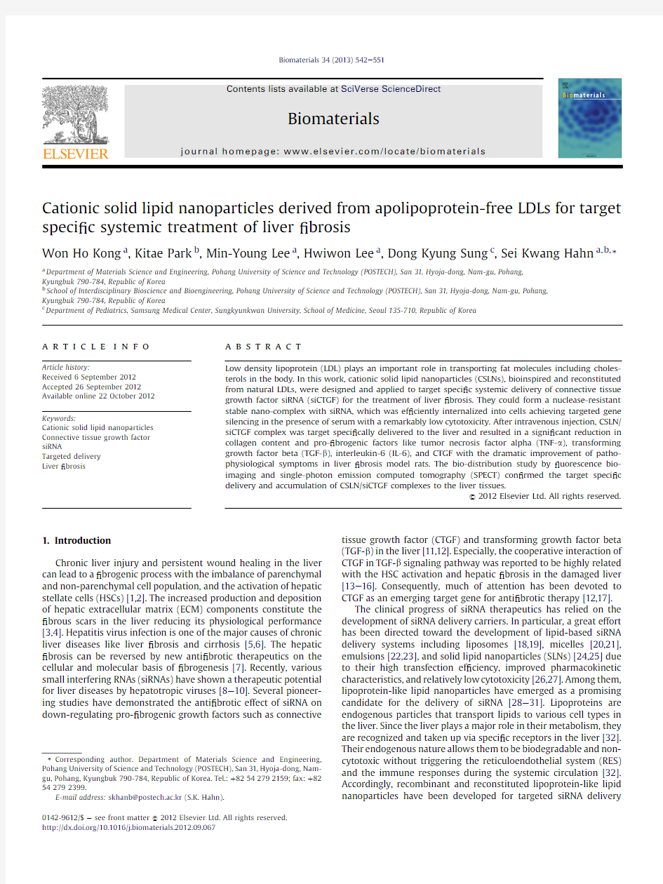
We have designed apolipoprotein free LDL-like solid lipid nanoparticles to prepare highly uniform and stable nano-sized particles with a PEG-protected and positively charged surface monolayer for targeted delivery of siRNA to the liver(Fig.1).Native human plasma LDLs are composed of a hydrophobic inner core and a surrounding surface monolayer of polar phospholipids.The inner core is known to be predominantly45%(w/w)of cholesterol ester and3%(w/w)of triglycerides.The outer-layer is10%(w/w)of cholesterol,22%(w/w)of phospholipids,and20%(w/w)of apoli-poproteins.As a bioinspired CSLN formulation,surface polar lipid constituents in natural lipoproteins were reconstituted with28% (w/w)cationic polar lipid DC-Chol and14%(w/w)fusogenic lipid DOPE to provide a positive surface charge,facilitate endosomal escape,and improve intracellular traf?cking.The resulting nano-particles maintained the same composition of ca.10%(w/w) cholesterol,and nonpolar lipids of45%(w/w)cholesteryl oleate and 5%(w/w)triglyceride in the inner core.Additionally,the surface monolayer of CSLN was further modi?ed with0.1%(w/w)PEGy-lated phospholipid of DSPE-PEG2k to improve hydrodynamic stability and protect their siRNA cargo from nuclease degradation during systemic circulation.As reported elsewhere[28,36],CSLNs were prepared by a modi?ed emulsi?cation and solvent evapora-tion method.All lipid components were initially dissolved in a co-solvent of chloroform/methanol(2/1).Then,the solvent mixture containing all lipid constituents was added to deionized water and ultra-sonicated to form a homogenous oil-in-water emulsion.The hydrophobic core lipid constituents of cholesteryl oleate and triglyceride might be localized inside the oil phase droplets. Subsequent solvent evaporation under a reduced pressure might result in the swift assembly of amphiphilic lipid building blocks around the hydrophobic core of nonpolar lipid constituents.A solid inner core was obtained by concomitant cooling down.The cho-lesteryl oleate has a high melting temperature(T m?52 C)above body temperature.The resulting highly stable CSLNs were thought to be more advantageous than conventional DC-Chol/DOPE lipo-some formulations.The hydrodynamic size distribution,z-poten-tial,and surface topology of CSLNs were analyzed by DLS and AFM. CSLNs had a narrow size distribution with a diameter in the range of106.2?5.4nm showing a positive z-potential of64.3mV.AFM image of CSLNs in Fig.2a also revealed the well-dispersed spherical
W.H.Kong et al./Biomaterials34(2013)542e551 544
CSLN
Component Component Cholesteryl oleate
45.03.045.03.010.020.022.09.928.014.00.1
Triolein S u r f a c e
C o r e
Cholesterol
+
-----
Cholesteryl oleate
Triolein Cholesterol Apolipoprotein Phospholipid
--
Cationic DC-Chol Fusogenic DOPE DSPE-PEG 2k
Portion (%, w/w)Portion (%, w/w)Natural LDL
a
b
Fig.1.(a)A schematic representation of the bioinspired cationic solid lipid nanoparticle (CSLN)and its composition in comparison with natural low density lipoprotein (LDL).(b)A schematic illustration for the formation of CSLN/siRNA complex by electrostatic interaction between positively charged CSLN and negatively charged siRNA.
1201008060402001251007550250
Nuclease -+--+--+
Heparine
siRNA
CSLN/siRNA Complex
CSLN/siRNA ratio (w/w)
siRNA
10
15
20
25
0.0
Average Size = 102 nm
2.2 μm
30
35
20V i a b l e C e l l s (%)
Concentration (μg/mL)
B a n d I n t e n s i t y (%)
406080
CLNPs PEI 25k
a
b
d
c
Fig.2.Characterization of CSLNs.(a)Atomic force microscopy (AFM)of CSLNs.(b)Cytotoxicity of CSLN in comparison with PEI 25k.(c)The agarose gel retardation at various weight ratios of CSLN to siRNA.(d)The effect of CSLN on the protection of siRNA from ribonuclease degradation.The protective effect was quanti ?ed by the densitometric analysis.
W.H.Kong et al./Biomaterials 34(2013)542e 551545
assemblies with an average diameter of 102.0?2.4nm.The size,z -potential,and homogeneity of resulting CSLNs were well main-tained at room temperature for the experimental period.Then,we examined the cytotoxicity of CSLN in comparison with PEI as a control with increasing concentration.CSLN exhibited no severe cytotoxicity up to a concentration of 72m g/mL,whereas PEI signi ?cantly reduced the cell viability to ca.5.1%under the same condition (Fig.2b).The IC 50value of CSLN (333.5m g/mL)was remarkably higher than that of PEI (6.7m g/mL).These results con ?rmed the biocompatibility of CSLNs as a safe delivery vehicle of siRNA.
3.2.Preparation and characterization of CSLN/siRNA complex The complex formation of CSLN with siRNA as schematically shown in Fig.1b was examined by agarose gel electrophoresis.When CSLNs were mixed with siRNA at various weight ratios from 10to 35in 0.1M PBS (pH 7.4),the electrophoretic migration of siRNA revealed a gradual retardation within the gel with increasing weight ratio of the complexes (Fig.2c).The migration of siRNA was completely retarded above a weight ratio of 30.The results indi-cated that positively charged CSLNs effectively interacted with negatively charged siRNA molecules via electrostatic interaction.On the basis of these results,the optimal composition for CSLN/siRNA complexes was determined to be a weight ratio of 30,which was used for the following experiments.According to DLS analysis,the hydrodynamic diameter of CSLN/siRNA complexes (113.0? 3.2nm)was slightly larger than that of CSLN alone (106.2?5.4).In addition,the z -potential value of CSLNs signi ?-cantly decreased from 6
4.3?2.5mV to 32.1?3.3mV upon addi-tion of siRNA re ?ecting the electrostatic complex formation.It was noteworthy that the siRNA binding ability to CSLN was not signif-icantly altered by the incorporation of PEGylated phospholipid DSPE-PEG on the surface of CSLN.Then,the effect of CSLN in CSLN/siRNA complexes was assessed on the protection of siRNA from nuclease attack.As shown in Fig.2d,CSLN/siRNA complex retained 7
5.2?3.3%of siRNA even after treatment with 10units of nuclease.The CSLN/siRNA complex was dissociated in the presence of
100020406080100
020*********
020*********
020*********
020*********
020*********
1011021.0%47.8%
62.2%73.6%87.7%
42.0%w/w = 15FAM-siRNA
Lipopfectamine
CSLN/FAM-siRNA complex
w/w = 20w/w = 25w/w = 30
FAM-siRNA Fluorescene
C e l l N u m b e r
103104100101102103104100101102103104100101102103104100101102103104100101102103104
a
b
Fig.3.(a)Confocal microscopic images of HepG2.2.15cells after incubation with CSLM/FAM e siRNA complex for 2h.(b)Flow cytometric analysis for the intracellular uptake of CSLN/FAM e siRNA complex at different weight ratios.
W.H.Kong et al./Biomaterials 34(2013)542e 551
546
negatively charged heparin molecules at a concentration of 50m g/mL and the siRNA was completely degraded in 60min.From the results,we could con ?rm the formation of stable nano-sized CSLN/siRNA complexes protecting the payload from enzymatic degradation.
3.3.In vitro gene silencing of CSLN/siRNA complexes
Before in vitro gene silencing tests,the intracellular delivery and subcellular localization of CSLN/siRNA complex were examined by confocal microscopy using ?uorescently labeled FAM e siRNA.CSLN/FAM e siRNA complexes were internalized evenly into HepG.2.2.15cells in 10%(v/v)serum containing medium after incubation for 2h (Fig.3a).The internalized complexes accumulated near the nucleus in many cells.The cellular uptake of CSLN/FAM e siRNA complexes was further evaluated quantitatively by ?ow cytometry at various weight ratios from 15to https://www.360docs.net/doc/657194119.html,mercially available Lipofectamine was used as a control.As shown in Fig.3b,the cellular uptake of the complex gradually increased with increasing weight ratio of the complex showing a maximum at a weight ratio of 30.The percentage of FAM-positive cells reached ca.87.7%.In contrast,only a moderate level of cellular uptake was observed for the Lip-ofectamine/siRNA complexes (47.8%).The ef ?cient internalization of CSLN/siRNA complexes to the cells was ascribed to their nano-scale dimension and positive z -potential promoting their initial
interaction with cell membrane through electrostatic interaction.Moreover,the fusogenic lipid components,DOPE and cholesterol,might also facilitate the cellular entry and subsequent endosomal escape of the complexes.The gene silencing ef ?ciency of CSLN/siRNA complexes was assessed using four different candidate siR-NAs speci ?c for overlapping HBV open reading frames (ORF)(Fig.4a).The release of HBsAg was drastically reduced to the statistically signi ?cant levels after treatment with 200n M of CSLN/si257and CSLN/si1826complexes (P <0.0001)in 10%(v/v)serum containing culture condition (Fig.4b and c).The results were thought to re ?ect that CSLN/siRNA complexes were readily inter-nalized to the cells and escaped from the endosome releasing siRNA into cytoplasm for the effective gene silencing.3.4.In vivo anti ?brotic effect of CSLN/siCTGF complex
We carried out the systemic delivery of CSLN/siCTGF complex for the treatment of hepatic ?brosis.CTGF is produced by a variety of different cell types including HSCs and hepatocytes,which promotes ?brogenesis and survival of activated HSCs [37].With the progress of liver ?brosis and cirrhosis,HSCs were reported to be proliferated by 10e 20times and squeezed through the fenestrae [38].According to our previous bioimaging study using Qdots,hyaluronic acid (HA)derivatives were signi ?cantly delivered to the HSCs re ?ecting the easy access to the protruded HSCs [39].
Because
Fig.4.The gene silencing effect of CSLN/siRNA complex on hepatitis B virus surface antigen (HBsAg)expression in HBV producing HepG2.2.15cells.(a)A map of the HBV genome indicating the location of siRNA target sequences and their sequences.The HBsAg expression in HepG2.2.15cells after treatment with (b)increasing concentration of CSLN/si257complex,and (c)200n M of CSLN/si257,CSLN/si1658,CSLN/si1826,and CSLN/si2422complexes.
W.H.Kong et al./Biomaterials 34(2013)542e 551547
of the paracrine stimulation,the gene silencing of CTGF in the liver might result in signi?cant anti?brotic effect.The biochemical analysis for ALT,AST,TBIL,and ALB revealed signi?cant improve-ment of?brotic symptoms in SD rats treated with CSLN/siCTGF complex2h after daily injection of NDMA at a dose of10mg/kg for 7days(Fig.5).Serum cytokine levels of TNF-a,TGF-b,and IL-6were also signi?cantly improved when compared with CSLN treated group(P<0.001)(Fig.6).In addition,quantitative evaluation of CTGF intensity in Western blot analysis also showed a more signi?cant decrease in the production of CTGF in the liver after treatment with CSLN/siCTGF complex than CSLN alone(P<0.001).Fig.7shows the histological and immunohistochemical analyses of dissected?brotic liver tissues after treatment with CSLN/siCTGF complexes.The control group showed a normal lobular architecture and a normal hepatic cell structure with central veins and radiating hepatic cords(Fig.7a).In contrast,?brosis and early cirrhosis with multifocal hepatocyte necrosis and neutrophilic in?ltration were observed in SD rats treated with CSLN after daily injection of NDMA at a dose of10mg/kg for7days.There was no signi?cant alternation or prevention in the livers treated with CSLN/scCTGF complexes after NDMA injection for7days showing extensive bridging,well developed?bers,severe centrilobular congestion,and dilatation
of
Fig.5.The anti?brotic effect of the control,CSLN/siCTGF complex,CSLN/scCTGF complex,and CSLN on the liver function after systemic delivery to?brotic SD rats treated by the daily injection of NDMA at a dose of10mg/kg for7days.(a)ALT,(b)AST,(c)TBIL,and(d)albumin levels in the
serum.
Fig.6.The anti?brotic effect of the control,CSLN/siCTGF complex,CSLN/scCTGF complex,and CSLN on(a)TNF-a,(b)TGF-b,(c)IL-6,and(d)CTGF/a-Tubulin ratio after systemic delivery to?brotic SD rats treated by the daily injection of NDMA at a dose of10mg/kg for7days.
W.H.Kong et al./Biomaterials34(2013)542e551
548
sinusoids with focal hemorrhage.Interestingly,only mild dilatation of sinusoids and neutrophilic in ?ltrations were observed in animals treated with CSLN/siCTGF complex 2h after the injection of NDMA (Fig.7a).Liver ?brosis is characterized by overproduction and irregular deposition of collagens by liver ?brogenic cells resulting in subsequent loss of the normal function.As shown in Fig.7b,Mas-son ’s trichrome staining showed no histological alteration and collagen deposition in the livers for the control group.In contrast,the massive bridging and deposition of mature collagen ?bers were observed in the livers treated with CSLN or CSLN/scCTGF complex after NDMA injection for 7days.Importantly,only mild collagen disposition was observed in the livers treated with CSLN/siCTGF complex,which was in good agreement with the results in Fig.7a.The immunohistochemistry for CTGF and a -SMA was performed to assess the liver ?brosis.While remarkable staining for CTGF and a -SMA was observed in the livers treated with CSLN or CSLN/scCTGF complex after the NDMA injection for 7days,only focal marginal staining was observed in the livers treated with CSLN/siCTGF complex (Fig.7c).The target speci ?c gene silencing in the livers were also con ?rmed by the confocal microscopy of liver cry-osections showing remarkable cellular uptake of CSLN/FAM e siRNA complexes into the liver parenchyma (Fig.8).From the results,apolipoprotein-free LDL-like CSLN was thought to target-speci ?cally deliver siCTGF to the liver for ef ?cient silencing of
Control a
b
c
NDMA + siCTGF NDMA + scCTGF
NDMA + CSLN
Fig.7.Histological and immunohistochemical analyses of dissected ?brotic liver tissues after systemic treatment with the control,CSLN/siCTGF complex,CSLN/scCTGF complex,and CSLN.(a)H&E staining.(b)Masson ’s trichrome staining.(c)Immunohistochemistry for a -SMA (green),CTGF (red),and nucleus (blue).(For interpretation of the references to color in this ?gure legend,the reader is referred to the web version of this
article.)
Fig.8.Confocal microscopic images of (a)left and (b)right lateral liver robe cryosections 12h after systemic delivery of CSLN/FAM e siRNA complex.
W.H.Kong et al./Biomaterials 34(2013)542e 551549
CTGF gene exhibiting the therapeutic effect in liver ?brosis model rats.
3.5.Biodistribution of CSLN/siRNA complex in the body
To corroborate the targeted delivery of CSLN/siRNA complexes to the liver,their bio-distribution was investigated after iv injec-tion of siRNA complexed with CSLN encapsulating Qdots and radioisotope-labeled CSLN,respectively (Fig.9).According to ex vivo ?uorescence bioimaging,Qdot-CSLN/siRNA complexes containing 20m g of siRNA per 20g of body weight were target-speci ?cally delivered to the liver after single iv injection.A large portion of complexes were also accumulated gradually in the kidney re ?ecting the renal clearance (Fig.9a).In contrast,there was only negligible NIRF signal intensity in spleen and lung during the entire experimental period.Fig.9b and c shows the quanti ?-cation of NIRF signal intensity in each organ recorded as photon per sec (p/s)and photons per sec per square centimeter per steradian (p/sec/cm 2/sr).The biodistribution of CSLN/siRNA complexes was further evaluated by noninvasive whole-body SPECT imaging using the radiolabeled CSLNs.In accordance with the ?uorescence bioimaging,the SPECT imaging revealed that radiolabeled-CSLN/siRNA complexes preferentially accumulated in the liver.The radioactive signal was also observed in the kidney
but not in the spleen and lung.These results strongly supported that CSLN/siRNA complex might be successfully exploited for target speci ?c treatment of various liver diseases as demonstrated above.
4.Conclusions
CSLNs were successfully developed for targeted delivery of siRNA to the liver.The CSLNs were prepared by emulsi ?cation and solvent evaporation of the lipid components reconstituted from natural apolipoprotein-free LDLs.The CSLN was able to form a stable nano-sized complex with CTGF siRNA via electrostatic interaction and protect the payload against enzymatic degradation.The CSLN/siHBV complex showed a signi ?cant in vitro gene silencing with a drastically reduced HBsAg expression.Further-more,after iv injection,CSLN/siCTGF complexes were target-speci ?cally delivered to the liver and resulted in a signi ?cant reduction in collagen content and pro-?brogenic factors like TNF-a and TGF-b ,IL-6,and CTGF showing dramatically improved patho-physiological symptoms in liver ?brosis model rats.The bio-distribution studies by ex vivo ?uorescence bioimaging and SPECT clearly con ?rmed that CSLN/siCTGF complexes were preferentially delivered and accumulated in the liver for the therapeutic gene silencing.
12 h L i v e r T o t a l F l u x (p /s x 107)
R a d i a n c e (p /s /c m 2 x 10⁷)
K i d n e y L u n g
S p l e e n
L i
v e r
K
i d n e y
L u
n g
S p
l e
e n
L i
v e
r
K
i d
n e
y
L u
n g
S p l e
e n
24 h 48 h 72 h 48 h
1.5
12 h 24 h 48 h 72 h
12 h 24 h 48 h 72 h
1.00.50
1.51.00.50
a
b
c
Fig.9.(a)Ex vivo ?uorescence imaging (left upper panel)of dissected organs and non-invasive SPECT imaging (right upper panel)of whole-body after systemic delivery of Qdot-or radioisotope-labeled CSLN/siRNA complexes.(b)Quanti ?ed total ?ux and (c)average radiance at dissected organs by the ex vivo ?uorescence bioimaging.
W.H.Kong et al./Biomaterials 34(2013)542e 551
550
Acknowledgments
This work was?nancially supported by the Converging Research Center Program through the National Research Foundation of Korea (NRF)funded by the Ministry of Education,Science and Technology (2009-0081871).This study was also supported by Mid-career Researcher Program through NRF grant funded by the MEST (No.2012R1A2A2A06045773).
References
[1]Li L,Wang H,Ong ZY,Xu K,Ee PLR,Zheng S,et al.Polymer-and lipid-based
nanoparticle therapeutics for the treatment of liver diseases.Nano Today 2010;5:296e312.
[2]Geerts A.History,heterogeneity,developmental biology,and functions of
quiescent hepatic stellate cells.Semin Liver Dis2001;21:311e35.
[3]Muddu AK,Guha IN,Elsharkawy AM,Mann DA.Resolving?brosis in the
diseased liver:translating the scienti?c promise to the clinic.Int J Biochem Cell B2007;39:695e714.
[4]Benyon RC,Iredale JP.Is liver?brosis reversible?Gut2000;46:443e6.
[5]Lai CL,Ratziu V,Yuen MF,Poynard T.Viral hepatitis https://www.360docs.net/doc/657194119.html,ncet2003;362:2089e94.
[6]Poynard T,Yuen MF,Ratziu V,Lai CL.Viral hepatitis https://www.360docs.net/doc/657194119.html,ncet2003;362:2095e100.
[7]Friedman SL.Mechanisms of hepatic?brogenesis.Gastroenterology2008;
134:1655e69.
[8]Chen Y,Cheng G,Mahato RI.RNAi for treating hepatitis B viral infection.
Pharm Res2008;25:72e86.
[9]Khaliq S,Khaliq SA,Zahur M,Ijaz B,Jahan S,Ansar M,et al.RNAi as a new
therapeutic strategy against HCV.Biotechnol Adv2010;28:27e34.
[10]Watanabe T,Umehara T,Kohara M.Therapeutic application of RNA interfer-
ence for hepatitis C virus.Adv Drug Del Rev2007;59:1263e76.
[11]Park K,Hong SW,Hur W,Lee MY,Yang JA,Kim SW,et al.Target speci?c
systemic delivery of TGF-beta siRNA/(PEI-SS)-g-HA complex for the treatment of liver cirrhosis.Biomaterials2011;32:4951e8.
[12]George J,Tsutsumi M.siRNA-mediated knockdown of connective tissue
growth factor prevents N-nitrosodimethylamine-induced hepatic?brosis in rats.Gene Ther2007;14:790e803.
[13]Paradis V,Dargere D,Bonvoust F,Vidaud M,Segarini P,Bedossa P.Effects and
regulation of connective tissue growth factor on hepatic stellate https://www.360docs.net/doc/657194119.html,b Invest2002;82:767e74.
[14]Sonnylal S,Shi-Wen X,Leoni P,Naff K,Van Pelt CS,Nakamura H,et al.
Selective expression of connective tissue growth factor in?broblasts in vivo promotes systemic tissue?brosis.Arthritis Rheum2010;62:1523e32. [15]Hayashi N,Kakimuma T,Soma Y,Grotendorst GR,Tamaki K,Harada M,et al.
Connective tissue growth factor is directly related to liver?brosis.Hepato-gastroenterol2002;49:133e5.
[16]Rachfal AW,Brigstock DR.Connective tissue growth factor(CTGF/CCN2)in
hepatic?brosis.Hepatol Res2003;26:1e9.
[17]Blom IE,Goldschmeding R,Leask A.Gene regulation of connective tissue
growth factor:new targets for anti?brotic therapy?Matrix Biol2002;21: 473e82.
[18]Halder J,Kamat AA,Landen Jr CN,Han LY,Lutgendorf SK,Lin YG,et al.Focal
adhesion kinase targeting using in vivo short interfering RNA delivery in neutral liposomes for ovarian carcinoma therapy.Clin Cancer Res2006;12: 4916e24.
[19]Merritt WM,Lin YG,Spannuth WA,Fletcher MS,Kamat AA,Han LY,et al.
Effect of interleukin-8gene silencing with liposome-encapsulated small
interfering RNA on ovarian cancer cell growth.J Natl Cancer Inst2008;100: 359e72.
[20]Jeong JH,Mok H,Oh YK,Park TG.siRNA conjugate delivery systems.Bioconjug
Chem2009;20:5e14.
[21]Wolfrum C,Shi S,Jayaprakash KN,Jayaraman M,Wang G,Pandey RK,et al.
Mechanisms and optimization of in vivo delivery of lipophilic siRNAs.Nat Biotechnol2007;25:1149e57.
[22]Luo MC,Zhang DQ,Ma SW,Huang YY,Shuster SJ,Porreca F,et al.An ef?cient
intrathecal delivery of small interfering RNA to the spinal cord and peripheral neurons.Mol Pain2005;1:29.
[23]Yoo HS,Kwon SM,Kim YJ,Chung H,Kwon IC,Kim J,et al.Cationic lipid
emulsions containing heavy oils for the transfection of adherent cells.
J Control Release2004;98:179e88.
[24]Lobovkina T,Jacobson GB,Gonzalez-Gonzalez E,Hickerson RP,Leake D,
Kaspar RL,et al.In vivo sustained release of siRNA from solid lipid nano-particles.ACS Nano2011;5:9977e83.
[25]Xue HY,Wong HL.Solid lipid-PEI hybrid nanocarrier:an integrated approach
to provide extended,targeted,and safer siRNA therapy of prostate cancer in an all-in-one manner.ACS Nano2011;5:7034e47.
[26]Schroeder A,Levins CG,Cortez C,Langer R,Anderson DG.Lipid-based nano-
therapeutics for siRNA delivery.J Intern Med2010;267:9e21.
[27]Oh YK,Park TG.siRNA delivery systems for cancer treatment.Adv Drug Deliv
Rev2009;61:850e62.
[28]Kim HR,Kim IK,Bae KH,Lee SH,Lee Y,Park TG.Cationic solid lipid nano-
particles reconstituted from low density lipoprotein components for delivery of siRNA.Mol Pharm2008;5:622e31.
[29]Jin H,Lovell JF,Chen J,Lin Q,Ding L,Ng KK,et al.Mechanistic insights into LDL
nanoparticle-mediated siRNA delivery.Bioconjug Chem2012;23:33e41. [30]Shahzad MM,Mangala LS,Han HD,Lu C,Bottsford-Miller J,Nishimura M,et al.
Targeted delivery of small interfering RNA using reconstituted high-density lipoprotein nanoparticles.Neoplasia2011;13:309e19.
[31]Yang M,Jin H,Chen J,Ding L,Ng KK,Lin Q,et al.Ef?cient cytosolic delivery of
siRNA using HDL-mimicking nanoparticles.Small2011;7:568e73.
[32]Rensen PC,de Vrueh RL,Kuiper J,Bijsterbosch MK,Biessen EA,van Berkel TJ.
Recombinant lipoproteins:lipoprotein-like lipid particles for drug targeting.
Adv Drug Del Rev2001;47:251e76.
[33]Kim SI,Shin D,Lee H,Ahn BY,Yoon Y,Kim M.Targeted delivery of siRNA
against hepatitis C virus by apolipoprotein A-I-bound cationic liposomes.
J Hepatol2009;50:479e88.
[34]Maranhao RC,Garicochea B,Silva EL,Dorlhiac-Llacer P,Cadena SM,Coelho IJ,
et al.Plasma kinetics and biodistribution of a lipid emulsion resembling low-density lipoprotein in patients with acute leukemia.Cancer Res1994;54: 4660e6.
[35]Rensen PCN,Herijgers N,Netscher MH,Meskers SCJ,vanEck M,vanBerkel TJC.
Particle size determines the speci?city of apolipoprotein E-containing triglyceride-rich emulsions for the LDL receptor versus hepatic remnant receptor in vivo.J Lipid Res1997;38:1070e84.
[36]Kong WH,Bae KH,Jo SD,Kim JS,Park TG.Cationic lipid-coated gold nano-
particles as ef?cient and non-cytotoxic intracellular siRNA delivery vehicles.
Pharm Res2012;29:362e74.
[37]Tong Z,Chen R,Alt DS,Kemper S,Perbal B,Brigstock DR.Susceptibility to liver
?brosis in mice expressing a connective tissue growth factor transgene in hepatocytes.Hepatology2009;50:939e47.
[38]Warren A,Bertolino P,Benseler V,Fraser R,McCaughan GW,Le Couteur DG.
Marked changes of the hepatic sinusoid in a transgenic mouse model of acute immune-mediated hepatitis.J Hepatol2007;46:239e46.
[39]Kim KS,Hur W,Park SJ,Hong SW,Choi JE,Goh EJ,et al.Bioimaging for tar-
geted delivery of hyaluronic Acid derivatives to the livers in cirrhotic mice using quantum dots.ACS Nano2010;4:3005e14.
W.H.Kong et al./Biomaterials34(2013)542e551551
(完整word)生物化学讲义第六章脂代谢222汇总,推荐文档
第六章脂类代谢 【目的和要求】 1.了解脂类的分布及主要生理功能。 2.详尽描述脂肪酸氧化过程、有关酶,能进行能量计算。 3.解释酮体概念。复述酮体代谢、生理意义。 4.了解脂肪合成过程,结合软脂酸合成途径,熟记脂肪酸合成部位、原料 ( 包括来源 ) 及辅助因子,乙酰辅酶 A 羧化酶、脂肪酸合成酶系的特点及脂酰基载体蛋白( ACP )在脂肪酸合成中的作用。 5.熟悉鞘磷脂和鞘糖脂的化学组成。 6.掌握胆固醇合成原料、部位及胆固醇在体内的转化与排泄。 7.叙述血浆脂蛋白的分类和生理功能、熟悉血浆脂蛋白代谢及异常。 【本章重难点】 1.脂酸分解代谢过程及能量计算,脂酸β氧化。 2.酮体生成部位、原料、过程,酮体生理意义。 3.脂酸合成过程、原料及来源。 4.胆固醇合成关键步骤。 5.胆固醇转化产物及意义。 6. 血浆脂蛋白的种类及功能。 学习内容 第一节三酯酰甘油的代谢 第二节磷脂和鞘糖脂的代谢 第三节胆固醇的代谢 第四节血浆脂蛋白的代谢 第一节三酯酰甘油的代谢
一、脂类物质的分类和生理功用 脂类是脂肪和类脂的总称,是一大类不溶于水而易溶于有机溶剂的化合物。 脂肪(甘油三酯,TG) 脂类磷脂(PL)(甘油磷脂和鞘磷脂) 类脂糖脂(脑苷脂和神经节苷脂) 胆固醇(Ch)及胆固醇酯(CE)。 脂类物质具有下列生理功用: ①贮存及氧化供能 ②构成生物膜 ③协助脂溶性维生素的吸收,提供必需脂肪酸。必需脂肪酸是指机体需要,但自身不能合成,必须要靠食物提供的一些不饱和脂肪酸。 ④保护内脏和保温作用 二、甘油三酯的分解代谢 ⒈脂肪动员:贮存于脂肪细胞中的甘油三酯,在脂肪酶的催化下水解为游离脂肪酸(FFA)及甘油并释放入血,供给全身各组织细胞摄取利用的过程称为脂肪动员。激素敏感性甘油三酯脂肪酶(HSL)是脂肪动员的关键酶。能促进脂肪动员的激素称为脂解激素,如胰高血糖素、肾上腺素、促肾上腺皮质激素(ACTH)和促甲状腺激素(TSH);胰岛素、前列腺素E2和烟酸等能抑制脂肪动员,是抗脂解激素。 一分子甘油三酯可分解生成三分子的游离脂肪酸(FFA)和一分子的甘油。脂肪酸进入血液后与清蛋白结合成为复合体再转运到全身各组织,甘油则转运至肝、肾、肠等组织,主要在肝甘油激酶作用下,磷酸化为3-磷酸甘油,再脱氢生成磷酸二羟丙酮,或彻底氧化分解,或转变成糖,因此甘油是糖异生的原料。 ⒉脂肪酸的β-氧化 除脑组织外,体内大多数的组织细胞均可循此途径氧化利用脂肪酸。其代谢反应过程可分为三个阶段: ⑴活化:在线粒体外膜或内质网进行此反应过程。在ATP、CoASH、Mg2+存在条件下,由脂酰CoA合成酶催化脂肪酸生成脂酰CoA。每活化一分子脂肪酸,需消耗两分子ATP。 ⑵转运:借助于线粒体内膜两侧的两种肉碱脂酰转移酶(酶Ⅰ和酶Ⅱ)催化的移换反应,脂酰CoA由肉碱(肉毒碱)携带进入线粒体。位于线粒体内膜外侧面的肉碱脂酰转移酶Ⅰ是脂肪酸β-氧化的关键酶,脂酰CoA进入线粒体是脂肪酸β-氧化的主要限速步骤。 ⑶β-氧化:由四个连续的酶促反应组成。 ①脱氢:脂酰CoA在脂酰CoA脱氢酶的催化下,生成FADH2和α,β-烯脂酰CoA。 ②加水:在水化酶的催化下,生成L-β-羟脂酰CoA。 ③再脱氢:在β-羟脂酰CoA脱氢酶的催化下,生成β-酮脂酰CoA和NADH及H+。 ④硫解:在硫解酶的催化下,分解生成1分子乙酰CoA和1分子少两个碳原子的脂酰CoA。后者可继续氧化分解,直至全部分解为乙酰CoA。 乙酰CoA进入三羧酸循环彻底氧化分解, FADH2和NADH+H+通过呼吸链经氧化磷酸化后产生能量。 ⒊脂肪酸氧化分解时的能量释放: 以16C的软脂酸为例来计算,其生成ATP的数目为:一分子软脂酸可经七次β-氧化全部分解为八分子乙酰CoA,故β-氧化可得5×7=35分子ATP,八分子乙酰CoA可得12×8=96分子ATP,故一共可得131分子ATP,减去活化时消耗的两分子ATP,故软脂酸可净生成129分子ATP。即对于偶数碳原子的长链脂肪酸,可按下式计算:ATP净生成数目=(碳原子数
氨基酸与核酸代谢
氨基酸与核酸代谢 1. 概述三大类营养物质代谢的联系。 答:在生物体内,糖类、脂质和蛋白质这三类物质的代谢是同时进行的,它们之间既相互联系,又相互制约,形成一个协调统一的过程。 1.糖类、脂质和蛋白质之间可以转化关系 (1)糖类代谢与蛋白质代谢的关系 ①糖类代谢的中间产物可以转变成非必需氨基酸; ②蛋白质可以转化成糖类。 几乎所有组成蛋白质的天然氨基酸均可转变成糖类。 (2)糖类代谢与脂质代谢的关系 ①糖类转变成脂肪 ②脂肪转变成糖类 脂质分解产生的甘油和脂肪酸能够转变成糖类。 (3)蛋白质代谢与脂质代谢的关系 ①一般来说,动物体内不容易利用脂肪酸合成氨基酸。植物和微生物可由脂肪酸和氮源生成氨基酸。某些氨基酸通过不同途径可转变成甘油或脂肪酸 ②某些氨基酸通过不同途径可转变成甘油或脂肪酸。 2.糖类、脂质和蛋白质之间转化的条件 糖类、脂质和蛋白质之间的转化是有条件的。例如,只有在糖类供应充足的情况下,糖类才有可能大量转化成脂质。不仅如此,各种代谢物之间的转化程度也是有明显差异的。例如,糖类可以大量转化成脂肪,而脂肪却不能大量转化成糖类。只有当糖类代谢发生障碍时才由脂肪和蛋白质来供能,当糖类和脂肪摄入量都不足时,蛋白质的分解才会增加 3.糖类、脂质和蛋白质之间除了能转化外,还相互制约 三大营养物质在代谢上的共同点; (1)物质的来源:从根本上说均是食物。 (2)这些物质在细胞内虽然均进行着许多种的生物化学反应,但是可以归纳为进行合成和分解这两方面的反应,并且这两方面的反应在细胞内是同时进行,相互联系的。 (3)三大营养物质的代谢均必须在酶的催化作用下才能够完成。 (4)这些物质彻底氧化分解时,代谢终产物里均有CO2和水,均能放释能量。 三大营养物质在代谢上的不同点 (1)糖类是主要的供能物质,脂肪是主要的储能物质,蛋白质的主要功能是构成生物体和调节生命活动。 (2)蛋白质质代谢的最终产物里还有尿素。 人体内的物质代谢是一个完整的统一过程 我们从糖类、氨基酸能够转变成脂肪,脂肪、氨基酸能够转变成糖类,可以看出各种物质代谢之间是相互联系的。这种联系说明,人体内的物质代谢是一个完整的统一过程,它使细胞内的成分不断地进行新旧更替。 2. 如何看懂肝功化验单? 临床上肝功能检查的主要项目包括:蛋白代谢检查、糖代谢检查、脂类检查、胆红素代谢检查、血清酶学检查等。 如何看血清酶学化验单 常见以下几种酶:谷丙转氨酶(英文简写为ALT或GPT)、谷草转氨酶(AST或GOT)、碱性
脂类代谢、蛋白质代谢、核酸代谢练习题
《脂类代谢、蛋白质代谢、核酸代谢》练习题 一、填空题 1.氨基酸的分解代谢中,转氨酶的辅酶是_____________ ;氨基酸脱羧酶的辅酶是_____________ 。 2.肝、肾组织中氨基酸脱氨基作用的主要方式是_____________ 。肌肉组织中氨基酸脱氨基作用的 主要方式是_____________ 。 3.肝细胞参与合成尿素中两个氮原子的来源,第一个氮直接来源于_____________ ;第二个氮直接 来源于_____________ 4.体内有三种含硫氨基酸,它们是甲硫氨酸、_____________ 和_____________ 。 5.脂类消化的主要部位是_____________ ,消化后吸收的主要部位是_____________ 。 6.脂肪酸的氧化方式有三种,分别为_____________ 、_____________ 和_____________ 。 7.β -氧化是在细胞的中进行_____________的,β -氧化的氧化反应是在脂酰辅酶A 的β - 碳原子 上进行脱氢,氢的接受体是_____________和_____________ 。 8.脂酰CoA经脂肪酸β-氧化酶系的催化作用,在脂酰基__________位碳原子上依次进行 _____________、_____________、_____________及_____________4步连续反应,使脂酰基在______位与____位碳原子间断裂,生成1分子____________和少____________个碳原子的____________。 9.脂肪酸的β-氧化每循环一次,生成一分子乙酰CoA、一分子___________、一分子___________和 一分子减少两个碳原子的___________。生成的乙酰CoA将进入___________彻底氧化分解。 10.脂肪酸生物合成的基本原料是_____________ 和_____________ 。脂肪酸生物合成的供氢体是 _____________ ,它来源于_____________ 。脂肪的生物合成有两条途径,分别是_____________ 和_____________ 。 11.脂肪酸生物合成在细胞的_____________ 中进行,关键酶是________________________ 12.按核酸酶的作用位置的不同,可将核酸酶分为_____________________和__________________两类 13.黄嘌呤核苷酸的缩写符号为,次黄嘌呤核苷酸的缩写符号为,5-磷 酸核糖焦磷酸的缩写符号为。 14.人体合成的尿素分子中一个N来自,另一个N来自,CO2来自 于。 15.联合脱氨基作用的一种方式是:氨基酸的氨基先借转氨基作用转移到分子上,生成 相应的和,然后后者在的作用下,脱去氨基又生成。 16.磷酸戊糖途径发生于细胞的中。 17.不仅是糖、脂类、蛋白质和核酸的共同代谢途径,而且也是它们之间相互联系的 渠道。 18.生物体内的代谢调节在三种不同水平上进行,即、 和 二、单选题 1.PRPP是下列哪些代谢选径中的重要中间代谢物:①嘌呤核苷酸的从头合成②嘧啶核苷酸的从头合成③嘌呤核苷酸的补救途径④NMP-NDP-NTP () A)①B)①②C)①②③D)④ 2.体内脱氧核苷酸生成的主要方式是() A)由核苷还原B)由一磷酸核苷还原C)由二磷酸核苷还原D)由三磷酸核苷还原 3.糖代谢中间产物中有高能磷酸键的是()
生物化学第六章脂代谢随堂练习与参考答案
生物化学(本科)第六章脂代谢 随堂练习与参考答案 第一节脂类在体内的分布与功能第二节脂类的消化与吸收第三节甘油三酯代谢第四节磷脂的代谢第五节胆固醇代谢第六节血浆脂蛋白代谢 1. (单选题)脂肪在体内的主要生理功能是 A. 细胞膜结构的骨架 B. 参与细胞间信号转导 C. 储能和氧化供能 D. 降低细胞膜的流动性 E. 转变为前列腺素、血栓素及白三烯 参考答案:C 2. (单选题)脂肪酸在血中与下列哪种物质结合运输 A.载脂蛋白 B.清蛋白 C.球蛋白
D.脂蛋白 E.磷脂 参考答案:B 3. (单选题)关于载脂蛋白(Apo)的功能,在下列叙述中不正确的是: A.与脂类结合,在血浆中转运脂类 B.Apo AⅠ能激活LCAT C.Apo B能识别细胞膜上的LDL受体 D.Apo CⅠ能激活脂蛋白脂肪酶 E.Apo CⅡ能激活LPL 参考答案:D 4. (单选题)12个碳以上的长链脂肪酰辅酶A进入线粒体基质的主要影响因素是 A.脂酰CoA合成酶活性 B.脂酰CoA脱氢酶活性 C.ATP含量 B.肉毒碱脂酰转移酶Ⅰ活性
E.β-酮脂酰CoA硫解酶活性 参考答案:B 5. (单选题)脂肪动员的关键酶是: A.组织细胞中的甘油三酯酶 B.组织细胞中的甘油二酯脂肪酶 C.组织细胞中的甘油一酯脂肪酶 D.组织细胞中的激素敏感性脂肪酶 E.脂蛋白脂肪酶 参考答案:D 6. (单选题)以下关于脂酸β-氧化的描述错误的是 A.β-氧化的产生部位是线粒体中 B.β-氧化中脱下的氢传递给NADPH+H+ C.β-氧化的原料是脂酰CoA D.β-氧化的产物是乙酰CoA E.β-氧化中脱下的氢可经氧化磷酸化生成ATP 参考答案:B 7. (单选题)维生素PP缺乏, 可影响脂酸β-氧化过程中
氨基酸与核苷酸代谢
氨基酸与核苷酸代谢 (一)名词解释 1.蛋白酶(Proteinase) 2.肽酶(Peptidase) 3.氮平衡(Nitrogen balance) 4.转氨作用(Transamination) 联合脱氨基作用 8.尿素循环(Urea cycle) 9.生糖氨基酸(Glucogenic amino acid) 10.生酮氨基酸(Ketogenic amino acid) 11.核酸酶(Nuclease) 12.限制性核酸内切酶(Restriction endonuclease) 13.一碳单位(One carbon unit) (二)英文缩写符号 1.GOT 2.GPT 3.APS 4.PAL 5.PRPP 6.SAM 7.GDH 8.IMP (三)填空 1.生物体内的蛋白质可被和共同作用降解成氨基酸。 2.多肽链经胰蛋白酶降解后,产生新肽段羧基端主要是和氨基酸残基。3.胰凝乳蛋白酶专一性水解多肽链由族氨基酸端形成的肽键。 4.氨基酸的降解反应包括、和作用。 5.转氨酶和脱羧酶的辅酶通常是。 6.谷氨酸经脱氨后产生和氨,前者进入进一步代谢。 7.尿素循环中产生的和两种氨基酸不是蛋白质氨基酸。 8.尿素分子中两个N原子,分别来自和。 9.芳香族氨基酸碳架主要来自糖酵解中间代谢物和磷酸戊糖途径的中间代谢物。 13.组氨酸合成的碳架来自糖代谢的中间物。 14.氨基酸脱下氨的主要去路有、和。 15.胞嘧啶和尿嘧啶经脱氨、还原和水解产生的终产物为。 16.参与嘌呤核苷酸合成的氨基酸有、和。 17.尿苷酸转变为胞苷酸是在水平上进行的。 18.脱氧核糖核苷酸的合成是由酶催化的,被还原的底物是。19.在嘌呤核苷酸的合成中,腺苷酸的C-6氨基来自;鸟苷酸的C-2氨基来自。 20.对某些碱基顺序有专一性的核酸内切酶称为。 21.多巴是经作用生成的。 22.生物体中活性蛋氨酸是,它是活泼的供应者。 23.转氨基作用是沟通和桥梁; 24.尿素循环中涉及的天然蛋白质氨基酸是; 25.氨的去路有、和降解;脱氨产生的生理作用是和。 26.氨基酸通过、和降解,脱羧后产生和,此过程需——作辅酶。 27.Tyr脱NH3,然后脱羧后生成。 28.Tyr羟化后生成,后者经脱羧生成。
氨基酸代谢复习题-带答案
第八章氨基酸代谢 一、名词解释 86、转氨基作用 答案:(transmination)是α-氨基酸与α-酮酸之间在转氨酶的作用下氨基转移作用。 87、必需氨基酸 答案:(essential amino acids EAA)人类及哺乳动物自身不能合成,必需通过食物摄取得到的组成蛋白质的氨基酸,有Lys,Ile,Leu,Met,Trp,Phe,Val,Thr以及His和Arg。 88、尿素循环 答案:又称鸟氨酸循环(urea cycle)是生物体(陆生动物)排泄氨以维持正常生命活动的一种代谢方式。高等植物可将复杂的氨以酰胺的形式贮存起来,一般不进行尿素循环。整个循环从鸟氨酸开始经瓜氨酸精氨酸再回到鸟氨酸,循环一圈消耗2分子氨,1分子CO2和3分子ATP,净生成1分子尿素。 89、生酮氨基酸 答案:(ketogenic amino acid)可以降解为乙酰CoA或乙酰乙酰CoA,而生成酮体的氨基酸称生酮氨基酸。有Leu、Ile、Lys、Phe、Trp、Tyr,其中后5种为生酮生糖氨基酸。 90、生糖氨基酸 答案:(glucogenic amino acid)降解产物可以通过糖异生途径生成糖的氨基酸。组成蛋白质的 20种氨基酸中,除了生酮氨基酸外,其余皆为生糖氨基酸。 91、脱氨基作用 答案:(deamination)氨基酸失去氨基的作用,是生物体内氨基酸分解代谢的第一步,分氧化脱氨和非氧化脱氨两种方式。 92、联合脱氨基作用 答案:(dideamination)概括地说即先转氨后脱氨作用。分两个内容,一个指氨基酸先转氨生成谷氨酸和相应的α-酮酸,再在谷氨酸脱氢酶的催化下脱氨基,生成α-酮戊二酸,同时释放氨。另一个指嘌呤核苷酸循环,即天门冬氨酸与次黄嘌呤核苷酸作用生成腺苷酸代琥珀酸,后者被裂解酶催化,生成AMP和延胡索酸,AMP在腺苷酸脱氢酶作用下,脱去氨,生成次黄嘌呤核苷酸。 93、蛋白酶 答案:(proteinase)又称内肽酶,主要作用于肽链内部肽键,水解生成长度转短的多肽链。 94、肽酶 答案:(Peptidase)水解多肽链羧基末端肽键(羧肽酶)或氨基末端肽键(氨肽酶)。 二、填空题 124、氨的同化途径有合成途径、合成途径。 答案:谷氨酸;氨甲酰磷酸 125、由无机态的氨转变为氨基酸,首先是形成,然后由它通过作用形成其它们氨基酸。
第六章 脂类代谢
第六章脂类代谢 第一节生物体内的脂类 第二节脂肪的降解 第三节脂肪的合成 第四节类脂代谢 1
第一节生物体内的脂类 脂类:是脂肪、类脂及其衍生物的总称,不溶于水而溶于有机溶剂一类生物分子。 功能:(1)生物膜的成分磷脂、糖脂及胆固醇是膜脂类的三种 主要类型。 (2)重要能源 (3)具有营养、代谢及调节功能V A、V D、V E、V E、胆酸及固醇类激素等。 (4)保护作用防止机械损伤、热量散失 (5)与细胞识别、种特异性及组织免疫等有密切关系 脂类按化学结构和组成可分为三大类: 一、单纯脂质 是脂肪酸(C4以上)和醇(甘油醇和高级一元醇)构成的酯。 又分为脂肪(室温下:液态→油;固态→脂): 甘油+3个不同脂肪酸(多为偶数碳原子→脂肪) 蜡:高级脂肪酸(C12—C32)+高级醇(C26—C28)或固醇→蜡 二、复合脂质 单纯脂质+非脂溶性物质 1、磷脂 含磷酸的单纯脂质衍生物,生物膜的主要成分 2、糖脂 即糖脂酰甘油,糖苷与甘油分子第三个羟基以糖苷键相连,甘油的另两个羟基被脂肪酸脂化。 主要存在于:动物神经系统、植物叶绿体及代谢活跃部位。 三、非皂化脂质 特点:大都不含脂肪酸 包括萜类、类固醇类及前列腺素等 (一)萜类 萜类和类固醇类(除胆固醇外)都是不含脂肪酸的非皂化脂质,而且均为异戊二烯的衍生物,又称异戊二烯的脂质。 2
3 异戊二烯的结构: 由二个异戊二烯构成的萜为单萜 例柠檬苦素(柠檬油主成分) 三个异戊二烯构成的萜为倍半萜 法尼醇(昆虫保幼激素) 四个异戊二烯构成的萜为二萜 叶绿醇(叶绿素组分) 单萜结构 (二)固醇类 为环状高分子一元醇,可离态或与脂肪酸结合成酯的形式存在,都含环戊烷多氢菲母核。 菲 环戊烧多氢菲 固醇类基本结构 第二节 脂肪的降解 脂肪是由甘油的三个羟基与三个脂肪酸缩合而成,也称为甘油三酯。 脂肪是动物体内重要的贮能物质,当机体需要时,贮存在脂肪细胞中的脂肪被脂肪酶逐步水解为游离脂肪酸和甘油并释放进入血液,被其他组织氧化利用,这一过程也称为脂肪动员作用。水解产物脂肪酸和甘油在动物体内经扩散作用进入肠黏膜细胞,再经淋巴系统进入血液。 一、 脂肪的酶促降解 脂肪酶 甘油二酯酶 甘油单酯酶 1 、动物:甘油三酯 甘油二酯 甘油单酯 甘油 2、 植物:由α—脂酶完成。 二、甘油命运 C H 2=C -C H =C H 2 C H 3
氨基酸代谢核苷酸代谢
组卷测试 一:填空题 1.哺乳动物产生1分子尿素需要消耗________________分子的A TP。 2.褪黑激素来源于________________氨基酸,而硫磺酸来源于________________氨基酸。 3.γ-谷氨酰循环的生理功能是________________。 4.核苷酸的合成包括________________和________________两条途径。 5.转氨酶的辅基是________________。 6.________________酶的缺乏可导致人患严重的复合性免疫缺陷症(SCID),使用________________治疗可治愈此疾患。 7.痛风是因为体内________________产生过多造成的,使用________________作为黄嘌呤氧化酶的自杀性底物可以治疗痛风。 8.不能使用5-溴尿嘧啶核苷酸代替5-溴尿嘧啶治疗癌症是因为________________。 9.脑细胞中氨的主要代谢去向是________________。 10.从IMP合成GMP需要消耗________________,而从IMP合成AMP需要消耗________________作为能源物质。 11.Arg可以通过________________循环形成。 12.氨基酸共有的代谢途径有________________和________________。 13.人类对嘌呤代谢的终产物是________________。 14.羟基脲作为________________酶的抑制剂,可抑制脱氧核苷酸的生物合成。 15.脱氧核苷酸是由________________还原而来。 16.HGPRT是指________________,该酶的完全缺失可导致人患________________。 17.人类对氨基代谢的终产物是________________,鸟类对氨基代谢的终产物是________________,植物解除氨的毒害的方法是________________。 18.重亮氨酸作为________________类似物可抑制嘌呤核苷酸的从头合成。 19.通过________________的脱羧可产生β-丙氨酸。 20.细菌嘧啶核苷酸从头合成途径中的第一个酶是________________。该酶可被终产物 ________________抑制。 21.PAPS是指________________,它的生理功能是________________。 二:是非题 1.[ ]真核细胞内参与嘧啶核苷酸从头合成的酶都位于细胞质。 2.[ ]参与尿素循环的酶都位于线粒体内。 3.[ ]既然谷氨酸上的N原子可经过转氨基作用重新分布,那么谷氨酸应该可作为很好的营养品而弥补蛋白质缺乏。 4.[ ]嘧啶合成所需要的氨甲酰磷酸合成酶与尿素循环所需要的氨甲酰磷酸合成酶是同一个酶。 5.[ ]氨基酸脱羧酶通常也需要吡哆醛磷酸作为其辅基。 6.[ ]一般来说,在哺乳动物体内由蛋白质氧化分解产生的能量效率低于糖或脂肪的氧化分解。 7.[ ]嘌呤核苷酸的从头合成是先闭环,再在形成N糖苷键。 8.[ ]氨基酸经脱氨基作用以后留下的碳骨架进行氧化分解需要先形成能够进入TCA循环的中间物。 9.[ ]Arg是哺乳动物的一种非必需氨基酸,因为在它们的肝细胞之中,含有足够的合成Arg的酶。 10.[ ]动物产生尿素的主要器官是肾脏。 11.[ ]黄嘌呤氧化酶既可以使用黄嘌呤又可以使用次黄嘌呤作为底物。 12.[ ]L-氨基酸氧化酶是参与氨基酸脱氨基作用的主要酶。
第六章脂类代谢
第六章脂类代谢 一、选择题 1、线粒体基质中脂酰CoA脱氢酶的辅酶是()。 A、FAD B、NADP+ C、NAD+ D、GSSG 2、在脂肪酸的合成中,每次碳链的延长都需要()直接参加。 A、乙酰CoA B、草酰乙酸 C、丙二酸单酰CoA D、甲硫氨酸 3、合成脂肪酸所需的氢由下列()递氢体提供。 A、NADP+ B、NADPH+H+ C、FADH2 D、NADH+H+ 4、脂肪酸活化后,β-氧化反复进行,不需要下列()酶参与。 A、脂酰CoA脱氢酶 B、β-羟脂酰CoA脱氢酶 C、烯脂酰CoA水合酶 D、硫激酶 5、软脂酸的合成及其氧化的区别为()。 (1)细胞部位不同;(2)酰基载体不同;(3)加上及去掉2C?单位的化学方式不同;(4)?β-酮脂酰转变为β-羟酯酰反应所需脱氢辅酶不同;(5)β-羟酯酰CoA的立体构型不同 A、(4)及(5) B、(1)及(2) C、(1)(2)(4) D、全部 6、在脂肪酸合成中,将乙酰CoA?从线粒体内转移到细胞质中的载体是()。 A、乙酰CoA B、草酰乙酸 C、柠檬酸 D、琥珀酸 7、β-氧化的酶促反应顺序为()。 A、脱氢、再脱氢、加水、硫解 B、脱氢、加水、再脱氢、硫解 C、脱氢、脱水、再脱氢、硫解 D、加水、脱氢、硫解、再脱氢 8、胞浆中合成脂肪酸的限速酶是()。 A、β-酮酯酰CoA合成酶 B、水化酶 C、酯酰转移酶 D、乙酰CoA羧化酶 9、脂肪大量动员时肝内生成的乙酰CoA主要转变为()。 A、葡萄糖 B、酮体 C、胆固醇 D、草酰乙酸 10、乙酰CoA羧化酶的变构抑制剂是()。 A、柠檬酸 B、ATP C、长链脂肪酸 D、CoA 11、脂肪酸合成需要的NADPH+H+主要来源于()。 A、TCA B、EMP C、磷酸戊糖途径 D、以上都不是 12、生成甘油的前体是()。 A、丙酮酸 B、乙醛 C、磷酸二羟丙酮 D、乙酰CoA 13、卵磷脂中含有的含氮化合物是()。 A、磷酸吡哆醛 B、胆胺 C、胆碱 D、谷氨酰胺 14、哺乳动物不能从脂肪酸净合成葡萄糖是因为缺乏转化()的能力。 A、乙酰CoA到乙酰乙酸 B、乙酰CoA到丙酮酸 C、草酰乙酸到丙酮酸 D、乙酰CoA到丙二酰CoA 15、葡萄糖和脂肪酸代谢的共同代谢中间物是( )。 A、草酰乙酸 B、乳酸 C、乙醇 D、乙酰CoA
生化核苷酸代谢和生物转化
本科-核苷酸代谢-非营养物质代谢1 一、单5选1(题下选项可能多个正确,只能选择其中最佳的一项) 1、参加肠道次级结合胆汁酸生成的氨基酸是 A:鸟氨酸 B:精氨酸 C:甘氨酸 D:蛋氨酸 E:瓜氨酸 考生答案:C 标准答案:C 满分:4 得分:4 2、体内脱氧核苷酸是由下列哪种物质直接还原而成的 A:核糖 B:核糖核苷 C:一磷酸核苷 D:二磷酸核苷 E:三磷酸核苷 考生答案:D 标准答案:D 满分:4 得分:4 3、关于生物转化作用,下列哪项是不正确的 A:具有多样性和连续性的特点 B:常受年龄、性别、诱导物等因素影响 C:有解毒与致毒的双重性 D:使非营养性物质极性降低,利于排泄 E:使非营养性物质极性增加,利于排泄 考生答案:D 标准答案:D 满分:4 得分:4 4、甲氨蝶呤(MTX)在临床上用于治疗白血病的依据是 A:嘌呤类似物 B:嘧啶类似物 C:叶酸类似物 D:二氢叶酸类似物
E:氨基酸类似物 考生答案:C 标准答案:C 满分:4 得分:4 5、嘧啶分解代谢的终产物正确的是 A:尿酸 B:尿苷 C:尿素 D:α-丙氨酸 E:氨和二氧化碳 考生答案:E 标准答案:E 满分:4 得分:4 6、胸腺嘧啶在体内合成时甲基来自 A:N10-甲酰四氢叶酸 B:胆碱 C:N5,N10-甲烯四氢叶酸 D:S-腺苷甲硫氨酸 E:肉碱 考生答案:C 标准答案:C 满分:4 得分:4 7、下列哪种物质是结合胆红素 A:胆红素—清蛋白 B:胆红素—Y蛋白 C:胆红素—Z蛋白 D:双葡糖醛酸胆红素 E:胆红素-结合珠蛋白 考生答案:D 标准答案:D 满分:4 得分:4 8、嘧啶环中两个氮原子是来自 A:谷氨酰胺和氮 B:谷氨酰胺和天冬酰胺 C:谷氨酰胺和氨甲基鳞酸 D:天冬酰胺和氨甲基磷酸
脂类代谢等作业
脂类代谢,氨基酸代谢,代谢调控 一、名词解释 1.脂肪酸的β-氧化 2.必需脂肪酸 3.脂肪酸从头合成 4.氧化脱氨 5.转氨作用 6.联合脱氨基作用 7.限制性核酸内切酶 8.酶的化学修饰 9.反馈抑制 二、填空题:(25%) 1.甘油在酶催化下,与作用生成,经脱氢生成磷酸二羟丙酮进入糖代谢。 2.在线粒体外膜脂酰CoA合成酶催化下,游离脂肪酸与和反应,生成活化形式的,再经线粒体内膜的携带进入线粒体衬质。 3.含n个碳原子的脂肪酸经次β-氧化,产生个乙酰辅酶A,在此过程中可生成个 FADH2和个 NADH+H+。 4.1分子的软脂酸(16碳)彻底氧化分解成CO2和H2O,可产生分子A TP。 5.合成脂肪所需要的3-磷酸甘油可通过和方式生成。6.饱和脂肪酸从头合成的C2供体需通过穿梭作用才能将其由转运到中去。 7.脂肪酸合成中的缩合、两次还原和脱水反应,脂酰基均连在上,它有一个与蛋白质结合的长臂。 8.体内脂肪酸的去路有、 和。 9.乙酰辅酶A羧化酶的主要功能是合成,为脂肪酸合成提供化合物。 10.20中基本氨基酸中,能够经过转氨基一步反应生成EMP-TCA途径中间代谢物的氨基酸是、和。
11.生物体内脱氨基作用产生NH3的去路有、 、。12.两栖类和哺乳类动物尿素的生成是在中经循环过程完成的。13.氨基酸脱氨生成的α-酮酸去路有、 和。 14.丙氨酸族氨基酸的共同碳架是来源于糖酵解的中间代谢物、天冬氨酸族氨基酸的共同碳架是来源于TCA中间代谢物、谷氨酸族氨基酸的共同碳架是来源于TCA中间代谢物。 15.芳香族氨基酸的共同碳架是来自糖酵解的中间代谢物和磷酸戊糖途径的。 16.糖酵解的中间代谢物为丝氨酸族氨基酸的合成提供共同碳架。17.不同生物嘌呤降解的最终产物不同,灵长类、鸟类、爬行类的最终产物为,除了灵长类外的哺乳动物为,多数鱼类为和。18.酶水平的调节包括的调节和的调节。 19.在有些反应过程中,终产物可对反应序列前头的酶发生抑制作用,这种抑制作用叫。 20.是三大营养物质共同的中间代谢物,是糖类、脂类、蛋白质最后分解的共同代谢途径。 三、选择题 (20%) 1.下列辅助因子,参与脂肪酸的β氧化过程的是()(多选) A.CoASH ;B.FAD ;C.生物素;D.NAD+ 。 2.脂肪酸β-氧化的细胞定位是() A.细胞浆;B.微粒体;C.线粒体;D.内质网。 3.下列关于脂肪酸的β-氧化的论述,错误的是() A.在脂酰CoA合成酶催化下,脂肪酸活化成脂酰CoA,同时消耗A TP的两个高能磷酸键; B.脂酰基必须在肉碱脂酰转移酶(Ⅰ、Ⅱ)的帮助下,才能透过线粒体内膜进入线粒体; C.β-氧化经脱氢、水化、再脱氢、硫解4个循环步骤; D.脂酰CoA每经一次β-氧化可生成一分子乙酰辅酶A和比原来少两个碳原子的脂肪酸,后者必须再度活化后才可进行下一轮的β-氧化过程。
第20章氨酸代谢与核苷酸代谢
第十一章氨基酸代谢与核苷酸代谢 一:填空题 1.氨基酸共有的代谢途径有________________和________________。 2.转氨酶的辅基是________________。 3.人类对氨基代谢的终产物是________________,鸟类对氨基代谢的终产物是________________,植物解除氨的毒害的方法是________________。 4.哺乳动物产生1分子尿素需要消耗________________分子的A TP。 5.脑细胞中氨的主要代谢去向是________________。 6.通过________________的脱羧可产生β-丙氨酸。 7.人类对嘌呤代谢的终产物是________________。 8.痛风是因为体内________________产生过多造成的,使用________________作为黄嘌呤氧化酶的自杀性底物可以治疗痛风。 9.________________酶的缺乏可导致人患严重的复合性免疫缺陷症(SCID),使用________________治疗可治愈此疾患。 10.核苷酸的合成包括________________和________________两条途径。 11.脱氧核苷酸是由________________还原而来。 12.Arg可以通过________________循环形成。 13.重亮氨酸作为________________类似物可抑制嘌呤核苷酸的从头合成。 14.HGPRT是指________________,该酶的完全缺失可导致人患________________。 15.从IMP合成GMP需要消耗________________,而从IMP合成AMP需要消耗________________作为能源物质。 16.羟基脲作为________________酶的抑制剂,可抑制脱氧核苷酸的生物合成。 17.不能使用5-溴尿嘧啶核苷酸代替5-溴尿嘧啶治疗癌症是因为________________。 18.细菌嘧啶核苷酸从头合成途径中的第一个酶是________________。该酶可被终产物 ________________抑制。 19.褪黑激素来源于________________氨基酸,而硫磺酸来源于________________氨基酸。 20.PAPS是指________________,它的生理功能是________________。 21.γ-谷氨酰循环的生理功能是________________。 二:是非题 1.[ ]对于苯丙酮尿患者来说酪氨酸也是必需氨基酸。 2.[ ]氨基酸脱羧酶通常也需要吡哆醛磷酸作为其辅基。 3.[ ]动物产生尿素的主要器官是肾脏。 4.[ ]参与尿素循环的酶都位于线粒体内。 5.[ ]L-氨基酸氧化酶是参与氨基酸脱氨基作用的主要酶。 6.[ ]黄嘌呤氧化酶既可以使用黄嘌呤又可以使用次黄嘌呤作为底物。 7.[ ]嘌呤核苷酸的从头合成是先闭环,再在形成N糖苷键。 8.[ ]IMP是嘌呤核苷酸从头合成途径中的中间产物。 9.[ ]严格的生酮氨基酸都是必需氨基酸。 10.[ ]Lys的缺乏可以通过在食物中添加相应的α-酮酸加以纠正。 11.[ ]能刺激固氮酶的活性。 12.[ ]氨基酸经脱氨基作用以后留下的碳骨架进行氧化分解需要先形成能够进入TCA循环的中间物。 13.[ ]真核细胞内参与嘧啶核苷酸从头合成的酶都位于细胞质。
氨基酸代谢复习题带答案教学提纲
氨基酸代谢复习题带 答案
第八章氨基酸代谢 一、名词解释 86、转氨基作用 答案:(transmination)是α-氨基酸与α-酮酸之间在转氨酶的作用下氨基转移作用。 87、必需氨基酸 答案:(essential amino acids EAA)人类及哺乳动物自身不能合成,必需通过食物摄取得到的组成蛋白质的氨基酸,有Lys,Ile,Leu,Met,Trp,Phe,Val,Thr以及His和Arg。 88、尿素循环 答案:又称鸟氨酸循环(urea cycle)是生物体(陆生动物)排泄氨以维持正常生命活动的一种代谢方式。高等植物可将复杂的氨以酰胺的形式贮存起来,一般不进行尿素循环。整个循环从鸟氨酸开始经瓜氨酸精氨酸再回到鸟氨酸,循环一圈消耗2分子氨,1分子CO2和3分子ATP,净生成1分子尿素。 89、生酮氨基酸 答案:(ketogenic amino acid)可以降解为乙酰CoA或乙酰乙酰CoA,而生成酮体的氨基酸称生酮氨基酸。有Leu、Ile、Lys、Phe、Trp、Tyr,其中后5种为生酮生糖氨基酸。 90、生糖氨基酸 答案:(glucogenic amino acid)降解产物可以通过糖异生途径生成糖的氨基酸。组成蛋白质的 20种氨基酸中,除了生酮氨基酸外,其余皆为生糖氨基酸。
91、脱氨基作用 答案:(deamination)氨基酸失去氨基的作用,是生物体内氨基酸分解代谢的第一步,分氧化脱氨和非氧化脱氨两种方式。 92、联合脱氨基作用 答案:(dideamination)概括地说即先转氨后脱氨作用。分两个内容,一个指氨基酸先转氨生成谷氨酸和相应的α-酮酸,再在谷氨酸脱氢酶的催化下脱氨基,生成α-酮戊二酸,同时释放氨。另一个指嘌呤核苷酸循环,即天门冬氨酸与次黄嘌呤核苷酸作用生成腺苷酸代琥珀酸,后者被裂解酶催化,生成AMP 和延胡索酸,AMP在腺苷酸脱氢酶作用下,脱去氨,生成次黄嘌呤核苷酸。 93、蛋白酶 答案:(proteinase)又称内肽酶,主要作用于肽链内部肽键,水解生成长度转短的多肽链。 94、肽酶 答案:(Peptidase)水解多肽链羧基末端肽键(羧肽酶)或氨基末端肽键(氨肽酶)。 二、填空题 124、氨的同化途径有合成途径、合成途径。 答案:谷氨酸;氨甲酰磷酸 125、由无机态的氨转变为氨基酸,首先是形成,然后由它通过作用形成其它们氨基酸。 答案:谷氨酸;转氨 126、氨基酸降解的主要方式有作用,作用,作用。
生物化学第六章 脂类代谢.
第六章脂类代谢 1.必需脂肪酸:为人体生长所必需但有不能自身合成,必须从事物中摄取的脂肪酸。在脂肪中有三种脂肪酸是人体所必需的,即亚油酸,亚麻酸,花生四烯酸。 2.α-氧化:α-氧化作用是以具有3-18碳原子的游离脂肪酸作为底物,有分子氧间接参与,经脂肪酸过氧化物酶催化作用,由α碳原子开始氧化,氧化产物是D-α-羟脂肪酸或少一个碳原子的脂肪酸。 3. 脂肪酸的β-氧化:脂肪酸的β-氧化作用是脂肪酸在一系列酶的作用下,在α碳原子和β碳原子之间断裂,β碳原子氧化成羧基生成含2个碳原子的乙酰CoA 和比原来少2 个碳原子的脂肪酸。 4. 脂肪酸ω-氧化:ω-氧化是C5、C6、C10、C12脂肪酸在远离羧基的烷基末端碳原子被氧化成羟基,再进一步氧化而成为羧基,生成α,ω-二羧酸的过程。 5. 乙醛酸循环:一种被修改的柠檬酸循环,在其异柠檬酸和苹果酸之间反应顺序有改变,以及乙酸是用作能量和中间物的一个来源。某些植物和微生物体内有此循环,他需要二分子乙酰辅酶A的参与;并导致一分子琥珀酸的合成。 6. 柠檬酸穿梭:就是线粒体内的乙酰CoA 与草酰乙酸缩合成柠檬酸,然后经内膜上的三羧酸载体运至胞液中,在柠檬酸裂解酶催化下,需消耗ATP 将柠檬酸裂解回草酰乙酸和,后者就可用于脂肪酸合成,而草酰乙酸经还原后再氧化脱羧成丙酮酸,丙酮酸经内膜载体运回线粒体,在丙酮酸羧化酶作用下重新生成草酰乙酸,这样就可又一次参与转运乙酰CoA 的循环。 7.乙酰CoA 羧化酶系:大肠杆菌乙酰CoA 羧化酶含生物素羧化酶、生物素羧基载体蛋白(BCCP和转羧基酶三种组份,它们共同作用催化乙酰CoA 的羧化反应,生成丙二酸单酰-CoA。
脂类代谢思维导图
思维导图: 生物化学课程体系能量代谢(能量变化)能量释放反应,能量吸收反应(耦合)生物化学静态生物化学(生物大分子的结构和功能)动态生物化学(物质代谢和调控)基础分子生物学(基因表达和调控)糖,脂质,蛋白质,核酸(酶,维生素,激素)组成:元素组成特征,组分分子组成特征(可修饰性)结构:一级结构,空间结构,作用力(共价和非共价),主链的单调重复性,分支的可变性链,异构和构象,以及一级和二级结构。性质:物理,化学和生物功能:生物功能的主要和次要经验:生物大分子是生物信息的载体(携带,反射,传递和表达);秩序是信息载体的基础;链的长度,数量和缠绕方式是信息承载能力的基础。葡萄糖代谢,脂质代谢,氨基酸代谢,核苷酸代谢:细胞定位,关键酶,代谢产物,反应特性,调节。合成代谢:从头合成,半合成(补救合成)分解代谢:水解,磷酸化,硫水解,焦磷酸水解:各种代谢途径的意义和生理功能。复制,转录,翻译(DNA合成,RNA合成,蛋白质合成)的定义,核酸和蛋白质生物合成的系统(模板,酶,原料,辅因子),方向,模式,特征,过程(起始和延伸)。终止),处理修改。基因表达的调控,操纵子模式(概
念,结构,调控模式)。经验:基因表达的内容,调控和意义。重要的多糖组成特征:二糖单元,方向,糖苷键,分支重要的二糖结构:单糖类型,构型,序列,糖苷键肽聚糖:组成,功能蛋白聚糖:组成,功能化学性质:可还原性,氧化,糖苷形成,酯形成,显色反应,鉴定和其他物理特性:旋光(比旋光),可变旋光单糖衍生物:复合多糖,例如磷酸糖,氨基糖,糖醇,糖苷,脱氧糖等。多糖:类型,组成,功能性碳水化合物化学糖蛋白:组成,功能性低聚糖重要的二糖性质:光学活性,氧化还原性质,重要的单糖结构的分析和鉴定:构象的书写方式(D,l,α,β),直链和环结构
生物化学氨基酸代谢知识点总结
第九章氨基酸代谢 第一节:蛋白质的生理功能和营养代谢 蛋白质重要作用 1.维持细胞、组织的生长、更新和修补 2.参与多种重要的生理活动(免疫,酶,运动,凝血,转运) 3.氧化供能 氮平衡 【 1.氮总平衡:摄入氮 = 排出氮(正常成人) 氮正平衡:摄入氮 > 排出氮(儿童、孕妇等) 氮负平衡:摄入氮 < 排出氮(饥饿、消耗性疾病患者)2.意义:反映体内蛋白质代谢的慨况。 蛋白质营养价值 1.蛋白质的营养价值取决于必需氨基酸的数量、种类、量质比 2.必需氨基酸-----甲来写一本亮色书、假设梁借一本书来 3.蛋白质的互补作用,指营养价值较低的蛋白质混合食用,其必需 氨基酸可以互相补充 ~ 而提高营养价值。 第二节:蛋白质的消化、吸收与腐败 外源性蛋白消化 1.胃:壁细胞分泌的胃蛋白酶原被盐酸激活,水解蛋白为多肽和氨基
酸,主要水解芳香族氨基酸 2.小肠:胰液分泌的内、外肽酶原被肠激酶激活,水解蛋白为小肽和氨基酸;生成的寡肽继续在小肠细胞内由寡肽酶水解成氨基酸 氨基酸和寡肽的主动吸收 1.吸收部位:小肠,吸收作用在小肠近端较强 2.吸收机制:耗能的主动吸收过程 、 ○1通过转运蛋白(氨基酸+小肽):载体蛋白与氨基酸、Na+组成三联体,由ATP供能将氨基酸、Na+转入细胞内,Na+再由钠泵排出细胞。○2通过r-谷氨酰基循环(氨基酸):关键酶----r--谷氨酰基转移酶, 具体过程参P199图 !
【 大肠下段的腐败作用 1.产生胺:肠道细菌脱羧基作用生成胺,其中 假神经递质:酪胺和苯乙胺未能及时在肝转化,入脑羟基化成β-羟酪胺,苯乙醇胺,其结构类似儿茶酚胺,它们可取代儿茶酚胺与脑细胞结合,但不能传递神经冲动,使大脑发生异常抑制。 2.产生氨: 3.产生其他物质:有害(多),如胺、氨、苯酚、吲哚; 可利用物质(少),如脂肪酸、维生素 :
第六章 脂类代谢
第六章脂类代谢(2) 点击:338次| 回复:0次| 发布时间:2012-09-05 08:17:38 第二节类脂代谢 一、甘油磷脂的代谢 甘油磷脂由一分子的甘油,两分子的脂肪酸,一分子的磷酸和X基团构成。 其X基团因不同的磷脂而不同,卵磷脂(磷脂酰胆碱)为胆碱,脑磷脂(磷脂酰乙醇胺)为胆胺,磷脂酰丝氨酸为丝氨酸,磷脂酰肌醇为肌醇。 1.甘油磷脂的合成代谢:甘油磷脂的合成途径有两条。 ⑴甘油二酯合成途径:磷脂酰胆碱和磷脂酰乙醇胺通过此代谢途径合成。合成过程中需消耗CTP,所需胆碱及乙醇胺以CDP-胆碱和CDP-乙醇胺的形式提供。 ⑵CDP-甘油二酯合成途径:磷脂酰肌醇、磷脂酰丝氨酸和心磷脂通过此途径合成。合成过程中需消耗CTP,所需甘油二酯以CDP-甘油二酯的活性形式提供。 2.甘油磷脂的分解代谢:甘油磷脂的分解靠存在于体内的各种磷脂酶将其分解为脂肪酸、甘油、磷酸等,然后再进一步降解。 磷脂酶A1存在于蛇毒中,其降解产物为溶血磷脂2,后者有很强的溶血作用。 二、鞘磷脂的代谢 鞘脂类化合物中不含甘油,其脂质部分为鞘氨醇或N-脂酰鞘氨醇(神经酰胺)。体内含量最多的鞘磷脂是神经鞘磷脂,是构成生物膜的重要磷脂。 三、胆固醇的代谢 胆固醇的基本结构为环戊烷多氢菲。胆固醇的酯化在C3位羟基上进行,由两种不同的酶催化。存在于血浆中的是卵磷脂胆固醇酰基转移酶(LCAT),而主要存在于组织细胞中的是脂肪酰CoA胆固醇酰基转移酶(ACAT)。 1.胆固醇的合成:胆固醇合成部位主要是在肝脏和小肠的胞液和微粒体。其合成所需原料为乙酰CoA。每合成一分子的胆固醇需18分子乙酰CoA,54分子ATP和10分子NADPH。 ⑴乙酰CoA缩合生成甲羟戊酸(MVA):此过程在胞液和微粒体进行。2×乙酰CoA→乙酰乙酰CoA→HMG-CoA→MVA。HMG-CoA 还原酶是胆固醇合成的关键酶。 ⑵甲羟戊酸缩合生成鲨烯。 ⑶鲨烯环化为胆固醇。
核苷酸代谢与遗传性疾病
核苷酸代谢与遗传性疾病 ●摘要: 核苷酸是遗传物质核酸的基本结构单位,它具有多种生物学功用,如作为核酸合成的原料;.构成能量物质,如A TP、GTP、CTP等;参与代谢和生理调节,如cAMP是体内重要第二信使物质,参与信号转导;.组成辅酶,如腺苷是多种辅酶的组成成分;组成活性中间代谢物,核苷酸是多种活性中间代谢物的载体如UDP葡萄糖,CDP-甘油二酯,SAM等。鉴于核苷酸有如此重要的生理意义,因此它在代谢过程中的异常情况往往造成严重的后果,近年来不断发现由于核苷酸代谢而造成的一系列遗传性疾病。本文将以核苷酸的基本代谢情况为基础,分别从嘌呤和嘧啶代谢异常的典型疾病出发探讨有关核苷酸代谢与遗传性疾病。 ●关键词: 核苷酸代谢嘌呤代谢遗传病嘧啶代谢遗传病 ●核苷酸 核苷酸是核酸的基本结构单位,分为脱氧核糖核苷酸和核糖核苷酸。而核苷酸则由碱基、戊糖和磷酸三种成分连接而成。构成核苷酸的碱基有五种,分别属于嘌呤和嘧啶。戊糖作为核苷酸的另一重要成分,脱氧核糖核苷酸中的戊糖是β-D-2-脱氧核糖,核糖核苷酸中的戊糖是β-D-核糖。核苷酸在体内分布广泛,细胞中主要以5‘-核苷酸形式存在。核苷酸具有多种生物学功用:1.作为核酸合成的原料;2.构成能量物质,如A TP、GTP、CTP等;3.参与代谢和生理调节,如cAMP是体内重要第二信使物质,参与信号转导;4.组成辅酶,如腺苷是多种辅酶的组成成分;5.组成活性中间代谢物,核苷酸是多种活性中间代谢物的载体如UDP 葡萄糖,CDP-甘油二酯,SAM等。 ●核苷酸的代谢 核苷酸的合成代谢 一、嘌呤核糖核苷酸的合成 (一)从头合成途径 1.IMP的合成:其磷酸核糖部分由PRPP提供,由5-磷酸核糖与A TP在磷酸核糖焦磷酸激酶催化下生成。IMP的合成有10步,分两个阶段,先生成咪唑环,再生成次黄嘌呤。首先由谷氨酰胺的氨基取代焦磷酸,再连接甘氨酸、甲川基,甘氨酸的羰基生成氨基后环化,生成5-氨基咪唑核苷酸。然后羧化,得到天冬氨酸的氨基,甲酰化,最后脱水闭环,生成IMP。 2. AMP的合成:IMP与天冬氨酸生成腺苷酸琥珀酸,由腺苷酸琥珀酸合成酶催化,GTP提供能量。腺苷酸琥珀酸裂解酶催化分解生成AMP和延胡索酸。 3.GMP的合成:IMP先由次黄嘌呤核苷酸脱氢酶氧化生成黄嘌呤,再由谷氨酰胺提供氨基,生成GMP。 (二)补救途径: 1. 碱基与核糖-1-磷酸在特异的核苷磷酸化酶催化下生成核苷,再由其核苷磷酸激酶生成核苷酸。 2.嘌呤与PRPP在磷酸核糖转移酶催化下生成核苷酸。 (三)调控 从头合成途径受AMP和GMP的反馈抑制,第一步转酰胺酶受二者抑制,分枝后的第一步只受自身抑制。