Fine structure, enzyme histochemistry and immunohistochemistry of liver in zebrafish
Fine Structure,Enzyme Histochemistry, and Immunohistochemistry of Liver in
Zebrafish
YILIN YAO,1JINXING LIN,2*PING YANG,1QIUSHENG CHEN,1*
XIAOHONG CHU,1CHENG GAO,2AND JIANHUA HU2 1Laboratory of Histology and Embryology in College of Veterinary Medicine,
Nanjing Agricultural University,Nanjng,China
2Shanghai Laboratory Animal Research Centre,Shanghai Institutes for Biological
Sciences,Shanghai,China
ABSTRACT
The fine structures,enzyme histochemical,and immunohistochemical characterization of liver in zebrafish were investigated using light microscopy
and electron microscopy.The results showed that liver was separated into
three lobes and each lobe had a central vessel comparable to the mammalian
central vein.However typical hepatic lobules,portal areas,and hepatic
arteries were not observed.A portal vein entered the liver and its tributaries
were connected directly to the sinusoids,which then converged to the central
vessel.Three central vessels in lobes finally carried the blood out of liver.The
polygonal and bilayered hepatocytes were arranged as twisting,branching,
and anastomosing cords.Ultrastructurally,they showed apparent morpholog-
ical features of protein synthesis and secretion.Bile entered the biliary tree
through the intracellular canaliculus,the ramifications of intercellular canali-
culi that originated near the hepatic nucleus and then extended to the hepato-
cyte surface where two adjacent hepatocyte membranes formed intercellular
canaliculi,and then ran sequentially through bile preductules,bile ductules,
and bile ducts to be secreted out of the liver.Bile preductular epithelial cells
(BPs)were cells located between bilayered hepatocytes in one hepatic cord.
Occasionally,some tight junctions were detected forming the link between
BPs and hepatocytes,which led us to assume that BPs might have a close
relationship with hepatocytes during evolution.The present results indicate
that zebrafish liver has its own specific fine structure.Anat Rec,295:567–576,
2012.V C2012Wiley Periodicals,Inc.
Key words:zebrafish liver;hepatocytes arrangement and
ultrastructure;intrahepatic circulatory system;
intrahepatic biliary system
INTRODUCTION
The structure of the liver of vertebrates has been studied over centuries,accompanied by the development of research methods and technology.So far there are two major models of vertebrate liver structure.The first one, representing mammalian liver,is named lobule model;it is described as a hexagonal structure,called‘‘lobule,’’with portal tracts at the hexagon corners and a central hepatic venule in the center,which is determined as a functional unit.Hepatocytes are irregularly shaped po-lygonal cells that form predominantly a one-to two-cell-thick muralium,which anastomoses throughout the liver
Grant sponsor:Shanghai Science and Technology Commission in People’s Republic of China;Grant number:11140902302. Yilin Yao,Jinxing Lin,and Ping Yang are cofirst authors.
*Correspondence to:Jinxing Lin,Shanghai Laboratory Animal Research Centre,Shanghai Institutes for Biological Sciences, Shanghai,China.E-mail:linjinxing83@https://www.360docs.net/doc/74106080.html, or Qiusheng Chen,Laboratory of Histology and Embryology in College of Veterinary Medicine,Nanjing Agricultural University,Nanjng, China.E-mail:chenqsh305@https://www.360docs.net/doc/74106080.html,
Received9May2010;Accepted16November2011.
DOI10.1002/ar.22416
Published online24January2012in Wiley Online Library (https://www.360docs.net/doc/74106080.html,).
THE ANATOMICAL RECORD295:567–576(2012)
V C2012WILEY PERIODICALS,INC.
(Elias and Bengelsdorf,1952).Each portal tract is com-posed of a portal vein radicle,a hepatic artery radicle, and one or two interlobular bile ducts.The biliary sys-tem arises within the liver lobule.Bile enters the biliary tree through the canaliculus which is contiguous with the canals of Hering,the hybrid tubules comprised of cholangiocytes and hepatocytes.The canals of Hering join the interlobular bile ducts which in turn join larger ducts within the liver that carry bile to the common bile duct,gallbladder and,ultimately,the intestine.
The second model,describing the lower vertebrates liver,is reported as a tubule model.In longitudinal view, two rows of hepatocytes form a tubule lumen or bile passageway at their apical membranes,while basal membranes border sinusoidal space.Bile secreted by he-patocytes enters the centrally located ducts through canaliculus,which then join large ducts that carry bile to the extrahepatic biliary system and the gallbladder. Portal vein and hepatic vein branches are indistinguish-able from one another(Hinton and Couch,1998).As a matter of fact,the latter model is widely believed to be a common architecture existing in all embryonic verte-brates.The only difference between the higher and lower vertebrates is that the liver of the former changes its architecture from a tubular structure to muralium, which is complete in humans by the age of5(Arias, 1988).
The liver of various piscine species has been studied extensively,giving rise to a number of discrepancies in describing hepatobiliary structure of different species (Tanuma,1980;Hampton et al.,1985;Robertson et al., 1992;Rocha et al.,1994).Hardman et al.(2007)investi-gated the Medaka liver architecture using three-dimensional reconstructions and found that the intrahe-patic biliary system of the medaka liver was an interconnected network of canaliculi and bile preduc-tules that was more complex than that observed in other lower vertebrates.The three-dimensional approach clearly demonstrated the accurate structure,and as such was a dramatic advantage to two-dimensional methods.Zebrafish,just like medaka,is a commonly used species in many areas of biological research.Most studies of zebrafish liver were focused on liver initiation and development during embryogenesis(Field et al., 2003;Wallace and Pack,2003;Kikuchi et al.,2004;Lor-ent et al.,2004;Chu and Sadler,2009)and toxicology (Burkhardt-Holm et al.,1999;Paris-Palacios et al., 2000),while a few studies dealt with morphology(Peute et al.,1978).Tao and Peng have summarized these recent advances in zebrafish liver research in a review article(Tao and Peng,2009).Lorent et al.(2004)argued that the liver structure features of zebrafish were analo-gous to those in the lower vertebrates liver,which was in contrast with observations in medaka.We therefore used classic morphological methods to elucidate the healthy adult zebrafish liver architecture to allow a bet-ter comparison of the various conceptual models applied to vertebrate liver structure.
MATERIAL AND METHODS
Animals Processing
Ten AB(MN)strain wild-type zebrafish(Brachydanio rerio)at130dpf,including seven females and three males(average length:3.8cm),were obtained in Febru-ary from Shanghai laboratory animal research center where they were raised at25 C aeration running water, pH7.4,and fed by aquatic particle feed every2–3days. They were anesthetized with MS-222(tricaine methane sulfonate,150mg Là1)to death.After opening the ab-dominal cavity by cutting one side of body wall off,the liver was found caudal to the heart,encircling the cra-nial digestive tract(the initial intestine)and divided into three lobes ventrally and dorsolaterally projecting to the posterior part of the cavity,occupying positions between loops of the intestine.Because the liver was too small and too closely connected with intestine to sepa-rate,a complete visceral mass composed of liver and intestine were excised as a whole.All fish were proc-essed followed the same procedure for observations by light microscopy and transmission electron microscopy. H&E Staining
Six samples(the complete visceral masses)were fixed by immersion for24hr in neutral buffered10%formalin (0.01M PBS as buffer,pH7.4)and paraffin-processed: dehydration using75%alcohol for1.5hr,85%alcohol for1hr,95%alcohol twice for0.75hr each,100%alcohol twice for0.5hr each and infiltration using dimethylben-zene for3min,The samples were embedded in transverse(4)and longitudinal(2)direction.After being sliced into5-l m-thick sections by a rotary microtome (RM2015,Leica),mounted onto glass slides and dried overnight at37 C,80%of the tissues were stained by hematoxylin and eosin(H&E)undergoing the routine process,observed at40?,100?,200?,and1,000?mag-nifications by light microscopy(BH-2,Olympus)and photographed with professional equipment(Video Re-corder DS,I-O.DATA DEVICE.INC).
Transmission Electron Microscopy
Two samples(one female and one male)were fixed in 2.5%glutaraldehyde diluted by0.02M PBS(pH7.2–7.4) for24hr at4 C.After being washed repetitively in PBS, the liver lobes separated under the stereomicroscope were individually fixed in1%osmic acid overnight at 4 C,then washed in0.02M PBS again and dehydrated, and finally embedded in Epon812.Sections were cut to 70-nm-thick and mounted on copper grids,and stained with uranyl acetate and lead citrate.Stained sections were examined under HITACHI H-7650transmission electron microscope.
Enzyme Histochemistry
Three enzymes were localized in serial frozen liver sections:magnesium-dependent adenosine triphospha-tase(Mg2t-ATPase),acid phosphatase(ACPase),and alkaline phosphatase(ALPase).The rationale for selec-tion included activity in a variety of hepatic cell membranes(hepatocytes,biliary epithelium,and endo-thelial cells),and association with subcellular processes (lysosomal degradation).Two fresh samples(one female and one male)were frozen atà20 C immediately. Twelve micrometers cryostat sections were cut directly with a frozen microtome(CM1850,Leica).Following air drying and rehydrations,sections were respectively incu-bated at37 C in different incubation liquid,but each
568YAO ET AL.
using b -glycerophosphate sodium as substrate for differ-ent time.The ACPase assay was performed for 1hr and deposits of lead sulfide (brown)marked sites of enzyme activity,whereas the ALPase assay lasted only half an hour and deposits of lead sulfide were black.In the ATP assay,sections were incubated at 37 C for 40min at pH 7.2using adenosine 50-triphosphate disodium salt as substrate.Enzyme activity sites were marked with brown deposits of lead sulfide.
Immunohistochemistry
Sections prepared synchronously with H&E staining were dewaxed and rehydrated.They were not ready for immunohistochemistry until immersion in 3%H 2O 2to inactivate the endogenous enzyme for 5min;subsequen-tially,antibody repair were done.To reduce unspecific binding,sections were treated with PBS (0.02M,pH 7.4)containing 5%bovine serum albumin for 30min at 37 C.Thereafter,the sections were incubated overnight at 4 C with mouse monoclonal (FIS 2F11/2)antiserum specially labeling zebrafish intrahepatic bile ducts (Abcom,USA)
diluted at 1:100and washed repetitively in PBS.The FIS 2F11/2antiserum was detected by incubation with biotinylated goat anti-rabbit IgG for 30min at 37 C.Af-ter several rinses in PBS,the sections were incubated with Strept Avidin-Biotin complex for 30min at 37 C.Finally,the specificity of the reactions obtained was colored by using DAB (3,30-diaminobenzidine).The nega-tive control group was synchronously processed following the same procedure,except that antiserum was replaced by PBS.
RESULTS
Anatomical Position of Liver
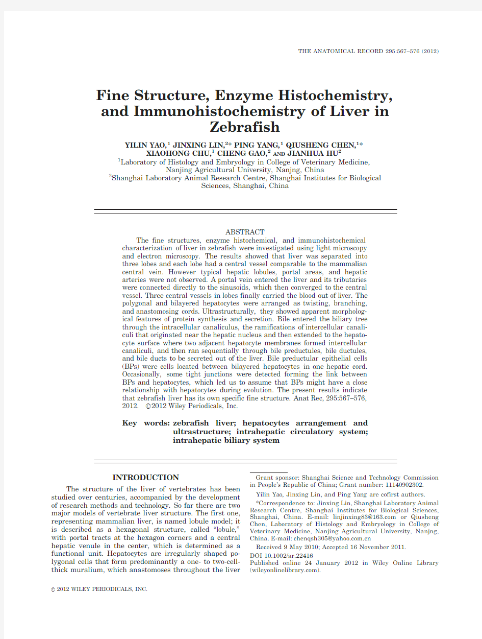
The liver of the zebrafish was composed of three nar-row,long lobes,two longer in the dorsum and one shorter in the center.It was situated in cranial part of abdominal cavity starting from the position caudal to the heart,encircling the whole digestive tract;it was divided into three lobes ventrally and dorsolaterally pro-jecting to the posterior part of cavity,occupying positions between loops of the intestine (Fig.
1A).
Fig.1.Zebrafish liver anatomy characteristics and architecture.A:The liver situated in anterior (A)part of abdominal cavity stating from the position caudal to heart,encircling the whole digestive tract and di-vided into three lobes ventrally and dorsolaterally projecting to the pos-terior (P)part of cavity,occupying positions between loops of the intestine.B:This schema reveals the architecture of the zebrafish liver.Zebrafish hepatocytes usually arranged as bilayered structures to form cords with bile preductule cells (BPs)between two layers,and the cords distributed radially around a central vein.The sinusoids were the passages that transported the blood entering the liver through the por-tal vein and its branches to the central vein,which carried the blood out of the liver.The zebrafish biliary system started from the intracellular canaliculi,extended through intercellular canaliculi,bile preductules,bile ductules,bile duct,and finally to the common bile duct.
CHARACTERISTICS OF LIVER IN ZEBRAFISH
569
H&E Staining
The parenchyma of liver was covered with simple squa-mous epithelium,which formed the serosa.Along the serosa a vessel with nucleated red blood cells in its lumen was found directly connected to the sinusoids in the paren-chyma.The other ends of the sinusoids converged to the vessel in the longitudinal center of each lobe,which was equivalent of the mammalian central vein (Fig.2A,B).
Our observations revealed no typical hepatic lobule and portal area in the zebrafish liver.In addition,there is lit-tle evidence to support the existence of a hepatic artery.The parenchyma was composed of liver cords,which were
specially formed by bilayered hepatocytes and distributed around a central intrahepatic vein (Fig.2C,D).Hepatocyte had a big and spherical single nucleus distinctly located in the center of cell body or close to the sinusoid side membrane,which was supposed to be the basal mem-brane.The cytoplasm was apparently eosinophilic,having a large vacuole-like structure next to the opposite surface of the hepatocyte,which was identified as an apical mem-brane.Bilayered hepatocytes had their apical membranes opposite to one another,and bile preductular cells were located between them (Fig.2E).Additionally,a transverse section of revealed tributaries of the biliary tree formed by cuboidal epithelium (Fig.
2F).
Fig.2.H&E staining.(A )Liver at low magnification (?40).Simple squamous epithelium formed the serosa covering the liver parenchyma connecting with a tenuous blood vessel (:).The radially distributed sinu-soids converged to the central vein ().(
Electron Microscopic Observations Hepatocytes
Hepatocytes had polygonal cell body and flat mem-branes connecting to adjacent hepatocytes where desmosomes were occasionally seen.The nucleus was spherical (diameter:ca.5l m)located in the center of the cell body within which perinuclear cisterna,a double nuclear membrane,and many nuclear pores,were dis-tinctly observed.Well-developed nucleoli (diameter:ca.
2l m),which were sometimes reticulated,were com-posed of an apparent granulose component,fibrillar centers,and a dense fibrillar component.The electron-lucent euchromatin accounted for a large percentage of chromatin in the nucleus (Fig.3A,B).
The cytoplasm was mainly characterized by numerous stacks of nonfenestrated lamellae of rough endoplasmic reticulum (RER)of considerable length,which were arranged in close parallel arrays (sometimes up to 20arrays).Many electron-dense and homogenic
spherical
Fig.3.Ultrastructure of hepatocyte and sinusoid.(A )Many nuclear pores (:)were distinctly observed in the hepatocyte nuclear mem-brane.The well-developed,reticulated nucleoli were comprised of an apparent granulose component ($),and fibrillar centers (~).(B –D)The hepatocyte cytoplasm had numerous stacks of rough endoplas-mic reticulum (RER,~)arranged in close parallel arrays near the nu-cleus ($).Many electron-dense and homogenic spherical lysosomes were distributed at both ends of the RERs.Mitochondria (~)were dis-tributed at the periphery of the RERs.Some of them had scare mito-chondrial cristae or a pseudo myelin structure (white :).Desmosome (:)coule also been seen.(E )An endothelial cell (~)was seen in the si-nusoidal lumen with a red cell ($)in it.The numerous microvilli of the hepatocytes extended into the subendothelial space of Disse (white :).(F )Fat-storing cells (~)located in the space of Disse had an irreg-ular-shaped nucleus,and many lipid droplets in the cytoplasm.Some organelles with membranes (white :)found in the hepatic cytoplasm were presumed as phagolysosomes.(G ,H )Glycogens ($)were dis-covered in a concentrated region at the periphery of the hepatic cyto-plasma,while desmosomes (white :)were also seen between the membranes of hepatocytes.
CHARACTERISTICS OF LIVER IN ZEBRAFISH
571
lysosomes(diameter:ca.0.5l m)were distributed at both ends of the RERs,whereas a few different types of RERs with tubular cisternae were dispersed in the cytoplasm.A large number of mitochondria(diameter:ca.1.5l m)were distributed at the periphery of the lamellar RERs.Some of these structures had scarce mitochondrial cristae or a pseudo myelin structure inside(Fig.3B–D).Glycogens were also discovered in some concentrated regions in the periphery of the hepatic cytoplasm(Fig.3G,H).
Liver sinusoids
Endothelial cells with spindle-shaped nuclei,small amount of cytoplasm,were fenestrated lining the major part of the sinusoidal lumen.Numerous microvilli from hepatocytes extended into the subendothelial space of Disse,which was also the location of fat-storing cells. Fat-storing cells had an irregular-shaped nucleus and many lipid droplets in the cytoplasm.Lymphocytes and erythrocytes were observed in the liver sinusoids(Fig. 3E,F).
Bile canaliculi
Bile canaliculi comprised of the intracellular canaliculi and the intercellular canaliculi.Intracellular canaliculi were formed by invaginated hepatocyte membranes, which were connected to the intercellular canaliculi lined by adjacent hepatocyte membranes.Although named as the intracellular canaliculi,they were ramifi-cations of the intercellular canaliculi to facilitate bile transport,not really inside the hepatocyte.The lumen of the intracellular canaliculi had a large number of https://www.360docs.net/doc/74106080.html,paratively the intercellular canaliculi contained relatively small amounts of microvilli(Fig. 4A,B).
Bile preductules
Preductule,situated between bilayered hepatocytes, formed gaps constituted by hepatocytes and a new cell type called bile preductular cells(BPs),which collected bile from the intercellular canaliculi.The BPs had no ba-sal lamina,but contained a single,elongated nucleus and a long narrow apophysis,which sometimes were linked to hepatocytes by tight junctions(Fig.4C,D). Bile ductules
When the wall of the biliary channels was integrally constituted by two biliary epithelial cells,they became bile ductules.These cells were attached by tight junc-tions and desmosomes to seal the lumen.The ductule cells had no basal lamina and were directly contacted by hepatocytes(Fig.4E).Additionally,a ductule that was composed of a single cell and was embedded between two adjoining liver cells was found by TEM.The lumen of this ductule was very narrow and formed by the infolding of the plasma membrane of the BPs(Fig.4F). Bile ducts
The wall of bile ducts was formed by several cuboidal epithelial cells and a distinct basal lamina separating epithelium from the periphery connective tissue.Apical and lateral surfaces of adjacent epithelial cells were attached by tight junctions.There were a sparse number of long but branching luminal microvilli(Fig.4G).As the dusts became larger,the epithelium turned into columnar epithelial cells,surrounded by thicker connec-tive tissue.
Enzyme Cytochemistry
ATPase
Inside the hepatic cord,bile preductular cells(BPs) located between the bilayered hepatocytes displayed a strongly positive ATPase reaction,as did the intracellu-lar and intercellular canaliculus within the adjacent hepatocytes.The cavosurface of the biliary ductule epi-thelium also showed a positive reaction.The above results demonstrated that intracellular and intercellular canaliculus were connected to the bile preductules which joined the bile ductules,thus revealing the distinct branched structure.The hepatic sinusoid endothelium expressed a powerful ATPase-positive reaction and com-municated with the central vein endothelium,which was positive as well.The ATPase-positive bile ducts epithe-lium had connections with every level of bile ductules and canaliculus to shape the biliary tree in transsection direction(Fig.5A–C).
ACPase
Moderate ACPase activity was observed in the hepatic cells,whose nuclei showed moderate ACPase activity, while the cytoplasm contained one to three positive granules.These positive granules were mainly adjacent to the surface of biliary channels in the liver cells(Fig. 5D,E).
ALPase
An intense ALPase-positive reaction was only observed in the liver sinusoidal endothelium in both the longitudinal sections and cross-sections,whereas the bile system and the hepatic cells were negative(Fig.5F,G). Immunohistochemical Reaction
The antibody(FIS2F11/2)-labeled hepatopancreatic ducts and intrahepatic bile ducts;in contrast to the neg-ative control group,brown positive reactions were found between bilayered hepatocytes representing the primary biliary system that stated from the bile preductules to the bile ductules(Fig.5H,I).
DISSUSSION
Hepatocytes Arrangement and Intrahepatic Circulatory System
As reported,four types of arrangement of parenchy-mal cells are found in the liver of fish.In the first type, hepatocytes are organized radially in tubules made up by five to eight basiconic hepatocytes whose basal sur-face is faced to the sinusoid,with biliary passages(a segment of the biliary tree,with or without epithelial cells)running through the center of this structure.The classes of fish belonging to this type include Atlantic salmon(Salmo salar)(Rocha et al.,1994),rainbow trout (Salmo gairdneri)(Hampton et al.,1985),brown trout
572YAO ET AL.
(Salmo trutta fario )(Robertson and Bradley,1992),and lamprey (Lampetra lamottenii )(Li et al.,2007).In the second arrangement,8–10hepatocytes revealed a tubu-lar arrangement with sinusoids located at the center of the tubule.This type is found in Mullet (Mugil cephalus )(Ma and Huang,1987)and Atlantic croaker (Micropogon undulatus L)(Eurell and Haensly,1982).In the third arrangement,occurring in the tigerfish (Hydrocynus for-skahlii)(Geyer et al.,1996)and golden ide (Leuciscus idus melanotus L )(Braunbeck et al.,1987),the liver is composed of anastomosing bilayered hepatocyte laminae separated by sinusoids.In the fourth arrangement,occurring in the medaka (Oryzias latipes )(Hardman et al.,2007)and pangolin (Cirrhinus molitorella )
(Ma
Fig.4.Ultrastructure of biliary system.(A,B )Bile canaliculi com-prised of intracellular canaliculi
()and intercellular canaliculi (:)with a large number of microvilli in their lumen.(C,D )Long narrow BP cell located between bilayered hepatocytes was discovered with many cell connections (white :)between BP cell and hepatocytes.A bile preduc-tule ($)was formed by BPs and hepatocytes.(E )Bile ductule ($)integrally constituted by two BP cells attached by tight junctions and desmosomes.(F )Transsection of bile ductule (~)composed by a sin-gle infolding BP cell’s apophysis (:).Inside one hepatocyte an intracel-lular canaliculi ($)was located.(G )The wall of the bile duct was formed by several cuboidal epithelial cells,a distinct basal lamina and periphery connective https://www.360docs.net/doc/74106080.html,teral surfaces of adjacent epithelial cells were attached by tight junctions,and a small number of long branching luminal microvilli could be seen.
CHARACTERISTICS OF LIVER IN ZEBRAFISH
573
and Huang,1987),the interconnecting bilayered hepato-cyte cords separated by sinusoids surround the central vein.It can be concluded that the arrangement of liver cells in fish is diversified.
Paris-Palacios et al.(2000)reported that the arrange-ment of hepatic cells in zebrafish was typically consisted of tubules with centrally located sinusoids.Lorent et al.(2004),however,argued that the biliary passages lay in the centre of the tubules instead of sinusoids.We stud-ied the arrangement of hepatic cells in zebrafish by light and electron microscopy and enzyme histochemistry in this article,and reached a different conclusion.We believe that zebrafish hepatocytes are usually arranged in cords consisting of bilayered hepatocytes between which the BPs are located,and cords distributed radially around a central vein,which is approximately in line with type four arrangement of fish hepatocytes.
Additionally enzyme cytochemistry revealed that the structure of hepatocytes and enzymatic reactions have obvious polarity .The sinusoidal sides of the hepatocytes disclosed a basal surface,absorbing materials from blood appearing ALPase-positive,while the bile preductules sides had an apical surface secreting the bile appearing ACPase-positive.The nuclei of hepatocytes were situated at the basal surface,whereas the top part contained abundant cytoplasm and inclusions.These specific features suggest that hepatocytes have apparent directivity of absorption and secretion,requiring a typical strict
arrangement.
Fig.5.Enzyme histochemistry and immunohistochemical reaction.(A )The epithelium of both the biliary system and vasculature showed a strong ATPase-positive reaction.The biliary tree (:?200)in the trans-sected direction showed every level of the biliary system.(B )Bile pre-ductules ()
j
)(?
The zebrafish hepatocyte arrangement and the partly hepatic blood circulatory system are depicted in a sche-matic presentation which displays the sinusoids where the passages transporting the blood from the intestine enter the liver parenchyma through the portal vein and its branches to the large central vessel,which carries blood out of the liver(Fig.1B).Comparison with the mammalian liver structure revealed a number of simi-larities between the zebrafish liver lobe and mammalian liver lobule.First,in both case,the hepatocyte cords are separated by sinusoids.Second,they both secrete the bile through a distinct bile ductule.Third,they both had a vessel in the center called the central vein,which in mammalian is surrounded by hepatic cords and collects blood from the sinusoids.The above three points lead us to presume that zebrafish liver lobes resemble mamma-lian liver lobules in function,which is in agreement with Hardman’s view(Hardman et al.,2007)and is in accord-ance with the rules of evolution.Therefore,the vessel in the longitudinal middle of the elongated zebrafish liver lobe was designated as central vein.However,the gross anatomy of the extrahepatic vein receiving the exiting blood was not clear because of insufficient sample proc-essing,which could be improved in the future. Ultrastructure Characteristic of Hepatocytes The liver is the largest gland inside fish body,and car-ries out bile synthesis,glycogen storage and lipid metabolism,which is also related to vitellogenin synthe-sis.Vitellogenin is the prosoma of vitellogenin in both oviparous vertebrates and invertebrates;its principal function is to provide energy sources for embryonic de-velopment.Female zebrafish started to produce mature eggs at the fifth day of the genesial cycle.Peute et al. (1978)observed ultrastructure changes in the zebrafish liver during these5days.In the first day of production, the number of nuclear pores increased sharply,followed by proliferation of RERs.During the first2days after production,glycogen disappeared,lipid droplets appeared abundantly,and golgi complexes had a rich content and started to secret dense bodies.The liver began to restore its features before production.These changes in the liver occurring during the egg production showed that the liver is in an active phase of vitelloge-nin synthesis,during which fatty storage materials are mobilized and subsequently transported to the gonad.In the spawning period of female zebrafish,the increase in the number of nuclear pores was the first significant morphological character of vitellogenin synthesis.The synthesis of steroid hormones in the liver introduced the vitellogenin production and hence induced yolk forma-tion in the ovaries.This article disclosed a large number of nuclear pores and developed RERs in one hepatocyte sampled as female,which might constitute evidence sup-porting the above study.
Intrahepatic Biliary System
The study so far revealed a number of species differ-ences in the composition and structure of osteichthyes hepatic biliary system.Atlantic salmon(Salmo salar) lacked canaliculus inside hepatocytes(Robertson and Bradley,1992),while crucian(Carassius carassius), which have no intercellular biliary tracts,had canalicu-lus inside each hepatocyte(Tanuma,1980);lamprey (Petromyzon marinus)is a very special kind of fish in that its biliary system and gall bladder appear during childhood but disappear in adults(Sidon et al.,1980; Youson et al.,1985).Medaka’s(Oryzias latipes)(Hard-man et al.,2007)intrahepatic biliary system is an interconnected network of canaliculi and bile preduc-tules.Yamamoto(Yamamoto,1965)and Rocha et al. (1994)discovered directional reticular cytoplasmic fila-ments surrounding the intracellular canaliculi in the peripheral cytoplasm,which even inserted into the mi-crovilli of the intracellular canaliculi to facilitate gall secretion and transport.
Lorent et al.(2004)observed the adult zebrafish liver and concluded that the interhepatic biliary system which comprises many small bile ducts surrounded by hepato-cytes,anastomose with canaliculi from several adjacent hepatocytes,which is in agreement with previous reports in other teleost fish.However,they could not dis-tinguish these small bile ducts.The results of the present study combing enzyme histochemistry,immuno-histochemistry,and electron microscopy,revealed that zebrafish(Brachydanio rerio)biliary system started from the intracellular canaliculi,to extend through the intercellular canaliculi to the bile preductules which were formed by BP cells and the hepatocytes(Fig.1B). Then the bile preductules converged into the bile duc-tules shaped by two BP cells and the latter went off the hepatic cord to join a bile duct surrounded by several cu-boidal epithelial cells.The bile duct gradually thickened until many layers of connective tissue cells and smooth muscle cells surrounded the basement membrane of the epithelium.This structural organization of the zebrafish (Brachydanio rerio)biliary system was mainly in accord-ance with that of the most teleost species,such as Medaka(Oryzias latipes)(Hardman et al,2007),Gold-fish(Torao Y amamoto)(Yamamoto,1965),Rainbow trout (Hampton et al.,1988)and Carp(Cyprinus carpio)(Fish-elson and Becker,2001),but differed from that of Atlantic salmon(Salmo salar),Crucian(Carassius car-assius),and Lamprey(Petromyzon marinus).Electron microscope observation further showed that the intracel-lular canaliculus of zebrafish hepatocytes started near the hepatic nucleolus to extend to the cell surface and connect to the intercellular biliary tracts,and as such apparently differed from that of birds and mammalian. Enzyme cytochemistry revealed ATPase-positive struc-tures in the intracellular canaliculus,intercellular biliary tracts,and cavosurface of the biliary system, indicative of active material transport and biliary secretion.
The Relationship Between BP Cells and Hepatocytes
Opie(1944)discovered a kind of minicell with a small shape,little cytoplasm,and oval nucleus in the rat liver during chemically induced carcinogenesis.Farber later designated these cells as oval cells(Farber,1956).The oval cell is a type of stem cell in the mammalian liver with a strong reproductive activity and bilateral differ-entiation potential(i.e.,it is capable of differentiating into both bile duct epithelium and hepatocytes).Using partial hepatectomy,bile duct ligation,enzyme cyto-chemistry and immunocytochemistry of cytokeratin,
CHARACTERISTICS OF LIVER IN ZEBRAFISH575
Okihiro et al.(2000)found that BP cells could develop into hepatocytes or bile system epithelium in the rain-bow trout,and as such correspond to the oval cells in mammalian liver,while Hardman et al.(2007)advocated the presumption that BPs correlated to mammalian bipotential stem cells.The BP cells in zebrafish had a small shape,little cytoplasm but apparent cell junctions with hepatocytes,leading us to assumed that BP cells and hepatocytes might be closely related and have the same origin.This observation morphologically supports Okihiro’s hypothesis that BP cells might be germ cells of the fish liver.
LITERATURE CITED
Arias IM.1988.The liver.In:Arias IM,Jakoby WB,Popper H, Schachter D,Shafritz DA,editors.Biology and pathobiology.Bal-timore,Maryland:Raven Press.
Braunbeck T,Gorgas K,Storch V,V€o lkl A.1987.Ultrastructure of hepatocytes in golden ide(Leuciscus idus melanotus L.;Cyprini-dae:Teleostei)during thermal adaptation.Anat Embryol(Berl) 175:303–313.
Burkhardt-Holm P,Oulmi Y,Schroeder A,Storch V,Braunbeck T. 1999.Toxicity of4-chloroaniline in early life stages of zebrafish (Daniorerio).Arch Environ Contam Toxicol37:85–102.
Chu J,Sadler KC.2009.New school in liver development:lessons from zebrafish.Hepatology50:1656–1663.
Elias H,Bengelsdorf H.1952.The structure of the liver of verte-brates.Acta Anat14:297–337.
Eurell JA,Haensly WE.1982.The histology and ultrastructure of the liver of Atlantic croaker Micropogon undulates.L.J Fish Biol 21:113–125.
Farber E.1956.Similarities in the sequence of early histological changes induced in the liver of the rat by ethionine,2-acetyla-mino-fluorene,and30-methyl-4-dimethylaminoazobenzene.Cancer Res16:142–148.
Field HA,Ober EA,Roeser T,Stainier DY.2003.Formation of the digestive system in zebrafish.I.Liver morphogenesis.Dev Biol 253:279–290.
Fishelson L,Becker K.2001.Development and aging of the liver and pancreas in the domestic carp,Cyprinus carpio:from embryo-genesis to15-year-old fish.Environ Biol Fishes61:85–97.
Geyer HJ,Nel MN,Swanepoel JH.1996.Histology and ultrastruc-ture of the hepatopancreas of the tigerfish,Hydrocynus forskahlii. J Morphol227:93–100.
Hampton JA,Lantz RC,Goldblatt PJ,Lauren DJ,Hinton DE. 1988.Functional units in rainbow trout(Salmo gairdneri, Richardson)liver.II.The biliary system.Anat Rec221:619–634. Hampton JA,McCuskey PA,McCuskey RS,Hinton DE.1985.Func-tional units in rainbow trout(Salmo gairdneri)Liver:I.Arrange-ment and histochemical properties of hepatocytes.Anat Rec213: 166–175.Hardman RC,Volz DC,Kullman SW,Hinton DE.2007.An in vivo look at vertebrate liver architecture:three-dimensional recon-structions from Medaka(Oryziaslatipes).Anat Rec290:770–782. Hinton DE,Couch JA.1998.Architectural pattern,tissue and cellu-lar morphology in livers of fishes:relationship to experimentally induced neoplastic responses.EXS86:141–164.
Kikuchi K,Verkade H,Reiter JF,Kim CH,Chitnis AB,Kuroiwa A, Stainier DY.2004.Notch signaling can regulate endoderm.Dev Dyn229:756–762.
Li W,Liu R,Jiang Y,Wan Y,Xiong L,Xu J,Hu Q.2007.The influ-ence on the activity of SOD and ATPase in zebrafish liver and branchia by DBP.J Hydroecol27:15–18.
Lorent K,Yeo SY,Oda T,Chandrasekharappa S,Chitnis A,Mat-thews RP,Pack M.2004.Inhibition of Jagged-mediated Notch sig-naling disrupts zebrafish biliary development and generates multi-organ defects compatible with an Alagille syndrome pheno-copy.Development131:5753–5766.
Ma S,Huang R.1987.A comparative study on the microscopic structure of the hepatopancreas in four species of fresh-water fish.Acta Sci Nat Univ Sunyatseni3:106–112.
Okihiro MS,Hinton DE.2000.Partial hepatectomy and bile duct li-gation in rainbow trout(Oncorhynchus mykiss):histologic,immu-nohistochemical and enzyme histochemical characterization of hepatic regeneration and biliary hyperplasia.Toxicol Pathol28: 342–356.
Opie EL.1944.The pathogenesis of tumors of the liver produced by butter yellow.Exp Med80:231–246.
Paris-Palacios S,Biagianti-Risbourg S,Vernet G.2000.Biochemical and(ultra)structural hepatic perturbations of Brachydanio rerio (Teleostei,Cyprinidae)exposed to two sublethal concentrations of copper sulfate.Aquat Toxicol50:109–124.
Peute J,van der Gaag MA,Lambert JG.1978.Ultrastructure and lipid content of the liver of the zebrafish,Brachydanio rerio, related to vitellogenin synthesis.Cell Tissue Res186:297–308. Robertson JC,Bradley TM.1992.Liver ultrastructure of juvenile Atlantic salmon(Salmo salar).J Morphol211:41–54.
Rocha E,Monteiro RA,Pereira CA.1994.The liver of the brown trout,Salmo trutta fario:a light and electron microscope study. Anal185(Part2):241–249.
Sidon EW,Peek WD,Youson JH,Fisher MM.1980.Fine structure of the liver in the larval lamprey,Petromyzon marinus L.:bile ducts and gall bladder.J Anat131(Part3):499–517.
Tanuma Y.1980.Electron microscope observations on the intrahe-patocytic bile canalicules and sequent bile ductules in the crucian, Carassius carassius.Arch Histol Jpn43:1–21.
Tao T,Peng J.2009.Liver development in zebrafish(Danio rerio). J Genet Genom36:325–334.
Wallace KN,Pack M.2003.Unique and conserved aspects of gut de-velopment in zebrafish.Dev Biol255:12–29.
Yamamoto T.1965.Some observations on the fine structure of the lntrahepatic biliary passages in goldfish.Z Zellforsch Mikrosk Anat65:319–330.
Youson JH,Sidon EW,Peek WD,Shivers RR.1985.Ultrastructure of the hepatocytes in a vertebrate liver without bile ducts.J Anat 140(Part1):143–158.
576YAO ET AL.