生物可降解材料聚乙二醇_聚乳酸_聚谷氨酸三_省略_段共聚物与人脐静脉内皮细胞的相
Basic Medicine
中国组织工程研究与临床康复 第 12 卷 第 10 期 2008–03–04 出版
Journal of Clinical Rehabilitative Tissue Engineering Research March 4, 2008 Vol.12, No.10
Cytocompatibility of biodegradable poly(ethylene glycol)-b-poly(L-lactide)-b-poly(L-glutamic acid) tri-block copolymer with human umbilical vein endothelial cells☆
Liu Bin1, Song Xian-jing2,3, Liu Jie-yu3, Jiang Feng4, Shi Yong-feng1, Shuang Dong-si1, Li Zhi-bo1
Second Hospital, Jilin University, Changchun 130041, Jilin Province, China; 2 China-Japan Union Hospital, Jilin University, Changchun 130041, Jilin Province, 3 China; Changchun Centre Hospital, Changchun 130041, Jilin Province, China; 4 Chengde Medical College Affiliated Hospital, Chengde 067000, Hebei Province, China Liu Bin☆, Doctor, Professor, Second Hospital, Jilin University, Changchun 130041, Jilin Province, China Songxianjing2007@2008.sin https://www.360docs.net/doc/931442897.html, Received: 2007-11-03 Accepted: 2008-01-14 (07-50-11-6029/YWY) Liu B, Song XJ, Liu JY, Jiang F, Shi YF, Shuang DS, Li ZB. Cytocompatibility of biodegradable poly(ethylene glycol)-b-poly (L-lactide)-b-poly (L-glutamic acid) tri-block copolymer with human umbilical vein endothelial cells.Zhongguo Zuzhi Gongcheng Yanjiu yu Linchuang Kangfu 2008;12(10): 1950-1953(China) [https://www.360docs.net/doc/931442897.html,/ zglckf/ejournal/ upfiles/12-10/ 10k-1950(ps).pdf]
1
Abstract
BACKGROUND: Poly(ethylene glycol)-b-poly(L-lactide)-b-poly(L-glutamic acid) (PEG-PLA-PGL) tri-block copolymers have good applied foreground in constructing tissue engineering scaffold materials. Whether endothelial cells survive and grow on the materials has a direct influence on the application as a biodegradable material for the scaffold of endothelial cell vector. OBJECTIVE: To explore the cytocompatibility of PEG-PLA-PGL tri-block copolymers with human umbilical vein endothelial cells (HUVECs). DESIGN: Randomized control observation. SETTING: the Second Hospital of Jilin University. MATERIALS: The experiment was carried out in the Department of Pathobiology, School of Basic Medical Sciences, Jilin University from February to October in 2006. Human umbilical cord about 20 cm length came from one neonatal infant who was delivered normally after enough months in the Department of Gynecology and Obstetrics, the Second Hospital of Jilin University. Human umbilical cord was sampled in the informed consents of the infant's family member. The experimentation was authorized by the medical ethic committee of the hospital. PEG-PLA-PGL membranes were provided by Changchun Institute of Applied Chemistry, Chinese Academy of Sciences. Inverted microscope and phase-contrast microscope were bought from Olympus Company (Japan). METHODS: HUVECs cultivated and grew steadily, were inoculated onto PEG-PLA-PGL membranes, serving as the experiment group. While the culture medium without PEG-PLA-PGL membranes were taken as the control group.① Cytocompatibility of PEG-PLA-PGL membranes with HUVECs was evaluated by observing cellular growth through phase-contrast microscope.②The proliferation index of cells was detected by MTT method in 1, 3, 5 and 7 days after inoculation. MAIN OUTCOME MEASURES: ①Cytocompatibility of PEG-PLA-PGL membranes with HUVECs;②The proliferation index of cells in 1, 3, 5 and 7 days after inoculation RESULTS: ①Cytocompatibility of PEG-PLA-PGL membranes with HUVECs: The observation result of phase contrast microscopy showed that, endothelial cells planted on the PEG-PLA-PGL membranes began to attach and stretch after being planted 4-6 hours. Three days later, cells grew in colonies rapidly, after 5 days, colonies began to fuse and seemed like cobble-stone. The cells were shuttle or polygon in shape after passages. There were no significant differences between the experiment and control group. Cells cultured on PEG-PLA-PGL membranes for 15 days grew in inserts with membranes, but they didn't grow into patches through scanning electron microscope.②The proliferation index of cells: No significant differences of the proliferation index of cells were detected by MTT method in 1, 3, 5 and 7 days after inoculation between experiment group and control group (P > 0.05). CONCLUSION: Endothelial cells grow well in PEG-PLA-PGL membranes, and the two have good cytocompatibility.
INTRODUCTION
In the field of new materials about biomedicine, the research related to therapeutic materials, especially materials for coating intracoronary stent have been developed rapidly. From the point of biomedicine, the potential prospects in coronary heart disease and acute myocardial infarction are to find or design a kind of macromolecule material that has good biocompatibility, can carry drugs, genes or cell factors, meanwhile can be biodegradable and release drugs steadily. This study was aimed to evaluate the cytocompatibility of biodegradable poly (ethylene glycol)-b-poly(L-lactide)-b-poly(L-glutamic acid) (PEGPLA-PGL) membrane by observing the growth of human umbilical vein endothelial cells (HUVECs) cultured on the membrane.
MATERIALS AND METHODS
Materials The experiment was carried out in the National Key
Laboratory of Pathobiology Department, School of Basic Medical Sciences, Jilin University from February to October in 2006. Human umbilical cord about 20 cm long came from one neonatal infant, who was delivered normally after enough months in the Department of Gynecology and Obstetrics of the Second Hospital, Jilin University. Human umbilical vein was sampled in the informed consents of the infant's family member. The experimentation was authorized by the medical ethic committee of the hospital. PEG-PLA-PGL membranes were provided by Changchun Institute of Applied Chemistry, Chinese Academy of Sciences. Super-clean desk was bought from Labconco Company (America). CO2 incubator was bought from Jouan Company (France). Inverted microscope and phase-contrast microscope were bought from Olympus Company (Japan). Mean reagents: Phosphate buffered solution (PBS) was prepared by the laboratory of Pathobiology Department, School of Basic Medical Sciences, Jilin University. Penicillin, streptomycin, M199 medium (without Earle's salt) and EDTA were bought from Gibco Company. Fetal blood serum
1950
P.O.Box 1200,Shenyang
110004
kf23385083@https://www.360docs.net/doc/931442897.html,
刘斌,等.生物可降解材料聚乙二醇-聚乳酸-聚谷氨酸三嵌段共聚物与人脐静脉内皮细胞的相容性
https://www.360docs.net/doc/931442897.html,
(FBS) was bought from Hangzhou Sijiqing Company. Separation liquid of lymphocyte (Ficoll paque, 1.077 g/L) was bought from TBD Company (Tianjin). Pancreas enzyme was a product from Sigma Company. Factor Ⅷ-related antigen (FⅧ R-Ag) Immunohistochemistry Kit was purchased from Zhongshan Biological Technology Co., Ltd. (Beijing). Methods Isolation and culture of HUVECs: The isolated human umbilical cord (20 cm) was cultured within 2 hours after the baby was born, then washed at 37 ℃ with PBS (0.01 mmol/l, pH=7.6) until remnant blood in umbilical vein cord was washed down. Detailed approaches were described by Kou BJ and his colleagues[16]. HUVECs were inoculated onto a 25-cm2 culture bottle, which was placed in a culture case at 37 ℃ with 0.05 volume fraction of CO2. Eight hours later, the medium was exchanged for the first time, to eliminate unattached cells; afterwards the medium was altered once every 2 days. When cells fused to a monolayer about 4 days later, foregoing culture medium were abandoned and digested with above digest liquid (2 mL) after washing with PBS. Although digestive liquid was removed 1 minute later, the digestion was continued until the cells shrank and isolated or monolayer cells appeared mesh under inverted microscope. Culture medium were added to culture bottles for preparing cell suspension, which was inoculated in culture bottle at a ratio of 1∶2 (V/V), which was placed in culture case at the same conditions as above. The cells covered the whole monolayer and then generated continually until the seventh generation. Identification of HUVECs by FⅧ R-Ag immunohistochemistry: SP immunohistochemistry method was applied. HUVECs suspension (1 mL) at the cell concentration of 1× 10⁴ was inserted into a glass slide on a 10 cm × 10 cm culture dish, which was placed in a culture case at 37 ℃ with 0.05 volume fraction of CO2. After 24 hours of culture, the glass slide was fetched out and fixed in cool acetone for 10 minutes at 40 ℃. Then the cells were cultured at room temperature with 0.03 volume fraction of H2O2 to eliminate the activity of endogenous peroxidase. Cells were incubated for 30 minutes in medium, which consisted of 100 m L /L PBS dilution of normal goat serum, FⅧ antibody fluid, and biotin-labeled antibody fluid. Horseradish-labeled streptavidin fluid was added to continue the culture, which was followed for 30 minutes at 37 ℃, restaining and mounting. Disposal with PEG-PLA-PGL membranes before HUVECs culture: PEG-PLA-PGL membranes (4 mm×4 mm×2 mm, bore diameter 150-200 μm, porosity 85%-95%) were purged of by tri-distilled water in ultrasonic oscillation, and then dried naturally. Membranes were immerged into 1% NaOH for 2 hours after suffumigation with ethylene oxide, washed by PBS, 50% grain alcohol for 2 hours, polylysine at the ratio of 1∶10 (V/V) for 24 hours, and then dried naturally. The sterilized β-TCP was also cut into a size of 4 mm×4 mm×2 mm. Compound of HUVECs and membranes: Eight PEG-PLA-PGL membranes were put onto 24-pore culture dishes, and dipped with M199 culture medium for 24 hours, then dried naturally for further use. HUVECs of the third generation were digested, centrifuged, isolated and seeded into membranes in a density of 1.0×10⁹ cells/L, serving as ex-
periment group, while equivalent suspensions seeded into other 8 culture pores were taken as controls. Both groups were cultured for 15 days at 37 ℃ with 0.05 volume fraction of CO2 culture dishes. Every day cells were counted by trypan blue method. Growth curve of HUVECs: The third generation HUVECs steadily growing were digested by 2 mL trypsin at 37 ℃, and the density was adjusted to 1.0×10⁵ cells/L. 100 μL cell suspension was added to 96 cores of culture dishes before PEG-PLA-PGL membranes were placed down. Then 100 μL M199 medium containing 20% FBS was add. The experiment was assigned into PEG-PLA-PGL membrane group and control group, each containing 5 pores. Exchanged mediums were given every 3 days. Grow curve was drew after every day counting cells. Assay of HUVECs proliferation: The fourth generation HUVECs steadily growing were seeded in 96-core culture dishes at the density of 1.0×10⁵ cells/L, 100 μL suspended liquids was put in every pore, together with 100 μL M199 medium containing 20% FBS. Then a culture at 37 ℃ with 0.05 volume fraction of CO2 was performed for 7 days. One culture plate was taken out and add with 20 μL MTT (5 g/L) to continue culture for 4 hours in days 1, 3, 5 and 7. After supernatant removal, 150 μL dimethyl sulfoxide (DMSO) was added into every pore and shake for 12 minutes before the absorptance (A) value was measured at 490 nm by enzyme-linked immunoassay, and higher A value indicated stronger cell activity. Statistical analysis: All data were analyzed by the first author by means of SPSS 12.0 analysis software. Results were expressed as Mean±SD. Cell counting and A value between groups were assessed by t test.
RESULTS
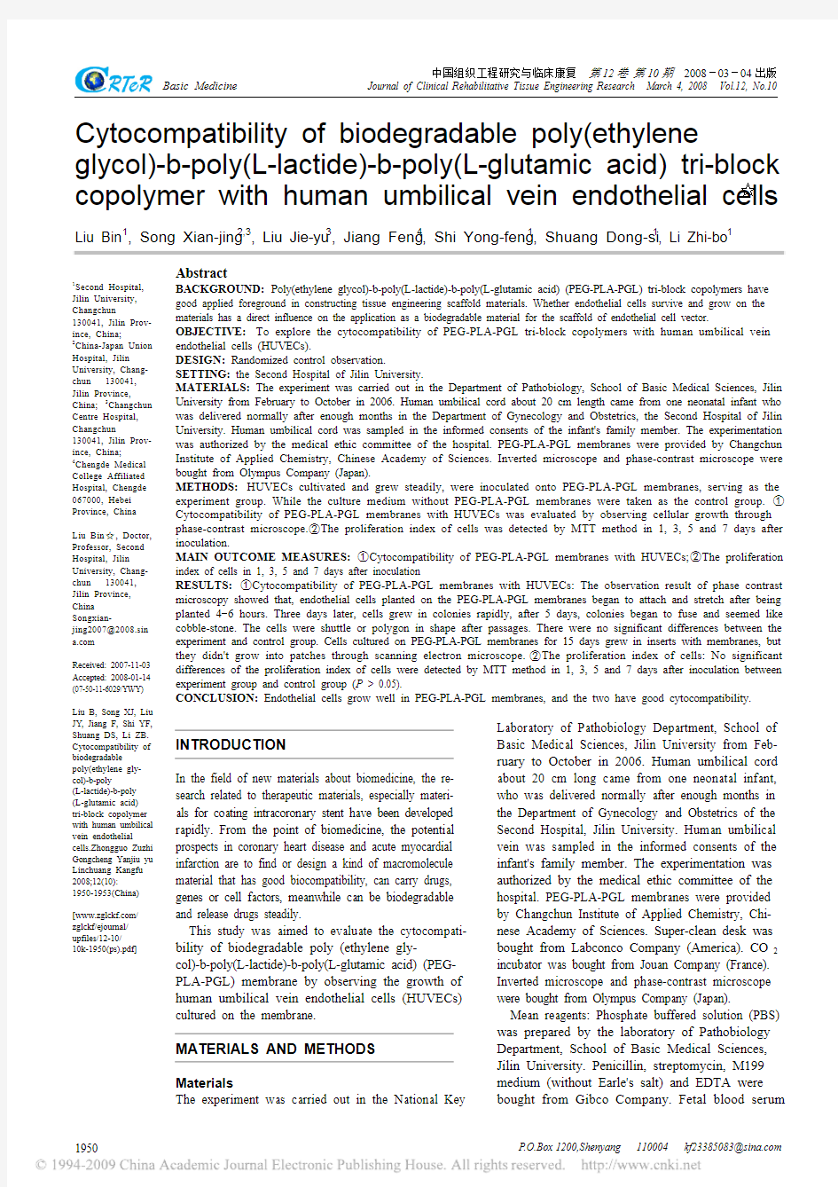
F Ⅷ R-Ag immunohistochemistry and indirect immuno- fluorescence staining Cells in dishes sending out red fluorescence were confirmed as HUVECs (Figure 1).
Figure 1
Factor Ⅷ-related antigen (+) on the cells cultivated in culture bottles
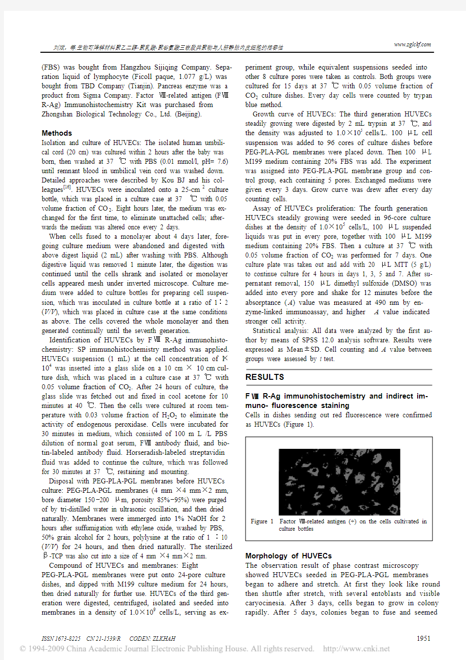
Morphology of HUVECs The observation result of phase contrast microscopy showed HUVECs seeded in PEG-PLA-PGL membranes began to adhere and stretch. At first they look like round then shuttle after stretch, with several entoblasts and visible caryocinesia. After 3 days, cells began to grow in colony rapidly. After 5 days, colonies began to fuse and seemed
1951
ISSN 1673-8225 CN 21-1539/R
CODEN: ZLKHAH
https://www.360docs.net/doc/931442897.html,
刘斌,等.生物可降解材料聚乙二醇-聚乳酸-聚谷氨酸三嵌段共聚物与人脐静脉内皮细胞的相容性
like cobble stone, presenting shuttle and polygon shape after passages (more than 7 generations). Cells cultured on PEG-PLA-PGL membranes for 15 days showed an interstital growth between membranes, but they did not grow into patches (Figure 2).
DISCUSSION
In the biomedical science field, biodegradable macromolecule materials are the most foregone in all world. In the medicine field, the PEG-PLA-PGL membranes have good applied foreground in constructing tissue engineering scaffold materials[1]. As carriers materials, biodegradable layer graft materials must have the characters of no inflammation, no thromboses, and pharmacokinetics that can be predicted, keeping surface integrity, drugs activity constantly and being easy to operate, etc. So the most frontline of preventing restenosis is to look for a kind of macromolecule material with good biocompatibility, carrying drugs, antibodies, cells and genes, etc[2-5], meanwhile the material should degrade and release drugs or antibodies steadily[6-8]. The PEG-PLA-PGL material has new characters and functions, such as hydrophilicity, flexibility, anticoagulant property, anti-macrophages phagocytosis, meanwhile increases the polyactive radical in the base of keeping the good fatty acids, polyamine acids, biocompatibility. It can be as the carrier materials for intracoronary stent[1]. But whether endothelial cells (ECs) can survive and steadily grow on the materials will influence their application in the surface biodegradable materials of ECs carrier. There are few reports about this topic. The experiment was designed to conduct a preliminary study on the cytocompatibility between biodegradable PEG-PLA-PGL materials and ECs, and has supported that PEG-PLA-PGL scaffold is a perfect biodegradable carrier material by observing HUVECs seeded onto the PEG-PLA-PGL membranes. It can be as good tissue engineering blood vessels with normal cytocompatibility. There are lots of methods observing biodegradable materials' compatibility, such as directly planting in vivo and compound cultivating in vitro[9-10]. Because of being influenced by many factors in body, the method in vivo can not reflect compatibility of biomaterial and tissue cells. By the method of culturing cells in vitro, we can directly observe cells' growing, it is more sensitive and objective than the former[11]. However, ECs are inside the blood vessels, which is disadvantaged of the observation and research. It is important to culture ECs in vitro. As the base of cultured ECs, many basic researches are valued gradually[12-13]. Whether ECs can attach availably to the surface of carrier materials will decide the success of planting techniques[14-15]. In the experiment we referred to relevant methods for isolating, culturing and identifying HUVECs, meanwhile improved related methods[16-17]. The results revealed that, HUVECs could steadily grow on the materials and maintained their bioactivity by the observation of their culture. Normal ECs layer covering the surface of blood vessels are the most important barrier of protecting blood from cruor. Blood stent materials seeded ECs can inhibit blood platelet's activity/thrombocyte[18]. Herring et al[19] have put forward to and approved that ECs can grow in blood vessel surface and form total intima. Artificial blood vessel endothelium emerges as the time require. Circulating endothelial progenitor cells (EPCs) have the characters of differentiating ECs and repairing scathed intima, as well as forming new blood vessels. So
Figure 2
Endothelial cells cultured for 15 days on poly(ethylene glycol)-b-poly(L-lactide)-b-poly(L-glutamic acid) under microscopy (×400)
Cells count and grow curve HUVECs grew in the speed of logarithm as time went on. During 2-5 days, cells grew most rapidly. The growing speed of HUVECs on PEG-PLA-PGL membranes wasn't lower than the controls in 7 days (Figure 3).
9 8 7 6 5 4 3 2 1 0 1 2 3 4 5 6 7 8 t(culture)/d
PEG-PLA-PGL: poly(ethylene glycol)-b-poly(L-lactide)-b-poly
(L-glutamic acid) Figure 3 Growth curve of human umbilical vein endothelial cells
Proliferation of HUVECs As time went on, A value increased. It suggested that HUVECs' proliferation activity increased when time went on. However, there was no statistical difference in the cell activity between the two groups at the same time (Table 1).
Table 1 Absorptance value of human umbilical vein endothelial _ cells (x±s)
t(post culture)/d 1 3 5 7
0.160±0.012 0.297±0.007 0.364±0.020 0.402±0.014 Control PEG- PLA- 0.154±0.032 0.265±0.009 0.347±0.040 0.398±0.022 PGL PEG-PLA-PGL:
Cell density (×105 /L)
Control PEG-PLA-PGL
Group
poly(ethylene
glycol)-b-poly(L-lactide)-b-poly
(L-glutamic acid)
1952
P.O.Box 1200,Shenyang
110004
kf23385083@https://www.360docs.net/doc/931442897.html,
刘斌,等.生物可降解材料聚乙二醇-聚乳酸-聚谷氨酸三嵌段共聚物与人脐静脉内皮细胞的相容性
https://www.360docs.net/doc/931442897.html,
EPCs are more and more concerned in treating coronary artery disease. The Healing's research (Healthy Endothelial Accelerated Lining Inhibits Neointimal Growth) that is under way is to aim at the CD34 antibody covering in support graft surface, which is a new method to prevent in-stent restenosis. It has obtained primary effects[20]. In the present experiment, we cultured ECs on PEG-PLA-PGL membranes and found ECs grow well on the membranes, becoming total ECs layer. This suggested that the material is an ideal graft material on which ECs grow well. Thus it provides the foundation for constructing the tissue engineering degradable scaffolds and further studying the biodegradable molecular carriers of EPCs CD34 antibody.
生物可降解材料聚乙二醇-聚乳酸-聚谷 氨酸三嵌段共聚物与人脐静脉内皮 细胞的相容性☆
刘 斌 ,宋显晶 ,刘婕妤 ,姜 锋 ,史永峰 ,双东思 ,李智博
1
1 2,3 3 4 1 1 1
吉林大学第二医学院,吉林省长春市 130041;
3
130041;
2
吉林大学中日联谊 吉林省长春市 067000
医院, 吉林省长春市 刘
长春市中心医院,
130041;4 承德医学院附属医院,河北省承德市
斌☆, 男,1964 年生,吉林省长春市人,汉族,2007 年吉林
大学第二医学院毕业,博士,教授,主要从事心脏病介入治疗与冠 心病研究。
REFERENCES
1 2 3 Guo Y, Li M, Mylonakis A, et al. Electroactive oligoaniline-containing self-assembled monolayers for tissue engineering applications. Biomacromolecules 2007;8(10):3025-3034 Walter DH, Cejna M, Diaz-Sandoval L, et al. Local Gene Transfer of phVEGF-2 plasmid by Gene-Eluting Stents: An Alternative Strategy for Inhibition of Restenosis. Circulation 2004;110(1):36-45 Matsumura G, Miyagawa-Tomita S, Shin'oka T, et al. First evidence that bone marrow cells contribute to the construction of tissue-engineered vascular autografts in vivo. Circulation 2003; 108(14):1729-1734 Costa MA, Simon DI. Molecular basis of restenosis and drug-eluting stents. Circulation 2005;111(17):2257-2273 Sekine H, Shimizu T, Yang J, et al. Pulsatile myocardial tubes fabricated with cell sheet engineering. Circulation 2006;114(1 Suppl):I87-93 Zhang AX, Lü DL, Zhong W, et al. Study advances in natural biomaterials used as scaffolds for tissue engineering 2005; 24(5): 387-389 Chen ZX, Zhang ZW, Tian H, et al. Application of biodegradable polymeric materials in medicine. Huaxue Tuijinji yu Gaofenzi Cailiao 2005;3(1):31-34 Hu XF, Liao YZ, Huang YD, et al. Primary study of characteristics and biocompatibility of the O,O-Dilauryl Chitosan/PLLA blend membrane. Zuzhi Gongcheng yu Chongjian Waike Zazhi 2006;2(6):304-307 Rotte KH, Kriedemann E. Possibilities and limitations of computed tomography in the diagnosis of advanced stomach cancer and tumor recurrence. Arch Geschwulstforsch 1983;53(5):449-457 Mohammadi M, Shokrgozar MA, Mofid R. Culture of human gingival fibroblasts on a biodegradable scaffold and evaluation of its effect on attached gingiva: a randomized, controlled pilot study. J Periodontol 2007;78(10):1897-1903 el-Ghannam A, Ducheyne P, Shapiro IM. Formation of surface reaction products on bioactive glass and their effects on the expression of the osteoblastic phenotype and the deposition of mineralized extracellular matrix. Biomaterials 1997;18(4):295-303 Cao HP, Shu ZB. Antiangiogenesis effect of thalidmide on mice bearing Lewis lung cancer cells. Jilin Daxue Xuebao: Yixueban 2003;29(3):298-300 Campbell GR, Camphell JH. Development of tissue engineered vascular grafts. Curr Pharm Biotechnol 2007;8(1):43-50 Xiong M, Xia WS. Effect of shear on the form and function of vascular endothelial cells on PLGA mesh. Disi Junyi Daxue Xuebao 2005:26(23) Campbell GR, Campbell JH. Development of tissue engineered vascular grafts. Curr Pharm Biotechnol 2007;8(1):43-50 Kou BJ, Li YL, Shi YA. Isolation,culture,and identification of human and mouse endothelial cells. Jilin Daxue Xuebao: Yixueban 2005;1(1):155-157 Xiao RD, Weng GX. Bio-specialty of culture endothelial cell of rabbit aorta. Fujian Yike Daxue Xuebao 2006;40(5):452-455 Heyligers JM, Arts CH, Verhagen HJ, et al. Improving small-diameter vascular grafts: from the application of an endothelial cell lining to the construction of a tissue-engineered blood vessel. Ann Vasc Surg 2005;19(3):448-456 Herring M, Smith J, Dalsing M, et al. Endothelial seeding of polytetrafluoroethylene femoral popliteal bypasses: the failure of low-density seeding to improve patency. J Vasc Surg 1994;20(4): 650-655 Aoki J, Serruys PW, van Beusekom H, et al. Endothelial progenitor cell capture by stents coated with antibody against CD34: the HEALING-FIM (Healthy Endothelial Accelerated Lining Inhibits Neointimal Growth-First In Man) Registry. J Am Coll Cardiol 2005;45(10):1574-1579
摘要
背景:聚乙二醇-聚乳酸-聚谷氨酸三嵌段共聚物膜片材料在构建组 织工程支架上具有良好的应用前景,内皮细胞能否在该材料上存活 和稳定生长将直接影响其作为载内皮细胞载体支架表面可降解材料 的应用。 目的:观察聚乙二醇-聚乳酸-聚谷氨酸三嵌段共聚物与人脐静脉内 皮细胞的相容性。 设计:随机对照观察。 单位:吉林大学第二临床医学院。 材料:本实验于 2006-02/10 于吉林大学基础医学院病理生理教研室 完成。长约 20 cm 脐带来自吉林大学第二医院妇产科 1 名正常足月 分娩的新生儿,标本采集经新生儿家属知情同意,本实验经过医院 伦理委员会批准。聚乙二醇-聚乳酸-聚谷氨酸三嵌段共聚物膜片由 中国科学院长春应用化学研究所提供。倒置显微镜及相差显微镜为 日本 Olympus 公司产品。 方法:将从脐带中分离培养的稳态生长的人脐静脉内皮细胞接种于 聚乙二醇-聚乳酸-聚谷氨酸三嵌段共聚物膜片上培养作为实验组, 对照组为未放聚乙二醇-聚乳酸-聚谷氨酸三嵌段共聚物膜片的培养 皿。①以相差显微镜下观察细胞生长情况评价人脐静脉内皮细胞与 聚乙二醇-聚乳酸-聚谷氨酸三嵌段共聚物膜片的细胞相容性。②采 用 MTT 法检测细胞种植后 1,3,5 及 7 d 的细胞增殖指数。 主要观察指标:①人脐静脉内皮细胞与聚乙二醇-聚乳酸-聚谷氨酸 三嵌段共聚物膜片的细胞相容性。②细胞种植后 1,3,5 及 7 d 的 细胞增殖指数。 结果:①人脐静脉内皮细胞与聚乙二醇-聚乳酸-聚谷氨酸三嵌段共 聚物膜片的细胞相容性:相差显微镜下种植在聚乙二醇-聚乳酸-聚 谷氨酸三嵌段共聚物膜片上的细胞 4~6 h 后即开始贴壁、伸展,3 d 后细胞开始呈集落样生长,生长较快,5 d 后细胞集落开始融合,呈 现特征性的鹅卵石样形态,经多次传代后,细胞呈长梭形或多角形, 与对照组相比无明显差异。扫描电镜下观察在聚乙二醇-聚乳酸-聚 谷氨酸三嵌段共聚物膜片上培养 15 d 的人脐静脉内皮细胞嵌入膜片 间隙生长,但没有连接成片铺在膜片上。②细胞增殖指数:MTT 法 检测结果显示实验组及对照组实验后 1,3,5 及 7 d 的细胞增殖指 数差异无显著性意义(P > 0.05) 。 结论:内皮细胞在聚乙二醇-聚乳酸-聚谷氨酸三嵌段共聚物上生长 良好,两者具有良好的细胞相容性。 关键词:细胞相容性;聚乙二醇-聚乳酸-聚谷氨酸;内皮细胞; 生 物相容性材料
中图分类号: R318.08 文献标识码: A 文章编号: 1673-8225(2008)10-01950-04
4 5 6 7 8 9 10
11
12 13 14 15 16 17 18
19 20
刘斌,宋显晶,刘婕妤,姜锋,史永峰,双东思,李智博.生物可降解 材料聚乙二醇-聚乳酸-聚谷氨酸三嵌段共聚物与人脐静脉内皮细胞的 相容性[J].中国组织工程研究与临床康复,2008,12(10):1950-1953 [https://www.360docs.net/doc/931442897.html,/zglckf/ejournal/upfiles/12-10/10k-1950(ps).pdf] (Edited by: H YC/Yang Y/Wang L)
ISSN 1673-8225 CN 21-1539/R
CODEN: ZLKHAH
1953