Directed differentiation of human pluripotent stem

LETTER
doi:10.1038/nature09691
Directed differentiation of human pluripotent stem cells into intestinal tissue in vitro
Jason R.Spence 1,Christopher N.Mayhew 1,Scott A.Rankin 1,Matthew F.Kuhar 1,Jefferson E.V allance 2,Kathryn Tolle 1,
Elizabeth E.Hoskins 3,Vladimir V.Kalinichenko 1,4,Susanne I.Wells 3,Aaron M.Zorn 1,Noah F.Shroyer 1,2&James M.Wells 1
Studies in embryonic development have guided successful efforts to direct the differentiation of human embryonic and induced pluripotent stem cells (PSCs)into specific organ cell types in vitro 1,2.For example,human PSCs have been differentiated into monolayer cultures of liver hepatocytes and pancreatic endocrine cells 3–6that have therapeutic efficacy in animal models of liver disease 7,8and diabetes 9,respectively.However,the generation of complex three-dimensional organ tissues in vitro remains a major challenge for translational studies.Here we establish a robust and efficient process to direct the differentiation of human PSCs into intestinal tissue in vitro using a temporal series of growth factor manipulations to mimic embryonic intestinal development 10.This involved activin-induced definitive endoderm formation 11,FGF/Wnt-induced posterior endoderm pattering,hindgut specification and morphogenesis 12–14,and a pro-intestinal culture system 15,16to promote intestinal growth,morphogenesis and cytodifferentia-tion.The resulting three-dimensional intestinal ‘organoids’con-sisted of a polarized,columnar epithelium that was patterned into villus-like structures and crypt-like proliferative zones that expressed intestinal stem cell markers 17.The epithelium contained functional enterocytes,as well as goblet,Paneth and enteroendo-crine https://www.360docs.net/doc/944716126.html,ing this culture system as a model to study human intestinal development,we identified that the combined activity of WNT3A and FGF4is required for hindgut specification whereas FGF4alone is sufficient to promote hindgut morphogenesis.Our data indicate that human intestinal stem cells form de novo during development.We also determined that NEUROG3,a pro-endocrine transcription factor that is mutated in enteric anendocrinosis 18,is both necessary and sufficient for human enteroendocrine cell development in vitro .PSC-derived human intestinal tissue should allow for unprecedented studies of human intestinal development and disease.
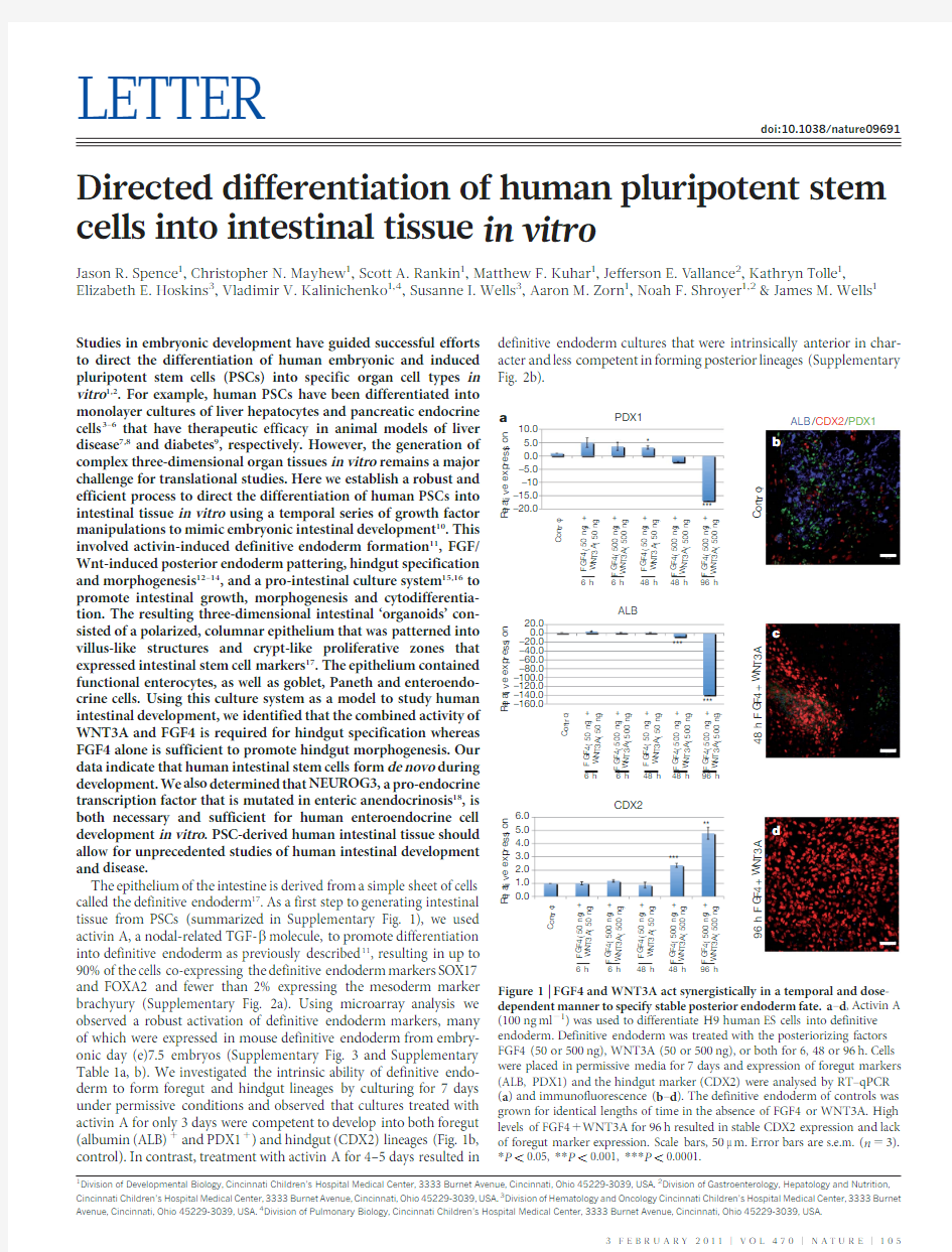
The epithelium of the intestine is derived from a simple sheet of cells called the definitive endoderm 17.As a first step to generating intestinal tissue from PSCs (summarized in Supplementary Fig.1),we used activin A,a nodal-related TGF-b molecule,to promote differentiation into definitive endoderm as previously described 11,resulting in up to 90%of the cells co-expressing the definitive endoderm markers SOX17and FOXA2and fewer than 2%expressing the mesoderm marker brachyury (Supplementary Fig.2a).Using microarray analysis we observed a robust activation of definitive endoderm markers,many of which were expressed in mouse definitive endoderm from embry-onic day (e)7.5embryos (Supplementary Fig.3and Supplementary Table 1a,b).We investigated the intrinsic ability of definitive endo-derm to form foregut and hindgut lineages by culturing for 7days under permissive conditions and observed that cultures treated with activin A for only 3days were competent to develop into both foregut (albumin (ALB)1and PDX11)and hindgut (CDX2)lineages (Fig.1b,control).In contrast,treatment with activin A for 4–5days resulted in
definitive endoderm cultures that were intrinsically anterior in char-acter and less competent in forming posterior lineages (Supplementary Fig.2b).
1
Division of Developmental Biology,Cincinnati Children’s Hospital Medical Center,3333Burnet Avenue,Cincinnati,Ohio 45229-3039,USA.2Division of Gastroenterology,Hepatology and Nutrition,Cincinnati Children’s Hospital Medical Center,3333Burnet Avenue,Cincinnati,Ohio 45229-3039,USA.3Division of Hematology and Oncology Cincinnati Children’s Hospital Medical Center,3333Burnet Avenue,Cincinnati,Ohio 45229-3039,USA.4Division of Pulmonary Biology,Cincinnati Children’s Hospital Medical Center,3333Burnet Avenue,Cincinnati,Ohio 45229-3039,
USA.
C o n t r o l
48 h F G F 4+W N T 3A 96 h F G F 4+W N T 3A
ALB /CDX2/PDX1
a
*
***
***
*****
10.05.00.0–5.0–10–15.0–20.0
C o n t r o l
F G F 4 (50 n g ) +W N T 3A (50 n g )
F G F 4 (500 n g ) +W N T 3A (500 n g )
F G F 4 (50 n g ) +W N T 3A (50 n g )
F G F 4 (500 n g ) +W N T 3A (500 n g )
F G F 4 (500 n g ) +W N T 3A (500 n g )
6 h 6 h 48 h 48 h
96 h
C o n t r o l
F G F 4 (50 n g ) +W N T 3A (50 n g )
F G F 4 (500 n g ) +W N T 3A (500 n g )
F G F 4 (50 n g ) +W N T 3A (50 n g )
F G F 4 (500 n g ) +W N T 3A (500 n g )
F G F 4 (500 n g ) +W N T 3A (500 n g )
C o n t r o l
F G F 4 (50 n g ) +W N T 3A (50 n g )
F G F 4 (500 n g ) +W N T 3A (500 n g )
F G F 4 (50 n g ) +W N T 3A (50 n g )
F G F 4 (500 n g ) +W N T 3A (500 n g )
F G F 4 (500 n g ) +W N T 3A (500 n g )
***
PDX1
ALB
CDX2
R e l a t i v e e x p r e s s i o n
R e l a t i v e e x p r e s s i o n
R e l a t i v e e x p r e s s i o n
20.00.0–20.0–40.0–60.0–80.0–100.0–120.0–140.0–160.06.05.04.03.02.01.00.0
*
6 h 6 h 48 h 48 h
96 h
6 h 6 h 48 h 48 h
96 h
Figure 1|FGF4and WNT3A act synergistically in a temporal and dose-dependent manner to specify stable posterior endoderm fate.a –d ,Activin A (100ng ml 21)was used to differentiate H9human ES cells into definitive endoderm.Definitive endoderm was treated with the posteriorizing factors FGF4(50or 500ng),WNT3A (50or 500ng),or both for 6,48or 96h.Cells were placed in permissive media for 7days and expression of foregut markers (ALB,PDX1)and the hindgut marker (CDX2)were analysed by RT–qPCR (a )and immunofluorescence (b –d ).The definitive endoderm of controls was grown for identical lengths of time in the absence of FGF4or WNT3A.High levels of FGF41WNT3A for 96h resulted in stable CDX2expression and lack of foregut marker expression.Scale bars,50m m.Error bars are s.e.m.(n 53).*P ,0.05,**P ,0.001,***P ,0.0001.
3F E B R U A R Y 2011|V O L 470|N A T U R E |105
Having identified the window of time when definitive endoderm fate was plastic (day 3of activin A treatment),we used WNT3A and FGF4to promote hindgut and intestinal specification.Studies in mouse,chick and frog embryos have demonstrated that Wnt and FGF signalling pathways are required for repressing anterior develop-ment and promoting posterior endoderm formation into the midgut and hindgut 12–14.Consistent with this,conditioned media containing WNT3A was recently shown to promote Cdx2expression in mouse embryonic stem (ES)-cell-derived embryoid bodies 19.In human defini-tive endoderm cultures,neither factor alone was sufficient to robustly promote a posterior fate (Supplementary Fig.2c);but high concentra-tions of both FGF4and WNT3A (FGF41WNT3A)induced expres-sion of the hindgut marker CDX2in the definitive endoderm after 48h (Supplementary Fig.4).However,48h of FGF41WNT3A treatment did not stably induce a CDX21hindgut fate and expression of anterior markers PDX1and albumin reappeared after cells were cultured in permissive media for 7days (Fig.1a,c).In contrast,96h of exposure to FGF41WNT3A resulted in stable CDX2expression and absence of anterior markers (Fig.1a,d).These findings indicate a previously unidentified requirement for the synergistic activities of both the FGF and Wnt pathways in specifying the CDX21mid/hindgut lineage.Remarkably,FGF41WNT3A-treated cultures underwent morpho-genesis that was similar to embryonic hindgut formation.Between 2and 5days of FGF41WNT3A treatment,flat cell sheets condensed into CDX21epithelial tubes,many of which budded off to form floating hindgut spheroids (Fig.2a–c,Supplementary Fig.5a–f and Sup-plementary Table 2a).Spheroids were similar to e8.5mouse hindgut and consisted of uniformly CDX21polarized epithelium surrounded by CDX21mesenchyme (Fig.2d–g).Spheroids were completely devoid
of albumin and PDX1-expressing foregut cells (Supplementary Fig.5h,i).In vitro gut-tube morphogenesis was never observed in control or WNT3A-only treated cultures.FGF4-treated cultures had a twofold expansion of mesoderm and generated 4–10-fold fewer spheroids (Supplementary Fig.2c and Supplementary Table 2a),which were weakly CDX21and did not undergo further expansion (data not shown).Together our data support a mechanism for hindgut develop-ment where FGF4promotes mesoderm expansion and morphogenesis,whereas FGF4and WNT3A synergy is required for the specification of the hindgut lineage.
Importantly,this method for directed differentiation is broadly applicable to other PSC lines,as we were able to generate hindgut spheroids from both H1and H9human ES cell lines and from four induced PSC (iPSC)lines that we have generated and characterized (Supplementary Figs 3,5and 6).The kinetics of differentiation and the formation of spheroids were comparable between these lines (Sup-plementary Table 2).Two other iPSC lines tested were poor at hindgut spheroid formation and line iPSC3.6also had a divergent transcrip-tional profile during definitive endoderm formation (Supplementary Fig.3and Supplementary Table 2c).
Whereas in vivo engraftment of PSC-derived cell types,such as pan-creatic endocrine cells,has been used to promote maturation 9,efficient development and maturation of organ tissues in vitro has proven more difficult.We investigated whether hindgut spheroids could develop and mature into intestinal tissue in vitro using recently described three-dimensional culture conditions that support growth and renewal of the adult intestinal epithelium 15,16.When placed into this culture system,hindgut spheroids developed into intestinal organoids in a staged manner that was notably similar to fetal gut development (Fig.3,Supplementary Fig.5g and Supplementary Fig.7).In the first 14days the simple cuboidal epithelium of the spheroid expanded and formed a highly convoluted pseudostratified epithelium surrounded by mesenchymal cells (Fig.3a–c),similar to an e12.5fetal mouse gut (Fig.3f).After 28days,the epithelium matured into a columnar epithelium with villus-like involutions that protrude into the lumen of the organoid (Fig.3d,e).Comparable transitions were observed during mouse fetal intestinal development (Fig.3f,g and Supplemen-tary Fig.7).The spheroids expanded up to 40fold in mass as they formed organoids (data not shown)and were split and passaged over 9additional times and cultured for over 140days with no signs of growth failure.The cellular gain during that time was up to 1,800fold (data not shown),resulting in a total cellular expansion of 72,000fold per hindgut spheroid.This directed differentiation was up to 50fold more efficient than spontaneous embryoid body differentiation meth-ods 20(Supplementary Fig.8)and resulted in organoids that were almost entirely intestinal (Supplementary Fig.2e–g)as compared to embryoid bodies that contained a mix of neural,vascular and epidermal tissues (Supplementary Fig.8).
Marker analysis showed that after 14days in culture,virtually all of the epithelium expressed the intestinal transcription factors CDX2,KLF5and SOX9broadly and was highly proliferative (Fig.3b,c).By 28days,CDX2and KLF5remained broadly expressed in over 90%of the epithelium (Supplementary Fig.2),whereas SOX9became localized to pockets of proliferating cells at the base of the villus-like protrusions (Fig.3d,e)similar to the intervillus epithelium of fetal mouse intestines at e16.5(Fig.3g and Supplementary Fig.9).5-bromodeoxyuridine (BrdU)pulse chase and analysis of organoids using a Z-stack series of confocal microscopic images showed that epithelial BrdU incorp-oration was largely restricted to SOX9-expressing cells in crypt-like structures that penetrated into the underlying mesenchyme (Sup-plementary Fig.9).At 28days,LGR5is not expressed and ASCL2(ref.21)is broadly expressed and not restricted to the SOX91prolif-erative zone.However,organoids cultured until 56days expressed both ASCL2and LGR5in restricted epithelial domains that appear to over-lap with the SOX91zone (Fig.3h–j and Supplementary Fig.10).This domain is similar to developing intestinal progenitor domains in vivo
,
c d
b
e
f Nuclei CDX2
g Nuclei
CDX2Nuclei
Laminin Nuclei
E-cadherin
a
Nuclei /CDX2
Figure 2|Morphogenesis of posterior endoderm into three-dimensional,hindgut-like spheroids.a ,Bright-field images of definitive endoderm cultured for 96h in media,FGF4,WNT3A or FGF41WNT3A.FGF41WNT3A
cultures contained three-dimensional epithelial tubes and free-floating spheres (black arrows)b ,CDX2immunostaining (green)and nuclear stain (DRAQ5,blue)on cultures shown in a .Insets show CDX2staining alone.c ,Bright-field image of hindgut-like spheroids.a –c ,Scale bars,50m m.d –f ,Analysis of CDX2,basal-lateral laminin and E-cadherin expression demonstrates an inner layer of polarized,cuboidal,CDX21epithelium surrounded by non-polarized
mesenchymal CDX21cells.Scale bar in e is 20m m.g ,CDX2expression in an e8.5mouse embryo (sagittal section).Inset is a magnified view showing that both hindgut endoderm (E;outlined with a red dashed line)and adjacent mesenchyme (M)are CDX2positive (green).FG,foregut;HG,hindgut.Scale bar,100m m.
RESEARCH LETTER
106|N A T U R E |V O L 470|3F E B R U A R Y 2011
which ultimately give rise to the stem cell niche in the crypt of Lieberku ¨hn 15.iPSCs were equally capable of forming intestinal progeni-tor domains (Supplementary Fig.9e).Thus,PSC-derived intestinal epithelium continued to mature in vitro and develop proliferative domains with nascent intestinal stem cells.
Between 18and 28days in culture,we observed cytodifferentiation of the stratified epithelium into a columnar epithelium containing brush borders and all of the major cell lineages of the gut as determined by immunofluorescence and quantitative polymerase chain reaction with reverse transcription (RT–qPCR)(Fig.4a–d and Supplementary Fig.11).By 28days of culture,villin (Fig.4a)and DPPIV (not shown)were localized to the apical surface of the polarized columnar epithe-lium and transmission electron microscopy revealed a brush border of apical microvilli indistinguishable from those found in mature intest-ine (Fig.4d and Supplementary Fig.1).Enterocytes had a functional peptide transport system and were able to absorb a fluorescently
labelled dipeptide (Fig.4e)22.Cell counting revealed that the epithe-lium contained approximately 15%MUC21goblet cells,which secrete mucins into the lumen of the organoid,18%lysozyme-positive cells,which are indicative of Paneth cells,and ,1%chromogranin-A-expressing enteroendocrine cells (Fig.4and Supplementary Fig.11g).MUC2and lysozyme staining indicated that the goblet and Paneth cells in 28-day organoids are immature (Fig.4a,b).However,in organoids that were passaged over 100days,all cells had acquired a more mature phenotype and Paneth cells were often localized in crypt-like structures (Supplementary Fig.12b,c).RT–qPCR confirmed the presence of additional markers of differentiated enterocytes (FABP2;also known as IFABP )and Paneth cells (MMP7)(Supplementary Fig.11).Indivi-dual organoids seemed to be a mix of proximal intestine (GATA41/GATA61)and distal intestine (GATA42/GATA61;HOXA13-expressing)(Supplementary Figs 11and 13)23.Thus,directed differ-entiation of PSCs into intestinal tissue in vitro is highly efficient in generating three-dimensional intestinal tissue containing crypt-like progenitor niches,villus-like domains and all of the differentiated cell types of the intestinal epithelium.
Intestinal organoids contained a mesenchymal layer that developed along with the epithelium in a staged manner similar to embryonic development 10,24(Supplementary Fig.14).Mesenchyme probably came from the 2%of mesoderm cells that were present after activin differentiation,which expanded up to 10%in FGF4-treated hindgut cultures (Supplementary Fig.2).At 14days,organoids broadly expressed mesenchymal markers including FOXF1and vimentin
MUC2/VIL
CDX2/CHGA Nuc /CDX2/LYZ
Nuc /CDX2/CHGA
ECad
d
28-d a y i P S C
h
k
Ala-Lys-AMCA/CDX2/B-Cat 76Ad-GFP Control shRNA NEUROG3shRNA
Ad-NEUROG3
543210
21.81.61.41.20.80.60.40.210
C D X 2 + C H G A + c e l l s (%)
C D X 2 + C H G A + c e l l s (%)
*
*
Figure 4|Formation and function of intestinal cell types and regulation of enteroendocrine differentiation by NEUROG3.a –c ,Twenty-eight-day iPSC-derived organoids were analysed for villin (VIL)and the goblet cell marker mucin (MUC2)(a ),the Paneth cell marker lysozyme (LYZ)(b ),or the endocrine cell marker chromogranin A (CHGA)(c ).Nuc,nuclei.d ,Electron micrograph showing an enterocyte cell with a characteristic brush border with microvilli (inset).e ,Epithelial uptake of the fluorescently labelled dipeptide d-Ala-Lys-AMCA (arrowheads)indicating a functional peptide transport system.f –h ,Adenoviral expression of NEUROG3(Ad-NEUROG3)causes a fivefold increase in CGA 1cells compared to a GFP control (Ad-GFP).n 54biological samples;*P 50.005.i –k ,Organoids were generated from human ES cells that were stably transduced with shRNA-expressing lentiviral https://www.360docs.net/doc/944716126.html,pared to control shRNA organoids,NEUROG3shRNA organoids had a 95%reduction in the number of CHGA 1cells.n 53for shRNA controls and n 55for NEUROG3-shRNA;*P 50.018.Scale bar in a is 10m m;all others are 20m m.Error bars are s.e.m.
d014-day organoid C D X 2/N u c /K i 67
K L F 5/C D X 2/S O X 9
K L F 5/C D X 2/S O X 9
28-day organoid
d1
d2
d3
d4
d9
d13
a
d
e
g
H 9 o r g a n o i d s
SOX9LGR5ASCL2
h
i j
e12.5 mouse
e16.5 mouse
Figure 3|Human ES cells and iPSCs form three-dimensional intestine-like organoids.a ,A time course shows that intestinal organoids formed highly convoluted epithelial structures surrounded by mesenchyme after 13days (d).b –e ,Intestinal transcription factor expression (KLF5,CDX2,SOX9)and cell proliferation on serial sections of organoids after 14and 28days (serial sections are b and c ,d and e ).Ki67,nuclear proliferation antigen.Nuc,nuclei.
f ,
g ,Expression of KLF5,CDX2,and SOX9in mouse fetal intestine at e14.5(f )and e16.5(g )is similar to developing intestinal organoids.The right panels show separate colour channels for d ,e and g (bracket highlights the region shown in the panels on the right).
h –j ,Whole mount in situ hybridization of 56-day-old organoids showing epithelial expression of SOX9(h )and restricted ‘crypt-like’expression of the stem cell markers LGR5(
i )and ASCL2(
j ).Insets show sense controls for each probe.Scale bars,20m m.
LETTER RESEARCH
3F E B R U A R Y 2011|V O L 470|N A T U R E |107
(Supplementary Fig.14),similar to an e12.5embryonic intestine(Sup-plementary Fig.7).We also observed vimentin/smooth muscle actin (SMA;also known as ACTA2)double-positive cells indicative of intestinal subepithelial myofibroblasts25.By28days,we observed a layer of SMA1/desmin1double-positive cells,indicating smooth muscle, and desmin1/vimentin1fibroblasts26.The fact that intestinal mesench-yme differentiation coincided with differentiation of the overlying epithelium indicates that epithelial–mesenchymal crosstalk may be important in the development of PSC-derived intestinal organoids. The molecular basis of congenital malformations in humans is often inferred from functional studies in model organisms.For example, neurogenin3(NEUROG3)was investigated as a candidate gene responsible for congenital loss of intestinal enteroendocrine cells in humans18because of its known role in enteroendocrine cell develop-ment in mouse27–30.However,it has been impossible to directly investi-gate the role of NEUROG3during human intestinal development.We therefore performed gain-and loss-of-function analyses to investigate the role of NEUROG3during human enteroendocrine cell development (Fig.4and Supplementary Fig.15).NEUROG3was overexpressed in 28-day human organoids using adenoviral(Ad)-mediated trans-duction31.After7days,approximately5%of cells were GFP1and Ad-NEUROG3–GFP-infected organoids contained fivefold more chro-mogranin A1endocrine cells than control organoids(Ad-enhanced GFP(eGFP))(Fig.4f–h and Supplementary Fig.15),demonstrating that NEUROG3expression is sufficient to promote an enteroendocrine cell fate.To knockdown endogenous NEUROG3,we generated human ES cell lines by transducing cells with NEUROG3short hairpin (sh)RNA-expressing lentiviral vectors.NEUROG3mRNA levels were knocked down by63%and this resulted in a90%reduction in the number of enteroendocrine cells(Fig.4i–k and Supplementary Fig. 15d–f),demonstrating that intestinal enteroendocrine cell development is highly dependent on NEUROG3expression.This indicates that partial loss-of-function mutations in human NEUROG3would be suf-ficient to cause a marked reduction in enteroendocrine cell numbers. This is the first report,to our knowledge,demonstrating that human PSCs can be efficiently directed to differentiate in vitro into human tissue with a three-dimensional architecture and cellular composition remarkably similar to the fetal intestine.Moreover,PSC-derived human intestinal tissue undergoes maturation in vitro,developing intestinal stem cells and acquiring both absorptive and secretory func-tionality.This system allows for functional studies to investigate the molecular basis of human congenital gut defects in vitro and to generate intestinal tissue for eventual transplantation-based therapy for diseases such as necrotizing enterocolitis,inflammatory bowel diseases and short-gut syndromes.The ability to generate human intestinal tissues should also greatly facilitate future studies of intestinal stem cells and drug design to enhance absorption and bioavailability. METHODS SUMMARY
Generation of human intestinal organoids.Human ES cells and iPSCs were maintained on Matrigel(BD Biosciences)in mTesR1medium without feeders. Differentiation into definitive endoderm was carried out as previously described11. Briefly,a3-day activin A(R&D systems)differentiation protocol was used.Cells were treated with activin A(100ng ml21)for three consecutive days in RPMI1640 medium(Invitrogen)with increasing concentrations of0%,0.2%and2%HyClone defined fetal bovine serum(dFBS;Thermo Scientific).For hindgut differentiation, definitive endoderm cells were incubated in2%dFBS-DMEM/F12with500ng ml21FGF4and500ng ml21WNT3A(R&D Systems)for up to4days.Between2 and4days of treatment with growth factors,three-dimensional floating spheroids formed and were then transferred into three-dimensional cultures previously shown to promote intestinal growth and differentiation15,16.Briefly,spheroids were embedded in Matrigel(BD Bioscience)containing500ng ml21R-Spondin1(R&D Systems),100ng ml21Noggin(R&D Systems)and50ng ml21EGF(R&D Systems).After the Matrigel solidified,medium(advanced DMEM/F12; Invitrogen)supplemented with L-glutamine,10m M HEPES,N2supplement (R&D Systems),B27supplement(Invitrogen),and penicillin/streptomycin-containing growth factors was overlaid and replaced every4days.Full Methods and any associated references are available in the online version of the paper at https://www.360docs.net/doc/944716126.html,/nature.
Received22April;accepted23November2010.
Published online12December2010.
1.Mayhew,C.N.&Wells,J.M.Converting human pluripotent stem cells into b-cells:
recent advances and future https://www.360docs.net/doc/944716126.html,an Transplant.15,54–60 (2010).
2.Spence,J.R.&Wells,J.M.Translational embryology:using embryonic principles
to generate pancreatic endocrine cells from embryonic stem cells.Dev.Dyn.236, 3218–3227(2007).
3.Cai,J.et al.Directed differentiation of human embryonic stem cells into functional
hepatic cells.Hepatology45,1229–1239(2007).
4.D’Amour,K.A.et al.Production of pancreatic hormone-expressing endocrine cells
from human embryonic stem cells.Nature Biotechnol.24,1392–1401(2006).
5.Song,Z.et al.Efficient generation of hepatocyte-like cells from human induced
pluripotent stem cells.Cell Res.19,1233–1242(2009).
6.Zhang,D.et al.Highly efficient differentiation of human ES cells and iPS cells into
mature pancreatic insulin-producing cells.Cell Res.19,429–438(2009).
7.Basma,H.et al.Differentiation and transplantation of human embryonic stem cell-
derived hepatocytes.Gastroenterology136,990–999(2008).
8.Touboul,T.et al.Generation of functional hepatocytes from human embryonic
stem cells under chemically defined conditions that recapitulate liver
development.Hepatology51,1754–1765(2010).
9.Kroon,E.et al.Pancreatic endoderm derived from human embryonic stem cells
generates glucose-responsive insulin-secreting cells in vivo.Nature Biotechnol.26, 443–452(2008).
10.Zorn,A.M.&Wells,J.M.Vertebrate endoderm development and organ formation.
Annu.Rev.Cell Dev.Biol.25,221–251(2009).
11.D’Amour,K.A.et al.Efficient differentiation of human embryonic stem cells to
definitive endoderm.Nature Biotechnol.23,1534–1541(2005).
12.Dessimoz,J.,Opoka,R.,Kordich,J.J.,Grapin-Botton,A.&Wells,J.M.FGF signaling
is necessary for establishing gut tube domains along the anterior-posterior axis in vivo.Mech.Dev.123,42–55(2006).
13.McLin,V.A.,Rankin,S.A.&Zorn,A.M.Repression of Wnt/b-catenin signaling in the
anterior endoderm is essential for liver and pancreas development.Development 134,2207–2217(2007).
14.Wells,J.M.&Melton,D.A.Early mouse endoderm is patterned by soluble factors
from adjacent germ layers.Development127,1563–1572(2000).
15.Gracz,A.D.,Ramalingam,S.&Magness,S.T.Sox9expression marks a subset of
CD24-expressing small intestine epithelial stem cells that form organoids in vitro.
Am.J.Physiol.Gastrointest.Liver Physiol.298,G590–G600(2010).
16.Sato,T.et al.Single Lgr5stem cells build crypt-villus structures in vitro without a
mesenchymal niche.Nature459,262–265(2009).
17.de Santa Barbara,P.,van den Brink,G.R.&Roberts,D.J.Development and
differentiation of the intestinal epithelium.Cell.Mol.Life Sci.60,1322–1332(2003).
18.Wang,J.et al.Mutant neurogenin-3in congenital malabsorptive diarrhea.N.Engl.
J.Med.355,270–280(2006).
19.Cao,L.et al.Intestinal lineage commitment of embryonic stem cells.Differentiation
doi:10.1016/j.diff.2010.09.182(in the press).
20.Torihashi,S.et al.Gut-like structures from mouse embryonic stem cells as an in
vitro model for gut organogenesis preserving developmental potential after
transplantation.Stem Cells24,2618–2626(2006).
21.van der Flier,L.G.et al.Transcription factor achaete scute-like2controls intestinal
stem cell fate.Cell136,903–912(2009).
22.Groneberg,D.A.,Doring,F.,Eynott,P.R.,Fischer,A.&Daniel,H.Intestinal peptide
transport:ex vivo uptake studies and localization of peptide carrier PEPT1.Am.J.
Physiol.Gastrointest.Liver Physiol.281,G697–G704(2001).
23.Haveri,H.et al.Transcription factors GATA-4and GATA-6in normal and neoplastic
human gastrointestinal mucosa.BMC Gastroenterol.8,9(2008).
24.McLin,V.A.,Henning,S.J.&Jamrich,M.The role of the visceral mesoderm in the
development of the gastrointestinaltract.Gastroenterology136,2074–2091(2009).
25.Ormestad,M.et al.Foxf1and Foxf2control murine gut development by limiting
mesenchymal Wnt signaling and promoting extracellular matrix production.
Development133,833–843(2006).
26.Kosinski,C.et al.Indian hedgehog regulates intestinal stem cell fate through
epithelial-mesenchymal interactions during development.Gastroenterology139, 893–903(2010).
27.Jenny,M.et al.Neurogenin3is differentially required for endocrine cell fate
specificationintheintestinalandgastricepithelium.EMBOJ.21,6338–6347(2002).
28.Lee,C.S.,Perreault,N.,Brestelli,J.E.&Kaestner,K.H.Neurogenin3is essential for
the proper specification of gastric enteroendocrine cells and the maintenance of gastric epithelial cell identity.Genes Dev.16,1488–1497(2002).
29.Lopez-Diaz,L.et al.Intestinal Neurogenin3directs differentiation of a bipotential
secretory progenitor to endocrine cell rather than goblet cell fate.Dev.Biol.309, 298–305(2007).
30.Ootani,A.et al.Sustained in vitro intestinal epithelial culture within a Wnt-
dependent stem cell niche.Nature Med.15,701–706(2009).
31.Zhou,Q.,Brown,J.,Kanarek,A.,Rajagopal,J.&Melton,D.A.In vivo reprogramming
of adult pancreatic exocrine cells to b-cells.Nature455,627–632(2008). Supplementary Information is linked to the online version of the paper at
https://www.360docs.net/doc/944716126.html,/nature.
Acknowledgements We thank members of the laboratory,D.Wiginton and C.Wylie for input.We also thank M.Kofron,T.Stefader and https://www.360docs.net/doc/944716126.html,ng for assistance with imaging.
RESEARCH LETTER
108|N A T U R E|V O L470|3F E B R U A R Y2011
Vectors and antibodies were from D.Melton(Addgene no.19410,19413),
S.Yamanaka(17217–17220),C.Baum(OCT4,KLF4,SOX4,MYC lenti),and I.Manabe (KLF5antibody).This work was supported by the Juvenile Diabetes Research Foundation JDRF-2-2003-530(J.M.W.)and NIH,R01GM072915(J.M.W.);
R01DK080823A1and S1(A.M.Z.and J.M.W.);R03DK084167and R01CA142826 (N.F.S.),F32DK83202-01and T32HD07463(J.R.S.).We also acknowledge core support for viral vectors,microarrays(supported by P30DK078392),karyotyping and the Pluripotent Stem Cell Facility(supported by U54RR025216).
Author Contributions J.M.W.and J.R.S.conceived the study and experimental design, performedandanalysedexperimentsandco-wrotethemanuscript.S.A.R.,M.F.K.andJ.E.V.performedexperiments.C.N.M.,M.F.K.,K.T.,V.V.K.,J.E.V.,E.E.H.andS.I.W.providedreagents, conceptual and/or technical support in generating and characterizing iPSC lines and intestinal organoids.N.F.S.and A.M.Z.provided additional conceptual and experimental support and co-funded the project.All authors read and approved the final manuscript. Author Information Data have been deposited at NCBI under accession number GSE25557.Reprints and permissions information is available at https://www.360docs.net/doc/944716126.html,/ reprints.The authors declare no competing financial interests.Readers are welcome to comment on the online version of this article at https://www.360docs.net/doc/944716126.html,/nature. Correspondence and requests for materials should be addressed to J.M.W. (james.wells@https://www.360docs.net/doc/944716126.html,).
LETTER RESEARCH
3F E B R U A R Y2011|V O L470|N A T U R E|109
METHODS
Maintenance of PSCs.Human ES cells and induced pluripotent stem cells were maintained on Matrigel(BD Biosciences)in mTesR1medium32,33.Cells were passaged approximately every4days,depending on colony density.To passage PSCs,they were washed with DMEM/F12medium(no serum)(Invitrogen)and incubated in DMEM/F12with1mg ml21dispase(Invitrogen)until colony edges started to detach from the dish.The dish was then washed3times with DMEM/ F12medium.After the final wash,DMEM/F12was replaced with mTesR1. Colonies were scraped off of the dish with a cell scraper and gently triturated into small clumps and passaged onto fresh Matrigel-coated plates. Differentiation of PSCs into definitive endoderm.Differentiation into defini-tive endoderm was carried out as previously described11.Briefly,a3-day activin A (R&D systems)differentiation protocol was used.Cells were treated with activin A (100ng ml21)for three consecutive days in RPMI1640media(Invitrogen)with increasing concentrations of0%,0.2%and2%HyClone defined fetal bovine serum (dFBS;Thermo Scientific).
Differentiation of definitive endoderm in permissive media.After differenti-ation into definitive endoderm,cells were incubated in DMEM/F12plus2%dFBS with either0,50or500ng ml21FGF4and/or0,50or500ng ml21WNT3A(R&D Systems)for6,48or96h.Cultures were then grown in permissive medium consisting of DMEM plus10%FBS for an additional7days.
Directed differentiation into hindgut and intestinal organoids.After differ-entiation into definitive endoderm,cells were incubated in2%dFBS-DMEM/F12 with either50or500ng ml21FGF4and/or50or500ng ml21WNT3A(R&D Systems)for2–4days.After2days with treatment of growth factors,three-dimensional floating spheroids were present in the culture.Three-dimensional spheroids were transferred into an in vitro system to support intestinal growth and differentiation previously described15,16.Briefly,spheroids were embedded in Matrigel(BD Bioscience;no.356237)containing500ng ml21R-Spondin1 (R&D Systems),100ng ml21Noggin(R&D Systems)and50ng ml21EGF (R&D Systems).After the Matrigel solidified,medium(advanced DMEM/F12; Invitrogen)supplemented with L-glutamine,10m M HEPES,N2supplement (R&D Systems),B27supplement(Invitrogen),and penicillin/streptomycin-containing growth factors was overlaid and replaced every4days. Generation and characterization of iPSC lines.Normal human skin keratino-cytes(HSKs)were obtained from donors with informed consent(Cincinnati Children’s Hospital Medical Center(CCHMC)Institutional Review Board pro-tocol CR1_2008-0899).Normal HSKs were isolated from punch biopsies follow-ing trypsinization and subsequent culture on irradiated NIH3T3feeder cells in F medium34.For iPSC generation,normal HSKs were transduced on two con-secutive days with a1:1:1:1mix of recombinant RD114-pseudotyped retroviruses expressing OCT4,SOX2,KLF4and MYC35,36in the presence of8m g ml21poly-brene.Twenty-four hours after the second transduction the virus mix was replaced with fresh F medium and cells were incubated for an additional three days.Cells were then trypsinized and seeded into6-well dishes containing1.8753105irra-diated mouse fibroblasts per well and Epilife medium.On the following day, medium was replaced with DMEM/F1250:50medium supplemented with20% knockout serum replacement,1mM L-glutamine,0.1mM b-mercaptoethanol,13 non-essential amino acids,4ng ml21basic fibroblast growth factor,and0.5mM valproic acid.Morphologically identifiable iPSC colonies arose after2–3weeks and were picked manually,expanded and analysed for expression of human PSC markers NANOG,DNMT3B,and using the antigen antibodies Tra1-60and Tra1-8137,38.Early passage iPSC lines were adapted to feeder-free culture condi-tions consisting of maintenance in mTeSR1(Stem Cell Technologies)in culture dishes coated with Matrigel(BD Biosciences)and lines were karyotyped. Microarray analysis of human ES cells,iPSCs and definitive endoderm cul-tures.For microarray analysis,RNA was isolated from undifferentiated and3-day activin-treated human ES cell and iPSC cultures and used to create target DNA for hybridization to Affymetrix Human1.0Gene ST Arrays using standard proce-dures(Affymetrix).Independent biological triplicates were performed for each cell line and condition.Affymetrix microarray Cel files were subjected to RMA nor-malization in GeneSpring10.1.Probe sets were first filtered for those that are overexpressed or underexpressed and then subjected to statistical analysis for differential expression by2fold or more between undifferentiated and differen-tiated cultures with P,0.05using the Students t-test.Log2gene expression ratios were then subjected to hierarchical clustering using the standard correlation dis-tance metric as implemented in GeneSpring.
Adenoviral-mediated expression of NEUROG3.Adenoviral plasmids were obtained from Addgene and particles were generated as previously described31. Transduction was done on28-day organoids that were removed from Matrigel, manually bisected then incubated in Ad-GFP or Ad-NEUROG3viral supernatant and medium at a1:1ratio for https://www.360docs.net/doc/944716126.html,anoids were then re-embedded in Matrigel and incubated overnight with viral supernatant and medium at a1:1 ratio.The next day,fresh organoid medium was placed on the cultures and was changed as described until the end of the experiment.
shRNA knockdown human ES cell lines.GipZ shRNA lentiviral vectors were obtained from Open Biosystems(GipZ-NEUROG3Open Biosystems clone no. v2lhs_309089;v2lhs_309091;v2lhs_309093;v2lhs_309092and GipZ-Control; Openbiosystems clone no.RHS4346).The CCHMC Viral Vector Core produced high-titre lentiviral particles for each plasmid.Low-passage H9human ES cells were dissociated into a single-cell suspension using Accutase,were spun down and resuspended in mTesR1containing10m M Y-27632.Cells were plated at low density and incubated with lentivirus for24h.For the NEUROG3shRNA knockdown line, particles from all four vectors were used.mTesR1was replaced daily,and after72h selection for puromycin-(2–4m g ml21)resistant human ES cells was carried out. Puromycin-resistant colonies were routinely maintained and passaged in mTesR11puromycin(4m g ml21).
b-Ala-Lys-AMCA uptake.b-Ala-Lys-AMCA was purchased from BioTrend Chemicals and was resuspended in water.Intestinal organoids were cut in half using a scalpel and were incubated for four hours in advanced DMEM/F12plus24m M b-Ala-Lys-AMCA.Following incubation,organoids were washed several times in PBS,embeddedinOCTfreezingmediumandwerefrozenat270u C.Ten-micrometre cryosections were cut and processed for standard immunohistochemistry.
Tissue processing,immunohistochemistry and microscopy.Tissues were fixed for1h to overnight in4%paraformaldehyde or3%glutaraldehyde for transmis-sion electron microscopy(TEM).Cultured PSCs and definitive endoderm cells were stained directly.Hindgut and intestinal organoids were embedded in paraffin, epoxy resin LX-112(Ladd Research),or frozen in OCT.Sections were cut at 6–10m m for standard microscopy and0.1m m for TEM.TEM sections were stained with uranyl acetate.Paraffin sections were deparaffinized,subjected to antigen retrieval,blocked in the appropriate serum(5%serum in13PBS plus0.5% Triton-X)for30min,and incubated with primary antibody overnight at4u C. Slides were washed and incubated in secondary antibody in blocking buffer for 2h at room temperature(23u C).For a list of antibodies used and dilutions,see Supplementary Table3.Slides were washed and mounted using Fluormount-G. Confocal images were captured on a Zeiss LSM510and Z-stacks were analysed and assembled using AxioVision software.An Hitachi H7600transmission electron microscope was used to capture images.
RNA isolation,RT–qPCR.RNA was isolated using the Nucleospin II RNA isola-tion kit(Clonetech).Reverse transcription was carried out using the SuperScriptIII Supermix(Invitrogen)according to manufacturer’s protocol.Finally,qPCR was carried out using Quantitect SybrGreen MasterMix(Qiagen)on a Chromo4Real-Time PCR(BioRad).PCR primers sequences were typically obtained from qPrimerDepot(https://www.360docs.net/doc/944716126.html,/).Primer sequences are available upon request.
32.Ludwig,T.E.et al.Feeder-independent culture of human embryonic stem cells.
Nature Methods3,637–646(2006).
33.Ludwig,T.E.et al.Derivation of human embryonic stem cells in defined conditions.
Nature Biotechnol.24,185–187(2006).
https://www.360docs.net/doc/944716126.html,mbert,P.F.et https://www.360docs.net/doc/944716126.html,ing an immortalized cell line to study the HPV life cycle in
organotypic‘‘raft’’cultures.Methods Mol.Med.119,141–155(2005).
35.Takahashi,K.et al.Induction of pluripotent stem cells from adult human
fibroblasts by defined factors.Cell131,861–872(2007).
36.Takahashi,K.&Yamanaka,S.Induction of pluripotent stem cells from mouse
embryonic and adult fibroblast cultures by defined factors.Cell126,663–676 (2006).
37.Richards,M.,Tan,S.P.,Tan,J.H.,Chan,W.K.&Bongso,A.The transcriptome
profile of human embryonic stem cells as defined by SAGE.Stem Cells22,51–64 (2004).
38.Thomson,J.A.et al.Embryonic stem cell lines derived from human blastocysts.
Science282,1145–1147(1998).
RESEARCH LETTER
雇主品牌建立思路
雇主品牌建立思路-标准化文件发布号:(9456-EUATWK-MWUB-WUNN-INNUL-DDQTY-KII
二、雇主品牌建立项目初步思路 雇主品牌是雇主和雇员之间被广泛传播到其他的利益相关人、更大范围的社会群体以及潜在雇员的一种情感关系,通过各种方式表明企业是最值得期望和尊重的雇主。针对的主要是企业的目标人才(外部+内部)。打造雇主品牌带来的利益:提高整体竞争优势、带来优厚的财务回报、减少雇佣双方适配的风险、聚集优秀人才(吸引+留住)、增强企业品牌。 雇主品牌一般是通过企业的社会行为和社会责任的履行被认可的,也是通过员工的体会对外所传播和宣传的好的口碑。雇主品牌更多的源于员工对公司或雇主的个人感受的定位。因此,要做好雇主品牌建设,在企业做好对外公关、品牌打造、社会责任担当的基础上,更重点要打造良好的内部环境和谐、互相尊重的文化精神: 从外部宣传来讲,公司不仅需要鼓励员工参与社会公益活动,而且更应该注重企业的主营业务——食品生产及销售,因为产品本身就体现着一种社会责任——为大众提供安全可靠的食品,即雇主品牌的建立需要借力强大的产品品牌;另一面,在与外部潜在人才的接触中(校招+社招),需要在招聘环节设置雇主品牌宣传内容。从内部来讲,可以从以下三个方面进行: 1、建立完善的薪酬福利保障体系。有吸引力的薪酬福利体系不一定是薪酬水平最高的,而是相对比较完善的体系更具吸引力。例如每年固定的加薪、人性化的福利设计等; 2、为员工搭建完善的职业发展通道。每名员工都比较关注自我价值的实现,而在企业最关键的就是通过公司提供的平台个人能够获得更多的更完善的职业发展机会; 3、归属感的营造。个人认为,雇主品牌说到底就是归属感的打造。良好地归属感,能够吸引员工愿意主动的为团队付出,愿意主动的创新工作思路,提供新的发展建议。有了归属感,员工才会更主动的对内和对外去宣传公司的品牌。
雇主品牌打造方案
雇主品牌打造方案 一、打造雇主品牌的目标 一个好的雇主品牌可以最大程度的让员工发挥价值,帮助公司实现战略目标。通过打造雇主品牌,吸引潜在人才、节约招聘成本、保留关键员工、增加企业核心竞争力,形成健康可持续的人才竞争优势。 二、雇主品牌的现状 目前XX公司的雇主品牌不够清晰,员工价值主张不明确,媒体宣传不聚焦,活动多未形成体系,总体在当地人才市场以及全国性的人才市场较为缺乏竞争力。 四、打造雇主品牌的关键环节 (一)雇主品牌的宣传 品牌内涵、员工价值主张以及视觉呈现,构成了雇主品牌的骨骼。因此通过高度一致的视觉体验,对XX公司的雇主品牌内涵和员工价值主张进行宣传是打造雇主品牌的首要考虑点。 1. 提炼雇主品牌主题 对于不真实的雇主品牌,员工容易产生反感情绪,只有“真实”的雇主品牌才具有生命力。“真实”要求通过对XX公司目前雇主品牌实践的调研,与员工体验相结合,提炼出员工价值主张,定义雇主品牌主题并进行宣传。例如:“你的未来,我们的期望”、“给你舞台,让你发光”等等。 2.挖掘员工故事
员工故事是雇主品牌重要的载体,让雇主品牌更加丰富和生动。挖掘员工故事,选择有代表的人物和事件,通过拍摄短片的形式,每季度推出一集。 3.设立宣传专栏 目前对外宣传有《XX》书刊,企业文化有《XX》书刊,但是对于雇主品牌的展示没有固定的渠道。通过在视频网站中开辟专栏,上传宣传片、员工故事、企业文化以及日常视频短片,可以集中展示雇主品牌形象,直达潜在候选人。 4.拓展当地媒体传播 公司新闻在民航资源网比较常见,其他的媒体鲜见相关报道。重庆基地的雇主品牌影响力的提升应多借助重庆本地媒体,通过和华龙网、大渝网、今日头条等网络媒体合作,传播重要的新闻事件,扩大影响力,增加重庆本地雇主的知名度。同理也可推广到其他基地。 (二)应聘者体验提升 通过应聘者体验的提升,帮助潜在的人才了解、熟悉公司,理解公司文化,成为符合企业文化和价值主张的候选人。 1.面试流程设计 优化招聘作业指导书,设计高效和科学的面试流程。对HR进行培训,包括面试话术、邀约注意事项、识人要点等,体现专业的形象。面试间放置“伯乐卡”,面试完发放给求职者,正面印制的有公司的员工故事或者活动照片,反面印制联系方式以及面试后流程。面试间的墙面和桌面,通过文字和图片展示雇主形象。
人力资源论文选题
人力资源管理专业毕业论文选题 (以下选题仅供参考,也可以自拟但必须和本专业相关) 1、浅析企业员工培训与发展 2、试析XX企业绩效考评的难点与重点 3、浅谈薪酬水平与员工满意度 4、XX企业激励机制的分析 5、浅析人力资源管理中的沟通技巧 6、企业人才流失的思考 7、企业团队建设的开发与管理 8、外包:人力资源管理发展方向探索 9、服务外包企业职业生涯管理 10、股份制合作企业劳动关系的初探 11、浅析人员招聘与岗位分析设计 12、人才中介市场的现状与发展 13、企业劳资纠纷的初探 14、失业问题的现状与对策 15、儒家思想对企业人力资源管理的影响 16、企业人员招聘与岗位分析设计的思考 17、企业人际关系的沟通技巧分析 18、苏州民营企业人力资源管理问题及对策研究 19、某省农村劳动力转移与城市化问题研究 20、中国家族企业的人力资源管理问题研究 21、沟通在绩效管理中的体现研究 22、企业销售人员绩效评价体系 23、XX企业员工的培训与开发 24、知识经济时代人力资源的新发展 25、浅析我国国有企业人力资本投资 26、企业管理人员绩效考核体系研究 27、知识型员工薪酬激励机制研究 28、招聘面试的方案设计与研究 29、国有企业绩效考评问题研究 30、绩效评估中的信度问题研究 31、基于目标管理的企业绩效管理系统研究 32、XX(某单位)绩效管理的问题与对策分析 33、绩效评价结果的应用研究
34、XX绩效评价指标体系的构建 35、强制分配法在绩效管理中的运用 36、XX地区职工社会保险研究 37、中国企业劳资纠纷现状及管理对策 38、企业文化与人力资源管理 39、人力资本与经济增长的关系研究 40、我国《劳动法》与《劳动合同法》的比较研究 41、劳务派遣制度对企业同工同酬的影响研究 42、XX企业的绩效薪酬研究 一、人力资源管理: 1.论人力资源管理在企业管理(或国有企业、私营企业、中小企业等)中的重要性 2.如何在中小企业(或国有企业、私营企业、家族企业等)落实人力资源管理职能 3.论强化理论(或期望理论、需求层次理论、双因素理论等)在人力资源管理实践中的运用 4.人本管理理论探析(或如何在企业管理中落实以人为本的理念、以某企业为例分析人本管理在企业管理中的作用等) 5.论人力资源管理信息系统的开发与运用 6.如何在组织管理中建构个性化管理机制(或以某个组织为例探讨个性化管理在组织管理中的地位与作用等) 7.论某组织的员工忠诚度(或满意度)建设(或员工忠诚度与满意度在企业管理中的作用等) 8.如何提高知识型员工(企业核心员工)的忠诚度(与满意度) 9.论情绪管理在企业人力资源管理中的应用(与作用) 10.国营企业(或民营企业、大中小型企业、家族企业等)战略人力资源管理模式探究 11.区域性(如沿海经济发达地区、中国西部地区等)人力资源管理模式(及发展趋势)探析 12.员工差异化管理模式浅议 13.论大学生自主创业模式选择 14.某企业(或经济发达地区、中西部地区、国有企业、私营企业等)管理创新模式探析 15.论国营企业(或民营企业、中小型企业、某企业等)人力资源管理问题及对策
浅议雇主品牌塑造
浅议雇主品牌塑造 在人力资本创造财富的时代,雇主品牌作为企业的无形资产,在企业的发展过程中起到了越来越重要的作用。本文分析了雇主品牌的内涵与评价标准,阐述了雇主品牌的塑造策略。 标签:雇主品牌内涵评价标准塑造策略 2008年1月26日晚,央视经济频道发布了“领袖气质——2007CCTV”年度雇主调查结果。欧普照明、万科股份、伊利实业等13家优秀企业被授予最具“领袖气质”的年度雇主称号。中央主流媒体年度雇主调查标志着“雇主品牌”意识在国内人力资源市场被重视与唤醒。企业不仅需要塑造产品的品牌,而且需要打造雇主品牌,因为雇主品牌能够通过人力资本为企业创造优秀的业绩回报。 华信惠悦在全球的“卓越雇主调查”结果显示,在网络经济高涨的2000年,卓越雇主的3年总体股东回报率是108%,而普通雇主的回报率只有66%;到了全球经济低迷的2002年,对于卓越雇主这一数字是24%,而普通雇主则是8%,卓越雇主的财务回报是普通雇主的整整3倍。 一、雇主品牌的内涵与评价标准 1、雇主品牌的内涵雇主品牌这一重要概念,是伦敦商学院安博拉教授最早于20世纪90年代初提出。雇主品牌是公司员工和社会公众心目中的雇主形象和特征。它只是一种关系,而不是一种产品。其主要特点表现在以下几个方面:一是雇主品牌是一种无形资产。二是雇主品牌是公司或企业在潜在员工和在职员心目中的一种声望。三是雇主品牌的最大特点是其具备的强大的凝聚力,有助于企业吸引和保留人才。“雇主品牌”包含外部品牌和内部品牌两个部分:外部品牌就是在潜在的雇员中树立品牌,使他们愿意到公司来工作,为公司树立最佳工作地的形象;内部品牌则是在现有员工中树立品牌,它是公司对雇员做出的某种承诺。 2、雇主品牌的评价标准为了塑造雇主品牌,许多机构推出了不同标准的“最佳雇主”的评选,但对于最佳雇主的评选标准,无论采取什么样的参考指标,要成为最佳雇主,关键是应该关注员工的满意度与敬业度。特别是员工敬业度,是衡量最佳雇主的理想指标,敬业度指员工在情感和工作方面对企业的一种承诺和投入,主要体现在三个层次方面:第一是乐于宣传,就是员工经常会对同事、可能加入企业的人、客户或潜在客户说公司的好话;第二是乐意留下,就是员工有留在组织内的强烈欲望。第三是敬业的最高境界,全力付出,就是员工不但忘我地投入工作,并且愿意付出额外的努力促使企业成功。世界著名咨询公司翰威特通过长期研究调查的结果证实:最佳雇主=最佳员工=最佳绩效,即最佳雇主拥有最敬业的员工,而最敬业的员工为企业带来卓越的经营结果。 二、雇主品牌的塑造策略
2021年企业员工差异化管理模式浅议
自考本科生毕业论文 欧阳光明(2021.03.07) 论文题目企业员工差异化管理模式浅议 作者姓名张瑜洁 专业名称人力资源管理 准考证号050115152007 指导教师陈志广 2016 年 9月30日
内容摘要 一个良性发展的企业背后必定有一套科学的管理方案。科学的管理方案使得员工和企业时刻进行着良好的互动,员工了解企业的战略目标并为之奋斗,企业理解和尊重员工的个性与差异,以此为前提进行富有弹性的差异化管理。并依靠人性化的制度来监督企业的设计、生产、销售等各个环节,以维持企业的健康发展。 关键词:个性差异化管理人性化 目录 一、引言1 二、差异化管理模式存在的问题1 (一)个体差异性影响企业的凝聚力和绩效水平1 (二)个体差异性会导致矛盾与冲突2 (三)个体差异性与制度统一性3 三、差异化管理模式存在问题的原因4 (一)管理者的自身因素4 (二)员工的接受程度4 四、高效利用差异化管理的对策5 (一)根据不同员工的个性进行差异化管理5 (二)根据员工不同的工作类型进行差异化管理6 (三)建立差异化的薪酬制度8 (四)建立差异化的考核体系9 (五)开展差异化的岗位培训9 (六)创造差异化管理的环境10 (七)弹性管理策略10 五、结束语11
正文 一、引言 人是企业最宝贵的资源。其宝贵之处体现在,不仅能够创造巨额的剩余价值,还在于人不可复制的差异性上。从某种意义上说,世界上最稀缺的不是时间、不是矿产,而是人拥有不可复制的能力和智慧。每个人都有着不同的教育背景、思维方式、价值观念等,不同的经历促使其形成了不同的处事方式,而这种差异一直存在着,却又一直被忽视着1。实践证明,一视同仁的管理法则看起来简单明了,实质是一种低效耗能的粗放管理方式。因此如何利用员工的差异,运用科学的管理模式管理好员工,将是管理者需要认真思考的问题。差异化管理作为一种新兴的人力资源管理方式,如何平衡个体差异对企业的影响,利用差异化人力资源管理塑造高效的企业,使企业人力资源发挥最大的效用,将是我们本次研究的重要课题。 二、差异化管理模式存在的问题 (一)个体差异性影响企业的凝聚力和绩效水平 个体差异是指“个人在人事、情感、意志等心理活动过程中表现出来的相对稳定而又不同于他人的心理、生理特点”2。 每一个独立的个体都千差万别,就有形的方面来说,他们性别不同、身高不同、肤色不同、讲话速度、腔调不同等,就无形的差异来说,家庭与教养、成长过程、价值取向、教育程度、反应速度、个性敏感度、事物认知、甚至行为导向等均不一样。因此对其他成员某方面的不认可会造成差异性,组织中具有不同背景和观点的人难于相融合。研究认为组织中的个体差异性有可能会对群体凝聚力产生不利影响,普遍看法是个体差异性与凝聚力负相关3。 1张世国:《出路》,中国物价出版社,2004 2白万纲:《人力资源管控实操全解》,中国经济出版社,2014 3苏敬勤等:《中国首届MBA管理案例评选百优案例集锦》第三辑,科学出版社,2011
实现差异化战略的企业案例
实现差异化战略的企业案例 差异化战略就是企业设法使自己的产品或服务乃至经营理念、管理方法、技术等有别于其他企业,在全行业范围内树立起别具一格的经营特色,从而在竞争中获取有利地位。 一、海尔差异化战略的实施 差异化战略的运用取决于各种因素。一般来讲,当出现下述几种情况时,这一战略是大致可行的。第一,在行业内存在许多种可使产品或服务出现差异的方式或方法,同时顾客又认为这些差异具有价值。第二,顾客对产品的需求与使用经常出现变化。第三,只有极少数竞争者会采取与该企业类似的差异化行动。此外,当企业能够较迅速地实施这一战略或竞争者进行追随模仿须付出高昂代价时,差异化战略将会获取更好的效果。 成功运用差异化战略,离不开核心能力基础,海尔的差异化战略就建立在此基础上。从海尔成长历程看,其差异化战略的实施,主要经历了以下3个阶段。 1.品牌战略阶段 在1984年到1991年实施品牌战略期间,别的企业上产量,而海尔扑下身 子抓质量。此战略在海尔创立之初即以张瑞敏砸冰箱的戏剧化举动宣告推出。这在当时家电产品尚需凭票购买的卖方市场时代,无疑是一个极具超前意识的经营理念。 此后的六七年间,海尔完善了生产过程的全面质量管理,同时在销售方面推出星级服务的概念,在消费者心目中树立起质量超群的国产品牌形象。另一方面,海尔在早期就是一家极为重视顾客需求的企业,在计划经济向市场调节转轨的年代即完成了市场导向的定位。这种市场或顾客导向的经营路线在海尔的产品改进和新产品开发方面表现得尤为显着。 海尔在实践中,形成一套以人本主义为核心的企业文化。当然,这种文化是以企业管理者或经理人对生产过程和企业员工的权威为前提的,市场化的用工制度、赏罚分明的激励和约束以及各种严格的规章制度都是此种文化的必要条件或组成部分。在此基础上,海尔在上世纪90年代初提出了OEC工作法,即全方 位全过程的控制和清理;它由三个体系构成:目标体系(首先确立目标)、日清体系(日清是完成目标的基础工作)、激励机制(日清的结果必须与正负激励挂钩才有效),它的中文表述则为“日事日毕,日清日高”。 至此,海尔以其全面质量管理或OEC工作法、以星级服务为特色的营销方式和顾客导向的产品改进与开发,三位一体形成了一个高效率、高品质的经营管理体系。以这样一个运营系统为基础,并配合以“真诚到永远”一类的广告宣传,品牌的创立和提升是水到渠成的事。更重要的是,此种运营系统构成了海尔当时企业知识的主要基础或核心能力的基本平台,并在国内企业中处于领先位置。依托这样一个平台,海尔开展了以产品多元化和品牌扩张为中心的第二阶段成长。 2.多元化战略阶段
雇主品牌建设
提高招才引智的吸引力 招聘是企业向目标人才集中展示其雇主品牌的重要途径,也是企业与目标人才签订心理契约的过程。招聘前的宣传以及招聘过程中对企业情况的介绍会让应聘者对企业的特色、管理风格、对待人才的态度等都有初步的了解,这是企业在目标人才心目中树立雇主品牌形象的第一步。 虽然招聘是企业招才引智、推广雇主品牌的一种重要手段,但人力资源管理者利用好这一契机并不容易。为了吸引更多的人才,一些企业在招聘时往往夸大对应聘者有利的因素,轻易向应聘者承诺入职后的若干条件,而提供这些承诺的多是企业的人力资源工作者,而不是应聘者未来的直接管理者。这些直接管理者可能并不清楚人力资源部门在招聘中许下的承诺,结果导致新员工入职后由于享受不到企业之前承诺的一些条件而感到心理预期遭到破坏,进而对企业产生质疑,甚至会选择离开。因为这种原因离开企业的员工往往会在社会上传播对企业不利的言论,这对企业雇主品牌的建设非常不利。 在招聘工作中,人力资源管理者应当识别企业需要的核心人才,研究目标人才群体的需求,制订有针对性的招聘措施,使目标人才在应聘过程中就体会到企业对人才的关怀与尊重,为企业树立良好的雇主品牌形象打下基础。 协助员工提升发展空间 企业为员工提供足够的学习、培训与发展机会对员工具有极大的激励作用。企业满足员工发展的需求、帮助员工成功不但可以大幅提高内部员工的满意度,同时也可以通过员工对企业内部美誉度的宣传作用来影响外部目标人才对企业的认识与看法。这就要求企业在观念上不要把学习、培训当作一种成本性支出,而应当作一种战略性投资。从狭义上说,企业通过这种投资为内部培养了人才;从广义上讲,企业也为行业发展输送了人才,这对企业的内部建设和外部品牌形象提升都十分有利。 目前不少企业的人力资源部门在管理培训上花费很多精力,但是对员工的技能培训、研发培训却关注甚少。事实上,并非所有员工都有成为管理者的潜质。所以,企业要充分考虑到培训与员工挽留之间的关系,多为员工的专业素质提升提供条件。人力资源部门可以帮助员工制定职业生涯规划,让他们知道自己在哪些方面做得好,哪些方面做得不够,还需要做一些什么努力等等,并为他们提供配套的培训课程、阅读资料等等,藉此来帮助他们寻求发展空间,提高员工的内部满意度。 制定科学的薪酬绩效制度 薪酬和绩效管理影响着员工对企业的忠诚度。但并不是所有企业都知道怎么用薪酬驱动绩效,更不知道如何成功保留或者发展其最重要的资本——企业的人才。科学的薪酬绩效制度能为雇主品牌建设提供战略性保障,是雇主品牌构建的重要因素。 建立科学的薪酬绩效制度要求企业的人力资源部门采取适当的绩效评估方法,公平、公正、合理地定义和评估绩效,将个人绩效与薪酬挂钩,明确企业的价值标准,制订表彰与激励机制,要明确地告诉员工企业奖励什么、鼓励什么、表彰什么,从而增强企业的价值导向力,引导员工的行为。绩效优异的员工不会抵触绩效管理,因为科学的绩效管理制度能使他
人才差异化管理
谈人才的差异化管理 有一个故事讲的是一群商人在一条船上谈生意,船在行进中出了故障,渐渐下沉,必须让乘客跳水。船长深谙世事,知道这些商人的文化背景不同,必须采取不同的方式分别去说服他们。于是他对英国商人说:“跳水是一种体育运动”,英国人崇尚体育,听罢即跳;他对法国商人说:“跳水是一种时髦,你没看见已经有人在跳了吗?”法国人爱赶时髦,遂跟着跳下;他对德国商人说:“我是船长,我命令你跳水”,德国人严于纪律,服从了命令;他对意大利人说:“乘坐别的船遇险可以跳水,但在我的船上不行”,意大利人多有逆反心理,说不让跳他偏要跳;对非常现实的美国人,船长就说:“跳吧,反正有人寿保险,不跳就死定了”;对中国商人则说:“你家中还有80岁的老母,你不逃命怎么对得起她老人家的养育之恩!”从这一不无夸张的幽默中,我们可以悟出受不同文化、环境熏陶的人,其人生哲学、追求、价值观迥然各异,对其管理也应有所不同。 因才施管,才能有的放矢,提高人才管理的绩效。人才管理要因才施管,首先就要会识别人才。 识别人才的差异 关于人才,有各种各样的论述,各个时代有不同的标准。首都经贸大学黄津孚教授从学术的角度将人才定义为:“人才是指在对社会有价值的知识、技能和意志方面有超常水平,在一定条件下能做出较大贡献的人。”他认为,人才与一般人没有质的区别,只是有的在知识方面,有的在技能方面,有的在意志方面表现出超常水平,人才不一定是高学历的知识分子,优秀的普通劳动者也可以是人才。
这里讲的人才,不是人才学中突出其杰出性而定义的人才,不是以学历教育划分的人才,也不是人才预测、人才规划中为了统计方便,使用学历加职称的人才范围,而是指从事或有能力从事管理岗位工作或专业技术岗位工作的人,相当于人才资源概念涵盖的范围。正如管理学家泰勒所说,不同的人,只要工作对他适合或只要他能够胜任此岗位的工作,他就是第一流的工人,就是人才。按照这样的思想,我们可以将一个组织内的人才分为四类: 开拓型人才:指那些能吃苦,勤奋向上,对环境适应能力极强的人。 创新型人才:指那些思维敏捷、博学超群,自己有明确目标,并能努力实现目标的人。 实用型人才:指在既定条件下,能按照上级的目标、步骤努力完成任务的人。 平庸型人才:指给予了良好的条件、明确的目标与步骤,仍不能胜任工作的人。按照我们前面的界定,这类人本不能叫做人才,这里权且叫做人才吧。 这四类人才的特点是,开拓型人才由于具备相当的能力,能承担重任,在实践中显得有些恃才傲物,不善于与人合作,但对组织的贡献可能最大,对组织的忠诚度最高;创新型人才思维敏锐,富于想像,不安于现状,当环境条件变得不利于其专长发挥时,极容易跳槽,对组织的忠诚度较低;实用型人才比较循规蹈矩,也有用武之地,对组织的忠诚度仅次于开拓型人才,是最稳定的群体;平庸型人才懒于承担责任,不听指挥,很难与人合作,对组织的忠诚度也最低,但因自身的弱点和毛病在任何组织都不受欢迎,因此,跳槽倾向最弱。
如何对员工的差异化管理
如何对员工的差异化管理 正如在世界上找不到两片完全相同的叶子一样,企业里没有两个完全相同的管理者,也没有两个完全相同的员工。我认为对员工进行差异化管理是企业管理不可缺少的。那么,如何对员工进行差异化管理呢? 一、管理制度的一元性和管理方法的差异性 现代企业管理中,常听到一句话:“依靠制度管人,而不是由人来管人”。因为人有情绪的高低,价值判断的差异,人际关系的良否,甚至个人利益的差别,都会影响到管理者对员工管理时所下的判断及处理的方式。为避免管理者自身主观上的这些差异导致管理的失误,所以用制度加以规范,希望用制度的一元性来消除或降低管理者在管理决策或技巧的偏差。但管理制度的一元性与管理方法的差异化并不矛盾,在运用制度管理员工时,是不能忽视员工个人的差异,须用不同的方式或技巧处理相关问题。例如:对员工表扬,有的希望公诸于他人,有的希望上级主管的私下激励;对员工批评,有的轻轻一句责难的话,员工已羞愧难当,甚至泪流满面,有的即使声色俱历,员工却毫无悔意。因此差异化管理中强调管理技巧的把握,方式的灵活运用,是制度管理的有效补充。 二、差异化管理与逾越纪律 差异化管理虽是现代企业提倡的人性化管理的具体方法之一,是对制度的灵 活掌握,但它是与逾越纪律有区别的,差异化管理并不是不讲原则,差异化管理强调的是管理者在管理过程中要把原则性与灵活性巧妙地运用。例如对员工差异价值与行为无心之失的宽容与体谅,就并非纵容。其界线在于以下因素:一是从国家法律与企业纪律来看,若员工行为逾越国家法律或企业纪律则不在宽容限度之内。二是为员工第一次,且自知行为不当。此种因价值或行为所造成的差异,有的可能为正向,有的可能为不当,既然是无心之失,就可以宽容。三是工作态度及工作绩效均具成果,那么其它细微价值与行为差异就不必过分追根究底。管理技巧就是要根据员工差异把握好处理问题的尺度。 三、弹性管理与例外管理
差异化管理——员工管理不可“一视同仁”
差异化管理——员工管理不可“一视同仁” 对员工的管理,我们经常听到的一句话就是:“我对他们都是一视同仁”。这句话听起来似乎颇为公平、公正,实际上这句话根本就既不公平,亦不公正,因为一个企业的员工数多则上万,少则几位,无论上万还是几位,他们彼此、相互之间并非"一个模子"制造出来,因而有许多差异。 就有形的方面来说,他们性别不同、身高不同、体重不同、持重能力不同、肤色不同、讲话速度与腔调不同等,就无形的差异来说,家庭与教养、成长过程、价值取向、教育程度、反应速度、个性敏感度、事物认知、甚至行为导向等均不一样,我们如何以"一视同仁"方法来领导与管理他们;因此,笔者认为,任何组织内的领导者,或任何企业内的管理者均应对个别员工的管理不可一视同仁,而要因个别的差异,以不同的管理方法与管理技巧施以管理。 比如不同性别或不同教育程度的员工,当同样的绩效,或同样的过失发生后,我们应该用同样的方式处理吗?答案可能是否定的。因为女性对责难的承受度通常均与男性有别;教育程度较高的员工在沟通方式及技巧上与教育程度较低的员工亦不一样。 再以体质来说,一个体质强壮的员工与体质较弱的员工,我们可以要求他们做同样需要体力工作的量化成果吗? 为了探讨这一对领导与管理者相当重要的管理观念,笔者特提出如下意见:制度一元化与管理差异化 我们所谓"管理员工一视同仁"是从制度面来看。法律面前人人平等,而企业内的相关制度就是企业的法律,这是一个企业在管理上应有的管理工具。任何企业在论及管理时,都必须了解,管理是通过人建立管理工具﹙制度﹚,由这些工具来管理员工,而不是由人来管理人,这也是所谓法治,而非人治;如果人治,人有情绪的高低、价值判断的差异、人际关系的良否,以及甚至个人利益的差别,都会影响到对员工管理时所下的决定及处理的方式。为避免因管理者自身的这些差异,所以用制度加以规范,希望能以制度的一元化,而消除、或者降低管理者在管理决策或技巧的差异。 但是,制度虽有其一致性与一元性,但在运用这些制度管理员工时,则因员工个人的差异而运用不同的方式或技巧处理相关问题。诸如对员工表扬来说,有的希望公诸于他人,有的希望上级主管的私下激励;就处分来说,有的轻轻一句责难的话,员工已羞愧难当,甚至泪流满面;有的即使声色俱厉,员工却安之若素,毫无悔意。就管理者应尽及
企业员工差异化管理模式浅议
自 考 本 科 生 毕 业 论 文 论文题目企业员工差异化管理模式浅议 作者姓名 张瑜洁 专 业 名 称 人力资源管理 准 考 证 号 050115152007 指 导 教 师 陈 志 广 2016 年 9月30日
内容摘要 一个良性发展的企业背后必定有一套科学的管理方案。科学的管理方案使得员工和企业时刻进行着良好的互动,员工了解企业的战略目标并为之奋斗,企业理解和尊重员工的个性与差异,以此为前提进行富有弹性的差异化管理。并依靠人性化的制度来监督企业的设计、生产、销售等各个环节,以维持企业的健康发展。 关键词:个性差异化管理人性化
目录 一、引言 (1) 二、差异化管理模式存在的问题 (1) (一)个体差异性影响企业的凝聚力和绩效水平 (1) (二)个体差异性会导致矛盾与冲突 (2) (三)个体差异性与制度统一性 (4) 三、差异化管理模式存在问题的原因 (4) (一)管理者的自身因素 (4) (二)员工的接受程度 (5) 四、高效利用差异化管理的对策 (5) (一)根据不同员工的个性进行差异化管理 (5) (二)根据员工不同的工作类型进行差异化管理 (7) (三)建立差异化的薪酬制度 (9) (四)建立差异化的考核体系 (10) (五)开展差异化的岗位培训 (10) (六)创造差异化管理的环境 (11) (七)弹性管理策略 (11) 五、结束语 (12)
正文 一、引言 人是企业最宝贵的资源。其宝贵之处体现在,不仅能够创造巨额的剩余价值,还在于人不可复制的差异性上。从某种意义上说,世界上最稀缺的不是时间、不是矿产,而是人拥有不可复制的能力和智慧。每个人都有着不同的教育背景、思维方式、价值观念等,不同的经历促使其形成了不同的处事方式,而这种差异一直存在着,却又一直被忽视着1。实践证明,一视同仁的管理法则看起来简单明了,实质是一种低效耗能的粗放管理方式。因此如何利用员工的差异,运用科学的管理模式管理好员工,将是管理者需要认真思考的问题。差异化管理作为一种新兴的人力资源管理方式,如何平衡个体差异对企业的影响,利用差异化人力资源管理塑造高效的企业,使企业人力资源发挥最大的效用,将是我们本次研究的重要课题。 二、差异化管理模式存在的问题 (一)个体差异性影响企业的凝聚力和绩效水平 个体差异是指“个人在人事、情感、意志等心理活动过程中表现出来的相对稳定而又不同于他人的心理、生理特点”2。 每一个独立的个体都千差万别,就有形的方面来说,他们性别不同、身高不同、肤色不同、讲话速度、腔调不同等,就无形的差异来说,家庭与教养、成长过程、价值取向、教育程度、反应速度、个性敏感度、事物认知、甚至行为导向等均不一样。因此对其他成员某方面的不认可会造成差异性,组织中具有不同背景和观点的人难于相融合。研究认为组织中的个体差异性有可能会对群体凝聚力产生不利影响,普遍看法是个体差 1张世国:《出路》,中国物价出版社,2004 2白万纲:《人力资源管控实操全解》,中国经济出版社,2014
雇主品牌管理(英语)课程大纲
《雇主品牌管理》教学大纲 Employer Branding Management syllabus 课程编号:052432B 课程类型:□通识教育必修课□通识教育选修课 □专业必修课■专业选修课 □学科基础课 总学时:32 讲课学时:32 实验(上机)学时:0 学分:2 适用对象:人力资源管理(普通班)、人力资源管理(国际班)、人力资源管理(实验班) 先修课程:人力资源管理导论Human Resource Management Introduction 人力资源战略Human Resource Strategic 一、教学目标(黑体,小四号字) 近年来,雇主品牌研究已经成为人力资源管理研究的热点领域,雇主品牌管理是综合了品牌管理与人才管理的一种管理模式,通过雇主品牌管理可以扩大人力资源管理的战略视角,提高人力资源管理的效率。 在企业的人力资源管理与开发、企业间的竞争和人才管理中,雇主品牌也发挥越来越重要的作用,学习本课程能使人力资源管理专业的学生毕业后更好的理解和适应本专业的工作,也更好的理解人才竞争在人力资源管理与开发中的作用。 In recent years, the study of employer branding has become a hot research field
of human resource management research. Employer branding management is a kind of pattern which integrates brand management and talent management. Through employer branding management, we can expand the strategic perspective of human resource management and improve human resources management efficiency. In the enterprise human resources management and development, competition between enterprises and talent management, the employer branding also play an increasingly important role. Through this course, students can explore the application of employer branding in human resource management practice; Besides. they can better understanding and adapt their professional work after graduated and the role of talent competition in human resources management and development. 目标1:理解雇主品牌的概念及其与相关概念的关系 目标2:理解现代竞争环境下雇主品牌的意义 目标3:学习和掌握雇主品牌构建和传播的方法与策略 目标4:学习和掌握雇佣价值定位方法 目标5:学习优秀雇主的品牌塑造案例 Goal 1: Understand the concept of EB and its relationship with related concepts Goal 2: Understand the meaning of the EB in a modern competitive environment Goal 3: Learn and master the methods and strategies of EB building and dissemination Goal 4: Learn and master the employment value orientation(EVP) method Goal 5: Learn the case of excellent employers branding 二、教学内容及其与毕业要求的对应关系(黑体,小四号字) 对于人力资源管理专业的学生而言,雇主品牌是一个崭新的概念,雇主品牌
雇主品牌建立思路
二、雇主品牌建立项目初步思路 雇主品牌是雇主和雇员之间被广泛传播到其他的利益相关人、更大范围的社会群体以及潜在雇员的一种情感关系,通过各种方式表明企业是最值得期望和尊重的雇主。针对的主要是企业的目标人才(外部+内部)。打造雇主品牌带来的利益:提高整体竞争优势、带来优厚的财务回报、减少雇佣双方适配的风险、聚集优秀人才(吸引+留住)、增强企业品牌。 雇主品牌一般是通过企业的社会行为和社会责任的履行被认可的,也是通过员工的体会对外所传播和宣传的好的口碑。雇主品牌更多的源于员工对公司或雇主的个人感受的定位。因此,要做好雇主品牌建设,在企业做好对外公关、品牌打造、社会责任担当的基础上,更重点要打造良好的内部环境和谐、互相尊重的文化精神: 从外部宣传来讲,公司不仅需要鼓励员工参与社会公益活动,而且更应该注重企业的主营业务——食品生产及销售,因为产品本身就体现着一种社会责任——为大众提供安全可靠的食品,即雇主品牌的建立需要借力强大的产品品牌;另一面,在与外部潜在人才的接触中(校招+社招),需要在招聘环节设置雇主品牌宣传内容。从内部来讲,可以从以下三个方面进行: 1、建立完善的薪酬福利保障体系。有吸引力的薪酬福利体系不一定是薪酬水平最高的,而是相对比较完善的体系更具吸引力。例如每年固定的加薪、人性化的福利设计等; 2、为员工搭建完善的职业发展通道。每名员工都比较关注自我价值的实现,而在企业最关键的就是通过公司提供的平台个人能够获得更多的更完善的职业发展机会; 3、归属感的营造。个人认为,雇主品牌说到底就是归属感的打造。良好地归属感,能够吸引员工愿意主动的为团队付出,愿意主动的创新工作思路,提供新的发展建议。有了归属感,员工才会更主动的对内和对外去宣传公司的品牌。 关键步骤 分析雇主品牌现状(SWOT)定位、提炼理想的雇主品牌雇主品牌传播途径、各部门分工及协作内容雇主品牌建立效果评估、改进 通过调研及现有资料,分析公司雇主品牌现状,特别注意品牌的优势和不足,结合企业文化,提炼出适合企业的理想的雇主品牌(对人才需求的一种理念,旨在吸引人才)。人力资源部将雇主品牌建立工作分解到各部门及具体员工身上,并将此工作贯穿到整个人力资源
差异化战略方案
差异化战略 差异化战略概述 差异化战略又称别具一格战略,是将公司提供的产品或服务差异化.形成一些在全产业范围中具有独特性的东西。实现差异化战略可以有许多方式:设计或品牌形象(Mercedes Benz在汽车业中声誉卓着)、技术特点(Coleman在野营设备业中)、外观特点(Jenn-Air在电器领域中)、客户服务(Crown Cork及Seal在金属罐产业中)、经销网络(Caterpillar Tractor在建筑设备业中)及其他方面的独特性。最理想的情况是公司使自己在几个方面都差异化。例如.卡特皮勒推土机公司(Caterpillar Tractor)不仅以其经销网络和优良的零配件供应服务着称.而且以其极为优质耐用的产品享有盛誉。所有这些对于大型设备都至关重要.因为大型设备使用时发生故障的代价是昂贵的。应当强调.差异化战略并不意味着公司可以忽略成本.但此时成本不是公司的首要战略目标。 如果差异化战略成功地实施了,它就成为在一个产业中赢得高水平收益的积极战略,因为它建立起防御阵地对付五种竞争力量,虽然其防御的形式与成本领先有所不同。波特认为,推行差异化战略有时会与争取占有更大的市场份额的活动相矛盾。推行差异化战略往往要求公司对于这一战略的排它性有思想准备。这一战略与提高市场份额两者不可兼顾。在建立公司的差异化战略的活动中总是伴随着很高的成本代价,有时即便全产业范围的顾客都了解公司的独特优点,也并不是所有顾客都将愿意或有能力支付公司要求的高价格。 产品差异化带来较高的收益.可以用来对付供方压力.同时可以缓解买方压力.当客户缺乏选择余地时其价格敏感性也就不高。最后.采取差异化战略而赢得顾客忠诚的公司.在面对替代品威胁时.其所处地位比其他竞争对手也更为有利。 实现产品差异化有时会与争取占领更大的市场份额相矛盾。它往往要求公司对于这一战略的排它性有思想准备.即这一战略与提高市场份额两者不可兼顾。较为普遍的情况是.如果建立差异化的活动总是成本高昂.如:广泛的研究、产品设计、高质量的材料或周密的顾客服务等.那么实现产品差异化将意味着以成本地位为代价。然而.即便全产业范围内的顾客都了解公司的独特优点.也并不是所有顾客都愿意或有能力支付公司所要求的较高价格(当然在诸如挖土机械设备行业中.这种愿出高价的客户占了多数.因而Caterpillar的产品尽管标价很高.仍有着占统治地位的市场份额)。在其他产业中.差异化战略与相对较低的成本和与其他竞争对手相当的价格之间可以不发生矛盾。 差异化战略的类型 (1)产品差异化战略 产品差异化的主要因素有:特征、工作性能、一致性、耐用性、可靠性、易修理性、式样和设计。 (2)服务差异化战略服务的差异化主要包括送货、安装、顾客培训、咨询服务等因素。 (3)人事差异化战略 训练有素的员工应能体现出下面的六个特征:胜任、礼貌、可信、可靠、反应敏捷、善于交流。 (4)形象差异化战略 差异化战略的适用条件与组织要求 (1)可以有很多途径创造企业与竞争对手产品之间的差异,并且这种差异被顾客认为是有价值的; (2)顾客对产品的需求和使用要求是多种多样的,即顾客需求是有差异的; (3)采用类似差异化途径的竞争对手很少,即真正能够保证企业是“差异化”的; (4)技术变革很快,市场上的竞争主要集中在不断地推出新的产品特色。 除上述外部条件之外,企业实施差异化战略还必须具备如下内部条件: (1)具有很强的研究开发能力,研究人员要有创造性的眼光; (2)企业具有以其产品质量或技术领先的声望; (3)企业在这一行业有悠久的历史或吸取其他企业的技能并自成一体; (4)很强的市场营销能力; (5)研究与开发、产品开发以及市场营销等职能部门之间要具有很强的协调性; (6)企业要具备能吸引高级研究人员、创造性人才和高技能职员的物质设施;
差异化竞争战略
差异化竞争战略 差异化竞争战略是小企业适宜采用的一种战略,教材第六章介绍了三种差异化战略。请你结合实际分析这三种战略类型。 分析解答 小企业竞争战略的类型:通常采用三种基本的竞争战略,既低成本战略、差异化战略和市场细分战略。低成本战略,是所有战略中最容易理解也最容易接受的,它的目标就是要使小企业成为市场中成本最低的生产者,让小企业的产品或服务在市场中以成本的优势与他人竞争。它有三种方式:同样的质量较低的价格,同样的价格更好的质量,更好的质量较低的价格。差异化战略,指小企业对其生产或提供的产品和服务进行差异化以避开直接竞争,创造市场差别优势。也有学者把该竞争战略称为“别具一格”战略或差别化战略。小企业的差异化战略要求企业的产品或服务在除成本之外的其他方面与众不同,而不是靠成本取胜。市场细分战略,是指小企业根据购买者的特性的差异把整个市场划分为若干个子市场,把其中的一个或几个子市场作为目标市场并在其中经营,以获取竞争优势。 差异化战略的基础:作为企业存在的目的就是以满足顾客需求为最终目标,它是各种资源的集合体,这为企业实施差异化战略提供了基础。一方面由于各种原因,企业拥有的资源各不相同,具有异质性,决定了企业竞争力的差异。另一方面顾客本身在许多方面存在差异性,导致了顾客在需求上的差异性,企业就可以通过这种需求上的差异性为目标选择竞争中的重要战略。 差异化战略的本质:是企业在形成该企业提供的产品的要素上,或在提供产品过程的诸条件上,与其他提供同类产品或服务的企业相比,造成足以引诱买者的特殊性,以便买着将之同其他经营同类产品的企业项区别,并以差异化使同一产业内的不同企业的产品或服务减少了可替代性,这就意味着该产品市场垄断因素的加强。 差异化战略的目标:一个企业可以在产品、服务、技术、渠道等方面创造不同于竞争对手的差异化优势,获取一定程度的垄断,但如何长期保持甚至不断加强这种程度的垄断地位并不是容易的事情。因此,企业差异化战略的现实目标应该是培育并发挥核心能力,维持企业的可持续发展。企业核心能力的不断加强与延伸又可以提高企业实施差异化战略的能力,企业的产品差异化优势只是一种短期的比较优势,但当产品的差异化是建立在核实能力的基础上时,企业就获得了长期的可持续的竞争优势。 企业的差异化并不能完全阻止竞争模仿,但是可以限制竞争者的模仿行为,企业的持续创新可以产生产品的不可替代性,或者使竞争者反映滞后,来不及反映或作出反应需要很长时间,由实施差异化战略而形成的优势地位是能力的体现,有效率地使用时间的快速反应能力也是竞争能力的体现。 差异化战略的优劣:企业实行差异化战略可建立起稳固的竞争地位,从而使得企业取得高于行业水平的收益,并获得一定的优势,主要体现在建立起顾客对产品或服务的认识和信赖,降低顾客因价格变化时的敏感程度;顾客对商标的信赖和忠实形成强有力的行业进入障碍;差异化战略产生的高边际收益增强了企业对付供应商讨价还价的能力;企业通过差异化战略,使得购买者缺乏与之可比较的产品选择;企业通过差异化战略建立起顾客对产品的信赖,使得替代品无法与之竞争。 与其他竞争战略一样,实施差异化战略也有一定的风险,主要表现在实施差异化战略的企业成本可能很高;随着企业所处行业的发展进入成熟期,差异化产品的优点很可能为竞争对手所模仿,消弱产品优势,企业就要处于非常困难的境地;购买者由于自身情况变化,会降低在某一方面产品或服务差异化的要求。 差异化战略的实施途径:在差异化战略的实施过程中,可以通过采取途径有产品差异化、品牌差异化、渠道差异化和服务差异化。
雇主品牌建立思路
雇主品牌建立思路
二、雇主品牌建立项目初步思路 雇主品牌是雇主和雇员之间被广泛传播到其他的利益相关人、更大范围的社会群体以及潜在雇员的一种情感关系,通过各种方式表明企业是最值得期望和尊重的雇主。针对的主要是企业的目标人才(外部+内部)。打造雇主品牌带来的利益:提高整体竞争优势、带来优厚的财务回报、减少雇佣双方适配的风险、聚集优秀人才(吸引+留住)、增强企业品牌。 雇主品牌一般是通过企业的社会行为和社会责任的履行被认可的,也是通过员工的体会对外所传播和宣传的好的口碑。雇主品牌更多的源于员工对公司或雇主的个人感受的定位。因此,要做好雇主品牌建设,在企业做好对外公关、品牌打造、社会责任担当的基础上,更重点要打造良好的内部环境和谐、互相尊重的文化精神:从外部宣传来讲,公司不仅需要鼓励员工参与社会公益活动,而且更应该注重企业的主营业务——食品生产及销售,因为产品本身就体现着一种社会责任——为大众提供安全可靠的食品,即雇主品牌的建立需要借力强大的产品品牌;另
一面,在与外部潜在人才的接触中(校招+社招),需要在招聘环节设置雇主品牌宣传内容。从内部来讲,可以从以下三个方面进行: 1、建立完善的薪酬福利保障体系。有吸引力的薪酬福利体系不一定是薪酬水平最高的,而是相对比较完善的体系更具吸引力。例如每年固定的加薪、人性化的福利设计等; 2、为员工搭建完善的职业发展通道。每名员工都比较关注自我价值的实现,而在企业最关键的就是通过公司提供的平台个人能够获得更多的更完善的职业发展机会; 3、归属感的营造。个人认为,雇主品牌说到底就是归属感的打造。良好地归属感,能够吸引员工愿意主动的为团队付出,愿意主动的创新工作思路,提供新的发展建议。有了归属感,员工才会更主动的对内和对外去宣传公司的品牌。 关键步骤 分析雇主品牌现状(SWOT)定位、提炼理想的雇主品牌雇 主品牌传播途径、各部门分工及协作内容雇主品牌建立效果评估、改进