Macroautophagy
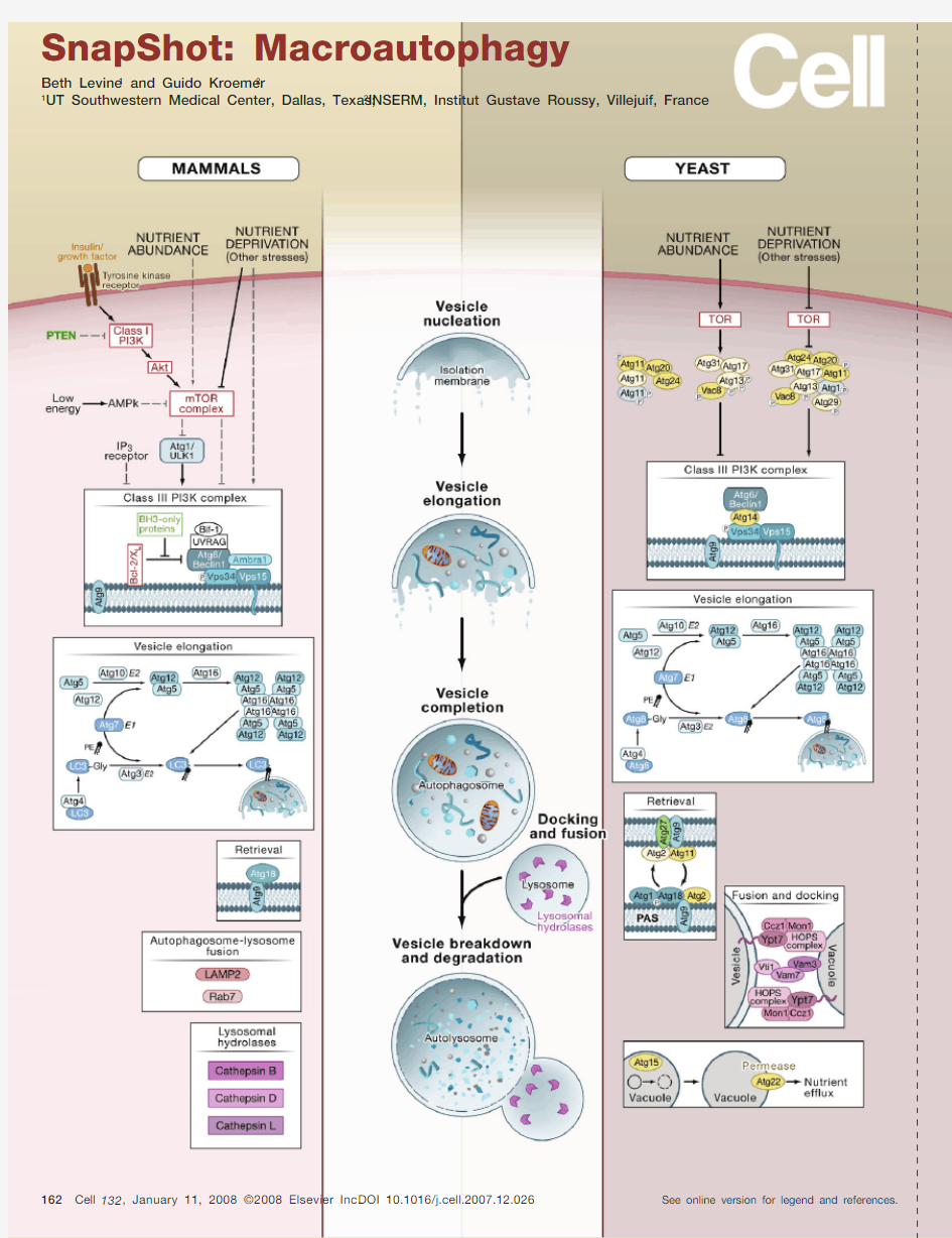
(Middle) Cellular Events in Macroautophagy
Macroautophagy (hereafter referred to as autophagy) is a catabolic process in which portions of the cytoplasm are sequestered within double- or multimembraned vesicles termed autophagosomes and then delivered to lysosomes for bulk degradation. The initial phases of macroautophagy consist of the formation of a phago-phore, which is also called an isolation membrane (vesicle nucleation step), the engulfment of cytoplasmic material—cytosol and/or organelles—by the phagophore, the elongation of the phagophore membrane, and fusion of its edges to form the autophagosome (vesicle elongation and completion steps). In yeast, the pre-au-tophagosomal structure (PAS), consisting of the phagophore or its precursor and autophagy (Atg) proteins, is thought to be the site for assembly of the autophago-some. The outer membrane of the autophagosome fuses with the vacuole in yeast to form the autophagic body or with the lysosome in mammalian cells to form the autolysosome, which is also called the autophagolysosome (docking and fusion steps). Inside the autophagic body/autolysosome, the luminal material including the internal membrane is degraded by vacuolar/lysosome hydrolases (vesicle breakdown and degradation steps). The resulting macromolecules are released through permeases and recycled in the cytosol. Alternatively (not depicted here), the autophagosome may fuse with an endosome to form an amphisome, prior to fusion with the lysosome.
(Left and Right) Molecular Events in Autophagy
Depicted are the molecular pathways of autophagy in yeast (right) and mammals (human or mouse) (left). Although many of the regulators and executors of yeast autophagy have orthologs in mammals (blue shaded ovals), not all yeast autophagy (ATG) genes have a known mammalian equivalent or vice versa. The autophagic machinery in other model eukaryotic organisms (e.g., Dictyostelium discoideum, Arabidopsis thaliana, Drosophila melanogaster, Caenorhabditis elegans) overlaps considerably, but lacks com-plete convergence, with that of yeast or mammals.
Initial Steps: Regulation and Nucleation
In yeast, autophagy is inhibited by the TOR serine/threonine kinase when nutrients are abundant. TOR may inhibit autophagy by indirectly or directly phosphorylating Atg13, which results in its reduced affinity for Atg1 kinase and dissociation from the autophagy-inducing complex. Upon nutrient starvation—the best-characterized physiological inducer of autophagy—TOR is repressed causing the association of dephosphorylated Atg13 with Atg1 and other factors, which stimulates Atg1 cata-lytic activity and induces autophagy.
Similarly, autophagy is stimulated in mammalian cells by nutrient deprivation, which may act through mTOR repression or other as-of-yet undefined mechanisms that activate the class III PI3K complex in an mTOR-independent manner. In mammals, autophagy is also stimulated by deprivation of insulin and other growth factors, which signal through receptor tyrosine kinases to activate the autophagy-inhibitory class I PI3K pathway and its downstream kinases including Akt and mTOR. (Class I PI3K is negatively regulated by the phosphatase PTEN.) In response to low energy, e.g., a cytosolic decrease in ATP and increase in AMP, the activation of AMPK inhibits mTOR, leading to autophagy activation. How mTOR inhibition stimulates autophagy in human or mouse cells is largely unknown, although one putative hu-
man Atg1 ortholog, ULK1, has been identified. An additional cytosolic factor, IP
3, can repress autophagy through the IP
3
receptor of the endoplasmic reticulum (ER).
Several other molecules, not depicted here, also play a role in autophagy regulation, including the eukaryotic initiation factor 2α (eIF2α); the tumor suppressor protein, p53; the stress-activated c-Jun-N-terminal kinase 1 (JNK1); GTPases, Erk1/2; ceramide; and calcium. Among the initial steps of vesicle nucleation, both in yeast and in mammals, is the activation of the class III PI3K (Vps34) to generate phosphatidylinositol 3-phosphate (PI3P). This activation depends on the formation of a multi-protein complex that includes, in yeast, Atg6/Beclin 1, the myristoylated Vps15 kinase, and Atg14 and, in mammals, Atg6/Beclin 1, Vps15, UVRAG, Bif-1 (also called
endophilin B1), and Ambra1. In mammals, Beclin 1 constitutively interacts with Bcl-2 or its close homolog Bcl-X
L and autophagy induction requires the dissociation
of Beclin 1 from its inhibitors Bcl-2 or Bcl-X
L . Given that Beclin 1 possesses a Bcl-2 homology 3 (BH3) domain that interacts with the BH3 receptor domains of Bcl-2
or Bcl-X
L , one mechanism leading to Beclin 1 release and autophagy activation is competition by BH3 domain-containing proteins (so-called “BH3-only” proteins).
Alternatively or in addition, phosphorylation of the flexible loop of Bcl-2 by JNK1 mediates the dissociation of Beclin 1 from Bcl-2 and subsequent autophagy activa-tion (not shown).
Vesicle Elongation, Vesicle Completion, and Membrane Retrieval
Two evolutionarily conserved ubiquitin-like conjugation systems are part of the vesicle elongation and vesicle completion processes in both yeast and mammals. The first pathway involves the covalent conjugation of Atg12 to Atg5, with the help of the E1-like enzyme Atg7 and the E2-like enzyme Atg10. This conjugate is organized into a complex by associating with Atg16 to form the Atg16·Atg5–Atg12 complex. The second pathway involves the conjugation of phosphatidylethanolamine (PE) to a glycine (Gly) residue of Atg8/LC3 (yeast and mammalian orthologs, respectively) by the sequential action of the protease Atg4, the E1-like enzyme Atg7, and the E2-like enzyme Atg3. This lipid conjugation leads to the conversion of the soluble form of Atg8 (in yeast) or LC3 (named LC3-I) (in mammals) to the autophagic vesicle-associated forms, Atg8-PE and LC3-II (or LC3-PE). Yeast Atg8 mediates tethering and hemifusion of liposomes containing Atg8-PE in an in vitro system. This membrane fusion process likely accounts for membrane elongation of phagophore/isolation membranes.
Atg9 (in yeast and mammalian cells) and Atg27 (in yeast) are the only known integral membrane proteins among the Atg proteins. The molecular complex organized around Atg9 may provide lipids to the nascent or expanding phagophore membrane and recycle back to cytoplasmic locations (including the PAS in yeast) to recruit lipids from peripheral sites. In yeast, the Atg9 cycling system includes the Atg1 kinase complex, Atg2, and Atg18 (which binds to PI3P), all of which are required for the retrieval of Atg9 from the PAS. The efficient delivery of yeast Atg9 to the PAS involves Atg9 transport factors Atg23 and Atg27. Atg11 serves as an adaptor that interacts with Atg9 and Atg1. In mammals, the only protein in this Atg9-dependent membrane retrieval complex that has been identified thus far is Atg18.
Docking and Fusion
Once autophagosomes are formed, they undergo progressive maturation by fusion with lysosomes, forming autolysosomes. In mammals, autophagolysosomal fusion is not well characterized but involves the integral lysosomal membrane protein LAMP-2 and the GTP-binding protein Rab7. In yeast, the autophagosome–vacuole fusion involves the Rab family GTPase Ypt7p; the SNAREs Vam3, Vam7 (which binds to PI3P), Vti1, and Ykt6; members of the HOPS complex (also called the class C Vps complex), and two other proteins involved in fusion, Mon1 and Ccz1.
Vesicle Breakdown and Degradation
In the autolysosome, the inner membrane as well as the luminal content of the autophagic vesicle is degraded by lysosomal enzymes that act optimally within this acidic compartment. One unique enzyme in this process is yeast Atg15, which is a putative lipase that is likely involved in the intravacuolar lysis of autophagic bodies. In mammalian cells, a series of lysosomal proteases (cathepsins B, D, and L) are required for the degradation of autophagosomal contents. Once macromolecules have been degraded in the lysosome/vacuole, monomeric units (e.g., amino acids, lipids) are exported to the cytosol for reuse. Yeast Atg22 serves as a putative amino acid efflux pump.
Abbreviations
Ambra1, activating molecule of Beclin 1-regulated autophagy; AMPK, AMP-activated kinase; Atg, autophagy genes/proteins; BH3, Bcl-2 homology 3; Ccz1, calcium
caffeine zinc sensitivity-1; Gly, glycine; HOPS, homotypic fusion and vacuole protein sorting; IP
3, myo-inositol-1,4,5-triphosphate; LAMP-2, lysosomal-associated
membrane protein-2; LC3, microtubule-associated protein light chain 3; Mon1, monensin sensitivity-1; mTOR, mammalian target of rapamycin; PAS, pre-autopha-gosomal structure; PE, phosphatidylethanolamine; PI3K, phosphatidylinositol 3 kinase; PTEN, phosphatase and tension homolog; SNARE, soluble NSF attachment receptor; TOR, target of rapamycin; ULK1, uncoordinated 51-like kinase 1; UVRAG, ultraviolet irradiation resistance-associated gene; Vps, vacuolar protein sorting.
ACkNowLEDgMENtS
We thank A. Diehl for expert scientific illustration. B.L. is supported by the National Institutes of Health, American Cancer Society, and Ellison Medical Foundation. G.K. is supported by the Ligue Nationale contre le Cancer, Agence Nationale de Recherche, Institut National contre le Cancer, Cancérop?le Ile-de France, and the European Union (Active p53, ApoSys, ChemoRes, DeathTrain, RIGHT, TransDeath).
(continued on next page)
REFERENCES
Gulati, P., and Thomas, G. (2007). Nutrient sensing in the mTOR/S6K1 signalling pathway. Biochem. Soc. Trans. 35, 236–238.
Klionsky, D.J. (2007). Autophagy: From phenomenology to molecular understanding in less than a decade. Nat. Rev. Mol. Cell Biol. 8, 931–937.
Levine, B., and Klionsky, D.J. (2004). Development by self-digestion: Molecular mechanisms and biological functions of autophagy. Dev. Cell 6, 463–477.
Levine, B., and Kroemer, G. (2008). Autophagy in the pathogenesis of disease. Cell, this issue.
Lum, J.J., DeBerardinis, R.J., and Thompson, C.B. (2005). Autophagy in metazoans: Cell survival in the land of plenty. Nat. Rev. Mol. Cell Biol. 6, 439–448.
Luzio, J.P., Pryor, P.R., and Bright, N.A. (2007). Lysosomes: Fusion and function. Nat. Rev. Mol. Cell Biol. 8, 622–632.
Maiuri, M.C., Zalckvar, E., Kimchi, A., and Kroemer, G. (2007). Self-eating and self-killing: Crosstalk between autophagy and apoptosis. Nat. Rev. Mol. Cell Biol. 8, 741–752. Meijer, A.J., and Codogno, P. (2006). Signalling and autophagy regulation in health, aging, and disease. Mol. Aspects Med. 27, 411–425.
Mizushima, N. (2007). Autophagy: Process and function. Genes Dev. 21, 2861–2873.
Mizushima, N., and Klionsky, D. (2007). Protein turnover via autophagy: Implications for metabolism. Annu. Rev. Nutr. 27, 19–40.
Nair, U., and Klionsky, D.J. (2005). Molecular mechanisms and regulation of specific and nonspecific autophagy pathways in yeast. J. Biol. Chem. 280, 41785–41788. Nakatogawa, H., Ichimura, Y., and Ohsumi, Y. (2007). Atg8, a ubiquitin-like protein required for autophagosome formation, mediates membrane tethering and hemifusion. Cell 130, 165–178.
Rubinsztein, D.C., Gestwicki, J.E., Murphy, L.O., and Klionsky, D.J. (2007). Potential therapeutic applications of autophagy. Nat. Rev. Drug Discov. 6, 304–312. Sarbassov, D.D., Ali, S.M., and Sabatini, D.M. (2005). Growing roles for the mTOR pathway. Curr. Opin. Cell Biol. 17, 596–603.
Suzuki, K., and Ohsumi, Y. (2007). Molecular machinery of autophagosome formation in yeast, Saccharomyces cerevisiae. FEBS Lett. 581, 2156–2161.
Xie, Z., and Klionsky, D.J. (2007). Autophagosome formation: Core machinery and adaptations. Nat. Cell Biol. 9, 1102–1109.