大肠杆菌纤溶酶基因的克隆和表达
Annals of Microbiology, 58 (1)95-98 (2008)
Gene clone and expression of a fibrinolytic enzyme (FE) in Escherichia coli
Shi-Hua WANG1,2*, Miao DIAO2, Yan-Ling YANG2, Wei-Zhu LIN2, Bi-Fang Huang2
1Key Laboratory of Biopesticide and Chemical Biology, Ministry of Education, 2College of Life Sciences, Fujian Agriculture and Forestry University, Fuzhou 350002, China
Received 2 April 2007 / Accepted 22 January 2008
Abstract -Bacillus subtilis LD-8547, a strong fibrinolytic enzyme (FE) producing strain, was obtained by the mutation from Bacillus subtilis LD-8, isolated in our laboratory. Based on the homology, the DNA fragment encoding the intact FE was amplified from total DNA of Bacillus subtilis LD-8547 by PCR. The vector pET32a (+) was used to construct the N-terminal thioredoxin (Trx) fusion expres-sion plasmid, and the recombinant FE was successfully expressed in Escherichia coli BL21 (DE3). The expressed protein showed high fibrinolytic activity in soluble fraction. This study provided evidences that FE could be actively expressed in E. coli, and large quanti-ties of FE could be produced for pharmacological and clinical research.
Key words: fibrinolytic enzyme (FE); gene clone; expression; activity.
INTRODUCTION
Thrombolytic therapy has been extensively researched as a means of medical treatment of the blood clot, and various fibrinolytic enzymes produced by different microorganisms were discovered (Kim et al.,1997; Choi and Shin, 1998) in recent years. The fibrinolytic activity was also found in douchi, a traditional Chinese fermented soybean food, from which some fibrinolytic enzyme producing strains were subsequently obtained (Peng et al.,2003; Wang et al., 2003; Wang et al.,2007).
Subtilisin fibrinolytic enzyme was purified from the broth by Bacillus amyloliquefaciens DC-4(Peng et al., 2003), and was actively expressed in the protease deficient strain Bacillus subtilis WB600 (Peng et al.,2004). When the native promoter of this subtilisin fibrinolytic enzyme was replaced by that of α-amylase gene, the expressed fibri-nolytic enzyme was increased (Xiao et al.,2004). But these fibrinolytic enzymes were insoluble aggregates. When this fibrinolytic enzyme was expressed as a fusion protein to thioredoxin (Trx), the fibrinolytic enzyme was highly obtained with fibrinolytic activity (Zhang et al.,2005).
Bacillus subt ilis LD-8, a fibrinolytic activity producing strain was isolated in our laboratory. This strain was mutat-ed by UV, NTG and γ-radiation, resulting in a higher fibri-nolytic enzyme (FE) producing strain Bacillus subt ilis LD-8547 (Wang et al.,2007). This purified fibrinolytic enzyme was about 30 kDa, and its optimal temperature and pH val-ues were 50 °C and 8.0, respectively. The enzyme activity was inhibited by phenylmethanesulfonyl fluoride (PMSF), but EDTA and EGTA did not affect the enzyme activity,showing that the enzyme was a serine protease (Wang et al.,2007).
When the DNA fragment encoding the intact FE was cloned into expression vector pET28a (+) and pET29a (+), the FE was highly expressed, but no fibrinolytic activity was detected at all in our laboratory. When pET32a (+) was used as expression vector, we successfully expressed FE in E. coli with fibrinolytic activity. In this paper,we describe the cloning and expression of the intact FE gene from Bacillus subtilis LD-8547 in ET32a (+)-E. coli BL21 (DE3) system.
MATERIALS AND METHODS
Bacterial strains and chemicals. Bacillus subt ilis LD-8547, a fibrinolytic enzyme producing strain, was mutated from isolated strain Bacillus subtilis LD-8 in our laboratory (Wang et al.,2007). Plasmid pET32a (+), purchased from Novagen, San Diego, CA, USA, was used as expression vector. Escherichia coli BL21 (DE3), stored in our laborato-ry, was used for expression of FE, and cultured in LB medi-um at 37 °C. Fibrinogen, thrombin, urokinase were pur-chased from Sigma, USA. Other chemicals were analytical grade.
Construction of expression vector. Bacillus subtilis LD-8547 genomic DNA was isolated and used as PCR template. To amplify DNA fragments encoding the intact FE, PCR was performed with forward primer P1 (AGGGAATTCGTGA-GAAGCAAAAAAT) and reverse primer P2 (ACCCTC-GAGTTGTGCAGCTGCT). PCR mixture was prepared in a total volume of 50 μL containing 20 ng of genomic DNA,
200 nmol/L each primer, 0.2 mmol/L dNTP, 1.5 mmol/L
MgCl2, 1x PCR buffer and 2.5 U Taq polymerase. The PCR product was digested with EcoR I and Xho I, and the isolat-ed fragment was inserted into EcoR I / Xho I linearised pET32a (+). The resulting plasmid was named as pET32FE, and transformed into E. coli BL21 (DE3) for recombinant protein expression. There was a His-tag (from vector pET32a) in the C-terminal of the recombinant FE for purifi-cation.
Gene expression of fib rinolytic enzyme (FE). Escherichia coli BL21 (DE3) harbouring the recombinant plasmid pET32FE was grown overnight at 37 °C in liquid LB medium containing 100 μg/mL ampicillin at 37 °C, and grown until an OD600of 0.6. Recombinant FE expression was induced by adding IPTG to a final concentration of 1 mmol/L, and the incubation was extended for additional 12 h at 25 °C. The cells were harvested by centrifugation, and used for SDS-PAGE analysis.
Purification of expressed FE. The recombinant FE pro-ducing strain was scaled up to one litre. After induction for 4 h, the bacteria were pelleted (8000 x g for 10 min at 4°C). The cells were washed and dissolved in PBS buffer. After sonicated, the supernatant was purified using Ni2+-chelation affinity resin (Bi et al.,2003) by His-tag at its C-terminal. The purified FE was identified by activity assay as description followed.
Fib rinolytic activity assay. Fibrinolytic activity was determined by the fibrin plate method with modification (Astrup and Mullertz, 1952; Zhang et al.,2005). In brief, 25 mL of 0.3% bovine fibrinogen solution in 100 mmol/L sodium phosphate buffer (pH 7.4) was mixed with 0.5 mL of thrombin solution (5 U/mL) and 25 mL of 1% agarose gel in a Petri dish. The dish was allowed to stand for 30 min at room temperature to form fibrin clot, and holes were made on the plate by suction using a capillary glass tube. Ten microlitres of sample solutions were added into each hole and the plate was then incubated for 20 h at 37 °C. The fibrinolytic activity was calculated by the lytic area. Urokinase was used as a standard. Amidolytic activity and kinetic constants of the fibrinolytic enzyme were estimated using synthetic substrate (Wang et al.,2007).
Blood clots lytic effect with FE. Blood clots from white rabbit were divided into two groups. One was heated at 80°C for 40 min to inactivate endogenous fibrinolytic factors, and the other group remained untreated. The heated and unheated blood clots were incubated with purified fibri-nolytic enzyme in Petri dish at 37 °C. At different time intervals, the lytic area was used for analysis.
Error prone PCR. We engaged to enhance the enzyme activity of FE by error prone PCR technology. Random mutations were introduced during the error prone PCR pro-cedure according to Xu et al.(2003).Bacillus subtilis LD-8547 genomic DNA was used as PCR template. PCR was performed with forward primer P1 and reverse primer P2. PCR mixture was prepared in a total volume of 50 μL con-taining 20 ng of genomic DNA, 200 nmol/L each primer, 0.2 mmol/L dATP, 0.2 mmol/L dGTP, 1 mmol/L dCTP, 1 mmol/L dTTP, 5 mmol/L MgCl2, 1 mmol/L MnCl2, 1x PCR buffer (Mg2+free) and 2.5 U Taq polymerase. The first amplified PCR product was used as the PCR template of the second time, and the second amplified product was used as the template of the third time. All the amplified products of the three times were digested with EcoR I and Xho I, followed by the ligation to the same digested vector pET32a (+). Escherichia coli BL21 (DE3) was transformed with the lig-ated mixture to create the mutant library for higher fibri-nolytic enzyme screening.
Bioinformatics analysis of this fib rinolytic enzyme gene. After sequencing, Primer Premier 5.0 was used to deduce the amino acid sequence. Homology analysis was performed using Blastx and SIB BLAST Network Service (https://www.360docs.net/doc/d31803879.html,/tools/blast/). The signal peptide cleavage site was predicted using SignalP 3.0 Server (http:// www.cbs.dtu.dk/services/SignalP/) and ANTHEP-ROT software. The secondary structure prediction and sol-vent ability analysis were carried out through PREDICT PROTEIN database online analysis and ANTHEPROT soft-ware. The prediction of conformation was performed using SWISS MODEL method.
RESULTS
Gene clone of Trx-FE
Bacillus subt ilis LD-8547 genomic DNA was used as PCR template, and PCR was performed with forward primer P1 and reverse primer P2. The intact DNA fragment of FE was 1146 bp, and there was a band of PCR product about 1146 bp. It was showed that gene encoding the intact FE was successfully obtained. The PCR product was digested with EcoR I and Xho I, and inserted into pET32a (+). The result-ing plasmid named pET32FE was used for production of recombinant FE.
Expression of recombinant FE
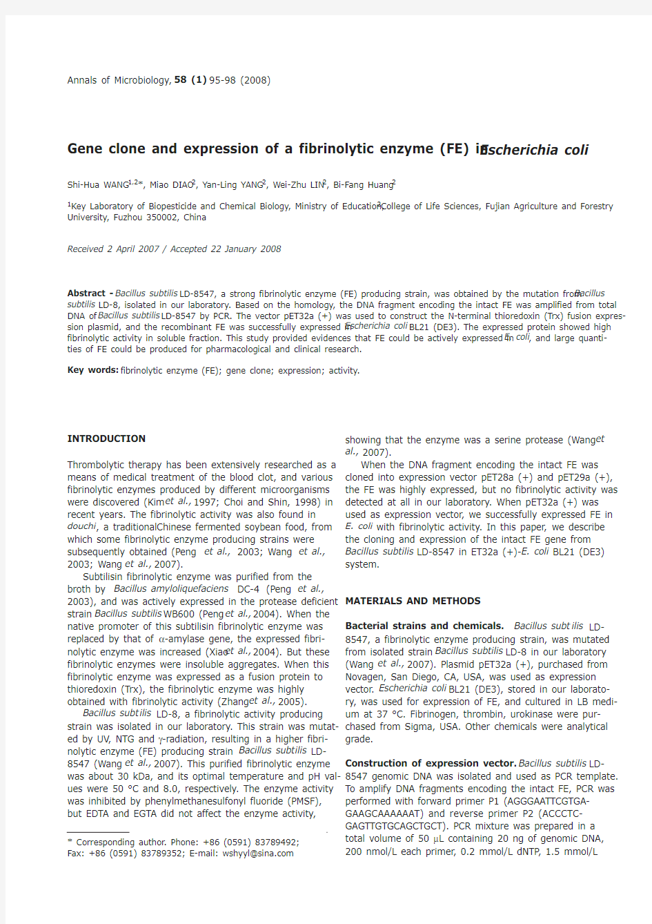
To obtain a sufficient amount of recombinant enzyme, FE was overproduced in E. coli BL21 (DE3) using the pET32a (+) vector. The expression of FE was induced with IPTG under the control of a T7-phage promoter. The fusion pro-tein Trx-pro-FE (about 54 kDa) was highly expressed (Fig. 1, lane 4). There was a thioredoxin in the N-terminal of the fusion protein, and it could increase the soluble yields to overcome the “inclusion body” problem. The C-terminal His-tag was used to facilitate the purification of the desired protein. Expression of recombinant FE was controlled by T7 promoter, and it was in both soluble (Fig. 1, lanes 3) and inclusion body fraction (Fig. 1, lanes 2). But fibrinolytic activity was only detected in the soluble fraction (Fig. 1, lane 3). This indicated that Trx-pro-FE was cleaved into mature FE (about 30 kDa) and only soluble mature FE had fibrinolytic activity.
Purification and characterisation of recombinant FE The cells were washed and dissolved in PBS buffer. After sonicated, the supernatant was purified using Ni2+-chela-tion affinity resin. To identify whether the expressed FE has the same characteristic as the native one, some character-istics of the purified FE were compared to that of the native FE. The result showed that the fibrinolytic activity was inhibited by 1 mM phenylmethanesulfonyl fluoride (PMSF), but not by EDTA or EGTA, and other results were in Table 1. All the characteristics identified above were almost the same as the native one produced by Bacillus subt ilis LD-
96S.-H. Wang et al.
8547. Specific activity of the enzyme was much higher than those of the fibrinolytic enzyme also isolated from douchi .Therefore, the FE can be easily prepared in large scale for further study.
Blood clots lytic effect with FE in vitro
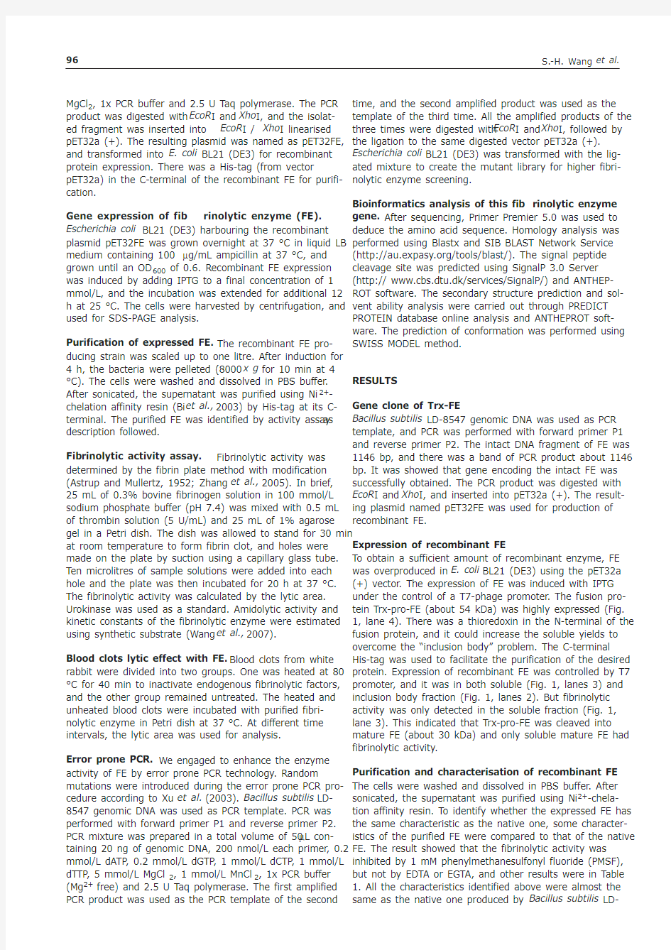
Blood clots from white rabbit were used to determine the lytic effect of this fibrinolytic enzyme. One group was heat-ed to inactivate endogenous fibrinolytic factors, and the other group remained untreated. The blood clots of two groups were incubated with purified FE in Petri dish at 37°C. The result showed that the heated blood clots dissolved within 2.5 h (Fig. 2 A3), but the non-heated blood clots dis-solved within 40 min (Fig. 2 B2), indicating that the enzyme was able to degrade blood clots in the absence of endogenous fibrinolytic factors.
Evolution strategy and screening of mutant library The strategy of FE evolution towards higher activity in this study was to use error prone PCR technology. The fibri-nolytic enzyme gene of LD-8547 was used as the parent template to generate the first generation error PCR prod-uct. Then the following two generations were successively created using the PCR products from the previous genera-tion as the new parents. All these error PCR products of the three generations were used to construct mutant library.After screening, about 100 colonies were random screened
and 35 clones gave positive signal. One clone giving the strongest activity was obtained, about 32% higher fibri-nolytic activity than that of the original recombinant FE (Detail data not shown).
Bioinformatics analysis
The results of DNA sequencing showed that the full length of the FE gene covered 1146 bp, with the GC contents of 46.25%, encoding a mature FE of 275 amino acids and a pro-peptide of 106 amino acids. ANTHEPROT analysis indi-cated that the isoelectric point of FE was 6.325, and the molecular weight was about 30 kDa.
Online analysis using PREDICE PROTEIN database indi-cated that the secondary structure was composed of 17.45% α-helix, 24.73% β-pleated sheet, and 57.82% ran-dom coil. Thus, the FE was a mixed protein. The prediction results of solvent accessibility composition indicated that the content of composition e (amino acid residues exposed to the surface of protein) was 41%, while the composition b (other residues) was 59%. ANTHEPROT and structural analysis revealed that the forth and the fifth amino acid residues were positive charged, and there was a distinct hydrophobic region from residue 6 to residue 25. SWISS-MODEL was applied to predict the conformation of FE, and the result showed that the peptide was composed of 7 α-helix connected by small peptides, and moved together to each other to form a spherical molecule (Fig. 3).
Ann. Microbiol., 58 (1), 95-98 (2008)
97
FIG. 1 - SDS-PAGE analysis of expression and purification of fib-rinolytic enzyme (FE). Lane 1: the purification result of the mature FE after induction; lane 2: the precipitation of the pET32FE after sonication and centrifugation; lane 3: the supernatant of the pET32FE after sonication and centrifugation; lane 4: the expression products of the whole pET32FE cells; lane 5: the control of empty vec-tor ET32a (+); lane 6: mid-range protein molecular
weight markers.
FIG. 2 - Blood clots from white rabbit were used to determine
the lytic effect of this fibrinolytic enzyme. A: all the three blood clot plats (A1, A2, A3) were heated at 80 °C for 30 min to inactivate endogenous fibrinolytic factors and incubated with purified fibrinolytic enzyme at 37 °C. B:all the three plats (B1, B2, B3), without heating, were incubated with fibrinolytic enzyme at 37 °C. A1 and B1:0 min incubation; A2 and B2: 40 min incubation; A3 and B3: 2.5 h incubation.
TABLE 1- Screening of plant growth promoting traits in Bacillus thuringiensis isolates Fibrinolytic Total protein
Total activity
Specific activity
Optimal Optimal temperature K m
V max
enzyme (mg)
(U)(U/mg)
pH (°C )(mmol/L )
(mmol/min)Native FE 3.088375862719448.0500.5210.049Expressed FE
2.56
696050
271895
8.0
50
0.527
0.047
DISCUSSION
When the DNA fragment encoding the intact FE was cloned into expression vector pET28a (+) and pET29a (+), target FE was expressed, but no fibrinolytic activity was detected at all. The possible reason was that the expression target protein formed “inclusion body”, resulting in no enzyme activity. So we turned to soluble expression vector pET32a (+). In this paper, recombinant plasmid pET32FE was con-structed to express FE in E. coli. The recombinant protein was expressed, and became more soluble when fused with the N-terminal Trx. It might yield soluble, active target pro-tein to overcome the “inclusion body” problem (Bi et al., 2003). C-terminal having a His-tag was used to facilitate the purification of the desired protein.
Soluble supernatant and “inclusion body” pellet of induced pET32FE were both used to test fibrinolytic activi-ty, and the result showed that only soluble supernatant had activity, whereas inclusion body had no activity. Purification result (Fig. 1, lane 1) only gave a 30 kDa FE with strong fibrinolytic activity, indicating that Trx-pro-FE was cleaved into mature FE. The purified FE has the same characteris-tics as native one produced by Bacillus subt ilis LD-8547, which showed that expressed FE can be easily prepared in large scale, and used for further pharmacological and clin-ical research.
Acknowledgement
We are sincerely thankful for the support from the Key Scientific and Technology Project of Fujian Province of China (2007N0094, 2007Y0002), Natural Sciences Foundation of Fujian Province of China (B0610008, X0750005), Postdoctoral Sciences Foundation of China (20060390183), and Program for New Century Excellent Talents in Fujian Province University. Dr. Yong Peng and Prof. Yi-Zheng Zhang (Sichuan University, Chengdu, China) are greatly appreciated for paper donation and technical discussion. REFERENCES
Astrup T., Mullertz S. (1952). The fibrin plate method for esti-mating fibrinolytic activity. Arch. Biochem. Biophys., 40: 346-351.
Bi L.J., Zhou Y.F., Zhang X.E., Deng J.Y., Zhang Z.P., Xie B., Zhang C.G. (2003). A muts-based protein chip for detection of DNA mutations. Anal. Chem., 75 (16): 4113-4119.
Choi H.S., Shin P.H. (1998). Purification and partial characteri-zation of a fibrinolytic protease in Pleuro us os rea us.
Mycologia, 4: 674-679.
Kim H.K., Kim G.T., Kim D.K., Choi W.A., Park S.H., Jeong Y.K., Kong I.S. (1997). Purification and characterization of a novel fibrinolytic enzyme from Bacillus sp. KA38 originated from fermented fish. J. Ferment. Bioeng., 84: 307-312.
Peng Y., Huang Q., Zhang R.H., Zhang Y.Z. (2003). Purification and characterization of a fibrinolytic enzyme produced by Bacillus amyloliquefaciens DC-4 screened from douchi, a tra-ditional Chinese soybean food. Comp. Biochem. Physiol. B., 134: 45-52.
Peng Y., Yang X.J., Xiao L., Zhang Y.Z. (2004). Cloning and expression of a fibrinolytic enzyme (subtilisin DFE) gene from Bacillus amyloliquefaciens DC-4 in Bacillus sub ilis. Res.
Microbiol., 155: 167-173.
Wang C.T., Ji B.P., Li B., Ji H. (2003). Isolation of Douchi fibri-nolytic enzyme producing strain DC33 from Chinese Douchi and primary analysis of the enzyme property. Food Sci.
(China), 6: 34-38.
Wang S.H., Zhang C., Yang Y.L., Diao M., Bai M.F. (2007).
Screening of a high fibrinolytic enzyme producing strain and characterization of the fibrinolytic enzyme produced from Bacillus subtilis LD-8547. World J. Microbiol. Biotechnol., 7: epub.
Xiao L., Zhang R.H., Peng Y., Zhang Y.Z. (2004). Highly efficient gene expression of a fibrinolytic enzyme (subtilisin DFE) in Bacillus subtilis mediated by the promoter of α-amylase gene from Bacillus amyloliquefaciens. Biotechnol. Lett.,26: 1365-1369.
Zhang R.H., Xiao L., Peng Y., Wang H.Y., Bai F, Zhang Y.Z.
(2005). Gene expression and characteristics of a novel fibri-nolytic enzyme (subtilisin DFE) in Escherichia coli. Lett. Appl.
Microbiol., 41: 190-195.
98S.-H. Wang
et al. FIG. 3 - The SWISS-MODEL analysis result of the 3-D structure
of the mature FE. The molecule consists of a-helix
(helix), sheet (arrow) and loops (thread).
木聚糖酶的基因克隆及表达
木聚糖酶的基因克隆及表达 木聚糖酶(xylanase)主要包括β-1,4-木聚糖酶、β-1,4-木聚糖内切酶等,是指能够将木聚糖降解为低聚木糖或单糖的一组酶的总称。木聚糖是植物半纤维素的主要成分,约占植物总糖的1/3,是自然界中除纤维素外含量最丰富的再生生物资源[1]。木聚糖酶在饲料、造纸、食品、医药、能源等领域应用较广。木聚糖酶广泛存于微生物中,目前已从不同来源的微生物中分离得到上百种木聚糖酶。在自然界中,绝大多数野生型木聚糖酶的最适温度为40~60℃,酶活性不高,热稳定性较差[2]。因此,研究者把研究重点放在了利用分子生物学手段对原始菌株的木聚糖酶基因进行克隆上,并对其进行表达,以期获得使用更加方便、特性更加优异的工程菌株。本文对聚糖酶特性等进行了回顾,对其基因克隆和表达作一综述,并对分子生物学技术在木聚糖酶上的应用前景进行了展望。1木聚糖酶的研究现状国外对木聚糖酶的研究开始较早,生产技术及应用已趋于成熟。研究者对细菌、真菌和放线菌木聚糖酶的研究更加深入和广泛,早在1992年就已经实现了木聚糖酶的工业化生产。国内对木聚糖酶的研究起步较晚,但发展迅速。20世纪80年代初期,中国科学院微生物所张树政院士首先从海枣曲霉(Aspergillusphoenicis)中纯化得到了木聚糖酶Ⅰ~Ⅳ,并深入研究了活力较高的组分酶Ⅲ的酶学性质。目前,由于从这些野生型菌株中获得的木聚糖酶活性并不高,并且受到酶稳定性和底物特异性等方面的限制[2],使研究者们将对木聚糖酶的研究重点放在了木聚糖酶基因克隆、表达和重组上,并在分子水平上对木聚糖酶进行改造。木聚糖酶基因克隆和表达的研究进展王丹丹1,2 综述,周晨妍1,付冠华1审校1.新乡医学院生命科学技术学院河南省医学遗传学与分子靶向药物高校重点实验室培育基地,河南新乡453003;2.新乡医学院三全学院,河南新乡453003 摘要:半纤维素分解微生物在自然界的物质循环过程中起着重要作用,半纤维素是植物多糖的重要成分之一,而木聚糖则是半纤维素的主要成分。木聚糖酶(xylanase)可催化木聚糖的水解,在各种生物体内均发现木聚糖酶。在过去几十年中,已有上百种木聚糖酶基因被克隆至同源或异源宿主内来表达木聚糖酶,以期改变宿主特性并适于商业应用。本文综述了木聚糖酶基因的克隆和表达,并对基因工程技术在木聚糖酶上的应用前景进行了展望。关键词:木聚糖酶;克隆;基因表达2木聚糖酶基因的克隆木聚糖水解酶系是一种复杂的复合酶系统,广泛分布于自然界的真菌和细菌中。已经报道的有细菌、真菌、酵母和放线菌等。在国内外研究最多的还是木霉、青霉、黑曲霉和棒曲霉等。到目前为止,已经有上百种来自细菌和真菌等微生物中的木聚糖酶基因被克隆,并在不同的表达系统中成功表达。从近几年克隆和表达出的木聚糖酶基因主要来自细菌、真菌、酵母和放线菌等,而研究最多的是木霉、青霉、黑曲霉和棒曲霉等。从这些野生菌种克隆出的木聚糖酶基因在不同宿主菌中已成功实现了异源表达。 木聚糖酶基因的表达3.1木聚糖酶基因在原核细胞内的表达木聚糖酶基因在原核细胞内的表达以大肠埃希菌的研究为热点。大肠埃希菌繁殖速度较快,是相对较理想的宿主细胞。将克隆得到的目的基因和原核载体经双酶切,胶回收产物与重组质粒经连接酶连接,转化合适的大肠埃希菌,选择培养基筛选阳性克隆子,并进行诱导表达。多数情况下,大肠埃希菌不但表达出目的蛋白,并且酶活力也有较大提高。见表2。 3.2木聚糖酶基因在真核细胞中的表达大肠埃希菌虽然繁殖速度较快,但由于其为原核生物,细胞外有一层厚厚的细胞壁,必须先破碎细胞壁,才能将目的蛋白释放出来,而真核细胞克服了原核细胞的这个缺点,可将表达的目的蛋白分泌到细胞外,便于分离纯化。能够表达木聚糖酶的真核细胞以酿酒酵母和毕赤酵母为代表,如水稻等真核细胞同样能够表达木聚糖酶,并且酶活也有一定程度的提高
小麦Rubisco 活化酶基因的克隆和表达特性
植物学通报 2005, 22 (3): 313 ̄319①国家重点基础研究发展规划项目(G1998010100)资助。 ②通讯作者。Author for correspondence. E-mail: zouqi@https://www.360docs.net/doc/d31803879.html, 收稿日期: 2004-02-17 接受日期: 2004-09-20 责任编辑: 孙冬花 小麦Rubisco 活化酶基因的克隆和表达特性① 张 国 李 滨 邹 琦② (山东农业大学生命科学学院植物系 泰安 271018) 摘要 Rubisco 活化酶是广泛存在于光合生物中调节Rubisco 活性的酶, 我们利用PCR 技术, 从小麦(Triticum aestivum )叶片cDNA 文库中克隆得到Rubisco 活化酶基因cDNA 片段, 该片段长度为850 bp, 编码201个氨基酸。Northern blot 表明, 小麦叶片在暗诱导衰老的条件下, 叶片中活化酶基因表达水平逐渐下降; 同时, 小麦叶片的光合特性、叶绿素含量和Rubisco 活性呈现下降趋势。这些结果表明, 衰老时小麦叶片Rubisco 活化酶基因表达水平下降与光合速率下降密切相关。关键词 Rubisco 活化酶, 小麦, 衰老 Cloning and Expression of Rubisco Activase Gene in Wheat ZHANG Guo LI Bin ZOU Qi ② (Department of Botany, College of Life Science, Shandong Agricultural University , Tai’an 271018) Abstract Rubisco activase is an ubiquitous enzyme for the activation of Rubisco in photosynthetic autotrophs. A cDNA fragment of Rubisco activase gene was cloned from wheat (Triticum aestivum ). Northern blot showed that expression of the gene was down-regu-lated in dark-induced senescence leaves, where photosynthetic rate, chlorophyll content and Rubisco activity also showed obvious decline. The results suggest that the decreased expres-sion level of the gene was related to the decline in photosynthetic rate.Key words Rubisco activase, Wheat, Senescence Rubisco 是光合生物进行光合碳同化关键的双功能酶, 它催化RuBP 的羧化-加氧反应,但效率很低。因为加氧反应除了消耗能量,还损失了羧化反应中固定的25%的有机碳; 同时, 各种磷酸糖类能抑制Rubisco 的活性, 如:底物RuBP 本身就是Rubisco 的强烈抑制剂, 且活化态Rubisco 易于脱氨甲酰化而失活, 这些因素使Rubisco 成为光合速率的限制因子, 因 而也成为提高作物光合效率的研究目标。 Salvucci 等(1985)发现了Rubisco 活化酶(Rubisco activase, RCA), 它能够活化Rubisco,同时具有ATPase 活性; 也有人认为RCA 是一种分子伴侣(Spreitzer and Salvucci, 2002)。植物中Rubisco 的活化状态实际是Rubisco 的失活速率和RCA 活化Rubisco 速率间的平衡状态(Crafts-Brandner and Salvucci, 2000)。人
基因克隆及转基因方法
基因克隆及转基因 一、基因克隆及转基因过程 1、设计引物 软件是https://www.360docs.net/doc/d31803879.html,sergene.v7.1,用到里面的PrimerSelect和EditSeq。 一般原则:1、长度:18-25; 2、GC含量:40-60%,正反向引物相差不要大于5%; 3、Tm值:55以上(到65),实在不行50以上也可以,正反向引物相差不要大 于5; 4、3’端结尾最好是GC,其次是T,不要A; 5、正反向引物连续配对数小于4; 6、在NCBI上的Primer Blast上看引物特异性如何; (如果克隆的话不能满足条件也没办法。) 不是必须条件,但可以考虑:多个基因设计引物时,可尽量使Tm值相似,方便PCR。 步骤: 一、打开PrimerSelect和EditSeq。 二、在EditSeq中输入你的序列。 引物有一对F和R 1、对于F是从5’到3’,在序列的前部分选择长度为18-25bp的碱基,如果你是要验证就随便选,如果你是要克隆就在最开始选,不符合原则就只能在你选的后边增或减碱基。 2、将选择的F引物输入到PrimerSelect中,在File中选择Enter New Primer,复制,OK,然后可以看到引物的情况,看看长度、Tm、GC含量是不是符合标准,不符合就继续选。 3、对于R是从3’到5’,选中序列,在EditSeq的Goodies中选择第一个“反向互补”,此时序列已反向互补,按照前面F的方法搜索R的引物。、 4、注意你想要的目的带的大小,比如序列是1000bp,你想PCR出来800大小的目的带,那就要看看F和R之间的长度在你想要的范围内。可以将R反向互补,在正向的序列中搜索R在的位置,就是在EditSeq中选择Search,点击第一个Find,开始搜寻。 5、搜索完引物在PrimerSelec中的Report中选择前两个查看二聚体情况。 6、在NCBI上的Primer Blast上看引物特异性如何。 7、因为是克隆,所以引物要有酶切位点,酶切位点的加入主要考虑所用到的表达载体,在NEBcutter网站中输入总序列查看可用的酶切位点。在引物上游加入酶切位点,注意加入时载体的表达的方向,前面的酶切位点在引物F上,后面的酶切位点在引物R上。一般在引物上游还要加上两个保护碱基。 2、提取醋栗DNA 3、PCR扩增与目的基因回收 PCR先找合适的退火温度,找到后回收时就可以多PCR几管,一般我们用20ul的体系,PCR5管就可以回收,就是琼脂糖凝胶回收,将目的基因用刀片切下来,用试剂盒回收。回收完可以再跑电泳检测一遍。 PCR: 20ul体系:灭菌水13.8ul,若模板为质粒灭菌水14.3ul; 2.5mMdNTP2.0ul;
基因克隆载体上的各种常用蛋白标签
基因克隆载体上的各种常用蛋白标签 蛋白标签(proteintag)是指利用DNA体外重组技术,与目的蛋白一起融合表达的一种多肽或者蛋白,以便于目的蛋白的表达、检测、示踪和纯化等。随着技术的不断发展,研究人员相继开发出了具有各种不同功能的蛋白标签。目前,这些蛋白标签已在基础研究和商业化产品生产等方面得到了广泛的应用。 美国GeneCopoeia(复能基因)为客户提供50多种蛋白标签,可以满足客户的不同需求,包括各种最新型的标签,如:SNAP-Tag?、Halo Tag?、AviTag?、Sumo等;也提供齐全的各种常用标签,如eGFP、His、Flag等等标签。 以下是部分蛋白标签的特性介绍,更加详细的介绍可在查询产品的结果列表里面看到各种推荐的蛋白标签和载体。 TrxHIS His6是指六个组氨酸残基组成的融合标签,可插入在目的蛋白的C末端或N末端。当某一个标签的使用,一是能构成表位利于纯化和检测;二是构成独特的结构特征(结合配体)利于纯化。组氨酸残基侧链与固态的镍有强烈的吸引力,可用于固定化金属螯合层析(IMAC),对重组蛋白进行分离纯化。使用His-tag有下面优点: 标签的量小,只有~0.84KD,而GST和蛋白A分别为~26KD和~30KD,一般不影响目标蛋白的功能; His标签融合蛋白可以在非离子型表面活性剂存在的条件下或变性条件下纯化,前者在纯化疏水性强的蛋白得到应用,后者在纯化包涵体蛋白时特别有用,用高浓度的变性剂溶解后通过金属螯和去除杂蛋白,使复性不受其它蛋白的干扰,或进行金属螯和亲和层析复性; His标签融合蛋白也被用于蛋白质-蛋白质、蛋白质-DNA相互作用研究; His标签免疫原性相对较低,可将纯化的蛋白直接注射动物进行免疫并制备抗体。 可应用于多种表达系统,纯化的条件温和; 可以和其它的亲和标签一起构建双亲和标签。 Flag标签蛋白 Flag标签蛋白为编码8个氨基酸的亲水性多肽(DYKDDDDK),同时载体中构建的Kozak序列使得带有FLAG的融合蛋白在真核表达系统中表达效率更高。FLAG作为标签蛋白,其融合表达目的蛋白后具有以下优点: FLAG作为融合表达标签,其通常不会与目的蛋白相互作用并且通常不会影响目的蛋白的功能、性质,这样就有利用研究人员对融合蛋白进行下游研究。 融合FLAG的目的蛋白,可以直接通过FLAG进行亲和层析,此层析为非变性纯化,可以纯化有活性的融合蛋白,并且纯化效率高。 FLAG作为标签蛋白,其可以被抗FLAG的抗体识别,这样就方便通过Western Blot、ELISA等方法对含有FLAG的融合蛋白进行检测、鉴定。
红豆杉中MYB家族基因克隆及表达分析 开题报告 于凯
毕业设计/论文 开题报告 课题名称红豆杉中MYB家族基因克隆及表达分析类别毕业论文 系别城市建设学院 专业班生物工程0701班 姓名于凯 评分 指导教师 华中科技大学武昌分校
华中科技大学武昌分校学生毕业论文开题报告
癌活性,对于治疗卵巢癌、乳腺癌等疗效突出。但是由于含量少、提取困难等诸多因素,高纯度紫杉醇价格昂贵,每公斤200万元人民币左右。因此,近年来国内外许研究人员、实验室和公司一直试图通过生物合成、化学合成、微生物提取、组织和细胞培养、寻找类似物等途径来解决紫杉醇的药源短缺问题。 研究紫杉醇的生物合成,尤其一些限速反应步骤机理的阐明对于人为定向的提高合成效率,克隆重组形成关键酶基因从而提高紫杉醇的产量意义重大。从理论上来说这是一个好方法,但是紫杉醇的合成途径非常复杂,涉及到多种酶以及很多分支途径,单纯依靠转化一、两种限速酶基因,只能保证转入的限速酶表达量提高,使之不再是限速因素,但其它阶段对于最终产量的限制依然存在,而且同时转入多种基因的可行性非常低,这种方法的缺陷很明显。 若采用化学合成,如从红豆杉植物中分离得到的巴卡亭Ⅲ经过四步化学过程可合成紫杉醇,为合成紫杉醇提供了新途径[5]。但化学合成从实质意义上说还没有取得彻底的突破,目前还不具备应用价值。 如果从共生真菌中直接提取紫杉醇,能够利用真菌生长速度快的优势,但目前分离的菌株无论从种类还是数量上都远不够工业化的要求,而且还存在很多不确定因素[1]。生产紫杉醇的微生物大多是与红豆杉共生的真菌,其紫杉醇含量极微,并且这些真菌的培养和大规模发酵困难,菌株衰退也是一个难题。 另外,红豆杉愈伤组织和细胞培养生产紫杉醇是研究的热点之一,是工厂化大规模生产紫杉醇的重要手段之一。但运用植物组织、细胞培养技术生产紫杉醇仍处在实验室阶段,如何获得高含量、产紫杉醇稳定的愈伤组织一直都是组织培养、细胞培养生产紫杉醇的关键。 1.1.3关于MYB基因 ①MYB基因 目前,在几乎所有的真核生物中都发现了与禽类逆转录病毒癌基因和细胞原癌基因c-MYB相似的基因,它们的编码产物在结构和功能上具有高度保守的DNA结合域,是一类转录因子[6]。在植物中首先从玉米中克隆了含有MYB结构域的转录因子C1基因,之后在植物中发现的MYB相关基因的数量迅速增加[7]。
基因的克隆、表达载体构建与功能验证
基因的克隆、表达载体构建及功能验证(一般性方法) 一、基因克隆 ★事前三问 a.克隆这个基因干什么?它有什么功能? b.这个基因在哪种材料中扩增? c.材料需要怎么处理? ◎实验前准备工作 a.设计引物,准备材料, b.购置试剂:Taq酶、反转录试剂盒、凝胶回收试剂盒、质粒提取试剂盒、连接试 剂盒 c.实验试剂及用具:枪头、离心管、培养皿、滤纸灭菌;Amp+ 、Kan+等抗生素准 备 ※基本流程 提取和纯化RNA—cDNA第一条链合成—PCR—凝胶电泳—胶回收—连接—转化—涂平板—挑单菌落—摇菌—提质粒—测序 1.总RNA的提取、纯化及cDNA第一链合成 1.1叶片、根总RNA的提取 Trizol是一种高效的总RNA抽提试剂,内含异硫氰酸胍等物质,能迅速裂解植物细胞,抑制细胞释放出的核酸酶,所提取的RNA完整性好且纯度高,以利于下一步的实验。 1)实验前准备 预先配制0.1%的DEPC水(ddH2O中含0.1%DEPC,V/V,37 ℃过夜处理12 h),高温灭菌后,用DEPC水配制75%乙醇,研钵、量筒、试剂瓶等需200℃灭菌至少4 h,所用枪头和枪盒均去RNA酶处理(直接购买)。 2)Trizol 法(小麦)叶片或根的总RNA实验步骤如下: (1)提前在1.5 ml离心管中加入1 mlTrizol,然后将200 mg样品液氮中研磨成白色粉末,
移入管内,用力摇15 s,在15-30℃温育5 min,使核酸蛋白复合物完全分离。 (2)4℃,12000g离心10min,取上清,离心得到的沉淀中包括细胞外膜、多糖、高分子量DNA,上清中含有RNA。 (3)吸取上清液加0.2 ml氯仿,盖好盖,用力摇15 s,15~30 ℃温育2~3 min。(4)在≤12000g,4℃离心10 min,样品分为三层:底层为黄色有机相,上层为无色水相和一个中间层,RNA主要在水相中,水相体积约为所用TRIzol试剂的60%。 (5)将上层水相转移到新的1.5 ml离心管中,加2倍体积的无水乙醇沉淀RNA,室温静止30 min。 (6)在≤12000g,4℃离心10 min,离心前看不出RNA沉淀,离心后在管侧和管底出现胶状沉淀。 (7)用≥1 ml的75%乙醇洗RNA,涡旋振荡样品,在≤7500g,4℃离心5 min,弃上清。(8)室温放置干燥或真空抽干RNA沉淀,大约晾5-10分钟,加无RNase的水100μl用枪头吸几次,55~60℃温育10 min使RNA溶解。 (9)配制以下体系: 10×DNase buffer 5 μl DNase I (RNase-free)(40 μg/μl) 1 μl RNasin Inhibitor(40 μg/μl) 1 μl Total RNA 70 μg 加去RNase水至总体积为50 μl (10)37 ℃水浴1h,加DEPC处理的水至总体积为100 μl,加入等体积氯仿抽提一次。(11)取上清,加入10 μl的3 mol/L NaAC溶液,200 μl的无水乙醇,-80 ℃沉淀30 min。 (12)2~8 ℃,12000g离心10 min,弃清液,干燥后取50μl无RNase的水溶解RNA。3)RNA的质量及纯度检测 (1)电泳检测取2ul RNA 与1 ul 10×Loading buffer上样缓冲液混合均匀在1% 的琼脂糖凝胶上电泳,在紫外灯下观察RNA 条带并记录实验结果。 (2)分光光度计RNA纯度检测 取1ul RNA液,以DEPC水为空白对照,测定A260/ A280 比值,估测RNA质 量。 4)cDNA第一条链的合成 按照以下体系将提取的总RNA反转录成第一链cDNA: 1)在Eppendorf管中配制下列混合液:
青杄PwUSP2基因的克隆和表达分析
青杄PwUSP2基因的克隆和表达分析 周xx,xx 班级 摘要 广泛逆境胁迫蛋白(USPs)参与碳缺乏、缺氧、干旱和高盐 等多种非生物胁迫, 但在植物中的研究尚不深入。本文通 过RACE-PCR 的方法获得青杄PwUSP2基因的cDNA 全长, 共987 bp ,其中编码区723 bp ,共编码240个氨基酸。利用 生物信息学工具对其理化性质、二级结构和三级结构进行 分析,结果显示,该蛋白理论分子质量为26.84 kDa ,理论等 电点为4.61,有丝氨酸和苏氨酸结合位点,为非跨膜的亲水 蛋白。PwUSP2具有USP 家族典型的UspA 结构域,但无典 型的A TP 结合位点G-2X-G-9X-G[S/T]。RT-qPCR 分析表明, PwUSP2在青杄花粉、果实、种子、成熟叶、幼叶、成茎中 均有表达,在果实中表达量较高。同时,PwUSP2在脱落酸 (ABA )、茉莉酸甲酯(MeJA )等非生物胁迫下表达量有明显 变化,推测PwUSP2可能参与青杄对逆境胁迫的响应。 材料与方法 青杄植 物材料 实验结果 通过RACE-PCR 方法获得PwUSP2基因的末端序列,与EST 序列拼接后获得完整的cDNA 序列全长。PwUSP2基因cDNA 序列全长共987 bp , 编码区共723 bp , 共编码240个氨基酸。 在85 bp 处为起始密码子ATG , 805 bp 处为终止密码子TGA , 968 bp 处为Poly(A)20尾巴。 青杄PwUSP2 全长cDNA 的获得 生物信息 学分析 组织特异 性表达 胁迫 处理 PwUSP2在不同非生物胁迫下的表达模式不同。PwUSP2受4℃低温诱导,表达量上调,且在12 h 表达量达到最高,在42℃热激胁迫下, PwUSP2呈现不同的表达模式,表达量呈整体下降趋势。 PwUSP2在ABA 胁迫下表达量出现下降, 与42℃热激胁迫模式相似,而在MeJA 胁迫下,PwUSP2基因受到诱导, 表达量显著上调。 ABA 和MeJA 胁迫下PwUSP2的表达分析 在NaCl 胁迫下, PwUSP2基因的表达量先上升后下降,同时PwUSP2基因的表达受干旱胁迫诱导上调。 温度胁迫下PwUSP2的表达分析 NaCl 和干旱胁迫下PwUSP2的表达分析 讨论 目前,在细菌和植物中,只有少数USPs 基因被克隆和分离,且部分参与了多种逆境胁迫。PwUSP2是广泛逆境胁迫蛋白,本研究结果显示其在多种逆境胁迫下存在表达差异,对不同胁迫的反应时间也存在差别,暗示其可能广泛参与多种逆境胁迫响应。PwUSP2在抗逆过程中的具体功能, 以及参与的信号转导路径和调控机制仍有待于研究。 林学院第五届学生学术论坛
青杄PwUSP2基因的克隆和表达分析
青杄PwUSP2基因的克隆和表达分析 周xx,xx班级 摘要 广泛逆境胁迫蛋白(USPs)参与碳缺乏、缺氧、干旱和高盐 等多种非生物胁迫, 但在植物中的研究尚不深入。本文通 过RACE-PCR的方法获得青杄PwUSP2基因的cDNA全长, 共987 bp,其中编码区723 bp,共编码240个氨基酸。利用 生物信息学工具对其理化性质、二级结构和三级结构进行 分析,结果显示,该蛋白理论分子质量为26.84 kDa,理论等 电点为4.61,有丝氨酸和苏氨酸结合位点,为非跨膜的亲水 蛋白。PwUSP2具有USP家族典型的UspA结构域,但无典 型的A TP结合位点G-2X-G-9X-G[S/T]。RT-qPCR分析表明, PwUSP2在青杄花粉、果实、种子、成熟叶、幼叶、成茎中均有表达,在果实中表达量较高。同时,PwUSP2在脱落酸(ABA)、茉莉酸甲酯(MeJA)等非生物胁迫下表达量有明显 EST 968 bp处为Poly(A)20尾巴。 PwUSP2全cDNA的核苷酸序列及推导的氨基酸序列PwUSP2在不同非生物胁迫下的表达模式不同。PwUSP2 受4℃低温诱导,表达量上调,且在12 h表达量达到最高,在 42℃热激胁迫下, PwUSP2呈现不同的表达模式,表达量呈整 体下降趋势。 ABA和MeJA胁迫下PwUSP2的表达分析 在NaCl胁迫下, PwUSP2基因的表达量先上升后下降, 同时PwUSP2基因的表达受干旱胁迫诱导上调。 温度胁迫下PwUSP2的表达分析 NaCl和干旱胁迫下PwUSP2的表达分析 讨论 目前,在细菌和植物中,只有少数USPs基因被克隆和 分离,且部分参与了多种逆境胁迫。PwUSP2是广泛逆境胁 迫蛋白,本研究结果显示其在多种逆境胁迫下存在表达差 异,对不同胁迫的反应时间也存在差别,暗示其可能广泛参 与多种逆境胁迫响应。PwUSP2在抗逆过程中的具体功能, 以及参与的信号转导路径和调控机制仍有待于研究。 林学院第五届学生学术论坛
绿色荧光蛋白基因克隆及表达结果分析
3 结果与分析 3.1质粒提取 用醋酸铵法提取pET-28a 和pEGFP-N3质粒后,进行琼脂糖电泳检测质粒是否提取成功。得到电泳结果,如图一所示,3、4号泳道有明显清晰的条带说明pEGFP-N3提取成功。1、2泳道同样有明显清晰的条带,说明pET-28a 提取成功。 3.2 双酶切 用BamH1和Not1分别对pEGFP-N3和pET-28a 双酶切。1、2号泳道为pEGFP-N3的酶切结果,如图二所示,电泳会得到两条带,说明pEGFP-N3酶切成功。4号泳道为pET-28a 的酶切产物的电泳有明显条带,证明酶切成功。 3.3 抗性筛选 通过氯化钙法制备DH5α感受态细胞,用热激发将pET-28a-GFP 转入DH5α感 图 1 pET-28a 和pEGFP-N3质粒提取电泳图 1、2泳道为pET-28a 电泳结果 3、4号泳道为pEGFP-N3电泳结果 图 2 BamH1、Not1双酶切 pEGFP-N3和pET-28a 1、2号泳道为pEGFP-N3酶切产物 3号泳道为pEGFP-N3原始质粒 4号泳道为pET-28a 酶切产物 5号用泳道为pET-28a 原使质粒
受态细胞。转化重组质粒后涂平板,进行重组质粒的抗性筛选。因为28a中含有 抗卡那基因,所以筛选后可以得到含28a的重组质粒。从图中可以看出1号平板 长出较多菌落,说明DH5α感受态细胞存活。2号平板无菌落生长,说明DH5α中 不含抗卡那基因。3号板生长出较少菌落,证明卡那有活性。4号板无菌落生长。 失败原因其一可能是在倒了第一个平板加入卡那后,由于倒平板速度太慢,导致 培养基凝固,影响了卡那的浓度和活性。其二可能是在转化过程中,离心后,弃 上清的过程中,将沉淀和上清混在了一起,影响了溶液的浓度。 图3重组质粒转化DH5α感受态细胞 1号图为不含卡那的阴性对照 2号图为含卡那的阴性对照 3号图为含卡那的自提pET-28a的阳性对照 4号图为含卡那的连接产物结果 3.4PCR鉴定 经PCR扩增后,进行琼脂糖凝胶电泳检测是否扩增成功,得到电泳结果如图 四所示,结果表明,1、2泳道的条带约为700bp,说明成功扩增出含有GFP的基 因。DNA电泳检验扩增片段,选出能够得到700bp左右片段的阳性克隆。 图4阳性重组菌的PCR鉴定 1、2号泳道为重组质粒转化结果
一个快速响应干旱的F-box基因的克隆和表达分析
作物学报 ACTA AGRONOMICA SINICA 2014, 40(6): 1027-1034 http://https://www.360docs.net/doc/d31803879.html,/ ISSN 0496-3490; CODEN TSHPA9 E-mail: xbzw@https://www.360docs.net/doc/d31803879.html, 本研究由辽宁省科技厅农业攻关项目(2011208001)资助。 * 通讯作者(Corresponding author): 李文利, E-mail: biolwl@https://www.360docs.net/doc/d31803879.html, 第一作者联系方式: E-mail: yh4018@https://www.360docs.net/doc/d31803879.html, Received(收稿日期): 2013-09-26; Accepted(接受日期): 2014-01-12; Published online(网络出版日期): 2014-03-24. URL: http://https://www.360docs.net/doc/d31803879.html,/kcms/detail/11.1809.S.20140324.1336.013.html DOI: 10.3724/SP.J.1006.2014.01027 一个快速响应干旱的F-box 基因的克隆和表达分析 尹 恒 余琴鸯 安利佳 李文利* 大连理工大学生命科学与技术学院, 辽宁大连 116024 摘 要: F-box 是Skp1-Cullin1-F-box (SCF)型泛素连接酶E3的重要组成部分, 在泛素化介导的蛋白质降解中选择性识别靶蛋白。本文从谷子苗期干旱胁迫条件下构建的转录组文库中克隆了与耐旱早期响应相关的F-box 基因, 命名为SiFBX (GenBank 登录号为KC252635.1)。该基因全长510 bp, 编码170个氨基酸。蛋白质结构预测表明, 该蛋白含有丰富的精氨酸、亮氨酸、丝氨酸, 缺少跨膜结构域及信号肽序列。系统进化分析表明, 该基因与已报道的EID1和FBW4亲缘关系较近。在该基因上游1.9 kb 序列处, 预测到启动子的核心序列及与多种逆境胁迫相关的调控序列。荧光定量PCR 分析表明, 该基因分别在正常干旱、PEG 和ABA 诱导下, 表达量出现显著变化。 关键词: 谷子; 干旱响应; F-box; gRT-PCR Cloning and Expression Analysis of an F-box Gene (SiFBX ) Rapidly Respon-sive to Drought Stress YIN Heng, YU Qin-Yang, AN Li-Jia, and LI Wen-Li * School of Life Science & Biotechnology, Dalian University of Technology, Dalian 116024, China Abstract: F-box proteins, components of the Skp1-Cullin1-F-box (SCF) protein E3 ubiquitin ligase complex, serve as the variable component responsible for substrate recognition and recruitment in SCF-mediated proteolysis. The anti-drought relative gene of SiFBX (GenBank accession number KC252635.1) which belongs to the F-box super family was cloned from foxtail millet (Se-taria italic ). The full-length cDNA of SiFBX was 510 bp, which encoded 170 amino acid residues. Protein analysis and structure predication showed that it has a higher proportion of arginine (R), leucine (L), and serine (S) and a lack of trans-membrane do-mains and signal peptide. Phylogenetic analysis demonstrated that SiFBX has similarity with EID1 and FBW4. Many abiotic stress-related cis -acting elements and transcription factors were discovered in the 1.9 kb upstream region of SiFBX . The results of real-time PCR showed that there were remarkable changes in the expectation level of SiFBX for the treatments with PEG , wa-ter-withholding, and ABA. Keywords: Setaria italica ; Drought response; F-box protein; qRT-PCR 研究表明, 泛素化蛋白连接酶E3对植物生长发育和逆境胁迫响应等过程中的关键步骤具有重要的调控作用[1], Skp1-Cullin1-F-box (SCF)型蛋白复合物是E3中研究最深入的一类。F-box 蛋白也是真核细胞中一大类蛋白质家族, 包含了一个35~60个氨基酸组成的F-box 结构域, 在SCF 型E3介导的蛋白降解中, 起着靶蛋白识别和稳定SCF 复合物的作用。F-box 蛋白结构域的N-端部分与SKP 结合, 通过其C-端部分与靶蛋白结合发挥作用。在F-box 蛋白结 构域的下游, 常常伴随一些重要的次级元件, 如LRR (leucine-rich repeat)、WD repeat 、亮氨酸拉链结构等[2]。 Shinozaki 等[3]首先在拟南芥基因组序列中发现了近700个编码F-box 蛋白的基因, 占基因组编码总蛋白的3%左右。Jain 等[4]也在水稻基因组中发现了687个F-box 蛋白, 根据F-box 蛋白C 端的不同将其分为10大类亚家族。对功能已知的F-box 蛋白深入研究表明, F-box 蛋白几乎参与所有的植物生长发
DNA结构与复制中的相关计算的三种常用方法
DNA结构与复制中的相关计算的三种常用方法 一、特值法: 先按照碱基比例假设DNA片段中碱基总数为100或200等整百数,再根据碱基互补配对原则(A-T,C-G)图解分析求解。 例:一个DNA分子中,G和C之和占全部碱基数的46%,又知在该DNA分子的一条链中,A和C分别占碱基数的28%和22%,则该DNA分子的另一条链中A和C分别占碱基数的()。 A.28%、22%B.22%、28%C.23%、27%D.26%、24% 【解析】假设DNA每条链的碱基数为100,依题意得:(图略) ∵甲链: A=28, C=22,G+C=46, ∴甲中G=24, T=100-28-46=26。则乙中A=26,C=24。故选D。 练习:分析某生物的双链DNA,发现腺嘌呤与胸腺嘧啶之和占全部碱基的64%,其中一条链上的腺嘌呤占该链全部碱基的30%,则对应链中腺嘌呤占整个DNA分子碱基的比例是() A.17%B.32%C.34%D.50%
二、首尾法: 根据DNA复制的过程与特点可以知道:一DNA分子复制n次后,将得到2n个DNA分子,其中保留原来母链的DNA 数目为2个。在处理与此相关的计算题过程中,我们只需要考虑开始和结尾的差异就可以顺利求解,笔者习惯于称之为首尾法。 例:假如一个DNA分子含有1000个碱基对(P元素只是32P),将这个DNA分子放在只含31P的脱氧核苷酸的培养液中让其复制两次,则子代DNA分子的相对分子量平均比原来( )。 A.减少1500 B.增加1500 C. 增加1000 D.减少1000 【解析】每个碱基对应一个脱氧核苷酸,含1个磷酸基,即1个磷原子。复制两次后形成4个DNA分子,8条单链。其中两条含32P,6条含31P,因而相对分子量减少6000,4 个DNA平均减少1500。故选A。 练习:已知14N-DNA和15N-DNA的相对分子量分别为a和b。现让一杂合DNA分子在含14N的培养基上连续繁殖两代,则其子代DNA的平均相对分子量为() A.(3a+b)/4 B.(a+3b)/4 C.(7a+b)/8 D.(a+7b)/8 三、公式法: 基于DNA的半保留复制,我们可以归纳出公式:X=m(2n-1)。
基因克隆的几种常见方法
基因克隆得几种常见方法 基因(gene)就是遗传物质得最基本单位,也就是所有生命活动得基础。不论要揭示某个基因得功能,还就是要改变某个基因得功能,都必须首先将所要研究得基因克隆出来。特定基因得克隆就是整个基因工程或分子生物学得起点。本文就基因克隆得几种常用方法介绍如下。 1 根据已知序列克隆基因 对已知序列得基因克隆就是基因克隆方法中最为简便得一种。获取基因序列多从文献中查取,即将别人报道得基因序列直接作为自己克隆得依据。现在国际上公开发行得杂志一般都不登载整个基因序列,而要求作者在投稿之前将文章中所涉及得基因序列在基因库中注册,拟发表得文章中仅提供该基因在基因库中得注册号(accession number),以便别人参考与查询。目前,世界上主要得基因库有1)EMBL,为设在欧洲分子生物学实验室得基因库,其网上地址为; (2)Genbank,为设在美国国家卫生研究院(NIH)得基因库,其网上地址为;(3)Swissport与TREMBL,Swissport就是一蛋白质序列库,其所含序列得准确度比较高,而TREMBL只含有从EMBL库中翻译过来得序列。目前,以Genbank得应用最频繁。这些基因库就是相互联系得,在Genbank注册得基因序列,也可能在Swissport注册。要克隆某个基因可首先通过Internet查询一下该基因或相关基因就是否已经在基因库中注存。查询所有基因文库都就是免费得,因而极易将所感兴趣得基因从库中拿出来,根据整个基因序列设计特异得引物,通过PCR从基因组中克隆该基因,也可以通过RT-PCR克隆cDNA。值得注意得就是,由于物种与分离株之间得差异,为了保证PCR扩增得准确性,有必要采用两步扩增法,即nested PCR。 根据蛋白质序列也可以将编码该蛋白质得基因扩增出来。在基因文库中注册得蛋白质序列都可以找到相应得DNA或cDNA序列。如蛋白质序列就是自己测定得,那么需要设计至少1对简并引物(degenerated primer),从cDNA文库中克隆该基因。以这种方法克隆得基因必须做序列测定才能鉴别所扩增产物得特异性。 另外,在基因克隆之后,如还要进一步做表达研究,所使用得PCR酶最好不用Taq DNA聚合酶,而采用其她有自我检测(reading proof)功能得酶,如pfu。这样可以避免由于扩增过程中出现得点突变或终止密码子而导致整个研究结论得错误。 2根据已知探针克隆基因 这也就是基因克隆得一种较直接得方法。首先将探针作放射性或非放射性标记,再将其与用不同内切酶处理得基因组DNA杂交,最后将所识别得片段从胶中切下来,克隆到特定得载体(质粒、噬菌体或病毒)中作序列测定或功能分析。这种方法不但可以将基因克隆出来,还能同时观察该基因在基因组中得拷贝数。
基因克隆的几种常见方法
基因克隆的几种常见方法 基因(gene)是遗传物质的最基本单位,也是所有生命活动的基础。不论要揭示某个基因的功能,还是要改变某个基因的功能,都必须首先将所要研究的基因克隆出来。特定基因的克隆是整个基因工程或分子生物学的起点。本文就基因克隆的几种常用方法介绍如下。 1 根据已知序列克隆基因 对已知序列的基因克隆是基因克隆方法中最为简便的一种。获取基因序列多从文献中查取,即将别人报道的基因序列直接作为自己克隆的依据。现在国际上公开发行的杂志一般都不登载整个基因序列,而要求作者在投稿之前将文章中所涉及的基因序列在基因库中注册,拟发表的文章中仅提供该基因在基因库中的注册号(accession number),以便别人参考和查询。目前,世界上主要的基因库有1)EMBL,为设在欧洲分子生物学实验室的基因库,其网上地址为 https://www.360docs.net/doc/d31803879.html,/ebi-home.html;(2)Genbank,为设在美国国家卫生研究院(NIH)的基因库,其网上地址为 https://www.360docs.net/doc/d31803879.html,/web/search/index.html;(3)Swissport和TREMBL,Swissport是一蛋白质序列库,其所含序列的准确度比较高,而TREMBL只含有从EMBL库中翻译过来的序列。目前,以Genbank的应用最频繁。这些基因库是相互联系的,在Genbank注册的基因序列,也可能在Swissport注册。要克隆某个基因可首先通过Internet查询一下该基因或相关基因是否已经在基因库中注存。查询所有基因文库都是免费的,因而极易将所感兴趣的基因从库中拿出来,根据整个基因序列设计特异的引物,通过PCR从基因组中克隆该基因,也可以通过RT-PCR克隆cDNA。值得注意的是,由于物种和分离株之间的差异,为了保证PCR 扩增的准确性,有必要采用两步扩增法,即nested PCR。 根据蛋白质序列也可以将编码该蛋白质的基因扩增出来。在基因文库中注册的蛋白质序列都可以找到相应的DNA或cDNA序列。如蛋白质序列是自己测定的,那么需要设计至少1对简并引物(degenerated primer),从cDNA文库中克隆该基因。以这种方法克隆的基因必须做序列测定才能鉴别所扩增产物的特异性。 另外,在基因克隆之后,如还要进一步做表达研究,所使用的PCR酶最好不用Taq DNA聚合酶,而采用其他有自我检测(reading proof)功能的酶,如pfu。这样可以避免由于扩增过程中出现的点突变或终止密码子而导致整个研究结论的错误。 2 根据已知探针克隆基因 这也是基因克隆的一种较直接的方法。首先将探针作放射性或非放射性标记,再将其与用不同内切酶处理的基因组DNA杂交,最后将所识别的片段从胶中切下来,克隆到特定的载体(质粒、噬菌体或病毒)中作序列测定或功能分析。这种方法不但可以将基因克隆出来,还能同时观察该基因在基因组中的拷贝数。但在探
基因克隆和表达
Cloning and expression of peroxisomal Ascorbate Peroxidase gene from wheat Yaping Chen,Huazhong Wang,Xiue Wang,Aizhong Cao&Peidu Chen* State Key Laboratory of Crop Genetics and Germplasm Enhancement,Nanjing Agricultural University, Nanjing210095,People’s Republic of China;*Author for correspondence(Phone:+86-25-84396026;E-mail: pdchen@https://www.360docs.net/doc/d31803879.html,) Accepted24October2005 Key words:peroxisomal ascorbate peroxidase,powdery mildew,SSH,wheat Abstract A full-length cDNA encoding wheat peroxisomal ascorbate peroxidase(pAPX)was cloned by Suppression Subtractive Hybridization(SSH)and in silico approach.The cDNA was1027bp in length and contained a complete ORF of876bp,which encodes a protein of292amino acid residues.Its deduced amino acids sequence had84%identity with that of pAPX from barley.The gene was designated as Ta-pAPX.The Ta-pAPX homologous genes were mapped on wheat chromosome7A and7D using Chinese Spring nulli-tetrasomic lines analysis.Northern analysis indicated that,after inoculation by Erysiphe graminis Dc.f.sp. tritici,the expression of Ta-pAPX gene in Yangmai5was enhanced,but its expression in wheat-Haynaldia villosa6VS/6AL translocation lines changed a little.The results implied that Ta-pAPX may be related to susceptibility of wheat to powdery mildew.The complete coding sequence of Ta-pAPX was cloned into an expression vector pET32(a+)and a protein with the same deduced molecular weight(MW)was expressed in E.coli BL21(DE3),which showed ascorbate peroxidase activity. Abbreviations:APX–ascorbate peroxidase;ESTs–expressed sequence tags;IPTG–isopropyl-beta-D-thiogalactopyranoside;MW–molecular weight;ORF–open reading frame;pAPX–peroxisomal ascorbate peroxidase;SSH–Suppression Subtractive Hybridization. Introduction Ascorbate peroxidase(APX),found in higher plants,cyanobacteria,and algae[1],is the key enzyme in degradation hydrogen peroxide.So far, at least?ve APX isoforms have been identi?ed in plants:cytosolic isoforms,mitochondria isoforms, peroxisomal/glyoxysomal isoform and two chlo-roplastie isoforms,one in stroma and the other associated with the thylakoid membranes,all of which catalyze the reaction: 2ascorbate peroxidasetH2O2! 2monodehydroascorbatet2H2O APXs activity increased in response to a num-ber of stress conditions,such as drought[2],salt [3],high temperature[4]and pathogen infection [5].Relationship between di?erent stress condi-tions and changes of APX activity were observed. Powdery mildew caused by E.graminis DC.f.sp.tritici is one of the most serious diseases of common wheat in China and many other countries.The Triticum aestivum(‘‘Yangmai5’’)–Haynaldia villosa6VS/6AL translocation line carrying powdery mildew resistance gene Pm:21 confers e?ective resistance to all current powdery mildew races.To investigate the mechanism of Molecular Biology Reports(2006)33:207–213 DOI10.1007/s11033-005-4536-1óSpringer2006