Genome-wide Analysis of PTB-RNA Interactions

Molecular Cell
Article
Genome-wide Analysis of PTB-RNA Interactions Reveals a Strategy Used by the General Splicing Repressor to Modulate Exon Inclusion or Skipping
Yuanchao Xue,1,4Yu Zhou,1,2,4Tongbin Wu,1Tuo Zhu,1Xiong Ji,1Young-Soo Kwon,2Chao Zhang,1Gene Yeo,2 Douglas L.Black,3Hui Sun,1Xiang-Dong Fu,1,2,*and Yi Zhang1,*
1State Key Laboratory of Virology,College of Life Sciences,Wuhan University,Wuhan,Hubei430072,China
2Department of Cellular and Molecular Medicine,University of California,San Diego,La Jolla,CA92093-0651,USA
3Department of Microbiology,Immunology,and Molecular Genetics,Howard Hughes Medical Institute,University of California,
Los Angeles,Los Angeles,CA90095-1662,USA
4These authors contributed equally to this work
*Correspondence:xdfu@https://www.360docs.net/doc/df4047271.html,(X.-D.F.),yizhang@https://www.360docs.net/doc/df4047271.html,(Y.Z.)
DOI10.1016/j.molcel.2009.12.003
SUMMARY
Recent transcriptome analysis indicates that>90% of human genes undergo alternative splicing,under-scoring the contribution of differential RNA process-ing to diverse proteomes in higher eukaryotic cells. The polypyrimidine tract-binding protein PTB is a well-characterized splicing repressor,but PTB knockdown causes both exon inclusion and skip-ping.Genome-wide mapping of PTB-RNA interac-tions and construction of a functional RNA map now reveal that dominant PTB binding near a competing constitutive splice site generally induces exon inclu-sion,whereas prevalent binding close to an alterna-tive site often causes exon skipping.This positional effect was further demonstrated by disrupting or creating a PTB-binding site on minigene constructs and testing their responses to PTB knockdown or overexpression.These?ndings suggest a mecha-nism for PTB to modulate splice site competition to produce opposite functional consequences,which may be generally applicable to RNA-binding splicing factors to positively or negatively regulate alternative splicing in mammalian cells.
INTRODUCTION
Alternative splicing has been increasingly appreciated as a major mechanism to generate structural and functional diversity of gene products in higher eukaryotic cells(Black,2003;Maniatis and Tasic,2002).A recent transcriptome analysis indicated that more than90%of human genes undergo alternative splicing and that many mRNA isoforms appear to be regulated in a tissue-speci?c manner(Wang et al.,2008).Differential RNA splicing is controlled by many RNA-binding proteins that recog-nize intronic and exonic cis-regulatory RNA elements,a second code of the genome for posttranscriptional regulation of gene expression(Black,2003).Characterized cis-acting elements can be generally classi?ed into intronic splicing enhancers (ISEs)or silencers(ISSs)and exonic splicing enhancers(ESEs) or silencers(ESSs),which act to positively or negatively in?uence the selection of alternative splice sites(Fu,2004).However, splicing regulators can often affect alternative splicing in a posi-tion-dependent manner,as has recently emerged from genome-wide analysis of RNA-binding splicing regulators(Licatalosi et al.,2008;Yeo et al.,2009).
The polypyrimidine tract-binding protein PTB(also known as hnRNP I)is a well-characterized splicing repressor on model minigene constructs(Spellman and Smith,2006).PTB binds to CU-rich elements,often overlapping with the U2AF65-binding sites near the30splice site.Therefore,one of the mechanisms for PTB-mediated splicing repression is thought to compete with U2AF65binding(Saulie′re et al.,2006;Singh et al.,1995). PTB also binds to CU-rich sequences in many exonic and intronic regions to in?uence splice site selection by interfering with the process of exon de?nition(Izquierdo et al.,2005), obstructing intron de?nition(Chou et al.,2000;Sharma et al., 2005)or preventing the transition from exon to intron de?nition (Sharma et al.,2008).
To explain how PTB prevents spliceosome assembly events across exons or introns,it was initially proposed that PTB homo-dimers might induce RNA looping to sequester the alternative exon from the splicing machinery(Oh et al.,1998;Pe′rez et al., 1997b).However,a later study indicates that PTB exists as a monomer in solution,capable of binding to RNA with high af?nity (Amir-Ahmady et al.,2005;Monie et al.,2005),and an NMR study suggests that PTB may use different RRMs(PTB has four)to contact CU-rich RNA elements at different locations to induce RNA looping(Oberstrass et al.,2005).Although no direct experimental evidence is available to demonstrate RNA looping mediated either by PTB dimers or by two RRMs within a single PTB molecule,both models predict extensive PTB-mediated RNA networks during regulated splicing,which is also consistent with the observation that mutating one PTB-binding site reduces PTB binding to another site in a model pre-mRNA substrate (Chou et al.,2000).
Although PTB is a well-known splicing repressor,recent splicing array analyses revealed both PTB-dependent exon
inclusion and skipping (Boutz et al.,2007;Xing et al.,2008).A recent observation indicates that PTB can promote exon inclu-sion by antagonizing an inhibitory binding event by a different splicing repressor (Paradis et al.,2007).However,it is unclear
how widely this ‘‘repression-of-repressor’’strategy is used by PTB to regulate alternative splicing.It has also been postulated that PTB may act in a similar fashion to the Nova and Fox families of splicing regulators to promote or suppress splice site selec-tion in a location-dependent manner (Boutz et al.,2007).Genome-wide analysis provides a unique opportunity to directly test this hypothesis,which is key to understanding the contribu-tion of PTB to the splicing code in mammals.
Here,we employed CLIP-seq to identify direct RNA targets for PTB in HeLa cells,?nding that PTB bound to intronic regions near the 50or 30splice site,regardless of whether the site is subject to regulation.About one-third of PTB-binding events in the human genome are linked to regulated splicing,consistent with PTB being a major splicing regulator in mammals,but the functional outcomes depend on the relative PTB binding frequency on the competing splice sites.Dominant PTB binding near the alternative splice site is correlated with exon skipping,whereas overriding PTB binding near a competing constitutive splice site is associated with exon inclusion.We further showed that PTB-mediated exon inclusion could be achieved by insert-ing a PTB-binding site near the ?anking constitutive splice sites,thereby elevating the competitiveness of the alternative splice sites.These ?ndings reveal a positional effect of PTB on regu-lated splicing through modulating the relative strength of competing splice sites,which is fundamentally distinct from the recently elucidated position-dependent activity of the Nova and Fox families of RNA-binding proteins in the regulation of alternative splicing.RESULTS
Evidence for an Extensive PTB-RNA Interaction Network In Vivo
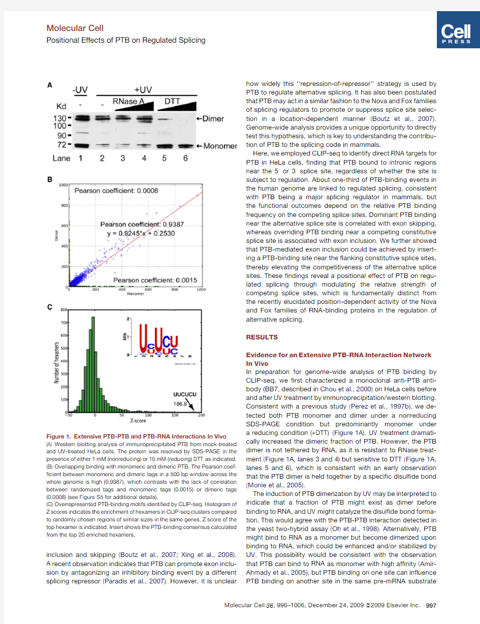
In preparation for genome-wide analysis of PTB binding by CLIP-seq,we ?rst characterized a monoclonal anti-PTB anti-body (BB7,described in Chou et al.,2000)on HeLa cells before and after UV treatment by immunoprecipitation/western blotting.
Consistent with a previous study (Pe
′rez et al.,1997b ),we de-tected both PTB monomer and dimer under a nonreducing SDS-PAGE condition but predominantly monomer under a reducing condition (+DTT)(Figure 1A).UV treatment dramati-cally increased the dimeric fraction of PTB.However,the PTB dimer is not tethered by RNA,as it is resistant to RNase treat-ment (Figure 1A,lanes 3and 4)but sensitive to DTT (Figure 1A,lanes 5and 6),which is consistent with an early observation that the PTB dimer is held together by a speci?c disul?de bond (Monie et al.,2005).
The induction of PTB dimerization by UV may be interpreted to indicate that a fraction of PTB might exist as dimer before binding to RNA,and UV might catalyze the disul?de bond forma-tion.This would agree with the PTB-PTB interaction detected in the yeast two-hybrid assay (Oh et al.,1998).Alternatively,PTB might bind to RNA as a monomer but become dimerized upon binding to RNA,which could be enhanced and/or stabilized by UV.This possibility would be consistent with the observation that PTB can bind to RNA as monomer with high af?nity (Amir-Ahmady et al.,2005),but PTB binding on one site can in?uence PTB binding on another site in the same pre-mRNA
substrate
Figure 1.Extensive PTB-PTB and PTB-RNA Interactions In Vivo
(A)Western blotting analysis of immunoprecipitated PTB from mock-treated and UV-treated HeLa cells.The protein was resolved by SDS-PAGE in the presence of either 1mM (nonreducing)or 10mM (reducing)DTT as indicated.(B)Overlapping binding with monomeric and dimeric PTB.The Pearson coef-?cient between monomeric and dimeric tags in a 500bp window across the whole genome is high (0.9387),which contrasts with the lack of correlation between randomized tags and monomeric tags (0.0015)or dimeric tags (0.0008)(see Figure S5for additional details).
(C)Overrepresented PTB-binding motifs identi?ed by CLIP-seq.Histogram of Z scores indicates the enrichment of hexamers in CLIP-seq clusters compared to randomly chosen regions of similar sizes in the same genes.Z score of the top hexamer is indicated.Insert shows the PTB-binding consensus calculated from the top 20enriched hexamers.
Molecular Cell
Positional Effects of PTB on Regulated Splicing
(Chou et al.,2000).In any case,the ability of PTB to simulta-neously engage in protein-protein and protein-RNA interactions suggests that PTB may nucleate extensive RNA-protein interac-tion networks,which are likely contributed by RNA-binding activities of individual RRMs in PTB(Oberstrass et al.,2005; Clerte and Hall,2009).
By32P labeling,we found that both monomeric and dimeric PTB are associated with RNA(Figure S1available online).To detect potential functional differences between the monomeric and dimeric forms of PTB,we separately isolated the two protein-RNA complexes and constructed two independent libraries for CLIP-seq analysis.We?rst determined the quality of the libraries by conventional cloning and sequencing,obtain-ing341and214unique tags associated with PTB monomer and dimer,respectively.Most of these tags($80%)were mapped to introns as expected(Figure S2),and the average size is$30nt in length(Figure S3),which is consistent with the previous report that the minimal PTB-binding sequence is30nt(Amir-Ahmady et al.,2005).This?nding suggests that the actual PTB-binding sites are likely to reside within,rather than nearby,the sequenced tags in most cases,therefore eliminating the need to computationally extend the tags for subsequent peak?nding (Yeo et al.,2009).We further con?rmed the speci?city of the CLIP assay by performing RIP-PCR analysis on a panel of anti-PTB enriched RNAs(Figure S4).
Having thoroughly characterized the libraries,we next subjected PCR amplicons to high-throughput sequencing on Solexa,resulting in2.44million tags for PTB monomer and 2.37million for PTB dimer that were uniquely mapped to the human genome(hg18).These high-density reads allowed us to ask?rst whether PTB monomeric and dimeric tags are dif-ferentially distributed in the genome.As shown in Figure1B, using a500bp window,most of the monomeric and dimeric tags are similarly distributed in the genome with a Pearson corre-lation coef?cient of0.9387,and the coef?cient between mono-mer and dimer increases with the increasing number of tags compared(Figure S5).We conclude that there are no two separate sets of sites for PTB binding as monomer or dimer in the cell.
We therefore used combined tags to determine overrepre-sented motifs in PTB-binding clusters(see Experimental Proce-dures),?nding that CU-rich hexamers are highly enriched (Figures1C and S6A).About21%of clusters(5.17%for random; p=0.00)contain the top-scored motif UUCUCU(Z score,186.5). The top20motifs are all CU enriched;83.56%of total clusters obtained contain at least one of the top20motifs(38.56%for random;p=0.00)(Figure S6B),and the consensus generated from the top20hexamers is UYUYU(insert in Figure1C).In fact,the C/U percentage is broadly elevated surrounding the PTB-binding sites,but not among randomly selected back-ground sequences(Figure S7),which fully corroborates with biochemically de?ned PTB-binding characteristics(Ashiya and Grabowski,1997;Pe′rez et al.,1997a).Of interest,$90%of identi?ed PTB-binding sites overlap with those predicted by an algorithm based on the biochemical properties of PTB(Gama-Carvalho et al.,2006),but the number of the experimentally detected sites represents only$1%of$5million sites predicted in the human genome by the algorithm(data not shown).Thus,biochemically deduced consensus may not be suf?cient to predict true binding sites because the recognition of some consensus motifs may be obstructed by competition of other RNA-binding proteins or by certain RNA secondary structures. Indeed,we did note a few underrepresented,A/G-rich motifs (Figure S6A),indicating that depletion of A/G-rich sequences may help maximize the single strandedness of PTB-binding sites by minimizing potential stem-loop structures due to base pair-ings between C/U and A/G rich sequences.These observations suggest that experimentally validated binding sites coupled with critical features in local genomic context will help to further improve prediction algorithms for RNA-binding splicing factors. Genomic Landscape of PTB Binding
By mapping the sequenced tags to the knownGene set from the UCSC genome database,we found that58.4%of the tags are localized in introns(Figure2A),with the relative density(counts per kb)17-fold higher in introns than in exons,indicating that most of the tags are derived from pre-mRNAs(a fraction of PTB-binding events may also be derived from excised lariats).
A sizable fraction of tags was mapped to antisense transcripts
(8.1%)and intergenic regions(28.4%),implying that PTB may also bind to many noncoding RNA and/or unannotated tran-scripts,which is subject to future studies.
We next focused on clustered PTB-binding events by identi-fying peaks above the gene-speci?c,randomized background as previously described(Yeo et al.,2009).The resulting64,314 peaks were further merged to51,394clusters by placing PTB peaks within a50nt window.Of interest,whereas more than half(56.5%)of PTB-binding clusters are separated by1to 10kb,as expected from independent binding events,a signi?-cant fraction(43.5%)of PTB clusters appears to be more closely positioned(<1kb)(Figure2B),likely re?ecting a concert action of multiple PTB-binding events in regulated splicing.Further anal-ysis revealed that PTB binds to10,372out of the24,378anno-tated human genes(30,986/66,803knownGene transcripts). This number might be an underestimate because our current sequencing density has not yet reached saturation according to power analysis(data not shown).Given the fact that most sequence tags contain PTB-binding consensus,indicating that contamination with other nonspeci?c RNA is minimal,this binding pro?le suggests that PTB is a major RNA-binding protein that may be widely involved in RNA metabolism in mammals. Association of PTB Binding with Alternative Splicing Events
We next explored how frequently PTB binding is linked to anno-tated alternative splicing events.We separately examined PTB association with several major modes of alternative splicing, including cassette exon,alternative50splice site,alternative30 splice site,and retained intron,based on the knownAlt track of the UCSC genome browser(Karolchik et al.,2008).This analysis revealed that28.3%of PTB-binding events are associated with annotated alternative splicing,and22.2%of all annotated alter-native splicing events are linked to PTB binding,thus suggesting a prevalent role of PTB as a splicing regulator in the human genome.PTB is involved in all common modes of alternative splicing(Table1),with cassette exons being the most frequent
Molecular Cell Positional Effects of PTB on Regulated Splicing
Molecular Cell
Positional Effects of PTB on Regulated Splicing
Figure2.Genomic Landscape of PTB Binding
(A)The distribution of PTB tags in the human genome(hg18).
(B)The distribution of PTB-binding clusters relative to one another in the same genes.
(C)Screen shot of PTB binding around the well-characterized nPTB exon10.
(D)Screen shot of PTB binding around TPM2exon7and the two alternative polyadenylation sites.
(E)Screen shot of PTB binding in the intron preceding the regulated exon9in PKM2gene.
(F)PTB-binding clusters associated with six major alternative RNA-processing modes.The patterns of PTB binding in250nt intronic and30nt exonic regions around splice sites were counted.The?lled black boxes indicate constitutive exons or exonic regions,whereas empty red boxes show alternative exons or exonic regions.The short blue lines mark the regions where PTB binding clusters were present(+)or absent(à).The number is the total events of PTB binding at each location.
targets for PTB regulation (Z score 14.11,compared to 100trials of randomly placed clusters).Many PTB-binding events are also found on ‘‘constitutive’’introns and exons,which might be asso-ciated with alternative splicing events that have not yet been annotated.Alternatively,PTB may function to repress decoy splicing signals within constitutively spliced genes,which deserves a close look in future studies.
We next determined how PTB binding might affect splice site selection on both known and newly identi?ed PTB target genes.As expected,a signi?cant number of tags were mapped to the PTBP2(also known as nPTB )gene,a well-known PTB target in which the alternative exon 10is repressed by PTB.Of note,PTB binds preferentially to the sequences upstream of the 30splice site of exon 10in nPTB as previously characterized (Boutz et al.,2007;Spellman et al.,2007).We also identi?ed a binding cluster near the downstream 50splice site and some distributive PTB binding in the upstream intron (Figure 2C),suggesting that PTB binds to multiple locations surrounding the regulated exon,which may collectively contribute to PTB-mediated exon repression,a situation similar to the well-characterized c-Src N1exon (Sharma et al.,2005).
In another example (Figure 2D),we identi?ed two PTB-binding clusters between the two mutually exclusive exons (exon 6and 7)in the TPM2gene,which is consistent with the observed
repression of exon 7in nonmuscle cells (Saulie
′re et al.,2006;Spellman et al.,2007).We also detected prevalent PTB binding near the polyadenylation site for E10,in agreement with the observed utilization of the E11polyadenylation site in nonmuscle cells.Of interest,we note multiple PTB-binding events between the regulated exons and polyadenylation sites,suggesting a potential RNA network that may underlie the coordinated regulation of both events as reported (Spellman et al.,2007).The high-quality PTB-RNA interaction map also helps to assign PTB as a regulator to previously uncharacterized alterna-tive splicing events.For example,the pyruvate kinase 2(PKM2)gene expresses two mutually exclusive isoforms,and such regu-lated splicing appears to be critical for cancer metabolism and
tumor growth (Christofk et al.,2008).Although PTB has been implicated in the regulation of PKM2splicing,critical cis -acting regulatory elements has remained unde?ned (Spellman et al.,2007).We found extensive PTB-binding clusters in the intron preceding the alternative exon 9(Figure 2E),and RT-PCR con?rmed PTB-dependent repression of PKM2exon 9in HeLa cells (data not shown).This ?nding raises the possibility that PTB may contribute to certain cancer phenotypes by regulating the alternative splicing of PKM2.
PTB binding appears to associate with regulated cassette exons more signi?cantly than do other modes of alternative splicing (Table 1).On the well-characterized c-Src gene,PTB binds to both sides of the regulated exon N1(Amir-Ahmady et al.,2005).To estimate how frequently PTB binds to both sides of alternative exons or exonic sequences,we analyzed a large number of annotated alternative splicing/polyadenylation events in comparison with mapped PTB-binding events (Figure 2F).This analysis revealed several interesting trends.First,whereas the bracket binding mode of PTB is clearly associated with many regulated RNA-processing events,PTB appears to bind either up-or downstream of the alternative splice site in the majority of cases.Second,among regulated cassette exons,PTB has the same tendency to bind to one of the competing (constitutive versus alternative)splice sites,implying that PTB does not always target the alternative splice site,which has distinct functional consequences (see below).Third,in most cases of regulated 50and 30splice site choices,PTB appears to prefer binding on the intronic side,predicting that PTB may favor the distal splice site by repressing the proximal site in general.PTB-Dependent Repression or Enhancement of Alternative Splicing In Vivo
Most minigene-based analysis focused on the consensus PTB-binding motif near a regulated exon(s),which leaves a general impression that PTB preferentially targets alternative splice sites for regulation.The PTB CLIP-seq data now offer an unbiased view on the actual location of PTB binding on PTB-regulated genes.We found both known and new locations for PTB binding on all 13previously documented PTB-regulated exons.This prompted us to examine additional candidates based on prevalent PTB-binding events.Of 32targets assayed,we found that 22altered splicing in response to PTB knockdown by RNAi (>5%absolute change),and among these,10showed PTB-dependent inclusion,and 12exhibited PTB-dependent exon skipping (Table S1).This ?nding con?rmed the previous observation that PTB regulates both exon inclusion and skipping in vivo (Boutz et al.,2007;Xing et al.,2008).
Although the mechanism for PTB-mediated exon skipping is well characterized on multiple minigene models in literature,it has been unclear how PTB enhances exon inclusion.We ?rst wished to establish sequence-dependent regulation of exon inclusion by PTB.For this purpose,we selected the CTTN gene that showed positive regulation by PTB to construct a minigene for analysis in transfected cells.The minigene,which contains the cassette exon 11and ?anking introns and exons,was expressed from the CMV promoter in pcDNA3(Figure 3A;note that this minigene was spliced less ef?ciently than was the endogenous gene [see Figure 4B],likely because
the
Table 1.PTB-Binding Clusters Associated with Different Modes of Alternative Splicing
#Total the UCSC knownAlt track.The observed number of PTB clusters associ-ated with each mode is the count of PTB clusters within the region covering the alternative exon,the ?anking intron(s),and constitutive exons.The expected number is the averaged number of associations in 100trials (random placement of PTB clusters).The column Z score shows the signi?cance of association.
Molecular Cell
Positional Effects of PTB on Regulated Splicing
minigene might miss some positive regulatory elements in the construct and/or impair ef?cient transcription/splicing coupling as on the endogenous gene).In response to simultaneous knockdown of PTB and nPTB by shRNAs (Figure 3B,lane 5),we detected a signi?cant reduction of exon 11inclusion in cells cotransfected with the minigene reporter (Figure 3C,compare lane 1treated with control shRNA with lane 5treated with combined shRNAs against PTB and nPTB ).
We next attempted to rescue the splicing defect by cotrans-fecting the cell with a plasmid expressing Flag-tagged PTB or nPTB,each of which contains a synonymous mutation that disrupts the shRNA target.By western blotting,these exoge-nous genes were robustly expressed (Figure 3B).We observed that the full-length PTB (PTB4;see below)was able to fully rescue the inclusion of the alternative exon 11(Figure 3C,lane 3).nPTB was also capable of rescuing exon 11inclusion to a signi?cant degree (Figures 3C and 3D).Previous studies showed that the PTB gene expresses two major isoforms,PTB4and PTB1,which differ by the presence or absence of the alternative exon 9(Wollerton et al.,2001).We found that PTB1had little activity in rescuing the inclusion of CTTN exon 11in comparison with the exon 9-containing PTB4,even though both proteins were expressed at comparable levels in transfected cells.This observation is consistent with the previous study that reported a stronger activity of PTB4than PTB1in regulated splicing (Wollerton et al.,2001).In these rescue experiments,we did not detect a further increase in exon 11inclusion even though the exogenous PTB or nPTB was overexpressed,indicating that PTB or nPTB is involved in the regulation but is not the only regulator(s)for this alterna-tive splicing event (as a result,it is no longer a rate-limiting factor in PTB -overexpressed
cells).
Figure 3.PTB-Dependent Inclusion of
Alternative Exon
(A)Schematic representation of the CTTN mini-gene constructs,showing both wild-type and the mutant that lacks the 44nt PTB-binding cluster.The mapped PTB-binding site is marked in gray,and the PCR primers used to detect alternatively spliced products are indicated by arrows.
(B)Exogenously expressed PTB isoforms and nPTB in double PTB/nPTB knockdown cells.
(C)Semiquantitative RT-PCR analysis of WT and mutant CTTN pre-mRNA splicing in response to PTB/nPTB knockdown with or without comple-mentation with exogenously expressed PTB isoforms or nPTB .
(D)Quanti?cation of the data as in (C)based on three independent experiments.Error bars are based on SEM;the statistical signi?cance is determined by Student’s t test (p <0.05).
To determine whether the regulation is dependent on the mapped PTB-binding site in the intron,we deleted the 44nt PTB-binding site in the reporter and found that the mutation abolished the response to exogenous PTB or nPTB
(Figures 3C and 3D).Deletion of the PTB-binding site renders levels of exon inclusion in the CTTN minigene similar to those caused in the wild-type by depletion of PTB/nPTB,further supporting the involvement of these proteins in regulation.Deletion of the PTB-binding sites also abolished the functional rescue by any PTB isoforms.We conclude from these experi-ments that PTB/nPTB is also directly involved in regulated exon inclusion in addition to its widely perceived role in exon skipping.
Mechanistic Insights into PTB-Regulated Alternative Splicing
In order to understand the mechanisms for PTB-dependent exon inclusion or skipping,we analyzed the PTB-binding pattern with respect to the functional consequence of alternative splicing and realized some general trends for PTB-regulated splicing (Fig-ure 4).Among PTB-mediated exon repression events,we note that PTB binding typically takes place near the alternative exon.This is clearly the case with both the MINK1and EIF4G2genes (Figure 4A,rows 1and 2).However,PTB also binds to other intronic locations besides around the alternative exon,as seen on the RBM27and FAM38A gene (Figure 4A,rows 3and 4).The remaining two examples (CCDC138and RBM15,rows 5and 6in Figure 4A)illustrate PTB binding on both sides of the regulated exon,although the upstream PTB-binding sites appear to vary in distance from the regulated exon.In these cases,we notice a relatively short intron after the alternative exon,indicating that PTB binding in the intron might obstruct the intron de?nition process (Fox-Walsh et al.,2005),thus result-ing in PTB-dependent skipping of the alternative exon.These examples agree in general with the established principle of PTB-dependent exon skipping,wherein PTB appears to mainly
Molecular Cell
Positional Effects of PTB on Regulated Splicing
act to interfere with the recognition of the splice sites associated with the alternative exon.
PTB-dependent exon inclusion seems to exhibit a different trend.As illustrated in Figure 4B,the ?rst three examples exhibited PTB-binding events that are far away from the alterna-tive exon and close to the competing constitutive 50(RILRA ,row 1)or 30splice site (NUF2,row 2).This trend may also be applicable to the CTTN gene (Figure 4B,row 3),despite a minor PTB-binding site near the alternative exon,and our mutagenesis study showed that the major site near the downstream constitu-tive 30splice site was responsible for PTB-dependent exon inclusion (Figure 3).The remaining three examples (RASSF8,EZH2,and PPP5C ,rows 4to 6)are not clear-cut.PTB clearly binds to both sides of the alternative exon in each case,which is similar to the situation with PTB-dependent exon skipping events.However,both PTB-binding sites appear closer to the competing constitutive 50and 30splice sites than to the alterna-tive exon.Together,these examples appear to point to the trend that the PTB-binding sites associated with PTB-dependent exon inclusion events are associated with competing constitutive splice sites.
To generalize the trend for both PTB-regulated exon inclusion and skipping,we collected a number of PTB-regulated exons,including 22identi?ed in the present study and 11that have been previously reported in humans (Table S1).In addition,we found that the CLIP tags are generally mapped to the
conserved
Figure 4.PTB Can Either Represses or Enhance Alternative Splicing In Vivo
(A)Examples of PTB-dependent exon skipping.Each is schematically diagramed (exon,black box;intron,black line)with mapped PTB-binding clusters as marked by blue boxes.PTB RNAi-induced splicing changes are shown on the right.Error bars are based on SEM from three indepen-dent experiments.All detected changes are signi?cant as determined by the Student’s t test (p <0.05).
(B)Examples of PTB-dependent exon inclusion with mapped PTB-binding clusters marked by brown boxes.For NUF2,PTB tags (not clusters)are shown under the intron line.
regions on PTB-regulated mouse genes as reported previously (Boutz et al.,2007),which strongly implicates similar regulation between mice and humans.We directly tested a subset of the human orthologs of several PTB-regulated genes previously characterized on mouse cells,including SPAG9,PTB ,nPTB ,TPM1,KTN1,TPM2,and MINK1,and found that these genes all similarly responded to PTB knockdown in HeLa cells.We therefore included additional PTB-regulated splicing events in mouse cells (Table S1),resulting in a total of 55PTB-regulated splicing events (41PTB-dependent exon skipping and 14
PTB-dependent exon inclusion)for further analysis.As controls,we selected 100groups of randomly sampled constitutive exons (each group contains 50exons)for similar analysis.
By integrating all PTB-binding events,we generated an RNA map associated with PTB-repressed,-enhanced,and -nonreg-ulated (constitutive)exons on a scaled pre-mRNA model,an approach that has been recently used for analysis of position-dependent activities of Nova (Licatalosi et al.,2008).Of interest,the map revealed that PTB binds to both the 50and 30splice sites of constitutive exons,as well as to both the 50and 30splice sites of alternative exons (Figure 5).Though PTB binding to the 30splice site is expected (because of the polypyrimdine tract as part of the splicing signal at the 30splice site),we were surprised by equally frequent PTB binding at the 50splice site.Most PTB-dependent exon-skipping events (bottom portion of Figure 5A)are associated with PTB binding near either side of the alternative exon,which is fully consistent with functional studies conducted so far on model minigenes.In contrast,the RNA map associated with PTB-dependent exon inclusion events (top portion of Figure 5A)suggests that PTB binds prev-alently to the ?anking constitutive splice sites,especially at the downstream constitutive 30splice site (Figure 5A).This most likely re?ects PTB interference with the recognition of the competing constitutive 30splice site,therefore in favor of the selection of the upstream alternative exon.On nonregulated exons,we found no clear bias in PTB binding to intronic regions
Molecular Cell
Positional Effects of PTB on Regulated Splicing
near any upstream or downstream splice sites (Figure 5B).Together,these ?ndings formally suggest a PTB-mediated splice site titration mechanism by which the relative binding frequency near the competing constitutive and alternative splice sites dictates the functional outcome,which appears to be neutralized on nonregulated constitutive exons (see further in Discussion ).
Induction of Exon Inclusion by Engineered PTB-Binding Sites
We demonstrated that the PTB-binding site near the constitutive 30splice site of the CTTN gene is responsible for PTB-dependent inclusion of the upstream alternative exon (Figure 3).To further test the hypothesis that PTB induces the inclusion of the alterna-tive exon by weakening the competing constitutive splice site(s),we engineered a different minigene containing a SIRT1exon (Figure 6A),which was previously used to screen for cis -acting splicing suppressors (Wang et al.,2004).To improve the PTB response range of the reporter,we made a minor modi?cation on the sequence in the SIRT1exon to reduce its inclusion level and selected four regions to insert a PTB-binding site (Figure 6B).In order not to directly interfere with U1binding,the positions for insertion in the upstream exon (UpE)or intron (UpI)are both $15nt away from the constitutive 50splice site.To avoid obstruction of 30splicing signals,the position for insertion in the downstream intron (DoI)is 15nt upstream of the branchpoint,whereas the position for insertion in the downstream exon (DoE)is 10nt from the 30AG
dinucleotide.
Figure https://www.360docs.net/doc/df4047271.html,posite Functional Map of PTB-Regulated Splicing
(A)PTB-regulated cassette exons are collected from previously reported cases and those that are validated in the present study.Among 55PTB-regulated splicing events compiled,14exhibited PTB-dependent splicing inclusion,and 41showed PTB-dependent exon skipping.
(B)RNA map on constitutive exons.Red line shows the average of normalized complexity of 100sets (each contains 50randomly selected exons)of constitutive exons;the upper and lower light-blue boundaries show the one standard deviation (see Experimental Procedures for further details).
We transfected the parental splicing reporter and the PTB site insertion deriv-atives into HeLa cell and analyzed the splicing products by semiquantitative RT-PCR.As shown in Figure 6B,insertion of a PTB-binding site near either the 50or 30constitutive splice site signi?cantly enhanced the inclusion of the alternative exon.RNAi knockdown of PTB and nPTB completely abolished the exon inclusion induced by inserted PTB-binding sites (Figure 6C),and overex-pression of PTB4further enhanced exon inclusion in a PTB binding site-dependent
manner (Figure 6D).These observations provide unequivocal support to the splice site titration mechanism for PTB-dependent exon inclusion in which weakening the constitutive 50or 30splice site enhances the competitiveness of the alternative 50or 30splice site.These ?ndings have therefore documented a posi-tional effect for a general splicing repressor to positively regulate alternative splicing in mammalian cells.DISCUSSION
Our global analysis of PTB-RNA interactions in the human genome provides mechanistic insights into PTB-regulated RNA processing.Besides competing directly with U2AF65binding to interfere with 30splice site recognition (Lin and Patton,1995;
Saulie
′re et al.,2006;Singh et al.,1995),PTB has been shown to use multiple mechanisms to regulate alternative RNA process-ing by binding to regions other than the core splicing signals on minigene models (Izquierdo et al.,2005;Sharma et al.,2008;Spellman and Smith,2006).We have now generalized and signif-icantly extended these ?ndings at the genome level.
Interference of Splice Site Recognition and
Communication by PTB-Mediated RNA Networks
RNA looping has been proposed as one of the mechanisms for PTB-mediated splicing repression to sequester the alternative exon from the splicing machinery (Chou et al.,2000;Wagner and Garcia-Blanco,2001).PTB dimerization was initially postu-lated to facilitate RNA looping (Oh et al.,1998;Pe
′rez et al.,Molecular Cell
Positional Effects of PTB on Regulated Splicing
1997b ),but a later structural analysis suggests potential induc-tion of RNA looping via RRM3and RRM4in the same PTB molecule to simultaneously bind to cis -acting RNA elements (Oberstrass et al.,2005).However,these two modes of RNA looping induced by inter-or intramolecular interactions do not have to be mutually exclusive.Although puri?ed PTB exists predominantly as a monomer in solution,which can bind to RNA with high af?nity,it has been suggested that PTB binding to RNA may create the spatial proximity for enhanced PTB-PTB interactions on RNA,which may be stabilized by the induced formation of a disul?de bond (Amir-Ahmady et al.,2005;Monie et al.,2005;Oberstrass et al.,2005).Our data are fully consistent with PTB binding to RNA as monomer and subsequent disul?de bond formation on closely spaced PTB molecules on target RNA.Of interest,we found that UV can further enhance or stabilize PTB-PTB interactions.Importantly,our data indicate that there are no separate sets of binding sties for monomeric and dimeric PTB in the human genome.However,this does not undermine the potential synergy between protein-protein and protein-RNA interactions that may be critical for induced RNA looping surrounding PTB-regulated exons as previously proposed (Wagner and Garcia-Blanco,2001).
Given frequent PTB binding in multiple locations in a single intron in many cases (e.g.,Figure S6B),we may envision an extensive RNA network nucleated by PTB,which may be the underlying mechanism for the observed interference of both exon de?nition and the transition from exon de?nition to intron de?nition during spliceosome assembly (Izquierdo et al.,2005;Sharma et al.,2005,2008).As there is no reason to believe that PTB-mediated RNA network has to be restricted within a single regulatory unit,we may further speculate that the network may spread on multiple intronic and exonic locations in the same pre-mRNA molecule,thereby allowing a coordinated regulation of multiple RNA processing events as evidenced on the TPM2gene (Spellman et al.,2007).Such a network may be more prevalent than what we can imagine at this point because initial PTB-RNA interactions may induce additional PTB binding to other sites that may not even contain a motif for high-af?nity
binding by PTB,which may be further enhanced by other PTB cofactors,such as Raver1(Gromak et al.,2003).
Mechanisms for Positive and Negative Regulation of Splice Site Selection by PTB
The PTB-RNA interaction map also suggests a potential mecha-nism for positive and negative regulation of splice site selection by PTB,depending on its binding relative to competing constitu-tive and alternative splice sites.Of interest,PTB not only binds to intronic locations near the 30splice site,but also to sites closer to the 50splice site regardless of whether the splice site is subjected to alternative choices.If predominant PTB binding occurs near a constitutive splice site,it may weaken the site,thereby raising the competitiveness of the competing alternative site.A minor modulation of splice site recognition may be translated into a major functional consequence as demonstrated by a recent kinetic analysis of splice site competition (Yu et al.,2008).This principle may be generally applicable to RNA-binding splicing regulators to give rise to either a positive or negative functional outcome that depends on where the factor binds.
A positional effect has clearly emerged from recent genome-wide studies of splicing regulators (Licatalosi et al.,2008;Yeo et al.,2009).However,the positional effect that we observed with PTB-regulated splicing appears to be fundamentally distinct from that exerted by Nova and Fox2.In those cases,Nova and Fox2binding to their cis -acting elements upstream and down-stream of the alternative exon generally represses or enhances the selection of the exon,respectively,but it is presently un-clearly how such opposite effects on splice site selection are achieved.In contrast,PT
B appears to be sampling multiple intronic locations in a pre-mRNA to exert a negative effect on the selection of the nearby splice site.PTB binding close to the intronic region near the 30and 50alternative splice sites likely results in skipping of the alternative exon,whereas PTB binding to sequences adjacent to constitutive exons tends to induce the inclusion of the alternative exon.
It is important to point out that potential composite effects may account for some apparent exceptions to this general trend.
For
Figure 6.Mechanism of PTB-Dependent Exon Inclusion
(A)The reporter construct.pM17is derived from pZW8-ESS17(Wang et al.,2004)with a cis -acting regulatory element disrupted by the inserted sequence in the alternative SIRT1exon.Four positions are selected for inserting a PTB-binding site as diagrammed.
(B)Splicing of the parental and mutant reporters in transfected HeLa cells was determined by RT-PCR (representative gel images shown as inserts)and quanti?ed.
(C and D)PTB-dependent exon inclusion through the inserted PTB-binding sites.The effect on exon inclusion was diminished by the shRNAs against PTB and nPTB in cotransfected HeLa cells (C).The effect on exon inclusion could be further enhanced by PTB overexpression (D).Together,these results demonstrate that PTB is directly involved in enhancing exon inclusion through the inserted binding sites.Error bars are based on SEM derived from three independent experiments.
Molecular Cell
Positional Effects of PTB on Regulated Splicing
example,the PTB-binding pattern was similar on both the CCDC138and EZH2genes,but PTB knockdown had opposite effects on these two genes.The PTB-binding events on the CCDC138gene are both far away from the regulated exon,yet the net effect is PTB-dependent exon skipping,perhaps because the exon skipping effect,due to strong PTB binding on both sides of the alternative exon,might be dominant over its in?uence on the downstream constitutive exon.Therefore, the?nal functional outcomes,in many cases,may be determined by the sum of those competing binding events.In addition,most alternative splicing events are likely subjected to regulation by multiple different splicing regulators,which may act synergisti-cally or antagonistically.Therefore,the possibility that other regulators may override the effect of PTB binding on certain regulated exons may account for various exceptions to the posi-tional effect observed,thus emphasizing the combinatory con-trol of alternative splicing that likely operate in mammalian cells. EXPERIMENTAL PROCEDURES
Cell Culture,Plasmids,and RNAi
HeLa cells were grown in Dulbecco’s modi?ed Eagle’s medium(DMEM)with 10%newborn bovine serum plus100U penicillin/streptomycin(Gibico)at 37 C in5%CO2.Oligofectamine and Lipo2000(both from Invitrogen)were used for siRNA and plasmid transfection,respectively,according to manufac-turer’s instructions.To construct the expression plasmids for PTB1,PTB4, and nPTB,FLAG-tagged primer sets were used to amplify the coding region of individual genes,and the PCR products were inserted into pcDNA3between the EcoR1and Not1sites.The CTTN minigene was constructed by amplifying Exon10to Exon12regions,which were inserted into pcDNA3between the EcoR1and Xho1sites.Validated siRNAs against PTB and nPTB were purchased from Ribbobio.
CLIP-seq,RIP-PCR,and Validation of PTB-Regulated Splicing
HeLa cells were UV irradiated at400mj and collected by scraping the cells from15cm plates.The CLIP procedure was performed as described(Yeo et al.,2009).Immunoprecipitated RNA was extracted using Trizol,and after DNase I(Promega)treatment,RNA was reverse transcribed using MMLV with N9random primer at37 C followed by inactivation of the reverse tran-scriptase at70 C for15min.The resulting cDNA was analysis by PCR.Table S2lists PCR primers for validation of PTB-dependent splicing.
Bioinformatics Analysis
The sequenced tags longer than18nt were mapped to the human genome sequence by allowing two mismatches and two insertions or deletions,and only those with maximal identity(R90%)were kept.To analyze genomic distribution of tags,known human genes(knownGene track from the UCSC genome browser)were chosen as the gene set,which contained66,803 entries.We arbitrarily de?ned promoter regions as5kb upstream of the transcriptional start site of the gene.For genes having multiple isoforms,the transcript with the longest length,the largest number of exons,the longest CDS,or the maximum length of all exons was chosen to analyze the tag distribution in exon,intron,50UTR,CDS,and30UTR.
The PTB-binding sites enriched of tags were detected by using the similar strategy as previously described(Yeo et al.,2009).The differences were:(1) tags were not extended;(2)peak identi?cation was independently done for each gene cluster of24,378clusters,which were grouped by using the program clusterGenes(-joinContained)on all known genes;(3)consecutive positions with the same height were counted only once;(4)control tags were randomly placed on gene cluster with repetitive elements masked;(5)for each height level h,the p value was assigned as the ratio of the number of heights higher than h divided by the total number of heights in100random placements,and the p values for all heights were adjusted by using Bonferroni correction to account for multiple hypotheses testing.The smallest height that gave an FDR<0.001was de?ned as the threshold height.Consecutive nucleotide posi-tions with height higher than the threshold were identi?ed as signi?cant PTB-binding peak.If multiple peaks were detected less than50nt from one another, they were merged to represent a single PTB-binding site or cluster.
The sequences extracted from genome according to PTB-binding clusters were used to detect overrepresented motifs(Defrance et al.,2008).For each cluster,it was extended to the two sides by25nt,as some clusters had small lengths.Background sequences include those from randomly selected inter-vals in genes or random sequences generated with respect to order0and 1Markov models(same single and dinucleotide frequencies)built from known-Genes(Ponty et al.,2006).Identi?cation of overrepresented k-mers(k=2,3,4, 5,6,and7)was based on random intervals.The pictogram was plotted according to WebLogo(https://www.360docs.net/doc/df4047271.html,/),which is based on the alignment of the top20motifs by ClustalW(https://www.360docs.net/doc/df4047271.html,/Tools/ clustalw/).
Normalized complexity map of PTB-RNA interactions was generated as described in(Licatalosi et al.,2008).The composite pre-mRNA was made by joining the longest upstream exon,upstream intron,middle exon(PTB-regulated exon),downstream intron,and downstream exon.The tags around PTB-regulated individual exons were mapped to the composite pre-mRNA according to their positions relative to the nearest splice site.The tags in one transcript were?rst normalized to their number across the region covering the PTB-regulated exon and?anking introns and exons and then to the number of different transcripts with tags at a given position as described(Licatalosi et al.,2008).For comparison,we extracted6460sets of three constitutive internal exons that are associated with PTB binding from knownGene set (hg18).Normalized complexity map was similarly created on50randomly selected constitutive exons to deduce both averaged PTB-binding events with standard deviation.
The analysis used the programs from Jim Kent’s source code(http://www. https://www.360docs.net/doc/df4047271.html,/$kent),bx-python library(https://www.360docs.net/doc/df4047271.html,/james_taylor/ bx-python/),pygr library(https://www.360docs.net/doc/df4047271.html,/p/pygr/),and homemade python codes.
ACCESSION NUMBERS
The CLIP-seq data for PTB monomer and dimer are available at the Gene Expression Omnibus under the accession number GSE19323.
SUPPLEMENTAL DATA
Supplemental Data include seven?gures and two tables and can be found with this article online at https://www.360docs.net/doc/df4047271.html,/molecular-cell/supplemental/ S1097-2765(09)00907-1.
ACKNOWLEDGMENTS
The authors are grateful to Chris Smith,Zefeng Wang,and Reuven Agami for sending us PTB expression plasmids and other regents;to Miriam Llorian for advice on PTB RNAi;and to Alain Denise for suggestions in computational analysis.We are also indebted to members of the Yi Zhang lab for cooperation and discussion during the course of this investigation.This work is supported by the China863program(2007AA02Z112)to Y.Z.,the China973program (2005CB724604)to Y.Z.and X.-D.F.,and US NIH grants(GM049369, HG004659,GM084317)to D.L.B.,G.Y.,and X.-D.F.
Received:May18,2009
Revised:August28,2009
Accepted:November3,2009
Published:December24,2009
REFERENCES
Amir-Ahmady,B.,Boutz,P.L.,Markovtsov,V.,Phillips,M.L.,and Black,D.L. (2005).Exon repression by polypyrimidine tract binding protein.RNA11, 699–716.
Molecular Cell
Positional Effects of PTB on Regulated Splicing
Ashiya,M.,and Grabowski,P.J.(1997).A neuron-speci?c splicing switch mediated by an array of pre-mRNA repressor sites:evidence of a regulatory role for the polypyrimidine tract binding protein and a brain-speci?c PTB coun-terpart.RNA3,996–1015.
Black,D.L.(2003).Mechanisms of alternative pre-messenger RNA splicing. Annu.Rev.Biochem.72,291–336.
Boutz,P.L.,Stoilov,P.,Li,Q.,Lin,C.H.,Chawla,G.,Ostrow,K.,Shiue,L.,Ares, M.,Jr.,and Black, D.L.(2007).A post-transcriptional regulatory switch in polypyrimidine tract-binding proteins reprograms alternative splicing in developing neurons.Genes Dev.21,1636–1652.
Chou,M.Y.,Underwood,J.G.,Nikolic,J.,Luu,M.H.,and Black,D.L.(2000). Multisite RNA binding and release of polypyrimidine tract binding protein during the regulation of c-src neural-speci?c splicing.Mol.Cell5,949–957.
Christofk,H.R.,Vander Heiden,M.G.,Harris,M.H.,Ramanathan,A.,Gerszten, R.E.,Wei,R.,Fleming,M.D.,Schreiber,S.L.,and Cantley,L.C.(2008).The M2 splice isoform of pyruvate kinase is important for cancer metabolism and tumour growth.Nature452,230–233.
Clerte,C.,and Hall,K.B.(2009).The domains of polypyrimidine tract binding protein have distinct RNA structural preferences.Biochemistry48,2063–2074.
Defrance,M.,Janky,R.,Sand,O.,and van Helden,J.(2008).Using RSAT oligo-analysis and dyad-analysis tools to discover regulatory signals in nucleic sequences.Nat.Protoc.3,1589–1603.
Fox-Walsh,K.L.,Dou,Y.,Lam,B.J.,Hung,S.P.,Baldi,P.F.,and Hertel,K.J. (2005).The architecture of pre-mRNAs affects mechanisms of splice-site https://www.360docs.net/doc/df4047271.html,A102,16176–16181.
Fu,X.D.(2004).Towards a splicing code.Cell119,736–738.
Gama-Carvalho,M.,Barbosa-Morais,N.L.,Brodsky,A.S.,Silver,P.A.,and Carmo-Fonseca,M.(2006).Genome-wide identi?cation of functionally distinct subsets of cellular mRNAs associated with two nucleocytoplasmic-shuttling mammalian splicing factors.Genome Biol.7,R113.
Gromak,N.,Rideau,A.,Southby,J.,Scadden,A.D.,Gooding,C.,Hu¨ttelmaier, S.,Singer,R.H.,and Smith,C.W.(2003).The PTB interacting protein raver1 regulates alpha-tropomyosin alternative splicing.EMBO J.22,6356–6364.
Izquierdo,J.M.,Majo′s,N.,Bonnal,S.,Mart?′nez,C.,Castelo,R.,Guigo′,R., Bilbao,D.,and Valca′rcel,J.(2005).Regulation of Fas alternative splicing by antagonistic effects of TIA-1and PTB on exon de?nition.Mol.Cell19, 475–484.
Karolchik,D.,Kuhn,R.M.,Baertsch,R.,Barber,G.P.,Clawson,H.,Diekhans, M.,Giardine,B.,Harte,R.A.,Hinrichs,A.S.,Hsu,F.,et al.(2008).The UCSC Genome Browser Database:2008update.Nucleic Acids Res.36(Database issue),D773–D779.
Licatalosi,D.D.,Mele,A.,Fak,J.J.,Ule,J.,Kayikci,M.,Chi,S.W.,Clark,T.A., Schweitzer, A.C.,Blume,J.E.,Wang,X.,et al.(2008).HITS-CLIP yields genome-wide insights into brain alternative RNA processing.Nature456, 464–469.
Lin,C.H.,and Patton,J.G.(1995).Regulation of alternative30splice site selec-tion by constitutive splicing factors.RNA1,234–245.
Maniatis,T.,and Tasic,B.(2002).Alternative pre-mRNA splicing and proteome expansion in metazoans.Nature418,236–243.
Monie,T.P.,Hernandez,H.,Robinson,C.V.,Simpson,P.,Matthews,S.,and Curry,S.(2005).The polypyrimidine tract binding protein is a monomer. RNA11,1803–1808.
Oberstrass,F.C.,Auweter,S.D.,Erat,M.,Hargous,Y.,Henning,A.,Wenter,P., Reymond,L.,Amir-Ahmady,B.,Pitsch,S.,Black,D.L.,and Allain,F.H.(2005). Structure of PTB bound to RNA:speci?c binding and implications for splicing regulation.Science309,2054–2057.Oh,Y.L.,Hahm,B.,Kim,Y.K.,Lee,H.K.,Lee,J.W.,Song,O.,Tsukiyama-Kohara,K.,Kohara,M.,Nomoto,A.,and Jang,S.K.(1998).Determination of functional domains in polypyrimidine-tract-binding protein.Biochem.J.331, 169–175.
Paradis,C.,Cloutier,P.,Shkreta,L.,Toutant,J.,Klarskov,K.,and Chabot,B. (2007).hnRNP I/PTB can antagonize the splicing repressor activity of SRp30c. RNA13,1287–1300.
Pe′rez,I.,Lin,C.H.,McAfee,J.G.,and Patton,J.G.(1997a).Mutation of PTB binding sites causes misregulation of alternative30splice site selection in vivo.RNA3,764–778.
Pe′rez,I.,McAfee,J.G.,and Patton,J.G.(1997b).Multiple RRMs contribute to RNA binding speci?city and af?nity for polypyrimidine tract binding protein. Biochemistry36,11881–11890.
Ponty,Y.,Termier,M.,and Denise,A.(2006).GenRGenS:software for gener-ating random genomic sequences and structures.Bioinformatics22,1534–1535.
Saulie`re,J.,Sureau, A.,Expert-Bezanc?on, A.,and Marie,J.(2006).The polypyrimidine tract binding protein(PTB)represses splicing of exon6B from the beta-tropomyosin pre-mRNA by directly interfering with the binding of the U2AF65subunit.Mol.Cell.Biol.26,8755–8769.
Sharma,S.,Falick,A.M.,and Black,D.L.(2005).Polypyrimidine tract binding protein blocks the50splice site-dependent assembly of U2AF and the prespli-ceosomal E complex.Mol.Cell19,485–496.
Sharma,S.,Kohlstaedt,L.A.,Damianov,A.,Rio,D.C.,and Black,D.L.(2008). Polypyrimidine tract binding protein controls the transition from exon de?nition to an intron de?ned spliceosome.Nat.Struct.Mol.Biol.15,183–191.
Singh,R.,Valca′rcel,J.,and Green,M.R.(1995).Distinct binding speci?cities and functions of higher eukaryotic polypyrimidine tract-binding proteins. Science268,1173–1176.
Spellman,R.,and Smith,C.W.(2006).Novel modes of splicing repression by PTB.Trends Biochem.Sci.31,73–76.
Spellman,R.,Llorian,M.,and Smith, C.W.(2007).Crossregulation and functional redundancy between the splicing regulator PTB and its paralogs nPTB and ROD1.Mol.Cell27,420–434.
Wagner,E.J.,and Garcia-Blanco,M.A.(2001).Polypyrimidine tract binding protein antagonizes exon de?nition.Mol.Cell.Biol.21,3281–3288.
Wang,Z.,Rolish,M.E.,Yeo,G.,Tung,V.,Mawson,M.,and Burge,C.B.(2004). Systematic identi?cation and analysis of exonic splicing silencers.Cell119, 831–845.
Wang,E.T.,Sandberg,R.,Luo,S.,Khrebtukova,I.,Zhang,L.,Mayr,C., Kingsmore,S.F.,Schroth,G.P.,and Burge,C.B.(2008).Alternative isoform regulation in human tissue transcriptomes.Nature456,470–476.
Wollerton,M.C.,Gooding,C.,Robinson,F.,Brown,E.C.,Jackson,R.J.,and Smith,C.W.(2001).Differential alternative splicing activity of isoforms of polypyrimidine tract binding protein(PTB).RNA7,819–832.
Xing,Y.,Stoilov,P.,Kapur,K.,Han,A.,Jiang,H.,Shen,S.,Black,D.L.,and Wong,W.H.(2008).MADS:a new and improved method for analysis of differential alternative splicing by exon-tiling microarrays.RNA14,1470–1479.
Yeo,G.W.,Coufal,N.G.,Liang,T.Y.,Peng,G.E.,Fu,X.D.,and Gage,F.H. (2009).An RNA code for the FOX2splicing regulator revealed by mapping RNA-protein interactions in stem cells.Nat.Struct.Mol.Biol.16,130–137.
Yu,Y.,Maroney,P.A.,Denker,J.A.,Zhang,X.H.,Dybkov,O.,Lu¨hrmann,R., Jankowsky,E.,Chasin,L.A.,and Nilsen,T.W.(2008).Dynamic regulation of alternative splicing by silencers that modulate50splice site competition.Cell 135,1224–1236.
Molecular Cell Positional Effects of PTB on Regulated Splicing