Human amniotic membrane as an alternative source of stem cells
Review
Human amniotic membrane as an alternative source of stem cells
for regenerative medicine
Silvia D?′az-Prado a,b,Emma Muin?os-Lo′pez c,Tamara Hermida-Go′mez c,Claudia Cicione b,c,
M.Esther Rendal-Va′zquez d,Isaac Fuentes-Boquete a,b,Francisco J.de Toro a,b,Francisco J.Blanco a,b,c,n
a Department of Medicine,INIBIC-University of A Corun?a,Spain
b CIBER-BBN-Cellular Therapy Area,Spain
c Rheumatology Division,INIBIC-Hospital Universitario A Corun?a,A Corun?a,Spain
d Tissu
e Bank,INIBIC-Hospital Universitario A Corun?a.A Corun?a,Spain
a r t i c l e i n f o
Keywords:
Amniotic membrane
Mesenchymal stem cell
Cartilage
Osteoarthritis
Differentiation pluripotent
Amnion-derived cells
a b s t r a c t
The human amniotic membrane(HAM)is a highly abundant and readily available tissue.This amniotic
tissue has considerable advantageous characteristics to be considered as an attractive material in the
?eld of regenerative medicine.It has low immunogenicity,anti-in?ammatory properties and their cells
can be isolated without the sacri?ce of human embryos.Since it is discarded post-partum it may be
useful for regenerative medicine and cell therapy.Amniotic membranes have already been used
extensively as
biologic dressings in ophthalmic,
abdominal
and plastic surgery.HAM contains two cell
types,from different embryological origins,which display some characteristic properties of stem cells.
Human amnion epithelial cells(hAECs)are derived from the embryonic ectoderm,while human
amnion the embryonic mesoderm.Both
populations have for in vitro differentiation into the
major HAM has been proposed as a
good candidate to be used in cell therapy or regenerative medicine to treat damaged or diseased
tissues.
&2011International Society of Differentiation.Published by Elsevier Ltd.All rights reserved. Contents
1.Mesenchymal stem cell therapy as a new clinical approach to treat osteoarthritis (162)
2.Structure of human amniotic membrane (164)
3.Potential applications of human amniotic membrane (164)
4.Localization of human amniotic membrane-derived cells (165)
5.Isolation and cultivation of cells from human amniotic membrane (166)
6.Characterization of cells isolated from human amniotic membrane (167)
7.Differentiation potential of amniotic cells (167)
8.Summary (169)
Acknowledgements (169)
References (169)
1.Mesenchymal stem cell therapy as a new clinical approach
to treat osteoarthritis
Osteoarthritis(OA)is a degenerative joint disease character-
ized by deterioration in the integrity of hyaline cartilage and
subchondral bone(Ishiguro et al.,2002).OA is the most common
articular pathology and the most frequent cause of disability.
Genetic,metabolic and physical factors interact in the pathogen-
esis of OA producing cartilage damage.The incidence of OA is
directly related to age and is expected to increase along with the
median age of the population(Brooks,2002).
The prevalence of OA in the human population underscores
the importance of developing an effective and functional articular
cartilage replacement.Recent research efforts have focused on
tissue engineering as a promising approach for cartilage
Contents lists available at ScienceDirect
journal homepage:https://www.360docs.net/doc/ee4100107.html,/locate/diff
Differentiation
0301-4681/$-see front matter&2011International Society of Differentiation.Published by Elsevier Ltd.All rights reserved.
Join the International Society for Differentiation(https://www.360docs.net/doc/ee4100107.html,)
doi:10.1016/j.diff.2011.01.005
n Correspondence to:Osteoarticular and Aging Research Laboratory,Hospital
Universitario A Corun?a,C/As Xubias S/N,15.006A Corun?a,Spain.
Tel.:+34981176399;fax:+34981176398.
E-mail address:fblagar@sergas.es(F.J.Blanco).
Differentiation1(2011)162–171
regeneration and repair(Kuo et al.,2006).Cartilage tissue engineering is critically dependent on the selection of appropriate cells,suitable scaffolds for cell delivery and biological stimulation with chondrogenically bioactive molecules(Kuo et al.,2006).
Articular cartilage receives its nourishment through diffusion from the synovial?uid.The capacity for the self-repair of articular cartilage is very limited,mainly because it is an avascular tissue (Steinert et al.,2007;Mankin,1982;Resinger et al.,2004; Fuentes-Boquete et al.,2008).Consequently,progenitor cells in blood and marrow cannot enter the damaged region to in?uence or contribute to the reparative process(Steinert et al.,2007).
There are a lack of reliable techniques and methods to stimulate growth of new tissue to treat degenerative diseases and trauma(Wong et al.,2005).Currently,there are no effective pharmaceutical treatments for OA,although some medications slow its progression(Brandt and Mazzuca,2006;Steinert et al., 2007).There are also no surgical approaches to treat OA;how-ever,surgery is an important tool for the repair of cartilage injuries,which if left untreated may result in secondary OA.
To date,most efforts made to repair an articular cartilage injury are intended to overcome the limitations of this tissue for healing by introducing new cells with chondrogenic capacity(Koga et al., 2008)and facilitating access to the vascular system.Of the numerous treatments available nowadays,no technique has yet been able to consistently regenerate normal hyaline cartilage. Current treatments generate a?brocartilaginous tissue that is different from hyaline articular cartilage.To avoid the need for prosthetic replacement,different cell treatments have been devel-oped with the aim of forming a repair tissue with structural, biochemical and functional characteristics equivalent to those of natural articular cartilage.The overall objective is not only to heal the chondral defect(repair),but to generate new tissue identical to native articular cartilage in structure,biochemical composition and functional behavior(regeneration)(Fuentes-Boquete et al.,2007).
Cell therapy is a new clinical approach for the repair of damaged tissues.Cell therapy using MSCs(Koga et al.,2008)or differentiated chondrocytes(autologous chondrocyte implanta-tion,ACI)is one therapeutic option for the repair of focal lesions of articular cartilage,which is most successful in young people producing repair tissue of high quality(Brittberg et al.,1994; Minas and Chiu,2000).
MSCs are multipotent non-hematopoietic progenitors located within the stroma of the bone marrow and other organs that are phenotypically characterized by the expression of several markers (e.g.,CD73,CD90and CD105)and the lack of expression of CD14 or CD11b,CD19or CD79a,CD34,CD45and HLA-DR surface molecules(Mrugala et al.,2009;Kastrinaki et al.,2008).More-over,characteristics of MSCs are also the expression of surface markers like Stro-1,CD44,CD73,CD90,CD105and CD166 (Pittenger et al.,1999).According to a recent proposal of the International Society for Cellular Therapy(Dominici et al.,2006) there are three criteria to de?ne all types of stem cells:self-renewal,multipotency and the ability to reconstitute a tissue in vivo.Because there is no speci?c marker for MSCs,the principal criteria for identi?cation are?broblast-like morphology,adher-ence to the plastic of the tissue culture?ask(Prockop,1997),the prolonged capacity for proliferation and the potential to differ-entiate in vitro into cells of mesodermal lineage.
MSCs can be isolated by adherence to plastic,expanded ex vivo and induced,both in vitro or in vivo,to terminally differentiate into ectodermal(e.g.,neurons)and endodermal(e.g.,hepatocytes) lineages(Pasquinelli et al.,2007)and also into cell of mesodermal origin(e.g.,osteocytes,chondrocytes,adipocytes,tenocytes,myo-tubes,astrocytes and hematopoietic-supporting stroma)(Barlow et al.,2008;Minguell et al.,2000;Caplan,1991).MSCs derived from bone marrow show a higher potential for osteogenic differentiation(Muraglia et al.,2000),while MSCs of synovial origin show a greater tendency toward chondrogenic differentia-tion(Djouad et al.,2005).Under identical culture conditions for differentiation,MSCs isolated from the synovial membrane show more chondrogenic potential than those derived from bone marrow,periostium,skeletal muscle or adipose tissue(Sakaguchi et al.,2005).These results indicated that MSCs from different tissue sources can have biologic distinctions.Studies of cartilage injury repair in animal models using MSCs embedded in collagen gel(Wakitani et al.,1989)or injected into defects closed with periosteal membrane(Im et al.,2001)indicate that MSCs can differentiate in vivo into a number of cell types in different biologic environments.
The recent use of autologous or allogenic stem cells has been suggested as an alternative therapeutic approach for treatment of cartilage defects(Jung et al.,2009).MSCs have the capability to self-renew and are responsible for repair and repopulation of damaged tissues in the adult(Hombach-Klonisch et al.,2008; Pittenger,2008;Tsai et al.,2007).The use of autologous MSCs represents the advantage of avoiding the problem of immunolo-gical rejection of the allotransplant and the ethical con?ict of using human embryonic stem cells(hESCs).Due to the low number of MSCs that can be isolated from a tissue biopsy, proliferation in vitro is necessary to obtain adequate cell numbers for their implant into the patient.Nevertheless,the number of mitotic divisions of MSCs in culture must be limited because MSCs age during in vitro culture,causing a reduction in their proliferative and multi-differentiation potential(Ban?et al., 2000;Bonab et al.,2006;Izadpanah et al.,2006).The conservation of phenotype and differentiation capacity of MSCs is proportional to telomerization(Abdallah et al.,2005).Telomeres are normally shortened in successive cell divisions,however,in embryonic stem cells the telomere length is restored by telomerase enzyme activity.On the other hand,MSCs lack(Zimmermann et al.,2003) adequate levels of telomerase activity to achieve telomeric restoration(Izadpanah et al.,2006;Parsch et al.,2004;Yanada et al.,2006).Patient age also in?uences the characteristics of MSCs because their proliferative capacity is reduced by aging (Stenderup et al.,2003).
Human MSCs have been isolated from several tissues such as bone marrow(Kastrinaki et al.,2008;Yoo et al.,1998),articular cartilage(Alsalameh et al.,2004),synovial membrane(De Bari et al.,2001;Fickert et al.,2003),perichondrium(Dounchis et al., 1997),periostium(Nakahara et al.,1990),connective tissue of dermis and skeletal muscle(Young et al.,2001),adipose tissue (Zuk et al.,2001,2002),peripheral blood(Villaron et al.,2004; Kuznetsov et al.,2001;Zvai?er et al.,2000),liver(Le Blanc et al., 2005),lung(In tAnker et al.,2003),placenta(Barlow et al.,2008, Steigman and Fauza,2007;Fauza,2004:Matikainen and Laine, 2005),umbilical cord(Baksh et al.,2007;McGuckin et al.,2005; Samuel et al.,2008),umbilical cord blood(Mareschi et al.,2001), amniotic?uid(You et al.,2008;Steigman and Fauza,2007;Fauza, 2004)and amniotic membrane(D?′az-Prado et al.,2010a,2010b; Alviano et al.,2007).Moreover,the list of tissues with the potential for tissue engineering is increasing because of recent progress in stem cell biology(Bianco and Robey,2001).
Although bone marrow is the traditionally used tissue source of adult MSCs,it has some limitations.Among the most important limitations are accessibility and that the procedure required to obtain this kind of tissue is invasive,painful and possibility of donor site morbidity,that the number of MSCs obtained is low, and that the potential to proliferate and differentiate diminishes as the donor s age increases(Soncini et al.,2007;Baksh et al., 2007;Wei et al.,2009;Ilancheran et al.,2009).The identi?cation of alternative sources of MSCs would be bene?cial for both research and therapeutic purposes.
S.D?′az-Prado et al./Differentiation1(2011)162–171163
2.Structure of human amniotic membrane
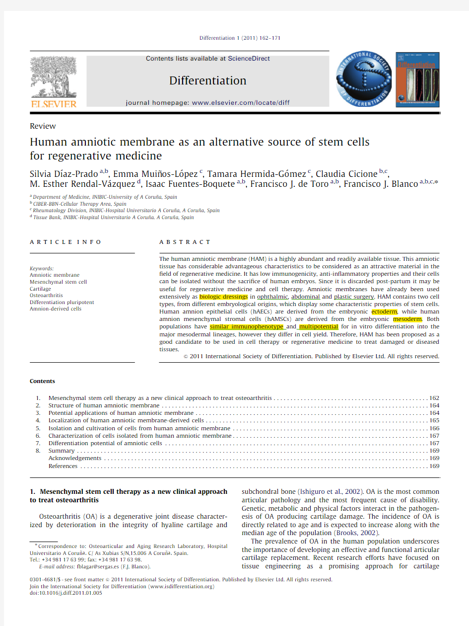
HAM develops from extra-embryonic tissue and consists of a
fetal component,the chorionic plate and a maternal component,the deciduas,which are comprised of an epithelial monolayer,a thick basement membrane and an avascular stroma (Niknejad et al.,2008;Jin et al.,2007).The amnion (Fig.1)is a thin (up to 2mm),elastic,translucent and semi-permeable fetal membrane attached to the chorionic membrane.Both the amnion and chorion form the amniotic sac ?lled with amniotic ?uid,providing and protecting the fetal environment.The outer layer,the chorion,consists of trophoblastic chorionic and mesenchymal tissues.The inner layer,the amnion,consists of a single layer of ectodermally derived epithelium uniformly arranged on the base-ment membrane,which is one of the thickest membranes found in any human tissue,and a collagen-rich mesenchymal layer (Wilshaw et al.,2006).This mesenchymal layer can be subdivided into the compact layer forming the main ?brous skeleton of the HAM,the ?broblast layer and an intermediate layer,which is also called the spongy layer or zona spongiosa (Niknejad,et al.2008).3.Potential applications of human amniotic membrane Recent studies have focused on tissue engineering as a pro-mising approach for cartilage regeneration (Kuo et al.,2006).
Cartilage tissue engineering is based on the selection of appro-priate cells,suitable scaffolds and stimulation with chondrogenic molecules (Kuo et al.,2006).Both natural and synthetic polymers have been fabricated for cartilage tissue engineering,such as ?brous structures,porous sponges,woven or non-woven meshes and hydrogels (Tritz et al.,2010;Yuan et al.,2010;Kuo et al.,2006).
In cartilage tissue engineering,recent studies have focused not only on appropriate cells but also on novel methods of manufac-ture allowing building strati?ed scaffolds.In this regard,Han et al.(2008)established and validated a molding technique for fabrica-tion of cartilaginous constructs that are anatomically shaped on one or two surfaces,targeting the spherically shaped hip and biomimetically strati?ed with super?cial and middle/deep zone chondrocyte subpopulations.On the other hand,Tritz et al.(2010)aimed to buildup complex biomaterials for reconstructing biolo-gical tissue with three dimensional cells construction for mimick-ing cartilage architecture.These authors published for the ?rst time that is possible to spray mixed alginate and chondrocytes with little damage for cells.Therefore,the sprayed hydrogel keeps not only the mechanical properties needed for cells,but also maintains the chondrocyte phenotype to induce cartilage.
HAM has some characteristics that highlight their clinical use as scaffold compared to other biocompatible products.In this regard HAM is anti-microbial,anti-tumorigenic,anti-?brosis,anti-angiogenic and has acceptable mechanical properties.It also reduces pain and in?ammation,inhibits scarring,shows little or no immunogenicity thus it does not represent transplantation risks,enhances wound healing and epithelialization and acts as an anatomical and vapor barrier (Dua et al.,2004;Ganatra,2003;Gomes et al.,2005;Hao et al.,2000).In vitro studies have demonstrated that cells isolated from amnion do not trigger immuno allogenic or xenogenic responses,actively suppress the proliferation of T lymphocytes and inhibit the differentiation of monocytes.The HAM survives for long periods in immunocom-petent animals and the cells are grafted persistently in various organs and tissues.Amnion expresses the non-classic and little polymorphic HLA-G molecules (class I b)and lacks the highly polymorphic antigens HLA A-B-C-(class I a),HLA DR (class II)and the T cell co-stimulatory molecule B-7.The HLA-G molecule displays at least four inhibitory functions relevant to immune responses (Insausti et al.,2010):(a)it can bind directly to inhibitory receptors found in NK cells and other leukocytes.(b)It possesses the appropriate leader peptide for binding to HLA-e,which will in turn inhibit the NK cells via their CD94/NKg2receptor.(c)It can induce apoptosis of activated CD8+T cell.(d)It can inhibit CD4+T cell proliferation.Moreover,in
mixed
Fig.1.Structure of the HAM.HAM was stained with HE,hematoxylin and eosin.AE:Amniotic epithelium;%:Basement membrane; :Compact layer;:Fibroblastic layer and SL:Spongy Layer or zona spongiosa
.
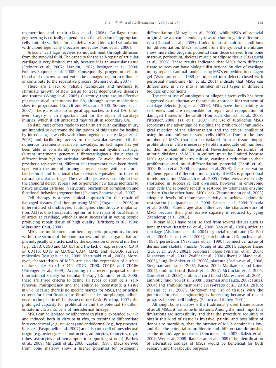
Fig.2.In vitro repair model of human OA articular cartilage.Human articular chondrocytes grown over HAM provided a super?cial cell cover that decreased the degree of damages of the OA articular cartilage surface (A,B,C and G).The newly-formed tissue showed high cell density and a thickening of the basement membrane of the HAM (D).Type I (E)and type II (F)collagen immunostainings indicated the ?brocartilaginous nature of the newly synthesized tissue.HE,hematoxylin and eosin;MM,Masson s trichrome;Col I,type I collagen;Col II,type II collagen.Original magni?cations:(A),(B)and (D)200?;(C),(E),(F)and (G)100?.
S.D?
′az-Prado et al./Differentiation 1(2011)162–171164
lymphocyte reactions using allogeneic cells,hAECs and hAMSCs, do not induce a cytotoxic response and inhibits lymphocyte proliferation(Ilancheran et al.,2009).
These properties enable surgeons to apply the HAM graft on various tissue surfaces without suturing.HAMs have been used as biologic dressings for ophthalmology,plastic surgery,derma-tology and gynecology procedures(Tejwani et al.,2007;Santos et al.,2005;Rinastiti et al.,2006;Meller et al.,2000;Morton and Dewhurst,1986).Moreover,the extracellular matrix(ECM)of the HAM has several components such as laminin,different types of collagens(I,III,IV,V and VI),nidogen,?bronectin,growth factors,hyaluronan and proteoglycans(Niknejad et al.,2008; Rinastiti et al.,2006;Jin et al.,2007)that are abundant on natural cartilage and are responsible of regulation and maintenance of normal chondrocyte metabolism(Jin et al.,2007);suggesting their use for cartilage tissue engineering(Niknejad et al.,2008). Indeed,a recent report showed the utility of the HAM as a scaffold to support human chondrocyte proliferation in cell therapy to repair human OA cartilage(D?′az-Prado et al.,2010c)(Fig.2). The low cost of HAM graft preparation and the very good clinical results in different applications have proposed the amnion as an alternative to other natural and synthetic wound dressings.
Published data suggest that HAM is a very attractive source of MSCs.Since the amniotic membrane arises from embryonic epiblast cells prior to gastrulation,it has been suggested that it may retain a reservoir of stem cells throughout pregnancy (Ilancheran et al.,2007).Pre-clinical and clinical studies have demonstrated multiple uses for amniotic membrane stem cells in tissue repair,such as corneal tissue(Shimmura and Tsubota, 2002),spinal cord injury(Sankar and Muthusamy,2003),brain infarction(Sakuragawa et al.,1997)and Parkinson s disease (Kakishita et al.,2003).Bailo et al.(2004)isolated and character-ized amniotic and chorionic cells from human full-term placentas, which suggested that both cell types may represent an advanta-geous source of progenitor cells.Indeed,the HAM is becoming appreciated as an alternative to bone marrow for adult MSCs for regenerative medicine.
Because fetal tissues are routinely discarded post-partum, HAMs have proved to be abundant,inexpensive and easily obtained with a virtually limitless availability,negating any need for mass tissue banking(Chang et al.,2010;Toda et al.,2007; Niknejad et al.,2008;Hennerbichler et al.,2007;Wilshaw et al., 2006).Moreover,this tissue provides ef?ciency in MSC recovery with non-invasive and safe procedures(Alviano et al.,2007).
A major advantage of cells isolated from the HAM is that they are harvested after birth and can be cryogenically stored to be
available in a timely manner for patient therapy after being thawed and expanded for use in tissue engineering,cell trans-plantation and gene therapy.Therefore,the HAM represents a very useful source of progenitor cells for a variety of applications. Because human embryos are not sacri?ced for the isolation of progenitor cells from HAMs,the current controversies associated with the use of human embryonic stem cells can be avoided (Chang et al.,2010;Insausti et al.,2010;Toda et al.,2007;Kim et al.,2009).
This fetal tissue expresses only moderate levels of the major histocompatibility complex(MHC)class I and MHC class II antigens on its surface.Therefore,hAECs and hAMSCs seem to be immune-privileged;thus,suitable for allo-transplantation and regenerative medicine(Wei et al.,2009;Kim et al.,2009).
In the present review,we focused the localization,isolation, quanti?cation and phenotypic characterization of HAM-derived cells(hAECs and hAMSCs)and summarized their in vitro differ-entiation potential useful for regenerative medicine and cell therapy.4.Localization of human amniotic membrane-derived cells
Location of HAM-derived cells in healthy human amniotic membranes stained with hematoxylin-eosin or Masson s Trichrome may be studied by histochemical and immunohistochemical techniques.The HAM contains two different cell types,hAECs and hAMSCs,from different embryological origins(Alviano et al., 2007;Wolbank et al.,2007).The hAECs forms a continuous monolayer of embryonic ectodermally derived epithelium uni-formely arranged on the basement membrane(BM)in contact with the amniotic?uid(Tamagawa et al.,2008)(Fig.3A and B). The hAECs are positive for the epithelial markers cytokeratin1,2, 3,4,5,6,7,8,10,13,14,15,16and19(D?′az-Prado et al.,2010a) (Fig.3C),which con?rms its epithelial nature.BM is one of the thickest membranes found in any human tissue that contains different types of collagens(Fig.3D),?bronectin,nidogen,lami-nin,proteoglycans and hyaluronan,as well as growth factors (Niknejad et al.,2008;Rinastiti et al.,2006;Jin et al.,2007). hAMSCs are derived from embryonic mesoderm(Tamagawa et
al., Fig.3.Histological characterization of human amnion-derived cells.Sections of healthy HAM stained with H-E(A)and MM(B),immunohistochemistry for CK(C) and Col II(D)and by immuno?uorescence for CD90and CD105(E).In(E)nuclei were counterstained with4’,6-diamidino-2-phenylindole(DAPI).BM indicates the thick basement membrane,and EC,the epithelial cells from extra-embryonic ectoderm.CK,cytokeratins.Original magni?cations:(A)100?,(B)400?,(C–E) 200?.
S.D?′az-Prado et al./Differentiation1(2011)162–171165
2008)and are sparsely distributed in the stroma underlying
the amnion epithelium (Bilic et al.,2008).An immuno?uores-cence study demonstrated that BM contains hAMSCs since they express common and well de?ned human MSC markers pre-viously described for bone marrow MSCs such as CD44,CD90,CD105and CD271indicating that BM contains cell having stem-cell characteristics (Fig.3E).The same study corroborated that hAECs contain positive cells for MSCs markers such as CD105(D?
′az-Prado et al.,2010b ),indicating that the HAM contains at least two different cell types having stem-cell characteristics.
5.Isolation and cultivation of cells from human amniotic membrane
Cells from the mesenchymal and epithelial regions of the amnion can be isolated easily.Different methods to isolate HAM-derived cells have been published (Dazzi and Marelli-Berg,2008;Parolini et al.,2008;Alviano et al.,2007;Soncini et al.,2007;Miki et al.,2007a;Miki and Strom,2006;Bailo et al.,2004).All of them start with a mechanical separation of the amniotic membrane from the underlying chorion through the spongy layer (Insausti et al.,2010),followed by a digestion with trypsin,dispase or other digestive enzymes,in different concentrations and for different periods of time,to release the hAECs from the basal membrane.hAMSCs can be subsequently released through subsequent digestion with collagenase (Parolini et al.,2009),alone or combined with DNAase (Steigman and Fauza,2007).hAECs are small-size cells that are easy to expand in vitro for at least 3passages without morphological changes,they grow in a lattice and have the typical cuboid morphology of epithelial cells (Fig.4A).These cells generally have a central or eccentric nucleus,one or two nucleoli and abundant cytoplasm,usually vacuolated
(Miki et al.,2007a ).The hAMSCs cells have ?broblast-like cell morphology (Fig.4B)and after 3to 4weeks of culture it is possible to obtain a population of adherent mesenchymal cells morphologically identical to MSCs isolated from bone marrow.These stromal cells are easy to expand in vitro for at least 9passages without morphological changes.hAECs and hAMSCs can be grown in Dulbecco modi?ed Eagle s media (DMEM)supplemented with 20%fetal bovine serum (FBS)and 1%penicillin–streptomycin (P/E)and seeded into culture ?asks.Both populations should be expanded in a humidi?ed 5%CO 2atmo-sphere at 371C.After the isolation of both cell types it is advisable to perform immunohistochemical stainings (e.g.for cytokeratin 7,CK7)to demonstrate the purity of both populations.In this regard,only hAECs may be
(Fig.
5).A recent
previously published (Soncini et al.,2007;Alviano et al.,2007),phenotypic characterization and in vitro potential for differentia-tion toward osteogenic,adipogenic and chondrogenic mesoder-mal lineages.Both protocols resulted in the isolation and culture of cells attached to the culture ?ask with ?broblast-like cell morphology.Quantitative studies showed that Soncini s protocol typically showed an increase in the hAMSCs isolation yield of almost ten fold with regard to Alviano s protocol.Also,the former protocol allowed the isolation and expansion of a larger number of cells in a very short time period.This ready and rapid availability of cells is one criteria required of a source of MSCs for it to be considered for cell transplantation.
There is a contradiction with the passage number at which HAM-derived cells stop proliferation.Based on the literature,proliferation slows down with every passage and For example,(2007a)Parolini et al.(2008)that hAECs Fig.4.Morphology of cultured hAECs (A)and hAMSCs (B)isolated from human amniotic membrane.Original magni?cations:100?.
Fig.5.Immunohistochemical stainings for CK7expression in cultured HAECs (A)and hAMSCs (B).Original magni?cations:400?.
S.D?
′az-Prado et al./Differentiation 1(2011)162–171166
for 2to 6passages before proliferation ceases.On the
contrary Bilic et al.(2008)con?rmed that hAECs and hAMSCs proliferation almost stops beyond passage 5whereas Toda et al.(2007)postulated that hAECs senescence is reached at lower passages,P3or P4.However Alviano et al.(2007)and Soncini et al.(2007)indicated that hAMSCs are easily expanded in vitro for at least 15passages without any visible morphological alterations but they used cells not exceeding P4for cell char-
derived mesenchymal stem cells (BM-MSCs)was the aim of the authors compared tissue included amnion,chorion and decidua)and human bone marrow-derived MSC in terms of cell characteristics,optimal growth conditions,mesodermal lineage differentiation and in vivo safety speci?cally to determine if human placenta-derived MSCs could represent a source of human MSC for clinical trials.They demonstrated that both populations were similar in terms of growth condition requirements and in terms of subsequent biological characteriza-tion.However both populations differed with respect to their proliferation capabilities at different seeding densities.In this regard human bone marrow-derived MSCs proliferated more slowly than human placenta-derived MSCs in every experiment.Also the latter had greater long-term growth ability than the former.Moreover MSCs from both sources exhibited similar morphology,size and cell surface phenotype and mesodermal differentiation ability with the exception that human placenta-derived MSC consistently appeared less able to differentiate to the adipogenic lineage.In line with the results obtained these authors suggested that human placenta is an acceptable alternative source for human MSC.
6.Characterization of cells isolated from human amniotic membrane
Immunophenotypic characterization of hAMSCs demonstrate the presence of the common well de?ned human MSC markers (CD90,CD44,CD73,CD166,CD105and CD29),described for bone marrow with the absence of the hematopoietic markers (CD34and CD45)and the concomitant lack of monocyte (CD114),macrophage (CD11)and ?broblast markers (Insausti et al.,2010;Kobayashi et al.,2008;Mihu et al.,2009).This antigen expression pattern is consistent with data published for stem cells isolated from various regions of the full-term placenta (Bilic et al.,2008;Bailo et al.,2004;Kobayashi et al.,2008;Barlow et al.,2008;Mihu et al.,2009).hAMSCs also express low levels of HLA-ABC,but do not express HLA-DR,indicating that these stromal cells may be useful in clinical transplantation procedures (Parolini et al.,2009).
hAECs are positive for desmin and vimentin (Toda et al.,2007)
and also for the same markers as hAMSCs (D?
′az-Prado et al.,2010b ).Therefore hAECs also had an antigen expression pro?le characteristic of culture-expanded MSCs (Bilic et al.,2008).Phenotypes of both cell populations,hAECs and hAMSCs,are
maintained from passage P0through passage P9(D?
′az-Prado et al.,2010b ).It is important to note that in culture although both populations show and maintain a similar marker pro?le for mesenchymal progenitors there are many differences between
them in cell shape and cell arrangement (D?
′az-Prado et al.,2010b;Bilic et al.2008).
Ilancheran et al.(2007)showed that hAECs expressed surface markers that are normally present on embryonic stem and cells such as SSEA3(stage speci?c embryonic antigen 3),TRA-1-60(tumor rejection antigen)and TRA-1-81and antigens such as the ABCG 2/BCRP (a member of the ATP-binding cassette superfamily),CD9,CD24,E-Cadherin,Integrin a 6and b and c-met (receptor growth factor of the hepatocyte)(Niknejad et al.,2008;Insausti et al.,2010).These epithelial cells also express transcription factors speci?c for pluripotential stem cells such as Oct4(octamer binding protein 4),NANOG,SOX2(SRY-related HMG-box gene 2)and REX-1(Parolini et al.,2009;Miki et al.,2007b,2005;Miki and Strom,2006).hAMSCs are also positive for these pluripotency markers but positivity for embryo-nic stem cell markers,SSEA-3or SSEA-4,remains debated (Parolini et al.,2009).
Parolini et al.(2009)published a comparison of key features of human amniotic membrane-derived cells and human BM-MSCs (Table 1).These authors postulated that BM-MSCs have a higher cell doubling time that hAECs,while for the hAMSC this time was not reported yet.Regarding the maximum number of passages it ranges from 5to 10for hAMSCs,10to 20for BM-MSCs and 30for hAECs.In vitro differentiation potential toward endodermal,mesodermal and ectodermal lineages was also reported for the three cell types.
7.Differentiation potential of amniotic cells
The pluripotency of hAECs was supported by a study by Tamagawa et al.(2004).These scienti?c created a xenogeneic chimera with hAECs and mouse embryonic stem cells in vitro.This chimera gives rise to cells of all germ https://www.360docs.net/doc/ee4100107.html,ter studies have corroborated the ability of hAECs to in vitro differentiate into cells from the three germ layers (Insausti et al.,2010;Ilancheran et al.,2007;Miki et al.,2005).
hAECs have characteristics of neural progenitor cells since these epithelial cells express some differentiation markers for neural stem,neuron and glial cells such as nestine,GAD (gluta-mate decarboxylase),GFAP (glial ?brillary acidic protein)and CNP (cyclic nucleotide phosphodiesterase)(Miki et al.,2005).Elwan and Sakuragawa (1997)and Kakishita et al.(2000),demonstrated the differentiation of the epithelial cells to neural cells (ectoder-mal lineage)with capacity to synthesize and release acetylcho-line,catecholamines and dopamine,suggesting their possible utility in the treatment of neural degenerative diseases.In this regard,several studies have already been published showing promising results in animal models with Parkinson s disease and Mucopolysaccaridosis type VII.Studies of intracerebral grafting of hAECs for the treatment of a mouse model of Parkinson s disease showed that these epithelial cells can synthesize and release
Table 1
Comparison of cell doubling time,maximum numbers of passages and differentiation potential of hAECs,hAMSCs and BM-MSCs.Data taken from Parolini et al.(2009).Cell type
Cell doubling time
Maximum number of passages Differentiation potential
hAECs At 5th passage:24h.30In vitro differentiation toward endodermal,mesodermal and ectodermal lineages.At 30th passage:18h.hAMSCs Not reported.5–10In vitro differentiation toward endodermal,mesodermal and ectodermal lineages.BM-MSCs 36–72h.
10–20
In vitro differentiation toward endodermal,mesodermal and ectodermal lineages.
S.D?′az-Prado et al./Differentiation 1(2011)162–171167
catecholamine and neurotrophic factors such as nerve growth
factor,neurotrophin-3and brain-derived neurotrophic factor (Kakishita et al.,2003,2000;Uchida et al.,2000).Kosuga et al.(2000)suggested that transplantation of hAECs transduced with adenoviral vectors can be employed for the treatment of con-genital lysosomal storage disorders.
Hepatic differentiation (endodermal lineage)of hAECs was also reported by Sakuragawa et al.(2000).These authors demon-strated that cultivated hAECs produced albumin and a -fetopro-tein;and that hepatocyte-like cells,positive for albumin and a -fetoprotein,could be identi?ed integrated in the liver parench-yma of mice with severe combined immunode?ciency (SCID),after the transplantation of hAECs in the https://www.360docs.net/doc/ee4100107.html,ter studies demonstrated that these epithelial cells also displayed other functional properties associated with hepatocytes,such as glyco-gen storage and expression liver-enriched transcription factors,such as hepatocyte nuclear factor (HNF)3g and HNF4a ,CCAAT/enhancer-binding protein (CEBP a and b )and several of the drug metabolizing genes (cytochrome P450)(Miki et al.,2005;Takashima et al.,2004;Davila et al.,2004).These ?ndings suggest the potential utility of hAECs for restore hepatic tissues that have been diseased or injured.Differentiation of hAECs to another endodermal lineage,pancreatic,was also reported.Wei et al.(2003)cultured these epithelial cells in the presence of nicotinamide to induce pancreatic differentiation and they observed that the result-ing cells expressed insulin.Subsequent transplantation of these insulin-expressing cells in the spleen of diabetic SCID mice normalized the levels of serum glucose for several months after the transplant,indicating the therapeutic potential of hAECs to treat diabetes mellitus type https://www.360docs.net/doc/ee4100107.html,ter,Miki et al.(2005)showed by RT-PCR analysis that,after pancreatic differentiation,hAECs express pan-creatic a and b cell markers such as the transcription factors PDX-1(pancreatic duodenum homeobox 1),PAX-6(paired box homeotic gene 6)and NKX2.2(NK2transcription factor-related locus 2)and the mature hormones insulin and glucagon.
The differentiation of hAECs to cardiac cells (mesodermal lineage)was ?rst evaluated by Miki et al.(2005).They demon-strated by RT-PCR that cardiac-speci?c genes atrial and ventricular myosin light chain 2(MLC-2A and MLC-2V)and the transcription factors GATA-4and Nkx 2.5are expressed or induced in hAECs cultured in media supplemented with ascorbic acid for 14days.The immunohistochemical analysis of alpha-actinin expression showed a staining pattern very similar to the one reported for hESC-derived cardiomyocytes.Differentiation of hAECs to another mesodermal lineages (Figs.6and 7)was reported by Ilancheran et al.(2007),who showed that native hAECs can differentiate into cells with a phenotype and markers characteristic of mesodermal-derived myocytes,osteocytes and adipocytes.
MSCs from different parts of the placenta have been shown to differentiate into chondrogenic,osteogenic,endothelial,hepatocytic,myogenic and neurogenic lineages,but with appreciable differences among cell types depending on the placental tissue source (Chang et al.,2010;Insausti et al.,2010;Portmann-Lanz et al.,2006;Tamagawa et al.,2007,2008;Kobayashi et al.,2008;In
tAnker
Fig.6.Osteogenic (DIF Osteo)and adipogenic (DIF Adipo)differentiation of human amnion mesenchymal stromal cells (hAMSCs)and human amnion epithelial cells
(hAECs)with their respective controls (C hAMSC and C hAEC)grown for 21days in Dulbecco’s Modi?ed Eagle Medium (DMEM 21).The presence of the calcium deposits characteristic of osteoblasts was detected using Alizarin Red (AR)stain (A).The presence of adipocytes was assessed by detection of lipid drops using Oil Red O (OR-O)stain (B).Original magni?cations:400?
.
Fig.7.Chondrogenic differentiation,assessed by micropellet formation,of hAMSCs [A]and hAECs [B].Original magni?cations:200x.
S.D?
′az-Prado et al./Differentiation 1(2011)162–171168
et al.,2004;Alviano et al.,2007;Wei et al.,2009;Mihu et al.,2009; Pasquinelli et al.,2007;Soncini et al.,2007;Sakuragawa et al.,2004).
hAMSCs differentiation to neuronal lineage have been demon-strated by the fact that these cells express neuronal markers (Nestin,Musashi1,neuron-speci?c enolase,neuro?lament med-ium,microtubule-associated protein[MAP]-2and Neu-N)and glial(GFAP)markers,after their culture in speci?c neural-induc-tion media(Sakuragawa et al.,2004;Portmann-Lanz et al.,2006; Kim et al.,2007;Tamagawa et al.,2008).
Tamagawa et al.(2007)showed that hAMSCs were able to differentiate into cells with characteristics of hepatocytes.In this regard,native cells expressed typical hepatocytic mRNA such as albumin,CK(cytokeratin)18,a-fetoprotein,a1-antitrypsin and HNF-4a but only glucose-6-phosphatase and ornithine transcar-bamylase expression and glycogen storage were observed after in vitro hepatic induction.
Regarding hAMSCs differentiation towards mesodermal line-age(Figs.6and7),In tAnker et al.(2004)demonstrated the potential of hAMSCs to differentiate into osteogenic and adipo-genic cells.After osteogenic differentiation hAMSCs suffered morphologic changes and showed calcium deposits when they were stained with von Kossa s dye.On the other hand,after adipogenic differentiation hAMSCs become multi vacuolated cells that were stained with Red oil O https://www.360docs.net/doc/ee4100107.html,ter,Portmann-Lanz et al. (2006)showed the capacity of these stromal cells for differentia-tion to chondrogenic and myogenic lineages.Chondrogenic dif-ferentiation of these cells was demonstrated by the presence of abundant collagen in the extracellular matrix by means of Alcino s blue dye.Myogenic differentiation of hAMSCs has been deter-mined by RT-PCR since Portmann-Lanz et al.(2006)demonstrated the mRNA expression of myogenic transcription factors such as Myo D and Myogenin and the protein expression of desmine in hAMSCs cultured in differentiation media.Alviano et al.(2007) con?rmed these results and also was the?rst in demonstrate the angiogenic differentiation potential of these cells.This latter study revealed that hAMSCs,after culture in induction media with VEGF,expressed endothelial-speci?c markers such as the receptors of the vascular endothelial growth factor1and2(FLT-1, KDR),ICAM-1as well as the appearance of CD34and von Will-ebrand Factor(vWF)positive cells.Regarding cardiomyogenic potential,it has been demonstrated that hAMSCs expressed cardiac-speci?c genes such as GATA4,MLC-2a(myosin light chain),MLC-2v,cTnI and cTnT(Tanaka et al.,1999;Zhao et al., 2005)after cardiomyogenic induction.Zhao et al.(2005)showed that after hAMSCs transplantation into the myocardial infarcts in rat hearts,these cells survived in the scar tissue for at least 2months and differentiated into cardiomyocyte-like cells.On the other hand,spontaneous differentiation of hAMSCs towards myo?broblasts has also been observed after their culture in standard medium(DMEM/FBS)within2passages(Li et al.,2008) 8.Summary
HAM,an abundant,inexpensive and easily obtained tissue that is discarded post-partum,represents a valuable material for tissue banking and a viable alternative to other synthetic or natural scaffolds.Amnion contains two different cell types,hAECs and hAMSCs,which display characteristic properties of stem cells. Both amnion-derived populations are easily isolated from HAM and have the capacity to differentiate in vitro into all three germ layers:endoderm,mesoderm and ectoderm,suggesting their great interest in the?elds of cell therapy and regenerative medicine.The unlimited availability of HAMs,the high ef?ciency in MSC recovery with non-invasive and safe procedures,the minimal ethical and legal issues associated with its use and the low immunogenicity of amnion-derived cells,make them an alternative source of MSCs.More studies should be carried out to determine whether such in vitro-differentiated cells can func-tion in vivo.
Acknowledgements
This study was supported by grants:Servizo Galego de Sau′de, Xunta de Galicia(PS07/84),Ca′tedra Bioiberica de la Universidade da Corun?a and Instituto de Salud Carlos III CIBER BBN CB06-01-0040; Ministerio Ciencia en Innovacion PLE2009-0144;Fondo Inves-tigacion Sanitaria-PI08/2028with participation of fundus from FEDER(European Community),Silvia Diaz-Prado is bene?ciary of an Isidro Parga Pondal contract from Xunta de Galicia,A Coruna, Spain.
References
Abdallah,BM,Haack-Sorensen,M,Burns,JS,Elsnab,B,Jakob,F,Hokland,P, Kassem,M.,2005.Maintenance of differentiation potential of human bone marrow mesenchymal stem cells immortalized by human telomerase reverse transcriptase gene despite[corrected]extensive proliferation.Biochem.
https://www.360docs.net/doc/ee4100107.html,mun.326,527–538.
Alsalameh,S,Amin,R,Gemba,T,Lotz,M.,2004.Identi?cation of mesenchymal progenitor cells in normal and osteoarthritic human articular cartilage.
Arthritis Rheum.50,1522–1532.
Alviano,F,Fossati,V,Marchionni,C,Arpinati,M,Bonsi,L,Franchina,M,Lanzoni,G, Cantoni,S,Cavallini,C,Bianchi,F,Tazzari,PL,Pasquinelli,G,Foroni,L,Ventura, C,Grossi,A,Bagnara,GP.,2007.Term amniotic membrane is a high throughput source for multipotent mesenchymal stem cells with ability to differentiate into endothelial cells in vitro.BMC Dev.Biol.7,11.
Bailo,M,Soncini,M,Vertua,E,Signoroni,PB,Sanzone,S,Lombardi,G,Arienti,D, Calamani,F,Zatti,D,Paul,P,Albertini,A,Zorzi,F,Cavagnini,A,Candotti,F, Wengler,GS,Parolini,O.,2004.Engraftment potential of human amnion and chorion cells derived from term placenta.Transplantation78,1439–1448. Baksh,D,Yao,R,Tuan,RS.,https://www.360docs.net/doc/ee4100107.html,parison of proliferative and multilineage differentiation potential of human mesenchymal stem cells derived from umbilical cord and bone marrow.Stem Cells25,1384–1392.
Ban?,A,Muraglia,A,Dozin,B,Mastrogiacomo,M,Cancedda,R,Quarto,R.,2000.
Proliferation kinetics and differentiation potential of ex vivo expanded human bone marrow stromal cells:implications for their use in cell therapy.Exp.
Hematol.28,707–715.
Barlow,S,Brooke,G,Chatterjee,K,Price,G,Pelekanos,R,Rossetti,T,Doody,M, Venter,D,Pain,S,Gilshenan,K,Atkinson,K.,https://www.360docs.net/doc/ee4100107.html,parison of human placenta-and bone marrow-derived multipotent mesenchymal stem cells.
Stem Cells Dev.17,1095–1108.
Bianco,P,Robey,PG.,2001.Stem cells in tissue engineering.Nature414,118–121. Bilic,G,Zeisberger,SM,Mallik,AS,Zimmermann,R,Zisch,AH.,https://www.360docs.net/doc/ee4100107.html,parative characterization of cultured human term amnion epithelial and mesenchymal stromal cells for application in cell therapy.Cell Transplant17,955–968. Bonab,MM,Alimoghaddam,K,Talebian,F,Ghaffari,SH,Ghavamzadeh,A,Nikbin,
B.,2006.Aging of mesenchymal stem cell in vitro.BMC Cell Biol.7,14–20. Brandt,KD,Mazzuca,SA.,2006.Experience with a placebo-controlled randomized clinical trial of a disease-modifying drug for osteoarthritis:the doxycycline trial.Rheum.Dis.Clin.North Am.32,217–234.
Brittberg,M,Lindahl,A,Nilsson,A,Ohlsson,C,Isaksson,O,Peterson,L.,1994.
Treatment of deep cartilage defects in the knee with autologous chondrocyte transplantation.N.Engl.J.Med.331,889–895.
Brooks,PM.,2002.Impact of osteoarthritis on individuals and society:how much disability?Social consequences and health economic implications.Curr.Opin.
Rheumatol.14,573–577.
Caplan,AI.,1991.Mesenchymal stem cells.J.Orthop.Res.9,641–650.
Chang,Y-J,Hwang,S-M,Tseng,C-P,Cheng,F-C,Huang,S-H,Hsu,L-F,Hsu,L-W, Tsai,M-S.,2010.Isolation of mesenchymal stem cells with neurogenic potential from the mesoderm of the amniotic membrane.Cells Tissues Organs 192,93–105.
Davila,JC,Cezar,GG,Thiede,M,Strom,S,Miki,T,Trosko,J.,https://www.360docs.net/doc/ee4100107.html,e and application of stem cells in toxicology.Toxicol.Sci.79,214–223.
Dazzi,F,Marelli-Berg, F.,2008.Mesenchymal stem cells for graft-versus-host disease:close encounters with T cells.Eur.J.Immunol.38,1479–1482.
De Bari,C,Dell’Acio,F,Tylzanowski,P,Luyten,FP.,2001.Multipotent mesench-ymal stem cells from adult human synovial membrane.Arthritis Rheum.44, 1928–1942.
D?′az-Prado,S,Mu?′n?os-Lo′pez,E,Hermida-Go′mez,T,Rendal-Va′zquez,ME,Fuentes-Boquete,I,de Toro,FJ,Blanco,FJ,2010a.Isolation and characterization of mesenchymal stem cells from human amniotic membrane.Tissue Eng.Part C Methods,1August.
D?′az-Prado,S,Mu?′n?os-Lo′pez,E,Hermida-Go′mez,T,Rendal-Va′zquez,ME,Fuentes-Boquete,I,de Toro,FJ,Blanco,FJ,2010b.Multilineage differentiation potential
S.D?′az-Prado et al./Differentiation1(2011)162–171169
of cells isolated from the human amniotic membrane.J.Cell Biochem.111, 846–857.
D?′az-Prado,S,Rendal-Va′zquez,ME,Mu?′n?os Lo′pez,E,Hermida-Go′mez,T, Rodr?′guez-Cabarcos,M,Fuentes-Boquete,I,de Toro,FJ,Blanco,FJ,2010c.
Potential use of the human amniotic membrane as a scaffold in human articular cartilage repair.Cell Tissue Bank11,183–195.
Djouad,F,Bony,C,H¨aupl,T,Uze′,G,Lahlou,N,Louis-Plence,P,Apparailly,F, Canovas,F,Re′me,T,Sany,J,Jorgensen,C,No¨el, D.,2005.Transcriptional pro?les discriminate bone marrow-derived and synovium-derived mesench-ymal cells.Arthritis Res.Ther.7,1304–1315.
Dominici,M,Le Blanc,K,Mueller,I,Slaper-Cortenbach,I,Marini,F,Krause,D, Deans,R,Keating,A,Dj,Prockop,Horwitz, E.,2006.Minimal criteria for de?ning multipotent mesenchymal stromal cells.The international society for cellular therapy position statement.Cytotherapy8,315–317.
Dounchis,JS,Goomer,RS,Harwood,FL,Khatod,M,Coutts,RD,Amiel,D.,1997.
Chondrogenic phenotype of perichondrium-derived chondroprogenitor cells is in?uenced by transforming growth factor-beta 1.J.Orthop.Res.15, 803–807.
Dua,H.S.,Gomes,J.A.,King,A.J.,Maharajan,V.S.,2004.The amniotic membrane in ophthalmology.Surv.Ophthalmol.49,51–77.
Elwan,MA,Sakuragawa,N.,1997.Evidence for synthesis and release of catecho-lamines by human amniotic epithelial cells.Neuroreport8,3435–3438. Fauza,D.,2004.Amniotic?uid and placental stem cells.Best Pract.Res.Clin.
Obstet.Gynaecol.18,877–891.
Fickert,S,Fiedler,J,Brenner,RE.,2003.Identi?cation,quanti?cation and isolation of mesenchymal progenitor cells from osteoarthritic synovium by?uorescence automated cell sorting.Osteoarthritis Cartilage11,790–800.
Fuentes-Boquete,IM,Arufe Gonda,MC,D?′az Prado,S,Hermida Go′mez,T,de Toro Santos,FJ,Blanco Garc?′a,FJ,2007.Tratamiento de lesiones del cart?′lago articular con terapia celular.Reumatol.Clin.3,S63–S693Suppl.
Fuentes-Boquete,IM,Arufe Gonda,MC,D?′az Prado,SM,Hermida Go′mez,T,de Toro santos,FJ,Blanco,FJ,2008.Cell and tissue transplant strategies for joint lesions.Open Transplantation J.2,21–28.
Ganatra,M.A.,2003.Amniotic membrane in surgery.J.Pak.Med.Assoc.53,29–32. Gomes,J.A.,Romano,A.,Santos,M.S.,Dua,H.S.,2005.Amniotic membrane use in ophthalmology.Curr.Opin.Ophthalmol.16,233–240.
Han,EH,Bae,WC,Hsieh-Bonassera,ND,Wong,VW,Schumacher,BL,G¨ortz,S, Masuda,K,Bugbee,WD,Sah,RL.,2008.Shaped,strati?ed,scaffold-free grafts for articular cartilage defects.Clin.Orthop.Relat.Res.466,1912–1920. Hao,Y.,Ma, D.H.,Hwang, D.G.,Kim,W.S.,Zhang, F.,2000.Identi?cation of antiangiogenic and antiin?ammatory proteins in human amniotic membrane.
Cornea19,348–352.
Hennerbichler,S,Reichl,B,Pleiner,D,Gabriel,C,Eibl,J,Redl,H.,2007.The in?uence of various storage conditions on cell viability in amniotic membrane.
Cell Tissue Bank8,1–8.
Hombach-Klonisch,S,Panigrahi,S,Rashedi,I,Seifert,A,Alberti,E,Pocar,P, Kurpisz,M,Schulze-Osthoff,K,Mackiewicz,A,Los,M.,2008.Adult stem cells and their trans-differentiation potential—perspectives and therapeutic appli-cations.J.Mol.Med.86,1301–1314.
Ilancheran,S,Michalska,A,Peh,G,Wallace,EM,Pera,M,Manuelpillai,U.,2007.
Stem cells derived from human fetal membranes display multilineage differ-entiation potential.Biol.Reprod.77,577–588.
Ilancheran,S,Moodley,Y,Manuelpillai,U.,2009.Human fetal membranes:
a source of stem cells for tissue regeneration and repair?Placenta30,
2–10.
Im,GI,Kim,DY,Shin,JH,Hyun,CW,Cho,WH.,2001.Repair of cartilage defect in the rabbit with cultured mesenchymal stem cells from bone marrow.J.Bone Joint Surg.Br.83,289–294.
Insausti,CL,Blanquer,M,Bleda,P,Iniesta,P,Majado,MJ,Castellanos,G,Moraleda, JM.,2010.The amniotic membrane as a source of stem cells.Histol.Histo-pathol.25,91–98.
In t Anker,PS,Scherjon,SA,Kleijburg-van der Keur,C,de Groot-Swings,GM, Claas,FH,Fibbe,WE,Kanhai,HH,2004.Isolation of mesenchymal stem cells of fetal or maternal origin from human placenta.Stem Cells22, 1338–1345.
In’t Anker,PS,Noort,WA,Kruisselbrink,AB,Scherjon,SA,Beekhuizen,W,Will-emze,R,Kanhai,HH,Fibbe,WE.,2003.Nonexpanded primary lung and bone marrow-derived mesenchymal cells promote the engraftment of umbilical cord blood-derived CD34(+)cells in NOD/SCID mice.Exp.Hematol.31, 881–889.
Ishiguro,N,Kojima,T,Poole,R.,2002.Mechanism of cartilage destruction in osteoarthritis.Nagoya J.Med.Sci.65,73–84.
Izadpanah,R,Trygg,C,Patel,B,Kriedt,C,Dufour,J,Gimble,JM,Bunnell,BA.,2006.
Biologic properties of mesenchymal stem cells derived from bone marrow and adipose tissue.J.Cell Biochem.99,1285–1297.
Jin,CZ,Park,SR,Choi,BH,Lee,KY,Kang,CK,Min,BH.,2007.Human amniotic membrane as a delivery matrix for articular cartilage repair.Tissue Eng.13, 693–702.
Jung,DI,Ha,J,Kang,BT,Kim,JW,Quan,FS,Lee,JH,Woo,EJ,Park,HM.,2009.
A comparison of autologous and allogenic bone marrow-derived mesenchymal
stem cell transplantation in canine spinal cord injury.J.Neurol.Sci.285, 67–77.
Kakishita,K,Elwan,MA,Nakao,N,Itakura,T,Sakuragawa,N.,2000.Human amniotic epithelial cells produce dopamine and survive after implantation into the striatum of a rat model of Parkinson s disease:a potential source of donor for transplantation therapy.Exp.Neurol.165,27–34.Kakishita,K,Nakao,N,Sakuragawa,N,Itakura,T.,2003.Implantation of human amniotic epithelial cells prevents the degeneration of nigral dopamine neurons in rats with6-hydroxydopaminen lesions.Brain Res.980,48–56. Kastrinaki,M-C,Andreakou,I,Charbord,P,Papadaki,HA.,2008.Isolation of human bone marrow mesenchymal stem cells using different membrane markers: comparison of colony/cloning ef?ciency,differentiation potential,and mole-cular pro?le.Tissue Eng.Part C Methods14,333–339.
Kim,J,Kang,HM,Kim,H,Kim,MR,Kwon,HC,Gye,MC,Kang,SG,Yang,HS,You,J., 2007.Ex vivo characteristics of human amniotic membrane-derived stem cells.Cloning Stem Cells9,581–594.
Kim,SS,Song,CK,Shon,SK,Lee,KY,Kim,CH,Lee,MJ,Wang,L.,2009.Effects of human amniotic membrane grafts combined with marrow mesenchymal stem cells on healing of full-thickness skin defects in rabbits.Cell Tissue Res.336, 59–66.
Kobayashi,M,Yakuwa,T,Sasaki,K,Sato,K,Kikuchi,A,Kamo,I,Yokoyama,Y, Sakuragawa,N.,2008.Multilineage potential of side population cells from human amnion mesenchymal layer.Cell Transplant.17,291–301.
Koga,H,Shimaya,M,Muneta,T,Nimura,A,Morito,T,Hayashi,M,Suzuki,S,Ju,YJ, Mochizuki,T,Sekiya,I.,2008.Local adherent technique for transplanting mesenchymal stem cells as a potential treatment of cartilage defect.Arthritis Res.Ther.10,R84.
Kosuga,M,Takahashi,S,Sasaki,K,Enosawa,S,Li,XK,Okuyama,S,Fujino,M, Suzuki,S,Yamada,M,Matsuo,N,Sakuragawa,N,Okuyama,T.,2000.Pheno-type correction in murine mucopolysaccharidosis type VII by transplantation of human amniotic epithelial cells after adenovirus-mediated gene transfer.
Cell Transplant.9,687–692.
Kuo,CK,Li,WJ,Mauck,RL,Tuan,RS.,2006.Cartilage tissue engineering:its potential and uses.Curr.Opin.Rheumatol.18,64–73.
Kuznetsov,SA,Mankani,MH,Gronthos,S,Satomura,K,Bianco,P,Robey,PG.,2001.
Circulating skeletal stem cells.J.Cell Biol.153,1133–1140.
Le Blanc,K,G¨otherstr¨om,C,Ringde′n,O,Hassan,M,McMahon,R,Horwitz,E, Anneren,G,Axelsson,O,Nunn,J,Ewald,U,Norde′n Lindeberg,S,Jansson,M, Dalton,A,Astr¨om,E,Westgren,M.,2005.Fetal mesenchymal stem-cell engraftment in bone after in utero transplantation in a patient with severe osteogenesis imperfecta.Transplantation79,1607–1614.
Li,W,He,H,Chen,YT,Hayashida,Y,Tseng,SC.,2008.Reversal of myo?broblasts by amniotic membrane stromal extract.J.Cell Physiol.215,657–664.
Mankin,HJ.,1982.The response of articular cartilage to mechanical injury.J.Bone Joint Surg.Am.64,460–466.
Mareschi,K,Biasin,E,Piacibello,W,Aglietta,M,Madon,E,Fagioli, F.,2001.
Isolation of human mesenchymal stem cells:bone marrow versus umbilical cord blood.Haematologica86,1099–1100.
Matikainen,T,Laine,J.,2005.Placenta-an alternative source of stem cells.Toxicol.
Appl.Pharmacol.207(2Suppl.),544–549.
McGuckin,CP,Forraz,N,Baradez,MO,Navran,S,Zhao,J,Urban,R,Tilton,R, Denner,L.,2005.Production of stem cells with embryonic characteristics from human umbilical cord blood.Cell Prolif.38,245–255.
Meller,D,Pires,RT,Mack,RJ,Figueiredo,F,Heiligenhaus,A,Park,WC,Prabhasawat, P,John,T,McLeod,SD,Steuhl,KP,Tseng,SC.,2000.Amniotic membrane transplantation for acute chemical or thermal burns.Ophthalmology107, 980–989.
Mihu,CM,Rus Ciuc?a,D,Sorit?a u,O,Sus-man,S,Mihu, D.,2009.Isolation and characterization of mesenchymal stem cells from the amniotic membrane.
Rom.J.Morphol.Embryol.50,73–77.
Miki,T,Lehmann,T,Cai,H,Stolz,DB,Strom,SC.,2005.Stem cell characteristics of amniotic epithelial cells.Stem Cells23,1549–1559.
Miki,T,Marongiu,F,Ellis,E,Strom,S.,2007a.Isolation of amniotic epithelial stem cells.Curr.Protoc.Stem Cell Biol.31E.3.1-1E.3.9.
Miki,T,Mitamura,K,Ross,MA,Stolz,DB,Strom,SC.,2007b.Identi?cation of stem cell marker-positive cells by immuno?uorescence in term human amnion.
J.Reprod.Immunol.75,91–96.
Miki,T,Strom,SC.,2006.Amnion-derived pluripotent/multipotent stem cells.
Stem Cell Rev.2,133–142.
Minas,T,Chiu,R.,2000.Autologous chondrocyte implantation.Am.J.Knee Surg.
13,41–50.
Minguell,JJ,Conget,P,Erices, A..,2000.Biology and clinical utilization of mesenchymal progenitor cells.Braz.J.Med.Biol.Res.33,881–887.
Morton,KE,Dewhurst,CJ.,1986.Human amnion in the treatment of vaginal malformations.Br.J.Obstet.Gynaecol.93,50–54.
Mrugala,D,Dossat,N,Ringe,J,Delorme,B,Coffy,A,Bony,C,Charbord,P,H¨aupl,T, Daures,J-P,No¨el,D,Jorgensen,C.,2009.Gene expression pro?le of multipotent mesenchymal stromal cells:identi?cation of pathways common to TGF b3/ BMP2-induced chondrogenesis.Cloning Stem Cells11,61–76.
Muraglia,A,Cancedda,R,Quarto,R.,2000.Clonal mesenchymal progenitors from human bone marrow differentiate in vitro according to a hierarchical model.
J.Cell Sci.113,1161–1166.
Nakahara,H,Bruder,SP,Haynesworth,SE,Holecek,JJ,Baber,MA,Goldberg,VM, Caplan,AI.,1990.Bone and cartilage formation in diffusion chambers by subcultured cells derived from the periosteum.Bone11,181–188. Niknejad,H,Peirovi,H,Jorjani,M,Ahmadiani,A,Ghanavi,J,Seifalian,AM.,2008.
Properties of the amniotic membrane for potential use in tissue engineering.
Eur.Cell Mater.15,88–99.
Parolini,O,Alviano,F,Bagnara,GP,Bilic,G,B¨uhring,HJ,Evangelista,M,Henner-bichler,S,Liu,B,Magatti,M,Mao,N,Miki,T,Marongiu,F,Nakajima,H, Nikaido,T,Portmann-Lanz,CB,Sankar,V,Soncini,M,Stadler,G,Surbek,D, Takahashi,TA,Redl,H,Sakuragawa,N,Wolbank,S,Zeisberger,S,Zisch,A,
S.D?′az-Prado et al./Differentiation1(2011)162–171 170
Strom,RN.,2008.Concise review:isolation and characterization of cells from human term placenta:outcome of the?rst international workshop on placenta derived stem cells.Stem Cells26,300–311.
Parolini,O,Soncini,M,Evangelista,M,Schmidt,D.,2009.Amniotic membrane and amniotic?uid-derived cells:potential tools for regeneative medicine?Regen.
Med.4,275–291.
Parsch,D,Fellenberg,J,Brummendorf,TH,Eschlbeck,AM,Richter,W.,2004.
Telomere length and telomerase activity during expansion and differen-tiation of human mesenchymal stem cells and chondrocytes.J.Mol.Med.82, 49–55.
Pasquinelli,G,Tazzari,P,Ricci,F,Vaselli,C,Buzzi,M,Conte,R.,2007.Ultrastruc-tural characteristics of human mesenchymal stromal(stem)cells derived from bone marrow and term placenta.Ultrastruc.Pathol.31,23–31.
Pittenger,MF,Mackay,AM,Beck,SC,Jaiswal,RK,Douglas,R,Mosca,JD,Moorman, MA,Simonetti,DW,Craig,S,Marshak,DR.,1999.Multilineage potential of adult human mesenchymal stem cells.Science284,143–147.
Pittenger,MF.,2008.Mesenchymal stem cells from adult bone marrow.Methods Mol.Biol.449,27–44.
Portmann-Lanz,CB,Schoeberlein,A,Huber,A,Sager,R,Malek,A,Holzgreve,W, Surbek,DV.,2006.Placental mesenchymal stem cells as potential autologous graft for pre-and perinatal neuroregeneration.Am.J.Obstet.Gynecol.194, 664–673.
Prockop,DJ.,1997.Marrow stromal cells as stem cells for nonhematopoietic tissues.Science276,71–74.
Resinger,C,Ve′csei,V,Marlovits,S.,2004.Therapeutic options in the treatment of cartilage defects.Techniques and indications.Radiologe44,756–762. Harijadi,Rinastiti M,Santoso,AL,Sosroseno,W,2006.Histological evaluation of rabbit gingival wound healing transplanted with human amniotic membrane.
Int.J.Oral Maxillofac.Surg.35,247–251.
Sakaguchi,Y,Sekiya,I,Yagishita,K,Muneta,T.,https://www.360docs.net/doc/ee4100107.html,parison of human stem cells derived from various mesenchymal tissues:superiority of synovium as a cell source.Arthritis Rheum.52,2521–2529.
Sakuragawa,N,Enosawa,S,Ishii,T,Thangavel,R,Tashiro,T,Okuyama,T,Suzuki,S., 2000.Human amniotic epithelial cells are promising transgene carriers for allogenic cell transplantation into liver.J.Hum.Genet.45,171–176. Sakuragawa,N,Kakinuma,K,Kikuchi,A,Okano,H,Uchida,S,Kamo,I,Kobayashi, M,Yokoyama,Y.,2004.Human amnion mesenchyme cells express phenotypes of neuroglial progenitor cells.J.Neurosci.Res.78,208–214Erratum in:J.
Neurosci.Res.2005;79,725.
Sakuragawa,N,Misawa,H,Ohsugi,K,Kakishita,K,Ishii,T,Thangavel,R,Tohyama, J,Elwan,M,Yokoyama,Y,Okuda,O,Arai,H,Ogino,I,Sato,K.,1997.Evidence for active acetylcholine metabolism in human amniotic epithelial cells: applicable to intracerebral allografting for neurologic disease.Neurosci.Lett.
232,53–56.
Samuel,GN,Kerridge,IH,O Brien,TA.,2008.Umbilical cord blood banking:public good or private bene?t?Med.J.Aust.188,533–535.
Sankar,V,Muthusamy,R.,2003.Role of human amniotic epithelial cell transplan-tation in spinal cord injury repair research.Neuroscience118,11–17. Santos,MS,Gomes,JAP,Ho?ing-Lima,AL,Rizzo,LV,Romano,AC,Belfort Jr.,R, 2005.Survival analysis of conjuctival limbal grafts and amniotic membrane transplantation in eyes with total limbal stem cell de?ciency.Am.J.Ophthal-mol.140,223–230.
Shimmura,S,Tsubota,K.,2002.Ocular surface reconstruction update.Curr.Opin.
Ophthalmol.13,213–219.
Soncini,M,Vertua,E,Gibelli,L,Zorzi,F,Denegri,M,Albertini,A,Wengler,GS, Parolini,O.,2007.Isolation and characterization of mesenchymal cells from human fetal membranes.J.Tissue Eng.Regen.Med.1,296–305.
Steigman,S.A.,Fauza, D.O.,2007E.Isolation of mesenchymal stem cells from amniotic?uid and placenta.Curr.Protoc.Stem.Cell.Biol.;Chapter1,Unit 1E.2.
Steinert,AF,Ghivizzani,SC,Rethwilm,A,Tuan,RS,Evans,CH,N¨oth,U.,2007.Major biological obstacles for persistent cell-based regeneration of articular carti-lage.Arthritis Res.Ther.9,213.
Stenderup,K,Justesen,J,Clausen,C,Kassem,M.,2003.Aging is associated with decreased maximal life span and accelerated senescence of bone marrow stromal cells.Bone33,919–926.
Takashima,S,Ise,H,Zhao,P,Akaike,T,Nikaido,T.,2004.Human amniotic epithelial cells possses hepatocyte-like characteristics and functions.Cell Struct.Funct.29,73–84.
Tamagawa,T,Ishiwata,I,Saito,S.,2004.Establishment and characterization of a pluripotent stem cell line derived from human amniotic membranes and initiation of germ layers in vitro.Hum.Cell17,125–130.
Tamagawa,T,Oi,S,Ishiwata,I,Ishikawa,H,Nakamura,Y.,2007.Differentiation of mesenchymal cells derived from human amniotic membranes into hepato-cyte-like cells in vitro.Hum.Cell20,77–84.
Tamagawa,T.,Ishiwata,I,Ishikawa,H,Nakamura,Y.,2008.Induced in vitro differentiation of neural-like cells from human amnion-derived?broblast-like cells.Hum.Cell21,38–45.Tanaka,M,Chen,Z,Bartunkova,S,Yamasaki,N,Izumo,S.,1999.The cardiac homeobox gene Csx/Nkx2.5lies genetically upstream of multiple genes essential for heart development.Development126,1269–1280.
Tejwani,S,Kolari,RS,Sangwan,VS,Rao,GN.,2007.Role of amniotic membrane graft for ocular chemical and thermal injuries.Cornea26,21–26.
Toda,A,Okabe,M,Yoshida,T,Nikaido,T.,2007.The potential of amniotic membrane/amnion-derived cells for regeneration of various tissues.J.Phar-macol.Sci.105,215–228.
Tritz,J,Rahouadi,R,de Isla,N,Charif,N,Pinzano,A,Mainard,D,Bensoussan,D, Netter,P,Stoltz,J-F,Benkirane–Jessel,N,Huselstein,C,2010.Designing a three-dimensional alginate hydrogel by spraying method for cartilage tissue engineering.Soft Matter6,5165–5174.
Tsai,MS,Hwang,SM,Chen,KD,Lee,YS,Hsu,LW,Chang,YJ,Wang,CN,Peng,HH, Chang,YL,Chao,AS,Chang,SD,Lee,KD,Wang,TH,Wang,HS,Soong,YK.,2007.
Functional network analysis on the transcriptomes of mesenchymal stem cells derived from amniotic?uid,amniotic membrane,cord blood,and bone marrow.Stem Cells25,2511–2523.
Uchida,S,Inanaga,Y,Kobayashi,M,Hurukawa,S,Araie,M,Sakuragawa,N.,2000.
Neurotrophic function of conditioned medium from human amniotic epithe-lial cells.J.Neurosci.Res.62,585–590.
Villaron,EM,Almeida,J,Lopez-Holgado,N,Alcoceba,M,Sa′nchez-Abarca,LI, Sanchez-Guijo,FM,Alberca,M,Pe′rez-Simon,JA,San Miguel,JF,Del Can?izo, MC,2004.Mesenchymal stem cells are present in peripheral blood and can engraft after allogenic haematopoietic stem cell transplantation.Haematolo-gica89,1421–1427.
Wakitani,S,Kimura,T,Hirooka,A,Ochi,T,Yoneda,M,Yasui,N,Owaki,H,Ono,K., 1989.Repair of rabbit articular surfaces with allograft chondrocytes embedded in collagen gel.J.Bone Joint Surg.Br.71,74–80.
Wei,JP,Nawata,M,Wakitani,S,Kametani,K,Ota,M,Toda,A,Konishi,I,Ebara,S, Nikaido,T.,2009.Human amniotic mesenchymal cells differentiate into chondrocytes.Cloning Stem Cells11,19–25.
Wei,JP,Zhang,TS,Kawa,S,Aizawa,T,Ota,M,Akaike,T,Kato,K,Konishi,I,Nikaido, T.,2003.Human amnion-isolated cells normalize blood glucose in streptozo-tocin-induced diabetic mice.Cell Transplant.12,545–552.
Wilshaw,SP,Kearney,JN,Fisher,J,Ingham,E.,2006.Production of an acellular amniotic membrane matrix for use in tissue engineering.Tissue Eng.12, 2117–2129.
Wolbank,S,Peterbauer,A,Fahrner,M,Hennerbichler,S,van Griensven,M,Stadler, G,Redl,H,Gabriel,C,2007.Dose-dependent immunomodulatory effect of human stem cells from amniotic membrane:a comparison with human mesenchymal stem cells from adipose tissue.Tissue Eng.13,1173–1183. Wong,BJ,Pandhoh,N,Truong,MT,Diaz,S,Chao,K,Hou,S,Gardiner,D.,2005.
Identi?cation of chondrocyte proliferation following laser irradiation,thermal injury,and mechanical https://www.360docs.net/doc/ee4100107.html,sers Surg.Med.37,89–96.
Yanada,S,Ochi,M,Kojima,K,Sharman,P,Yasunaga,Y,Hiyama, E.,2006.
Possibility of selection of chondrogenic progenitor cells by telomere length in FGF-2-expanded mesenchymal stromal cells.Cell Prolif.39,575–584. Yoo,JU,Barthel,TS,Nishimura,K,Solchaga,L,Caplan,AI,Goldberg,VM,Johnstone,
B.,1998.The chondrogenic potential of human bone-marrow-derived
mesenchymal progenitor cells.J.Bone Joint Surg.Am.80,1745–1757.
You,Q,Cai,L,Zheng,J,Tong,X,Zhang,D,Zhang,Y.,2008.Isolation of human mesenchymal stem cells from third-trimester amniotic?uid.Int.J.Gynaecol.
Obstet.103,149–152.
Young,HE,Steele,TA,Bray,RA,Hudson,J,Floyd,JA,Hawkins,K,Thomas,K,Austin, T,Edwards,C,Cuzzourt,J,Duenzl,M,Lucas,PA,Black Jr.,AC,2001.Human reserve pluripotent mesenchymal stem cells are present in the connective tissues of skeletal muscle and dermis derived from fetal,adult,and geriatric donors.Anat.Rec.264,51–62.
Yuan,T,Zhang,L,Feng,L,Fan,H,Zhang,X.,2010.Chondrogenic differentiation and immunological properties of mesenchymal stem cells in collagen type I hydrogel.Biotechnol.Prog.July28(Epub ahead of print).
Zhao,P,Ise,H,Hongo,M,Ota,M,Konishi,I,Nikaido,T.,2005.Human amniotic mesenchymal cells have some characteristics of cardiomyocytes.Transplanta-tion79,528–535.
Zimmermann,S,Voss,M,Kaiser,S,Kapp,U,Waller,CF,Martens,UM.,https://www.360docs.net/doc/ee4100107.html,ck of telomerase activity in human mesenchymal stem cells.Leukemia17, 1146–1149.
Zuk,PA,Zhu,M,Ashjian,P,De Ugarte,DA,Huang,JI,Mizuno,H,Alfonso,ZC,Fraser, JK,Benhaim,P,Hedrick,MH.,2002.Human adipose tissue is a source of multipotent stem cells.Mol.Biol.Cell13,4279–4295.
Zuk,PA,Zhu,M,Mizuno,H,Huang,J,Futrell,JW,Katz,AJ,Benhaim,P,Lorenz,HP, Hedrik,MH.,2001.Multilineage cells from human adipose tissue:implications for cell-based therapies.Tissue Eng.7,211–228.
Zvai?er,NJ,Marinova-Mutafchieva,L,Adams,G,Edwards,CJ,Moss,J,Burger,JA, Maini,RN.,2000.Mesenchymal precursor cells in the blood of normal individuals.Arthritis Res.2,477–488.
S.D?′az-Prado et al./Differentiation1(2011)162–171171
PEP六年级上册英语教案全册
Unit 1How can I get there? 第一课时 一、教学内容 Part A Let's try & Let's talk 二、教学目标 1.能够听、说、读、写句子:“Where is the museum shop?”“It's near the door.”。 2.能够听、说、认读单词ask、sir和句型“Is there a…?”“I want to…”“What a great museum!”。 三、教学重难点 1.学习句子“Where is the museum shop?”“It's near the door.”。 2.正确使用方位介词。 四、教学准备 单词卡、录音机、磁带。 五、教学过程 Step 1 热身(Warming-up) Let's do Go to the bookstore.Buy some books. Go to the post office.Send a letter. Go to the hospital.See the doctor. Go to the cinema.See a film.
Go to the museum.See some robots. Step 2 新课呈现(Presentation) 1.学习Let's try (1)打开课本读一读Let's try中呈现的问题和选项。 (2)播放录音,让学生听完后勾出正确的选项。 (3)全班核对答案。 2.学习Let's talk (1)播放Let's talk的录音,学生带着问题听录音:Where is the museum shop?Where is the post office?听完录音后让学生回答这两个问题,教师板书:It's near the door.It's next to the museum.教师讲解:near表示“在附近”,next to表示“与……相邻”,它的范围比near小。最后让学生用near和next to来讲述学校周围的建筑物。 (2)讲解“A talking robot!What a great museum!”,让学生说说这两个感叹句的意思。 (3)再次播放录音,学生一边听一边跟读。 (4)分角色朗读课文。 Step 3 巩固与拓展(Consolidation and extension) 1.三人一组分角色练习Let's talk的对话,然后请一些同学到台前表演。 2.教学Part A:Talk about the places in your city/town/village.
最新六年级英语上册期末试卷(含答案)
小学六年级英语上册期末试卷(含答案) 听力部分 一、L isten and choose. (根据你所听到的内容, 选择相符合的一项,并将其字母编号填在题号 前的括号内。)(10分) ( ) 1. A. always B.often C. aunt ( ) 2. A. actress B. actor C. active ( ) 3. A. buy B. bike C. bus ( )4. A. sell B. same C. say ( ) 5. A. bike B. kite C. side ( ) 6. A. difference B. different C. dictionary ( ) 7. A. writes a letter? B. write a letter C. writer ( ) 8. A. Amy’s uncle B. Lily’s uncle C. Billy’s uncle ( ) 9. A. What is your mother doing? B. What does your mother do? C. What is your mother going to do? ( ) 10. A. Mike works in a DVD company. B. Mike works in a VCD company. C. Mike works in a DVD factory. 二、Listen and judge. (根据所听到的内容, 判断图片或句子是否相符, 相符的在相应的题号前的括号内打“√”, 不相符的打“×”。)(10分) 1. 2. 3. ( ) ( ) ( ) 4. 5. ( ) ( ) 6. Chen Jie is going to be an artist. ( ) 7. Mike often does homework at 7:00. ( ) 8. Mr. Li likes playing the violin. ( ) 9. Feng Gang is a policeman. ( )
人教版六年级上册英语知识点总结
人教版六年级英语上册各单元知识点汇总 Unit 1 How do you go to school?一、重点短语: by plane 坐飞机 by ship 坐轮船 on foot 步行 by bike 骑自行车 by bus 坐公共汽车 by train 坐火车 traffic lights 交通灯 traffic rules 交通规则 go to school 去上学 get to 到达 get on 上车 get off 下车Stop at a red light. 红灯停Wait at a yellow light. 黄灯等Go at a green light. 绿灯行 二、重点句型: 1.How do you go to school?你怎么去上学? https://www.360docs.net/doc/ee4100107.html,ually I go to school on foot. Sometimes I go by bus. 通常我步行去上学。有时候骑自行车去。 3.How can I get to Zhongshan Park ?我怎么到达中山公园? 4.You can go by the No. 15 bus. 你可以坐 15 路公共汽车去。三、重点语法: 1、There are many ways to go somewhere.到一个地方去有许多方法。这里的 ways 一定要用复数。因为 there are 是There be 句型的复数形式。 2、on foot 步行乘坐其他交通工具大都可以用介词by…,但是步行只能用介词 on 。 4、go to school 的前面绝对不能加 the,这里是固定搭配。 5、USA 和 US 都是美国的意思。另外America 也是美国的意思。 6、go to the park 前面一定要加the. 如果要去的地方有具体的名字,就不能再加 the ,如果要去的地方没有具体名字,都要在前面加 the. ( go to school 除外。) 7、How do you go to …?你怎样到达某个地方?如果要问的是第三人称单数,则要用: How does he/she…go to …? 8、反义词: get on(上车)---get off(下车) near(近的)—far(远的) fast(快的)—slow(慢的) because(因为)—why(为什么) same(相同的)—different(不同的) 9、近义词: see you---goodbye sure---certainly---of course 10、频度副词: always 总是,一直 usually 通常 often 经常 sometimes 有时候 never 从来不 Unit 2 Where is the science museum?一、重点短语: library 图书馆 post office 邮局 hospital 医院 cinema 电影院
PEP人教版小学六年级上册英语期末分类专项复习试题及答案(全套)
分类专项复习卷(一) 听力 建议时间:40分钟满分:100分 一、听录音,选择你所听到的内容,将其序号填入题前括号里。(5分)( ) 1. A. afraid B. amazing C. attention ( ) 2. A. can’t go to school B. count to ten C. don’t be late ( ) 3. A. a TV reporter B. a sports reporter C. a PE teacher ( ) 4. A. I want to buy a postcard. B. I want to send the postcard. C. I want to go to the post office. ( ) 5. A. It’s next to the restaurant B. It’s near the museum shop. C. It’s behind the supermarket. 二、听录音,选择与你所听到的内容相符的图片,将其序号填入题前括号里。(5分) ( ) 1. A. B. C. ( ) 2. A. B. C. ( ) 3. A. B. C. ( ) 4. A. B. C. ( ) 5. A. B. C. 三、听录音,用数字给下列图片排序。(6分)
()()() ( ) ( ) ( ) 四、听录音,判断下列句子与你所听到的内容是否相符,相符的写“√”,不相符的写“×”。(5分) ( ) l. Mike should see a doctor in the morning. ( ) 2. Amy’s father works at sea. ( ) 3. Mike’s brother is a postman. ( ) 4. Peter likes doing kung fu and swimming. ( ) 5. We are going to Beijing next Tuesday. 五、听录音,判断下列图片与你所听到的内容是否相符,相符的打“√”,不相符的打“×”。(6分) 1. ( ) 2. ( ) 3. ( ) 4. ( ) 5. ( ) 6. ( ) 六、听录音,选择相应的答语,将其序号填入题前括号里。(7分) ( ) 1. A. He’s over there. B. Thank you. C. Great! ( ) 2. A. Here you are. B. Sure. C. Thanks. ( ) 3. A. Please turn left. B. It’s near the door.
新版pep六年级上册英语-各单元知识点总结
Unit 1 How can I get there? 一:重点单词和短语 Science科学, museum博物馆, post office, bookstore, cinema, hospital ,tasty, buy, London Eye伦敦银, stomach胃,crossing十字路口, turn left, turn right, go straight=walk straight直走.next to紧挨着/与。。。相邻, far from(离。。。远), near在。。。附近,behind(在。。。后面), in front of(在。。。前面), between…and…(在。。。和。。。之间) 二:按要求写单词: hot(反义词)cold , cool(反义词)warm,too(同音词)to/two can not(缩写)can’t right(反义词)left/wrong buy(同音词)by/bye sea(同音 词)see first(基数词)one four(序数词)fourth did (原形)do /does three(序数词)third give(过去式)gave 三:重点句型分析 1. Where is the museum shop? 此问句是由特殊疑问词where 引导的一个特殊疑问句,where意为“在哪里,到哪里”,用来询问地点,放在句子的开头。询问“某人或某物在哪里”的基本句型是:“ Where +is/are+ 主语?”,where is 后接名词或代词的单数形式,where are 后接名词或代词的复数形式。表示地点的词:museum博物馆, post office邮局, bookstore书店, cinema电影院, hospital医院restaurant餐馆bank银行bus stop 公交车站lake湖library图书馆zoo动物园school学校park公园garden花园hotel旅馆 2. It’s nea r the door. 此句中near是表示位置的介词,意为“旁边,附近” ,其同义句是:It’s next to the door.它在门的旁边。表示位置的短语:next to the bookstore挨着书店near the hospital在医院附近near the post office在邮局附近over there 在那边on Dongfang Street在东方大街上in front of the school在学校前面 3. How can we get there? 此句用来询问“怎样去某地”,后面直接跟地点。回答时,可以用“ Turn left, turn right, go straight.”等句来回答。 同义句:1.Can you tell me the way to+地点? 2.Where is the +地点? 3.Which is the way to +地点 4. Turn left at the bookstore. Then turn right at the hospital. 此句是指路的句型。常用到的句型有:turn left, 向左转turn right, 向右转go straight直着走。同时表示在某处的介词用at. 5.Is the Thames far from here? No, it isn’t. 此句是个be动词开头的一般疑问句,其回答要用Yes或No. 句中的far from意为“离……远”。反义词组为next to.
人教版小学六年级上册英语时态专项习题
人教版小学六年级上册英语时态专项习题 一般现在时专项练习 一.写出下列各词的复数 I _________him _________this ___________her ______ watch _______child _______photo ________diary ______ day________ foot________ book_______ dress ________ tooth_______ sheep ______box_______ strawberry _____ thief _______yo-yo ______ peach______ sandwich ______ man______ woman_______ paper_______ juice___________ water________ milk________ rice__________ tea__________ 二、写出下列动词的第三人称单数 drink ________ go _______ stay ________ make ________ look _________ have_______ pass_______ carry ____ come________ watch______ plant_______ fly ________ study_______ brush________ do_________ teach_______ 三、用括号内动词的适当形式填空。 1. He often ________(have) dinner at home. 2. Daniel and Tommy _______(be) in Class One. 3. We _______(not watch) TV on Monday. 4. Nick _______(not go) to the zoo on Sunday. 5. ______ they ________(like) the World Cup? 6. What _______they often _______(do) on Saturdays? 7. _______ your parents _______(read) newspapers every day? 8. The girl _______(teach) us English on Sundays. 9. She and I ________(take) a walk together every evening. 10. There ________(be) some water in the bottle. 11. Mike _______(like) cooking. 12. They _______(have) the same hobby. 13. My aunt _______(look) after her baby carefully. 14. You always _______(do) your homework well. 15. I _______(be) ill. I’m staying in bed. 16. She _______(go) to school from Monday to Friday. 17. Liu Tao _______(do) not like PE. 18. The child often _______(watch) TV in the evening. 19. Su Hai and Su Yang _______(have) eight lessons this term. 20. -What day _______(be) it today? It’s Saturday 四、按照要求改写句子 1. Daniel watches TV every evening.(改为否定句) ___________________________________________________ 2. I do my homework every day.(改为一般疑问句,作否定回答) ________________________________________________________ 3. She likes milk.(改为一般疑问句,作肯定回答)
人教版六年级上册英语语音专项练习题
1 一、 找出划线部分读音与其他三个不同的单词。 ( )1.A.how B.know C.yellow D.window ( )2. A.apple B.cat C.many D.mango ( )3. A.not B.doctor C.kilo D.orange ( )4. A.pears B.bananas C.apples D.grapes ( )5. A.boxes B.peaches C.buses D.apples ( )6. A.these B.thirsty C.those D.they 一、 选出划线部分读音与所给单词划线部分读音相同的一项。 ( )1.baseball A.table B.father C.banana ( )2. brother A.nose B.doctor C.son ( )3. which A.knife B.with C.white ( )4. this A.mouth B.three C.with ( )5. room A.too B.book C.good ( )6. ear A.pear B.bear C.hear 二、 找出划线部分读音与其他三个不相同的单词。 ( )1.A.hurry B.student C.puppet D.bus ( )2. A.that B.think C.mouth D.thank ( )3. A.guess B.she C.egg D.watermelon ( )4. A.right B.climb C.this D.pineapple ( )5. A.mother B.today https://www.360docs.net/doc/ee4100107.html,e D.some 三、 找出与所给单词划线部分读音相同的单词。 1.( )father A.mouth B.brother 2. ( )nose A.close B.seven 3. ( )cake https://www.360docs.net/doc/ee4100107.html,te B.father 4. ( )sister A.bike B.is 5. ( )boy A.climb B.brother 6. ( )mother A.sister B.her 四、 判断下列划线部分读音是否相同, ( )1.driver write ( )driver teacher ( )2.farmer her ( )knife big ( )3.cook good ( )drink dress ( )4.tree trousers ( )look school ( )5.their hair ( )where here ( )6.that mouth ( )they those 五、 将下列单词按th 的两种发音分类。 that mouth this three think thin thank thirsty those there these they [that] th [ ] mouth 六、 单词辨音,用“√”或“×”表示。 student ( ) bus ( ) bananas pear grapes ( ) ear ( ) teacher nose worker ( ) orange ( ) tree ( ) dress ( ) 八. 判断下列划线部分读音是否相同,用“√”或“×”表示。 1.hair airport ( ) 2.ship shop( ) 3.where who ( ) 4.good school ( ) 5.minibus student( ) 6.station library( ) 九.找出划线部分读音与其他不相同的单词。 ( )1.A.move B.noodle C.who D.coffee ()2. A.sweets B.friends C.cats D.biscuits ( )3. A.apple B.water C.at D.bag ( )4. A.juice B.cup C.bus D.much ( )5. https://www.360docs.net/doc/ee4100107.html,k B.it C.kite D.his ( )6. A.classroom B.bedroom C.room D.bathroom 十一、找出与所给单词划线部分读音相同的单词。 1.music A.bus B.student C.cup D.buy 2.chocolate A.goB.open C.too D.office 3.teacher A.school https://www.360docs.net/doc/ee4100107.html,e C.lunch D.cake 4.bookcase https://www.360docs.net/doc/ee4100107.html,te B.cap C.apple D.snack 5.desk A.grape B.bed C.she D.he 6.kilo A.library B.like C.tiger D.hospital 7.oranges A.booksB.cats C.teachers D.maps 8.chair A.pear B.here C.ear D.dear 十二、判断下列划线部分读音是否相同,用“√”或“×”表示。 1.bread egg ( ) 2.plate snack ( ) 3.where whose ( ) 4.try right ( ) 5.bowl how ( ) 6.chopstick school( )
PEP小学英语六年级上册各单元知识点复习资料
六年级上复习资料 Unit1 How Do You Go There? (你怎样去那里?) 重点单词: on foot= walk走路by bike骑自行车by bus坐公车by train坐火车by plane坐飞机by ship坐轮船by subway坐地铁near近的far远的usually通常sometimes有时候easy简单的the fifth floor第五层楼traffic lights交通灯traffic rules交通规则stop停止wait等待get to到达same相同的mean意思是driver司机right 右边left左边must必须know知道 重点句子: 1. How do you go to school? 你是怎样去上学的? 2. I go to school by bus.我是坐公交车去上学的。 3. Usually I go to school on foot. 我通常走路去上学。 4. Sometimes I go by bike. 有时候我骑自行车去。、 5. My home is near/ far. 我的家是近的。/ 远的。 6. Look at the traffic lights, remember the traffic rules. 看着交通灯,记住交通规则。 7. Stop at a red light. Wait at a yellow light. Go at a green light. 红灯停。黄灯等一等。绿灯行。 8. Red means stop, yellow means wait, green means go. 红色的意思是停止,黄色的意思是等待,绿色的意思是通行。 9. How can I get to the park? 我该怎样到达公园呢? 10. You can go by the No. 15 bus. 你可以坐15路公交车去。 重点知识: 1.坐某种交通工具用by,例如:by bike, by train。而走路用“on”例如on foot. 2.国家名字,地方名字第一个字母要大些:例如:Canada加拿大, China中国, America美 国,England英国,Australia澳大利亚 3.国家名缩写前面加the,缩写字母都要大写。例如:the USA=the US美国,the UK英 国,the CAN加拿大,the PRC中国。 4.频度副词是表示做的次数多少的词语。从多到少依次排列为:always总是,usually通 常,often经常,sometimes有时候,never从不。频度副词可以放在句首,也可以放在人称后面。例如:Usually I go to school by bus. = I usually go to school by bus. 5.near近的,far远的。这两个词是一对反义词。注意:not near= far, not far = near. 6.时间前面用at. 例如:在三点钟:at 3 o’ clock. 一段时间前面用for 7.表达第几层楼的时候我们要用序数词,前面还要有the。例如:第一、二、三、四、五 层楼分别是:the first floor. the second floor. the third floor. the fourth floor. the fifth floor. 8.交通灯traffic lights,交通规则:traffic rules。这两个词后面都一定要加s, 绝对不能少。 因为交通灯有红黄绿三盏,一定是复数,交通规则不可能只有一条,所以都一定要加s,考试的时候千万别忘了加s哦! 9.大部分的国家都是靠右行驶的:drivers drive on the right side of the road. 记住England and Australia, drivers drive on the left side of the road.英国和澳大利亚,司机是靠左行驶的。
人教版英语六年级上册 专题 阅读
一、阅读理解。 Jenny is from New York, America.She is twelve.Now she is in Shanghai with her parents.She can speak English very well and a little Chinese. Her father is a teacher.He works in a university.And her mother is a doctor.They go to work by subway.Jenny is studying in a school near her home.She walks to school from Monday to Friday.Her classmates are friendly to her.Her family are going to take a trip to Xi’an next summer holiday.She is very excited,because she loves China. ()1.Jenny is an girl. A.English B.American C.Australian ()2.Jenny’s father is a . A.teacher B.doctor C.coach ()3.Jenny goes to school . A.on foot B.by bike C.by subway ()4.Jenny goes to school days a week. A.three B.four C.five ()5.Jenny is going to next summer holiday. A.Shanghai B.Xi’an C.Beijing 二、阅读短文,判断正(T)误(F)。 Ben is my pen pal.He is tall and strong.He likes playing basketball and swimming.Ben is going to have a picnic next weekend.So he plans to(打算) go shopping tomorrow.He wants to buy a pair of sunglasses and a big water bottle.The supermarket is next to the museum.It’s very far.So he is going to take the No.19 bus to get there.After that,Ben is going to visit his uncle.His uncle’s home is near the supermarket.They will have dinner together.They Ben is going to play games with his cousin. ()1.Ben is tall and strong. ()2.Ben is going to have a picnic next weekend. ()3.The supermarket is next to the school. ()4.Ben is going to the supermarket by bike. ()5.Ben will play games with his cousin. 三、阅读理解。 Dear Frank, I’m very happy to be your new pen pal.I live in Guangzhou,China.It’s very hot in summer.So i like swimming very much.It’s cool to swim in the pool or in the sea in summer.I also like playing the piano. It’s interesting.My parents are scientists.They work in the same university.They go to work on foot.They think that’s good exercise.We often go hiking on the weekend. Tell me something about you,please. Yours, Fang Yan ()1.Fang Yan lives in . A.Shanghai B.Shenzhen C.Guangzhou ()2.Fang Yan likes . A.dancing and reading B.swimming and playing the piano
六年级上册英语知识点总结归纳(PEP新版)
PEP新版六年级上册英语知识点归纳总结 目录 Unit1How can I get there? (2) Unit2Ways to go to school (3) Unit3My weekend plan (4) Unit4I have a pen pal (5) Unit5What does he do? (6) Unit6How do you feel? (7)
Unit1How can I get there? library图书馆north(北) post office邮局 hospital医院turn left左转turn right右转places:cinema电影院 (地点)bookstore书店(东)east west(西) science museum科学博物馆 pet hospital宠物医院crossing十字路口 school学校south(南) shoe store/shop鞋店 supermarket超市go straight直行 一、问路 1.Where is the cinema,please?请问电影院在哪儿? next to the hospital.在医院的旁边。 in front of the school.在学校的前面. behind the park在公园的后面 It’s near the zoo.在动物园的附近. on the right/left of the bookstore.在书店的左/右边. east of the bank.在银行的东边. far from here.离这儿很远. 2.Excuse me,is there a cinema near here请问这附近有电影院吗? Yes,there is./No,there isn’t.有./没有。 3.How can I get to the hospital?我该怎样到达医院呢? Take the No.57bus.乘坐57路公汽。 二、指引路 1.You can take the No.312bus.你可乘坐312路公交车去那儿. 2.Go straight for three minutes.向前直走3分钟. 3.Turn right/left at the…在…地方向右/左转. 4.Walk east/west/south/north for…minutes.朝东/西/南/北/走…分钟. 三、Is it far from here?离这儿远吗? Yes,it is./No,it isn’t.是的,很远/不是,很远。
2016年人教版小学六年级上册英语时态专项习题
2016人教版小学六年级上册英语时态专项习题 look , read and write 1. There ( is / are ) some bananas ( on / near ) the table . 2. There ( is / are ) a football (in / under ) the bed . 3. There (is / are ) some tall buildings in the ( village /city ). 4. My teacher is ( strong / thin ) . And she’s(old / young ) . 5. I have ( onions / mutton ) and cabbage for lunch . 6. Mike ( has / have ) English and ( science / math ) on _________________ ( Friday / Sunday ) . When , Where , What , Who , How , Why 1. ---- Can I get to the zoo ?---- You can ride a bike there . 2. ---- do you go to school on foot ?---- Because my home is near . 3. ---- are you going after lunch ?---- I am going to the bookstore . 4. ---- are you going ? ---- I am going at 4 o’clock . 5. ---- are you going to do ? ---- I am going to play football . 6. ---- are you going to play with ? ---- My brother . 一般现在时专项练习 一.写出下列各词的复数 I _________him _________this ___________her ______ watch _______child _______photo ________diary ______ day________ foot________ book_______ dress ________ tooth_______ sheep ______box_______ strawberry _____ thief _______yo-yo ______ peach______ sandwich ______ man______ woman_______ paper_______ juice___________ water________ milk________ rice__________ tea__________ 二、写出下列动词的第三人称单数 drink ________ go _______ stay ________ make ________ look _________ have_______ pass_______ carry ____ come________ watch______ plant_______ fly ________ study_______ brush________ do_________ teach_______ 三、用括号内动词的适当形式填空。 1. He often ________(have) dinner at home. 2. Daniel and Tommy _______(be) in Class One.
pep最新六年级上册单元英语课文翻译
PEP7最新六年级下册Unit4-Unit6英语课文翻译 Unit4 I have a pen pal.单词表:学习的三单形式,迷,远足,笔友,业余爱好,茉莉, 想法主意,堪培拉,令人惊奇的,表示征求意见,射门,加入,俱乐部,分享 P38Let’s talk奥利弗:彼得的爱好是什么? 张鹏:他喜欢读故事。他住在一个农场上,所以有时他读给那些奶牛! 奥利弗:那是有趣的。张鹏:他喜欢练功夫和游泳。奥利弗:真的吗?我也是。张鹏:他也喜欢唱歌。奥利弗:哦,你也喜欢唱歌。张鹏:是的。我打算教他中国歌曲《茉莉花》!奥利弗:好主意!P39Let’s learn彼得:嘿,张鹏,你的爱好是什么?张鹏:我喜欢读故事。我也喜欢唱歌和练功夫。 跳舞唱歌读故事踢足球练功夫 P40Let’s talk约翰:嘿,一。你正在干什么?(现在进行时)吴一凡:我正在写一份电子邮件 给我的朋友在澳大利亚。约翰:他住在悉尼吗?吴一凡:不,他不是。他住在堪培拉。他的名字是约翰,也。约翰:真的吗?他喜欢猜字谜和去远足吗?吴一凡:是的,他是。约翰:太神奇了!我也喜欢这些。我也能成为他的笔友吗?吴一凡:当然。为什么不呢?约翰:酷! P41Let’s learn约翰:过来并且看看我的新的笔友。他的名字也是约翰!兄弟:真的吗?他也住在中国吗?约翰:不,他不在。他住在澳大利亚,但是他学语文。烧中国食物学语文猜字谜去远足 P42let’s read布告栏:我们能跳舞吗?有一个跳舞课在星期天在下午一点。我喜欢跳舞,兵器我需要一个搭档。打电话给艾米:3345567射门!射门!射门让我们一起读。你的爱好是什么?你喜欢阅读吗?我有很棒的书。我们能分享!打电话给迈克:4435678 科学俱乐部,你的俱乐部。你想学习关于机器人吗?来到科学房间。见见罗宾。他教学生制作机器人。robin Unit 5 What does he do?单词表:工厂,工人,邮递员,商人企业家,警察,渔民科学家, 飞行员,教练,国家,校长,大海,保持,大学,体育馆,如果,记者,使用,打字,快速地, 秘书 P48Let’s talk莎拉:你的爸爸是在这里今天吗?奥利弗:不,他在澳大利亚。莎拉:他是干什么的?奥利弗:他是一个商人。他经常去其它的城市。莎拉:并且你的妈妈是干什么的?奥利弗:她是一个班主任。莎拉:那是好的。奥利弗:是的,她将要是在这里今天。莎拉:你想成为一个班主任吗?奥利弗:不,我想成为一个出租车司机。 P49Let’s learn职业日工厂工人邮递员商人警察张鹏:你的爸爸是一个邮递员吗?奥利弗:不,他不是。张鹏:他是干什么的?:奥利弗:他是一个商人。 P50Let’s talk迈克:我的叔叔是一个渔夫。小雨:他在哪里工作?迈克:他工作在海上。他看 到很多鱼每天。小雨:我知道了。他怎么去工作的?乘小船?迈克:不,他工作在小船上。他骑自行车去工作。小雨:他有一个很健康的生活。迈克:是的。他工作非常努力并且保持健康。小雨:我们也应该努力学习并且保持健康。 P51Let’s learn渔民科学家飞行员教练奥利弗:我的叔叔是科学家。吴一凡:他在哪里工作?奥利弗:他工作在一个大学。 P52let’s read胡斌喜欢运动。他擅长足球,乒乓球和篮球。他经常去跑步放学后。他想工作在一个体育馆。提示:
人教版六年级英语上册期末重点知识点汇总--最新版
人教版六年级英语上册期末重点知识复习资料 六年级上册复习要点 Unit1 How can I get there? 一、重点单词: 地点:science museum科学博物馆post office 邮局bookstore 书店 cinema 电影院hospital 医院 动作:go straight 直走turn left/right 左转、右转 方位:in front of :在···前面behind 在···后面near在…旁边next to 紧挨着beside 在旁边 over 在…上方on the left 在左边on the right 在右边 二、重点句型: (1)Is / Are there…?某处有某物吗? 肯定回答:Yes, there is/are. 否定回答:No, there isn’t/aren’t. (2)Where is the + 地点?... ... 在哪里? It’s + 表示地点的名词. 它... ... 例句:Where is the cinema? 电影院在哪? It’s next to the bookstore. 在书店的旁边。 (3)How can + 主语+get(to)+ 地点? ... ...怎么到... ...? (如果get后面接的词为副词,则要省略介词to.) 例句:我们怎么到那儿? 同义句型:Can you tell me the way to + 地点? ( 4 )Where is + 地点?Which is the way to + 地点? 到书店左转。 Unit 2 Ways to go to school? 一、重点单词/短语: 交通方式:by bike /bus /plane /subway /train /ship /taxi /ferry 骑自行车/乘公共汽车/飞机/地铁/火车/船/出租汽车/轮渡take the No.57 bus 乘57路公共汽车 on foot 步行 其他:slow down慢下来pay attention to 注意traffic lights 交通信号灯look right 向右看cross the road 横穿马路at home 在家 二、重点句型: (1)How do you come (to) + 地点?你们怎么来... 的? 我通常/经常/有时... How do you go(to) + 地点?你们怎么去...的? How do you get (to) + 地点?你们怎么到达...的?