EV71 replication in PBMC
Journal of Medical Virology79:60–68(2007) Differences in Replication Capacity Between Enterovirus71Isolates Obtained From Patients With Encephalitis and Those Obtained From Patients With Herpangina in Taiwan
Chien-Min Kung,1,3Chwan-Chuen King,2Chun-Nan Lee,3,5Li-Min Huang,4
Ping-Ing Lee,4and Chuan-Liang Kao3,5*
1Department of Medical Technology,Yuanpei University,Hsinchu,Taiwan,Republic of China
2Institute of Epidemiology,College of Public Health,National Taiwan University(NTU),Taipei,Taiwan
3Department of Clinical Laboratory Sciences and Medical Biotechnology,
College of Medicine,NTU,Taipei,Taiwan
4Department of Pediatrics,NTU Hospital,Taipei,Taiwan
5Department of Laboratory Medicine,National Taiwan University Hospital,Taipei,Taiwan
The cellular-tropism and biological characteris-tics of enterovirus71(EV71)isolates in Taiwan (TW)were studied.Growth curve experiments were conducted using cell lines that were possi-bly exhibited pathogenesis,and RT-PCR and sequencing tests were undertaken to amplify the50non-coding region(50-NCR).The encepha-litis isolate EV71TW98NTU2078was PBMC-
tropic,temperature-resistant(Tr)at408C,and easier to replicate in HTB-14(astrocytoma)than the herpangina isolate EV71TW98NTU1186(The
viral yields were100-fold higher than those of the herpangina isolate EV71TW98NTU1186at96hr
post infection.).The herpangina isolate EV71 TW98NTU1186was non-PBMC-tropic,and tem-perature-sensitive(Ts)at408C.The replication of EV71TW98NTU1186in HTB-14was lower.No EV71isolate infected HTB-37(human colon adenocarcinoma cells).The encephalitis EV71 isolate exhibited better replication and transmis-sion in PBMCs and astrocytes than did the EV71 isolate without CNS involvement.J.Med.Virol.
79:60–68,2007.?2006Wiley-Liss,Inc.
KEY WORDS:enterovirus71;virus variation;
cellular tropism
INTRODUCTION
Enterovirus71(EV71),a member of the genus Enterovirus and the family Picornaviridae,is a main cause of hand-foot-mouth disease(HFMD),causing various serious neurological diseases,such as aseptic meningitis,encephalitis,poliomyelitis-like paralysis, and fatal meningoencephalitis in young children[Muir and Van Loon,1997].Since it was described in California in1969,EV71has been reported in several epidemics in Bulgaria,Australia,Europe,Malaysia, Japan,and Taiwan(TW)[Shindafov et al.,1979; Freymuth et al.,1981;Abubakar et al.,1999;Ho et al., 1999].In particular,the two largest epidemics of EV71 in TW were more severe than earlier epidemics,with 78fatalities in1998and26pediatric deaths in2000 [Ho et al.,1999].Severe clinical features included meningitis,encephalitis,poliomyelitis-like paralysis, and meningoencephalitis,and the most frequent clinical complications were pulmonary edema and hemorrhage [Chang et al.,1999a].However,the mechanism of viral pathogenesis remains unclear.
Like poliovirus,EV71transmitted by the fecal-oral route has an af?nity for cells in the central nervous system(CNS),and manifests as poliomyelitis-like paralysis[Huang et al.,1999;Nagata et al.,2004]. During the most serious epidemic of EV71in TW in 1998,EV71invaded the midbrain,the brain stem,the pons,the medulla oblongata,and the dentate nucleus of the cerebellum.In the most serious cases,the virus Chuan-Liang Kao is an Associate Professor in the Department of Clinical Laboratory Sciences and Medical Biotechnology, College of Medicine,National Taiwan University.
Grant sponsor:National Health Research Institute of the Republic of China,TW;Grant sponsor:Center for Disease Control in Taiwan;Grant numbers:NHRI-CN-CL8903P,DOH89-TD-1146.
*Correspondence to:Chuan-Liang Kao,Department of Clinical Laboratory Sciences and Medical Biotechnology,College of Medicine,National Taiwan University,No.1,Chang-Te Street, Taipei,Taiwan.E-mail:clkao@https://www.360docs.net/doc/e115493823.html,.tw
Accepted19July2006
DOI10.1002/jmv.20761
Published online in Wiley InterScience
(https://www.360docs.net/doc/e115493823.html,)
?2006WILEY-LISS,
INC.
invaded the spinal cord[Shen et al.,2000].However, how the cellular and molecular mechanisms of EV71 cause neurological damage,and whether some strains of EV71in CNS cases are more neurovirulent than others, are worthy of study.Two principal EV71isolates obtained from the1998outbreak in TW were used. These were the TW98NTU2078isolate derived from a patient with CNS involvement and the TW98NTU1186 isolate from a patient with herpangina(with mild symptoms).Their cellular tropisms have been demon-strated to be associated with viral replication[Bien-kowska-Szewczyk and Ehrenfeld,1988;Nugent et al., 1999],and were studied during this investigation.The disease progressed rapidly in fatal cases,and therefore several cell lines,including enteric,lung,neurological cells,and blood-adherent cells that are associated with viral transmission and clinical outcomes were used to test the growth yield of viruses.The results showed that the EV71TW98NTU2078isolate was temperature-resistant(Tr),able to grow in human peripheral blood mononuclear cells(PBMCs),and replicated more ef?-ciently in human astrocytoma(HTB-14)cells than was EV71TW98NTU1186isolate(>2log10PFU/ml).Addi-tionally,no isolate of EV71could infect human colon adenocarcinoma cells(HTB-37).
MATERIALS AND METHODS
Cells
African green monkey kidney(Vero)cells were propa-gated in Eagle’s minimal essential medium(MEM) supplemented with5%fetal bovine serum(FBS)(Gibco BRL,Life Technologies,Grand Island,NY).Human pharyngeal epidermoid carcinoma cells(HEp-2)and human lung carcinoma cells(A549)were replicated in Eagle’s MEM supplemented with10%FBS(MEM-10). PBMCs were propagated in RPMI-1640(Sigma Chemical Co.,St.Louis,MO)with10%FBS.Human neuroblas-toma cells(HTB-11,ATCC,Rockville,MD)and human astrocytoma cells(HTB-14,ATCC)were grown in Eagle’s MEM supplemented with1%L-glutamic acid,1%non-essential amino acid,and20%FBS.Human colon adenocarcinoma cells(HTB-37,ATCC)were grown in DMEM with1%pyruvic acid,1%L-glutamic acid,1%non-essential amino acid,and20%FBS.Vero cells were used for plaque assay and plaque puri?cation.
Virus
Two EV71isolates,TW98NTU2078from a patient with CNS and TW98NTU1186from a patient with herpangina,were isolated at National Taiwan Univer-sity Hospital during a single EV71outbreak from April to June of1998.Another nine EV71isolates,including ?vefrom patients with CNS involvement(TW80NTU3100, TW98NTU1107,TW98NTU1311,TW98CH35,TW98 PT142)and four from patients with herpangina (TW98NTU1334,TW99NTU1183,TW2KNTU0652, TW2KNTU1148),and other enteroviruses(poliovirus type1,poliovirus type2,coxsackie A16,coxsackie B2, and coxsackie B3)were also utilized to con?rm the results.Table I shows the details of all EV71isolates used in the study.Viruses were isolated originally in RD cells,and identi?ed by indirect?uorescent antibody staining(IFA)with EV71speci?c monoclonal antibody (3324,Chemicon International,Temecula,CA).The viruses were collected from infected cells in three freeze-thawing cycles,centrifuged at3,000rpm for10min to remove cell debris,and then treated with chloroform for 10min.The harvested virus stocks were stored at à808C.In all growth curve experiments,viruses were plaque-puri?ed on Vero cells triplely and their serotypes were con?rmed by a micro-neutralization assay.
Growth Curves of EV71Strains in Cells HEp-2,A549,HTB-11,HTB-14,and HTB-37infected with EV71isolates at a multiplicity of infection(MOI)of 0.01.The reason for using a low MOI of the added enterovirus is to elucidate the entire process of infection of the cycle of enterovirus propagation in cell culture, and particularly the steps involved in the release of the virus and the infection of new cells.Following adsorption for2hr at378C,non-adsorbed viruses were removed,and the maintenance medium that contained2%fetal calf serum(MEM-2)was added. The cells were incubated at378C in CO2for5days. During incubation,the viruses were harvested at the stated times(0,6,12,24,48,72,and96hr post infection)
J.Med.Virol.DOI10.1002/jmv
TABLE I.The Year of Isolation,Clinical Manifestations,Genotypes,Type of Clinical Specimens and Geographical Location
Isolated of EV71Isolates Used
EV71isolates Year of isolation
Clinical
manifestations Genotype Type of specimen
Geographical
location isolated
TW80NTU31001980Polio-like syndrome B Throat swabs Taipei
TW98NTU20781998Encephalitis C Throat swabs Taipei
TW98NTU11071998Meningitides C Throat swabs Taipei
TW98NTU13111998Meningitides C Throat swabs Taipei
TW98C0351998Encephalitis C Throat swabs Taichung TW98PT1421998Meningitides C Throat swabs Pin-tong TW98NTU11861998Herpangina C Throat swabs Taipei
TW98NTU13341998Herpangina C Throat swabs Taipei
TW99NTU11831999Herpangina C Throat swabs Taipei
TW2kNTU06522000Herpangina B Throat swabs Taipei
TW2kNTU11482000Herpangina B Throat swabs Taipei
EV71Variation From Encephalitis Versus Herpangina61
in three freeze-thawing cycles,and then quanti?ed by plaque assay on Vero cells.The viral incubation temperature was also increased to408C,to study the viral sensitivity or resistance to high temperatures.
Infectious Center Assay of EV71on
Human PBMC-Adherent Cells
The infectious center(IC)assay[Freistadt and Eberle, 1996]in vitro was conducted to determine whether human PBMCs can carry the EV71isolates and thus play a very important role in viral pathogenesis.PBMCs obtained from the venous blood of healthy persons were prepared by Ficoll-Paque TM plus(Amersham Pharma-cia Biotech,AB,Sweden)gradient centrifugation.The mononuclear cell fraction collected was washed three times in RPMI-1640with1%FBS and adhered to the plastic Petri plates(Costar.3513.Corning,NY)at378C for30min.The adherent cells were then washed three times to remove non-adhering cells.These adhering cells were collected using cell scrapers(Costar.3010. Corning,NY).The viability of these scraped cells was determined using trypan blue to be over95%.The harvested PBMC-adherent cells were counted and resuspended in RPMI-1640that contained10%FBS at a concentration of2?106cells/ml.PBMC-adherent cells were infected with EV71isolates(at an MOI of10)at 378C for30min.The non-adsorbed virus was removed by washing three times in a phosphate buffer saline(PBS, pH7).The infected PBMC-adherent cells were diluted serially from10à1to10à6in0.5%gelatin(in PBS).Each diluent(100m l)was added to con?uent Vero cells in duplicate.The non-infected PBMC-adherent cells were diluted serially and added to con?uent Vero cells as described above for the‘‘mock-infection’’control group. Following adsorption at378C for2hr,RPMI-1640(with 2%methylcellulose)medium was placed on the mono-layer,and incubated in a5%CO2incubator at378C for5 days.The same batch of PBMC-adherent cells was infected with various isolates of EV71to improve the consistence of the comparison.The cells were?xed in 10%formalin for60min and stained using5%crystal violet for10min;plaques were counted.The average number of plaques of duplicate wells represented the number of ICs/ml.
Replication of EV71in Human
PBMC-Adherent Cells
Human PBMCs were attached to6-well plastic Petri-dishes(Costar.3513Corning,NY)at378C for30min. The non-adherent cells were washed three times and the adherent cells were infected with EV71isolates at an MOI of0.1.Following adsorption at378C for2hr,the non-adsorbed viruses were removed by washing,and RPMI-1640that contained2%FBS medium was added to the cells and incubated at378C in a5%CO2incubator for4days.The viruses were harvested at72and96hr post infection in three cycles of freeze-thawing and plaque forming unit were quanti?ed by plaque assay on Vero cell.
RNA Extraction
Viral RNA was extracted using the TRI-ZOL method. Brie?y,TRI-ZOL1Reagent(Gibco BRL:15596)was added to a solution of the virus,mixed vigorously,and held at room temperature for3–5min.Chloroform was added to the mixture,which was mixed weakly and centrifuged at14,000g and48C for15min.Thereafter, the aqueous phase that contained the viral RNA was transferred to another fresh Eppendorff tube.Ice-cold isopropanol was added to the supernatant,mixed for 15sec,and left to stand at room temperature for10min. Following centrifugation at14,000g and48C for15min, the resulting pellet was washed in cold75%ethanol.The RNA pellets were dried by centrifugation,dissolved in 20m l of distilled water with0.01%diethyl pyrocarbo-nate,and stored atà808C.
RT-PCR and DNA Sequencing
RT-PCR was carried out using a Ready-to-go RT-PCR single-tube kit(Amersham Pharmaceutical Biotech, Amersham,UK).The total volume of the reaction mixture was100m l.Universal primers EV-1(50-TCCTCCGGCCCCTGAATGCG-30)(position449–468 upstream)and EV-2(50-ATTGTCACCATAAGC-AGCCA-30)(position569–603downstream)were employed to amplify the154bps-long part of the50NTR NTR region[Fujioka et al.,1995].EVP-4(50-CTACTTTGGGTGTCCGTGTT-30)(position547–566 upstream)and OL68-71R(50-GGGAACTTCCAGTAC-CAYCC-30)(position1182–1201downstream)were utilized to?ank a654bps-long part of the VP4–VP2 region[Shimizu et al.,1999].Brie?y,the?rst reverse transcription from RNA to cDNA was undertaken at428C by incubation for1hr,denatured at948C for4min,and ampli?ed in32cycles(model480;Perkin-Elmer Cetus, Foster City,CA)under the following conditions;948C for 40sec,558C for30sec,728C for45sec,and a?nal extension at728C for7min.The PCR products were analyzed by electrophoresis in a2%agarose gel and visualized by staining the gels with ethedium bromide. They were puri?ed further and sequenced using an ABI Model373A automated?uorescence sequencer with a Prism Ready Reaction Dideoxy Terminator Cycle sequencing kit(Perkin-Elmer,Norwalk,CT).Nucleotide sequence analysis and alignment were carried out using the GeneWork(2.01edition,Perkin-Elmer)and the DNAMAN software systems(4.15edition,Lynnon BioSoft,Quebec,Canada).
RESULTS
Cellular Tropism and Growth Curve of EV71 EV71isolates,TW98NTU2078(encephalitis)and TW98NTU1186(herpangina)were harvested on several pathogenically related cell lines at the stated times post infection and the viral yields were quanti?ed to determine whether the cellular tropisms of EV71in severe CNS and mild cases were similar.Human pharyngeal epidermoid carcinoma cells(HEp-2)were
J.Med.Virol.DOI10.1002/jmv
62Kung et al.
chosen because they were isolated from throat swabs. Human colon adenocarcinoma cells(HTB-37)were used to study possible local infection in the colon by viruses such as polioviruses[Masson et al.,2001].The role of human PBMCs in the transmission of a virus after viremia has been studied[Persidsky et al.,1999]. Human lung carcinoma cell(A549)was chosen as a candidate since it was associated with a high fatality rate in cases of severe lung edema in TW[Ho et al., 1999].The importance of human neuroblastoma cells (HTB-11)and human astrocytoma cells(HTB-14)in other viral diseases of the CNS has been documented [Brack-Werner and Bell,1999].
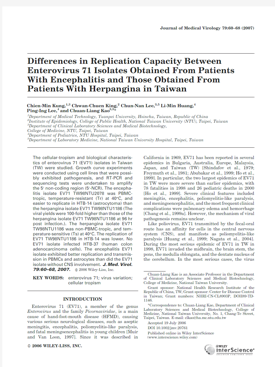
Figure1shows the growth curves obtained from both isolates.The two principal strains of EV71grew in all of the cells used in the experiments,except in the HTB-37 cells(human colon adenocarcinoma cells),which did not support the replication of both EV71isolates.However, the TW98NTU2078isolate from CNS patients grew much faster and generated almost100-fold PFU/ml greater viral yields than TW98NTU1186isolate in the mild case of herpangina in HTB-14(astrocytoma cell line)at12hr post infection(2log10PFU/ml vs.0.1?log10 PFU/ml).The numbers of plaques of both isolates in HEp-2cells(human epidermoid carcinoma from upper respiratory pharyngeal cells)and A549cells(human lung carcinoma Cells)72hr post infection were3.5and 4.3log10PFU/ml,showing that the EV71strains might multiply in both the upper and the lower respiratory tracts.
Another nine isolates of EV71(including?ve isolates from patients with CNS and four isolates from patients with herpangina)and other enteroviruses(poliovirus type1,poliovirus type2,coxsackie A16,coxsackie B2, and coxsackie B3)were used to infect HTB-37cells to con?rm the above?ndings on undetectable viral growth in HTB-37cells.The results demonstrate that no EV71 isolates and coxsackie A16viruses grew in HTB-37cells, whereas polioviruses types1and2and coxsackie viruses B2and B3grew very well in these cells. Growth Phenotype of EV71Isolates in Human PBMCs and Their Spreading Capacity
The growth of EV71TW98NTU2078isolated from a patient with severe encephalitis in human PBMC-adherent cells had high viral yields(5.8?0.4log10 PFU/ml at96hr post infection)whereas the EV71 TW98NTU1186did not replicate in these cells(Fig.2). Additionally,whether the virus can be carried by human PMBC adherent cells was determined using the IC assay.The results also showed that EV71 TW98NTU2078was indeed carried by human adherent cells more ef?ciently than was the EV71TW98NTU1186 isolate with a difference of over100-fold plaque numbers per milliliter(4.2?0.2vs.2.2?0.1)(Fig.3).
The CNS-Related Isolate of EV71Was
Temperature-Resistant at408C
The persistence of a high fever in EV71patients has been documented[Ng et al.,2001].The important biological characteristics of the sensitivity of EV71to temperature in monkeys have also been reported [Hashimoto and Hagiwara,1983].TW98NTU2078and TW98NTU1186isolates of EV71grown in Vero cells for 5days at37and408C,respectively,and the formation of plaques were studied.The results indicated that the TW98NTU2078isolate was a Tr strain that grew at both temperatures,whereas the TW98NTU1186isolate was a temperature-sensitive(Ts)strain that grew only at 378C.For further con?rmation,another?ve EV71 isolates,three of herpangina and two from patients with CNS,were tested.The results showed that both the TW80NTU3100isolate obtained from the patient with polio-like paralysis and the TW98NTUPT142isolate obtained from a patient with CNS-involvement were Tr strains.In contrast,all three herpangina EV71 isolates were Ts strains.
Comparison of Nucleotide Sequences
in50-NCR of EV71Isolates
Comparing the characteristics of neurovirulence showed that the nucleotides in the50non-coding region (50-NCR)had the following four characteristics similar to poliovirus serotype3;(1)neurovirulent cytidine nucleotide(at position475),(2)polypyrimidine tracts (at positions565–584),(3)upstream AUG codons(at positions593–595),and(4)a distance between the AAUAAA motif(at positions571–576)and the AUG codon in the G-loop of22nucleotides[Aurelia et al., 1992[.The other nine EV71isolates from TW,including ?ve from patients with CNS manifestations (TW80NTU3100,TW98NTU1107,TW98NTU1311, TW98C035,TW98PT142)and four herpangina patients (TW98NTU1334,TW99NTU1183,TW2KNTU0652, TW2KNTU1148),had all four phenomena described above.Restated,a total of11EV71TW isolates,from severe encephalitis and mild herpangina patients, exhibited features similar to the four features shown by the poliovirus.When compared to the EV71BrCr strain(ETU22521),an old isolate obtained from Cali-fornia,US in1969,all11Taiwanese isolates had two additional inserted nucleotides of uridine and cytidine at positions741and742.Interestingly,the50-NCR of all 8of the11tested EV71Taiwanese isolates exhibited a polyadenylation signal motif(AAUAAA)in the G loop, whereas the EV71TW98NTU1186,TW2KNTU0652, and TW2KNTU1148isolates,in the mild case of herpangina,had one point mutation(U!C)at positions of575or576at the polyadenylation site in the G loop (Fig.4).
DISCUSSION
Cellular tropism of a virus not only offers a site for the replication of an infectious agent but also ensures that the extent of replication may cause further successful spread of the virus and viral pathogenesis[Dow et al., 1999].EV71causes sudden death and the disease progresses rapidly,therefore several isolates obtained from patients with and without CNS involvement in TW were studied.The following three unique characteristics
J.Med.Virol.DOI10.1002/jmv
EV71Variation From Encephalitis Versus Herpangina63
were noted,which may explain important aspects of the epidemiology,laboratory diagnosis,and the mechanism of pathogenesis.First,no EV71isolate could replicate the HTB-37human colon carcinoma cell-line.Second,
the EV71isolate TW98NTU2078obtained from a pati-ent with encephalitis yielded a high virus in both HTB-14and human PBMC-adherent cells.Third,three EV71isolates obtained from patients with CNS involvement
J.Med.Virol.DOI
10.1002/jmv
012345678
012345678
a b
6
12
24
48
72
96
c
d
e
Fig.1.Cellular tropism of EV71isolates.Cells were infected with the TW98NTU1186(—&—)isolate from herpangina and TW98NTU2078(—*—)isolate from encephalitis at an MOI of 0.01,and the amounts of virus released at the indicated time points (0,6,12,24,48,72,and 96hr)were quanti?ed by plaque assay.The X-axis represents number of hours post infection,and the Y-axis represents log 10PFU/ml.The growth curves of viruses in HTB-14,HTB-11,HTB-37,HEp-2,and A549were shown in (a –e ),respectively.
64Kung et al.
exhibited greater resistance at 408C than did the EV71isolates obtained from the patient with herpangina.The inability of all EV71isolates to replicate in the HTB-37cells demonstrated that EV71may not invade or replicate in human colon cells.This result differs substantially from that obtained for poliovirus and
demonstrates that the binding of receptors of EV71to the colon cells may differ from that of the poliovirus.Poliovirus can bind to such cells via the CD155molecule on the cell membrane [Lange et al.,2001].Further investigations must be conducted to identify the binding sites,the receptors and the possible co-receptors on the cells.Moreover,the high viral yields of all EV71isolates in HEp-2cells (a pharyngeal cell line)may explain why the rates of EV71isolation from throat swabs exceeded markedly those from rectal swabs (91.7vs.64.8%)[Chuan et al.,1999],and differ from the high rates of isolation of polio viruses from fecal specimens [Wang et al.,2000].
EV71is known to be an infectious agent causing CNS disease and leading to poliomyelitis-like paralysis [Lum et al.,1998;Abubakar et al.,1999;Huang et al.,1999;Shen et al.,2000].The cellular and molecular mechan-isms of neurological invasion by EV71are unknown.However,the EV71isolate TW98NTU2078obtained from a patient with encephalitis was found in this study to exhibit three phenomena that were not shown by the TW98NTU1186isolate obtained from the patient with herpangina.First,TW98NTU2078yielded around 2log 10PFU/ml more plaques than the TW98NTU1186isolate on HTB-14cells obtained from human astro-cytoma.The other three EV71isolates (TW98NTU3100,TW98CH35,and TW98PT142)from patients with encephalitis and two EV71isolates (TW98NTU1183and TW99NTU1334)from patients with herpangina showed a similar biological difference (Data no shown).In fact,astrocytes are one of the most important constituent cells in the blood–brain barrier,a very important vasoneurotic part of the CNS [Abbruscato and Davis,1999].Moreover,the data also indicate that EV71can infect human neuroblasotma cells (HTB-11)(Fig.1).Hence,the higher viral yield of EV71on blood–brain barrier may allow the virus to break through the blood–brain barrier and then infect the neurons.Second,the TW98NTU2078isolate in the severe CNS case was a PBMC-tropic strain,which could not only be carried by PBMCs but also able to replicate in adherent cells of PBMCs,unlike the EV71TW98NTU1186isolate in the mild case,which was transported only by human PBMCs without further replication.After EV71viruses enter the blood stream,they may be carried by PBMCs and their subsequent productive infection may have the potential of infecting astrocytes in the blood–brain barrier and other neighboring neurons.Third,TW98NTU2078isolate was a Tr strain,which has been reported as being a neurovirulent strain of poliovirus [Freistadt and Eberle,1996].EV71patients have fever [Chang et al.,1999b],which is consistent with the fact that TW98NTU2078replicated at 408C,and so this isolate overcomes high body-temperature during fever in the host and continues to grow in the infected cells.Therefore,such marked temperature-resistance allows the severe EV71strain to infect cells in the CNS.The other two isolates of EV71obtained from patients with CNS involvement (TW80NTU3100and TW98PT142)also exhibited this biological characteristic.EV71can
J.Med.Virol.DOI
10.1002/jmv
Fig.2.Replication of two different EV71isolates (TW98NTU2078and TW98NTU1186)in human adherent cells infected with viruses with an MOI of 0.1.Incubated at 378C in CO 2for 5days,the viruses were harvested at the indicated times (72and 96hr post infection)following three cycles of freeze-thawing,and the supernatants were then quanti?ed with plaque assay on Vero cell
monolayer.
Fig. 3.Infectious center (IC)assay of Taiwan EV71isolates (TW98NTU2078and TW98NTU1186)in human blood mononuclear cells (PBMC).To investigate whether human PBMCs can carry EV71,the adhered PBMCs were infected with the EV71virus (TW98NTU2078and TW98NTU1186)at an MOI of 10at 378C for 30min,and washed immediately in PBS three times.The adsorbed cells were serially diluted,by a factor of 10each time,from 10à1to 10à6using 0.5%gelatin (in PBS),and then 100m l of each diluent was added onto the con?uent Vero cells,duplicated.The infected cells were incubated at 378C with 5%CO 2for 5days.The cells were then ?xed in 10%formalin solution,stained with 5%crystal violet,and the number of plaques were quanti?ed and represented as numbers of infection centers (ICs)/ml.The means and standard deviations of the numbers of infectious centers in duplicated wells per milliliter were presented.
EV71Variation From Encephalitis Versus Herpangina 65
J.Med.Virol.DOI 10.1002/jmv
also grow well in HEp-2(human pharyngeal epidermoid carcinoma)and A549(human lung carcinoma)cells, indicating that EV71may cause viremia immediately following local replication in the pharyngeal cells,and would then cause a more severe lung edema and CNS involvement[Lum et al.,1998;Chuan et al.,1999]. The internal ribosome entry site(IRES)of the50-NCR in enteroviruses is important as an internal ribosomal landing pad for the replication of viruses[Andino et al., 1990;Lee and Young,1998].EV7150-NCR includes F loop(at positions449–564)and G loop(at positions565–632),and a polyadenylation signal motif(AAUAAA)is present in the G loop,like that in the50-NCR of the poliovirus[Jackson et al.,1990].Although the eight EV71isolates that are similar to the TW98NTU2078 isolate had similar polyadenylation signal motifs in their G loops,which have been documented to be the motif that participates in accelerating viral replication [Hwang et al.,1998;Terhune et al.,1999],the TW98NTU1186,TW2KNTU0652,and TW2KNTU1148 isolates did not have this similar motif because of the point mutation(U!C)of the polyadenylation signal motif in each G loop(Fig.4).However,the mutation of polyadenylation sequences may affect the transduction of cellular sequences,reducing the number of viral transcripts[Swain and Cof?n,1989].Accordingly,the point mutation in this polyadenylation signal motif may reduce the capacity of the TW98NTU1186isolate to replicate in HTB-14cells(Fig.1a)and cause poor replication in the RD cells of TW2KNTU0652and TW2KNTU1148isolates(data not shown).
All11EV71isolates,regardless of CNS outcome or whether they were involved in mild herpangina,had a neurovirulent nucleotide(cytidine)at position475in the F loop of the50-NCR.The region has been reported to be a neurovirulent region of wild-type poliovirus,as deter-mined by comparison with the Sabin vaccine strain [Dildine and Semler,1992].However,EV71like other enteroviruses,including coxsackie viruses and ECHO viruses,has such a cytidine nucleotide position,which is important in the formation of the stem of an F-loop, which may bind with other host factors;therefore,a single nucleotide at this position cannot determine viral virulence.This?nding is unlike that of the neuro-virulence of poliovirus type3that involve the mutation of C to U at position472in50-NCR(the same stem position of the F-loop as EV71)and the viral load in neurological cells[Evans et al.,1985].
Based on the results obtained concerning phenotypic differences between encephalitis and herpangina EV71 isolates in human PBMCs and astrocytes in vitro,a possible pathogenic pathway of EV71from the oral route to CNS is hypothesized,although the cell culture experiments may not re?ect completely the situation in vivo.The virus would enter from the oral route by person-to-person transmission by the fecal-oral route, and it would be very likely to replicate initially in pharyngeal cells,increasing replication in PBMCs and viremia with a higher viral load,even during the fever stage at a high body temperature;therefore,the EV71pathogenic strain can infect the astrocytes(HTB-14)of the blood–brain barrier via PBMCs,?nally invading the neurons(HTB-11)in the brain.Further animal model investigations based on various strains of EV71inte-grated infectious clones may offer clues on the exact molecule that is responsible for viral pathogenesis.
ACKNOWLEDGMENTS
The sincere help of pediatricians and infection control nurses at National Taiwan University Hospital and Pingtung Christian Hospital is gratefully acknowl-edged.
REFERENCES
Abbruscato TJ,Davis TP.1999.Protein expression of brain endothelial cell E-cadherin after hypoxia/aglycemia:In?uence of astrocyte contact.Brain Res842:277–286.
AbuBakar S,Chee HY,Al-Kobaisi MF,Xiaoshan J,Chua KB,Lam SK.
1999.Identi?cation of enterovirus71isolates from an outbreak of hand,foot and mouth disease(HFMD)with fatal cases of encephalomyelitis in Malaysia.Virus Res61:1–9.
Andino R,Riechhof GE,Baltimore D.1990.A functional ribonucleo-protein complex forms around50end of poliovirus RNA.Cell63: 369–380.
Aurelia A,Haller XX,Semler BL.1992.Linker scanning mutagenesis of the internal ribosome entry site of poliovirus RNA.J Virol66: 5075–5086.
Bienkowska-Szewczyk K,Ehrenfeld E.1988.An internal50noncoding region required for translation of poliovirus RNA in vitro.J Virol 162:3068–3072.
Brack-Werner R,Bell JE.1999.Replication of HIV-1in human astrocytes.NeuroAids2:8.
Chang LY,Lin TY,Hsu KH,Huang YC,Lin KL,Hsueh C,Shih SR, Ning HC,Hwang MS,Wang HS,Lee CY.1999a.Clinical features and risk factors of pulmonary edema after enterovirus-71related hand,foot,and mouth https://www.360docs.net/doc/e115493823.html,ncet354:1682–1686.
Chang LY,Lin TY,Huang YC,Tsao K,Shih SR,Kuo ML,Ning HC, Chung PW,Kang https://www.360docs.net/doc/e115493823.html,parison of enterovirus71and coxsackie-virus A16clinical illnesses during the Taiwan entero-virus epidemic.Pediatr Infect Dis J18:1092–1096.
Chuan LH,Huang LM,Lee PI,Lee CY.1999.The epidemic of enterovirus infection in Taipei city.Formo J Med3:1.
Dildine SL,Semler BL.1992.Conservation of RNA-protein interac-tions among picornaviruses.J Virol66:4364–4376.
Dow SW,Mathiason CK,Hoover EA.1999.In vivo monocyte tropism of pathogenic Feline Immunode?ciency Virus.J Virol73:6852–6861. Evans DMA,Dunn G,Minor PD,Schild GC,Cann AJ,Stanway G, Almond JW,Currey K,Maizel JV.1985.Increased neurovirulence associated with a single nucleotide change in a noncoding region of the Sabin type3poliovaccine genome.Nature314:548–550. Freistadt MS,Eberle KE.1996.Correlation between Poliovirus Type1 Mahoney replication in blood cells and neurovirulence.J Virol70: 6486–6492.
Freymuth F,Duncombe C,Hardouin A,Boutard B,Guihard J,Leroy D.
1981.Isolation of enterovirus71in hand-foot-and mouth disease (letter).Nouv Presse Med10:2210.
Fujioka S,Koide H,Kitaura Y,Duguchi H,Kawamura K.1995.
Analysis of enterovirus genotypes using single-strand conforma-tion polymorphisms of polymerase chain reaction products.J Virol Methods51:253–258.
Hashimoto I,Hagiwara https://www.360docs.net/doc/e115493823.html,parative studies on the neuro-virulence of temperature-sensitive and temperature-resistant viruses of enterovirus71in monkeys.Acta Neuropathol60:266–270.
Ho M,Chen ER,Hsu KH,Twu SJ,Chen KT,Tsai SF,Wang JR,Shih SR.1999.An epidemic of enterovirus71infection in Taiwan.N Engl J Med341:929–935.
Huang CC,Liu CC,Chang YC,Chen CY,Wang ST,Yeh TF.1999.
Neurological complications in children with enterovirus71infec-tion.N Engl J Med341:936–942.
Hwang LN,Englund N,Pattnaik AK.1998.Polyadenylation of vesicular stomatitis virus mRNA dictates ef?cient transcription
J.Med.Virol.DOI10.1002/jmv
EV71Variation From Encephalitis Versus Herpangina67
termination at the intercistronic gene junctions.J Virol72:1805–1813.
Jackson RJ,Howell MT,Kaminski A.1990.The novel mechanism of initiation of picornavirus RNA translation.Trends Biochem Sci15: 477–483.
Lange R,Peng X,Wimmer E,Lipp M,Bernhardt G.2001.The poliovirus receptor CD155mediates cell-to-matrix contacts by speci?cally binding to vitronectin.Virol285:218–227.
Lee C,Young C.1998.Murine neurovirulence studies with a chimeric poliovirus:In vivo generation of a mutant base-paired stable attenuated poliovirus.Microb Pathogen25:215–225.
Lum LC,Wong KT,Lam SK,Chua KB,Goh AY.1998.Neurogenic pulmonary oedema and https://www.360docs.net/doc/e115493823.html,ncet 352:1391.
Masson D,Jarry A,Baury B,Blanchardie P,Laboisse C,Lustenberger P,Denis MG.2001.Overexpression of the CD155gene in human colorectal carcinoma.Gut49:236–240.
Muir P,Van Loon AM.1997.Enterovirus infections of the central Nervous System.Intervirol40:153–166.
Nagata N,Iwasaki T,Ami Y,Tano Y,Harashima A,Suzaki Y,Sato Y, Hasegawa H,Sata T,Miyamura T,Shimizu H.2004.Differential localization of neurons susceptible to enterovirus71and poliovirus type1in the central nervous system of cynomolgus monkeys after intravenous inoculation.J Gen Virol85:2981–2989.
Ng DK,Law AK,Cherk SW,Mak KL.2001.First fatal case of entero-virus71infection in Hong Kong.Hong Kong Med J7:193–196. Nugent CI,Johnson KL,Sarnow P,Kirkegaard K.1999.Functional coupling between replication and packaging of poliovirus replicon RNA.J Virol73:427–435.Persidsky Y,Ghorpade A,Rasmussen J,Limoges J,Liu XJ,Stins M,
Fiala M,Way D,Kim KS,Witte MH,Weinand M,Carhart L, Gendelman HE.1999.Microglial and astrocyte chemokines
regulate monocyte migration through the blood-brain barrier in
human immunode?ciency virus-1encephalitis.Amer J Path155: 1599–1611.
Shen WC,Tsai C,Chiu H,Chow K.2000.MRI of Enterovirus71
myelitis with monoplegia.Neuroradiology42:124–127.
Shimizu H,Utama A,Yoshii K,Yoshida H,Yoneyama T,Sinniah M, Yusof MA,Okuno Y,Okabe N,Shih SR,Chen HY,Wang GR,Kao CL,Chang KS,Miyamura T,Hagiwara A.1999.Enterovirus71 from fatal and nonfatal cases of hand,foot and mouth disease epidemics in Malaysia,Japan and Taiwan in1997–1998.Jpn J Infect Dis52:12–15.
Shindafov LM,Chumakov MP,Voroshelova MK,Bojinov S,Vasilenko
SM,Iordanov I,Kirov ID,Kamenov E,Leshchinskaya EV,Mitov G, Robinson IA,Vchev SS,Staikov ST.1979.Epidemiological,clinical, and pathomorphological characteristics of epidemic poliomyelitis-like disease caused by enterovirus71.J Hygiene Epidemio Microbiol&Immuno3:284–295.
Swain A,Cof?n JM.1989.Polyadenylation at correct sites in genome
RNA is not required for retrovirus replication or genome encapsida-
tion.J Virol63:3301–3306.
Terhune SS,Milcarek C,laimins LA.1999.Regulation of human papillomavirus type31polyadenylaiton during the differentiation-dependent life cycle.J Virol73:7185–7192.
Wang JR,Tsai HP,Chen PF,Lai YJ,Yan JJ,Kiang D,Lin KH,Liu CC, Su IJ.2000.An outbreak of enterovirus71infection in Taiwan,1998.
https://www.360docs.net/doc/e115493823.html,boratory diagnosis and genetic analysis.J Clin Virol17:91–99.
J.Med.Virol.DOI10.1002/jmv
68Kung et al.
体外细胞培养
第一讲体外细胞培养的基本技术 ●体外细胞培养条件和基本技术 ●体外细胞培养 ●体外培养物的生长生物学 ●细胞系和细胞株 ●培养物的冷冻与复苏 ●培养物的污染、检测和排除 ●一、体外细胞培养条件和基本技术 体外细胞培养的优缺点 优点:简化细胞的生长环境,方便施加实验因素,便于观测实验结果,便于获得均一细胞群,能够进行大规模生物制品的生产。 体外细胞培养不足之处:培养对象脱离了机体的整体支配和调控,细胞间在一定程度上失去组织联系及相互作用。体外培养条件下的生长发育情况与在体时的情况存在一定差异,分析实验结果时必须考虑这种差异。 一、体外细胞培养条件 (一)、体外培养实验室 1. 准备室 2. 培养室:基本条件要求:清洁、无菌、干燥、不通风,并具有适宜的光线。空气调节用中央空调或分体式空调机。室顶安装紫外灯等消毒装置。 3. 缓冲室 4. 其他实验室 (二)、体外培养的设备和器具 设备:1. 超静工作台,生物安全柜,净化室 2. 培养箱:温度,湿度,pH值 3. 倒置显微镜 4. 水净化装置:去离子水净化装置,石英玻璃蒸馏器, 超纯水装置 5. 冰箱 6. 离心机7. 冷冻保存装置 8. 高压蒸汽消毒装置:电热干燥箱,pH计,天平 培养器具:1. 过滤除菌装置:Zeiss 滤器,抽滤式玻璃简易型滤器, 针头式加压塑料小滤器 2. 培养器皿:(1)溶液瓶(2)培养瓶(3)培养皿 (4)多孔培养板(5)离心管 3. 移液器 4.筛网:金属筛网(不锈钢网、铜网),尼龙筛网 (三)培养用液 ?水和平衡盐溶液(balanced salt solution, BSS) 水:离子交换水,蒸馏水 平衡盐溶液:主要成分:无机盐和葡萄糖 常用BSS: PBS ,Ringer Earle ,D-Hanks,Hanks ?培养液:1.天然培养液:1)血清:小牛血清,胎牛血清(fetal bovine serum, FBS),马血清,2)水解乳蛋白,3)胚胎渗出液 2.合成培养液:合成培养基的种类MEM,RPMI 1640,McCoy 5A,HAM F12等
人体白细胞的种类和功能
人体白细胞的种类和功能 人体内白细胞为无色有核细胞,在血液中呈球形,它能以变形运动穿过毛细血管壁,进入结缔组织。正常5000—10000/mm3。 一、种类及特点 根据细胞质和细胞核的染色特点,可把白细胞分为两大类: 有颗粒白细胞:嗜中性粒细胞,嗜酸性粒细胞,嗜碱性粒细胞 无颗粒白细胞:淋巴细胞,单核细胞 正常时各种白细胞数目保持一定比例,称为白细胞分类计数,正常情况下数目如下: 疾病状况下,白细胞总数和分类计数都可发生改变。 二、白细胞的功能 ①中性粒细胞白细胞 : 吞噬和消化侵入微生物和机体本身的各种坏死细 胞,包括衰老和受损红细胞.是机体防御功能重要组成部分。细胞内溶酶体丰富,并做变形运动。 中性粒细胞可通过变形运动渗出毛细血管壁进入组织液,在组织液内游走(趋化性)。如果白细胞的的溶酶体释放的酶量过多,不仅能杀死和溶解细菌,本身也将致死,死亡的白细胞连同细菌分解物一起成为浓液组成部分。 ②嗜酸性粒细胞:能限制嗜碱性粒细胞和肥大细胞中的活性物质的合成和释放;当机体患某些过敏性疾病时,嗜酸性粒细胞数量增多,有人认为她和机体的过敏性反应有关。 ③嗜碱性粒细胞:含有多种生物活性物质,与肥大细胞相似,细胞颗粒中能合成组织胺和过敏性慢作用物质。
组织胺:使小动脉和毛细血管扩张,并使毛细血管和微静脉通透性增加,与人过敏反应有关。 过敏性慢作用物质:可使血管通透性增加,并使平滑肌收缩,特别是使支气管和细支气管的平滑肌收缩,从而引起哮喘。总括起来嗜碱性粒细胞所释放的活性物质可引起哮喘,寻麻疹,食物过敏等各种反应,同时又引起嗜碱性粒细胞聚集这一局部。 ④淋巴细胞:具有免疫功能 T细胞:骨髓中生成的淋巴系干细胞在胸腺中发育成熟,参与细胞免疫。血液中 80-90% 淋巴细胞属于T淋巴细胞。 B细胞:造血干细胞在骨髓中分裂分化成B细胞,多集中在淋巴结等淋巴器官中。参与体液免疫—抗体免疫。 ⑤单核细胞:从血管进入组织后,便分化成巨噬细胞,吞噬细菌和病毒等 三、白细胞测定临床意义 (一)白细胞增加:外周血液中白细胞数超过10×109/L称为白细胞增多症。白细胞增多可以是中性粒细胞、嗜酸性粒细胞、淋巴细胞及单核细胞增多。临床常见为淋巴细胞增多症。 1、中性粒细胞增多症 生理性:如饭后和剧烈运动后;冷水浴后及极度恐惧和疼痛后等、妇女经期及妊娠等。 病理性: (1)急性感染:某些细菌(葡萄球菌、肺炎球菌等)病毒(水痘病毒、狂犬病毒)引起的感染; (2)非感染性炎症,如急性风湿热、少年型类风湿关节炎等 (3)组织坏死:严重外伤、手术创伤、烧伤等。 (4)急性失血及贫血 (5)急性中毒:如化学毒物及药物中毒、尿毒症等。 (6)粒细胞异常增生:急性粒细胞白血病,骨髓增生性疾病 (7)其他:如脾脏切除后。 2、嗜酸性粒细胞增多症 外周血中嗜酸性粒细胞绝对值超过0.45×10⁹/L,称为嗜酸性粒细胞增多症. 嗜酸性粒细胞增多常与许多疾病有关,特别是寄生虫感染和过敏性疾病。 3、白血病 是骨髓和其他造血组织中原始和幼稚细胞异常增生的一种恶性疾病。临床表 现贫血、出血、感染及白血病细胞侵润机体各组织、器官所产生的相应表现。 ⑴病因: 病毒:人类T细胞白血病,已肯定是由人T细胞病毒-Ⅰ引起的 放射性核素:其致白血病的作用是肯定的,其作用与照射剂量的大 小和部位有关 化学因素:(如烷化剂、细胞毒药物)及化学毒物(苯)可诱发 遗传因素:家族性白血病约占7%。 其他血液病:骨髓增生性疾病、淋巴瘤等最终可发展为急性白血病。 ⑵分类: 按病程缓急及白细胞分化程度可分为 急性白血病:起病急,自然病程约半年以内。骨髓中以原始和早期幼稚细 胞增生为主,正常造血受到抑制的恶性疾病。成人以急性粒细胞白血病多见,儿童以急性淋巴细胞较多见。临床表现主要为贫血、出血、继发感染发热及白血病细胞侵润
胚胎干细胞体外培养
胚胎干细胞体外培养 (一)胚胎干细胞的来源 目前胚胎干细胞的主要来源有:①囊胚的ICM及受精卵发育至桑葚胚之前的早期胚胎细胞;②从胚胎生殖嵴及肠系膜中分离原始生殖细胞PGCs后培养建系的胚胎生殖细胞(embryonic germ cells,EG细胞),也具有ESCs的特性,可以分化为各种类型的成熟细胞;③体细胞核转移(somatic cell nuclear transplantation,SCNT)至去核卵母细胞后培育出来的全能细胞。其中囊胚的ICM最为常用。 (二)胚胎干细胞的分离 1.分离获取ESCs的时间:以既保证ESCs的全能性又要有足够的细胞数量为原则来确定ESCs分离获取的最佳时间。以ICM为ESCs来源时:小鼠取3~5天囊胚;猪取9~10天囊胚;羊取7~8天囊胚;牛取6~7天桑葚胚或早期囊胚;人取7~10天囊胚。以PGCs取ES细胞时:小鼠取1 2.5天胎儿生殖嵴;大鼠可取10.5天尿囊、中胚层组织块、12.5天背肠系膜或1 3.5~1 4.5天生殖嵴;牛取29~35天胎儿生殖嵴;人取35~63天的生殖嵴。 2.分离获取ESCs的方法:从PGCs分离ESCs的方法常为机械剪切与消化相结合法,即把采取的胚胎组织充分剪碎,采用EDTA、EDTA/胰酶消化。 从囊胚分离ICM的方法主要有三种: (1)免疫外科学方法:体外培养的小鼠胚泡去除透明带后,经兔抗JCR小鼠脾脏细胞抗血清(抗H-26)作用30分钟,移至1∶6稀释的新鲜豚鼠血清中作用30分钟,Hank’s液冲洗,此时胚泡的滋养外胚层呈空泡状,用眼科手术刀挑去死了的滋养层细胞,留存ICM 细胞用于培养。这种方法利用囊胚腔对抗体的不通透性,通过抗体、补体结合对细胞的毒性杀伤作用,去除滋养层细胞,保留ICM细胞进行培养。 (2)组织培养法:在小鼠受精2.5天后切除卵巢,给予外源激素,使胚胎继续发育,但延缓着床,4~6天后,由子宫冲取胚泡进行培养。结果滋养层细胞生长并推开饲养层细胞,在培养皿底壁上铺展;而ICM细胞增殖,垂直向上生长,形成卵圆柱状结构,在显微镜下用细玻璃针挑出这种柱状结构,消化传代。Evans和Kaufman采用这种方法第一个建立了小鼠ESCs系。 (3)显微外科学方法:小鼠受精后3~4天,由子宫冲取胚泡,利用显微操作系统直接从胚泡中吸出ICM细胞进行培养。 由于免疫外科学方法需要特殊的试剂去除透明带和滋养层,易对内细胞群造成损伤,而显微外科学方法需要专门的仪器设备,且对人员的技术水平要求较高,均难以推广应用。组织培养法将胚泡接种在饲养层上,模拟体内胚泡的着床,更接近自然发育过程,内细胞群增殖旺盛,较易获得胚胎干细胞样集落。 (三)胚胎干细胞的培养和建系 ESCs的分离培养和建系是其得以应用的前提。ESCs建系的原理是:将分离获取的ICM 或PGCs与饲养层共同培养,使之增殖而又保持其未分化状态,这样代代相传从而使ESCs
人体八种白细胞类型
白细胞亦称白血球,它的功能是使机体免受病原体(细菌和病毒)、癌细胞、异物侵入,是人体的守卫者。白细胞来源于骨髓中的造血干细胞,在骨髓中发育后进入血液和淋巴液循环,也存在于血管和淋巴管外的组织中。 人体中白细胞有八种类型,具体细胞类型如下。 1 巨噬细胞 巨噬细胞是个头最大的白细胞。 巨噬细胞由单核细胞分化发育而来,单核细胞几乎存在于所有组织中。巨噬细胞对细胞残体和病原体进行吞噬和消化,这一过程称为吞噬作用。巨噬细胞内的溶酶体会释放溶菌酶破坏病原体。吞噬细胞还是参与适应性免疫的专职抗原提呈细胞。除了免疫功能外,吞噬细胞还扮演者促进生殖 细胞发育、性激素、骨组织的再吸收以及血络的形成。巨噬细胞如下图所示: 2 树突状细胞 类似于吞噬细胞,树突状细胞也是一种单核细胞。树突状细胞成熟时会伸出许多树突样或伪足样突起,与神经元的树突相似。树突状细胞通常存在于跟外界接触的皮膜(黏膜)部位,比如皮肤、内层的鼻子、肺、胃与肠的内层。树突状细胞通过摄取、加工处理和递呈抗原到淋巴结和淋巴器官 中的淋巴细胞帮助机体识别病原体,启动特异性免疫应答。树突状细胞如下图所示: 3 B淋巴细胞 B细胞是淋巴细胞的成分之一。B细胞受抗原刺激后,会增合成和分泌一种特殊的蛋白质——抗体对抗病原体,抗体与抗原结合帮助机体识别病原体,并使之成为靶细胞被其他免疫细胞清除。被称为记忆细胞的B细胞可以保持对病原菌生物分子标记物的记忆,使得同一种抗原再次进入机体 时免受感染。这使得机体保持针对特定病原体的长期免疫力。B淋巴细胞如下图所示: 4 T淋巴细胞 类似于B细胞,T细胞也是淋巴细胞之一。T淋巴细胞来源于骨髓,迁移到胸腺内分化成熟。T 细胞通过直接杀伤靶细胞和释放淋巴因子参与免疫反应。T细胞包括细胞毒性T细胞、辅助性T细胞、抑制性T细胞、自然杀伤T细胞和记忆T细胞。细胞毒性T细胞直接杀伤靶细胞,辅助性T细胞协助B细胞产生抗体,抑制性T细胞抑制B细胞和其他T细胞对抗原的免疫应答。 NKT细胞具有T细胞和NK细胞两重性质,但是NKT细胞不是NK细胞而是T细胞。T淋巴细胞 如下图所示: 5 自然杀伤细胞 自然杀伤细胞是分布于血液循环的淋巴细胞,它能识别被感染细胞和衰老细胞。NK细胞含有化学颗粒,NK细胞能够识别肿瘤或病毒感染的细胞,通过释放化学颗粒吞噬破坏病变细胞。这些化学颗粒能溶解病变细胞的细胞膜,促使细胞凋亡,最终杀伤靶细胞。 同时应该区分NK细胞和某些T细胞,比如NKT细胞,不要相互混淆。自然杀伤细胞如下图所 示: 6 嗜中性粒细胞
造血干细胞分离培养方法
一造血干细胞分离 (一)小鼠骨髓采集与单个核细胞悬液的制备 1 HES(羟乙基淀粉)沉淀法 抽取髓液500 m L按4∶1比例加入HES,自然沉降红细胞后,分离上清。4℃400 g离心10 min 得细胞沉淀物,以1 %白蛋白盐水液洗涤细胞2次。 2 percoll液密度梯度离心法 ①按体积比为~2)∶1在骨髓液中加入淋巴细胞分离液。4℃,1 5 00 r / min,离心20 min。取单个核细胞层,以1%白蛋白盐水液洗涤3次。 ②取鼠股骨和胫骨 , 在两头关节处切开骨骼 , 反复用培养基冲洗骨髓腔 , 随后小心地逐滴将细胞悬液加在淋巴细胞分离液上 , 2 000 r / min 离心 20 min。吸取离心后相交液面处的白色细胞层即为单个核细胞。 3 Ficoll分离法 方法一在15ml分离管中加比重为的Ficoll液,缓慢移入等量骨髓细胞悬液,整体平衡后低温离心2000r/min×20min,吸取白细胞层,用RPMI1640液亲清洗后离心2000r/min×10min,取白细胞层加RPMI1640液到10ml制成造血干细胞悬液样本。 方法二取无菌离心管1支,预先添加3mlFicoll(与骨髓细胞悬液体积1:1),用滴管取单细胞悬液3ml,沿离心管壁小心缓慢叠加于分离液面上,注意保持清楚的界面,室温下水平离心2000rpm×20分钟,(后续在冰上进行)用毛细吸管插到云雾层,小心吸取单个核细胞,置入另一短中管中,加入5倍以上体积的磷酸缓冲液PBS,1500rpm×10分钟,L-DMEM洗涤细胞两次,每次以1000r/min 离心10min,去上清液。(除第一步的室温离心外,其余为低温离心)。 取骨髓细胞悬液的方法是:取出大鼠腿骨将肌肉尽可能剔除,并用PBS缓冲液冲洗干净。在超净台中腿骨浸泡在PBS缓冲液中5min,之后在两头关节处切开骨骼 , 反复用PBS缓冲液冲洗骨髓腔,从而得到骨髓细胞悬液。 (二)Linc-- kit+造血干细胞的分离纯化。 去除 Lin+细胞( 负筛选) : 带有生物素的混合抗体标记成熟细胞 ; 抗生物素的磁珠吸附抗体标记的成熟细胞 , 包括 T , B淋巴细胞、粒细胞、巨噬细胞以及它们的定向前体细胞 ; 细胞通过磁场不被柱子吸附的包括造血干细胞和骨髓间质干细胞。收集CD117+细胞( 正筛选):带有CD117抗体的磁珠吸附CD117+细胞 , 细胞通过磁场被柱子吸附的CD117+细胞(具体操作步骤参照M iltenyi公司MACS手册)。 (三)根据细胞表面CD34+抗原进行分离纯化 流式细胞仪检测骨髓CD34+细胞:取50μL细胞悬液 , 加入μL CD45- FITC和10μL CD34- PE 充分混合 , 避光孵育15 min。以PBS液洗涤并加多聚甲醛固定液450μL固定 , 上机检测。 磁性标记 CD34+细胞:在细胞悬液中加入100μL Fc - R阻滞剂 , 再加入100μLCD34 磁珠标记细胞 , 于4℃冰箱中充分混合孵育 30 min。用PBS 缓冲液洗涤细胞,离心10 min , 去上清液后再以500μL PBS缓冲液重新悬浮细胞。 MiniMACS磁性分离、纯化CD34+细胞:将MS分离柱放置在MACS分离器的磁场中,以500μL PBS 缓冲液漂洗。以孔径30μm的尼龙网或过滤器去除细胞悬液凝块 , 细胞通过分离柱 , 以500μL PBS
细胞体外培养时为什么会贴到培养皿壁上
生物学通报2007年第42卷第9期细胞体外培养时为什么会贴到培养皿壁上 刘梅芳徐国恒‘(北京大学医学都生理系北京100083) 北京市第22中学的生物学教师陈珊问:动物细胞培养时为什么会出现贴壁现象? 答:第1。绝大部分细胞在机体内的环境下是与其他细胞或者细胞外基质结合的,不是孤立存在的。第2,细胞与细胞或者细胞外基质结合的物质基础以及在体外培养中贴壁的物质基础是什么。 在体内,大部分细胞不是孤立存在的(血液中的细胞除外)。一般一种细胞具有一种特定的功能.不同种细胞有机组合在一起就构成能够完成较为完整的一项功能的器官。细胞之问,细胞与细胞外基质之间结合在一起,这使得每一部分组织或者器官具有一定的外形及特定的功能。血液在体内是起运输作用的,不停地流动,要通过只有几个“m的毛细血管,所以血液中的细胞是单个存在的。否则就会发生血管阻塞。但是它们受到刺激.其表面与黏附有关的蛋白会被激活然后与其他细胞或细胞外基质结合.如血小板接触到破损内皮下的胶原被激活而黏附在局部,从而发挥止血的功能;如巨噬细胞可以将衰老的红细胞、血液中凋亡的中性粒细胞结合并将其吞噬,这些都应该是属于细胞之间的结合。另外有些研究表明细胞必须与其他的细胞发生联系才能够生存.如果没有其他细胞或者细胞外基质与之结合.此细胞就会发生凋亡,称为失巢凋亡。 细胞在体外培养的条件下的贴壁,首先并不是所有的细胞在体外培养的情况下都会贴壁,但大部分来自实体组织器官的细胞都会贴壁。贴壁的过程可能是这样的。细胞首先分泌细胞外基质,这种细胞外基质黏附在支持物的表面(培养瓶,培养皿的底面),然后细胞通过其表面表达的黏附因子与这些细胞外基质结合。所以细胞贴壁与否与细胞本身分泌细胞外基质的能力以及细胞本身表达的黏附分子的数量有关,也与培养皿壁的表面结构有关。 细胞贴壁的过程使形态发生很大变化.细胞变形运动是细胞内骨架本身和细胞外基质共同作用的结果。细胞为什么要贴壁,可以想象一下.密度大于培养基的细胞(绝大部分细胞)会沉在底面上,那么细胞要生存必定改善这个环境。分泌细胞外基质,然后很自然就贴附在底面上了,而悬浮细胞.如淋巴细胞由于重力原因沉在培养瓶的底面,但是并不贴附,这可能与细胞表面的黏附分子少,以及分泌的细胞外基质过少有关.有人研究用细胞外基质处理培养器皿的底面.即增加细胞外基质,可以使悬浮的淋巴细胞贴壁。另外悬浮生长的细胞系THP一1受到某些药物刺激之后也可以贴壁.这可能与细胞本身分泌的细胞外基质以及细胞表面的黏附分子表达增多有关。密度小于水的细胞,如脂肪细胞,漂浮在液面上。所以不可能贴在底面上.但是如果将培养液加满,那么细胞能够与培养瓶上面的壁接触同样可以贴壁。因此可以说,细胞贴壁是适应环境的一种反应。 (E—IIlail:xug@bjwLI.edu.cn) (BH) 欢迎订阅2008年《生物学教学》杂志 《生物学教学)杂志是由华东师范大学主办,向国内外正式发行的全国教育类核心期刊。国内统一刊号:cN31一I009,c4.国际标准刊号:1004—7549。读者对象以中学生物学教师为主,兼顾高校和其他生物学工作者。主要栏目有:生物科学综述、国家课程标准与实验教材、现代教育论坛、教育教学研究、课堂教学设计与实践、信息技术、国外教育动态、考试与命题、实验教学、科技活动、教学参考、生物学科技信息、科学技术与社会、读者之窗等。另外,封面、封底刊登生物照片。 《生物学教学》杂志为80页,月刊,国际标准16K,2008年定价:7.00元,全年84元。国内订购:全国各地邮局,代号4—450。国外订购:中国国际图书贸易公司(北京399信箱,邮编:100044),代号:M5105。如果错过了邮局订购时间,可以与杂志社联系邮购。杂志社地址:华东师范大学《生物学教学》杂志社,邮编:200062.电话02l一62232225。电子信箱:swxiw@bio.ecnu.edu.cn。
体外培养细胞的种类和命名
体外培养细胞的种类和命名 体外培养细胞的名称,随培养细胞技术的发展和细胞种类的增多而演变。最早采用的名称为细胞株(Cell strain),以后又出现细胞系(Cell Line)一词,两者曾一度混用致概念不明确,导致文献中也很混乱。我国也曾有类似情况,在我国尚未制定出统一名词前,本书用的名词基本参考Schaeffer,W.I.(1979)和国内有关会议、以及国内外杂志常用名词为准。 (一)初代培养 初代培养又称原代培养,即直接从体内取出的细胞、组织和器官进行的第一次的培养物。一旦已进行传代培养(Subculture)的细胞,便不再称为初代培养,而改称为细胞系。 (二)细胞系 初代培养物开始第一次传代培养后的细胞,即称之为细胞系。如细胞系的生存期有限,则称之为有限细胞系(Finite Cell Line);已获无限繁殖能力能持续生存的细胞系,称连续细胞系或无限细胞系(Infinite Cell Line)。无限细胞系大多已发生异倍化,具异倍体核型,有的可能已成为恶性细胞,因此本质上已是发生转化的细胞系。无限细胞系有的只有永生性(或不死性),但仍保留接触抑制和无异体接种致癌性;有的不仅有永生性,异体接种也有致瘤性,说明已恶性化。这两种不同性质的无限细胞系,在国内外文献中对这些名词的应用上也常不十分严格。为概念上的明确,本书中对有恶性的无限细胞系采用“恶性转化细胞系”一词表示可能更妥。而对那些只具永生性而无恶性的细胞系,则用无限细胞系或转化细胞系即可。当前流传的NIH3T3、Rat-1、10T1/2等均属这类细胞系。 由某一细胞系分离出来的、在性状上与原细胞系不同的细胞系,称该细胞系的亚系(Subline)。 (三)克隆细胞株 从一个经过生物学鉴定的细胞系用单细胞分离培养或通过筛选的方法,由单细胞增殖形成的细胞群,称细胞株。再由原细胞株进一步分离培养出与原株性状不同的细胞群,亦可称之为亚株(Substrain) (四)二倍体细胞 细胞群染色体数目具有与原供体二倍细胞染色体数相同或基本相同(2n细胞占75%或80%以上)的细胞群,称二倍体细胞培养。如仅数目相同,而核型不同的即染色体形态有改变者为假二倍体。二倍体细胞在正常情况下具有限生命期,故属有限细胞系。但随供体年龄和组织细胞的不同,二倍体细胞的寿命长短各异。人胚肺成纤维细胞可传50代土10代,人胚肾只有8~10代,人胚神经胶质细胞15~30代;如从老龄个体取可传50则细胞生存期更短。由不同年龄供体取材建立的二倍体细胞系可供研究衰老之用。为保持二倍体细胞能长期被利用,一般在初代或2~5代即大量冻存作为原种(Stook Cells),用时再进行繁殖,用后再继续冻存,可供长期使用或延缓细胞的衰老。 (五)遗传缺陷细胞 从有先天遗传缺陷者取材(主要为成纤维细胞)培养的细胞,或用人工方法诱发突变的细胞,都属遗传缺欠细胞。这类细胞可能具有二倍体核型,也可呈异倍体。 (六)肿瘤细胞系或株 这是现有细胞系中最多的一类,我国已建细胞系主要为这类细胞。肿瘤细胞系多由癌瘤建成,多呈类上皮型细胞,常已传几十代或百代以上,并具有不死性和异体
细胞培养的基本方法-细胞分离技术
细胞培养的基本方法-细胞分离技术 细胞分离技术 一、从原代组织中分离细胞将组织块分离(散)成细胞悬液的方法有多种,最常用的是机 械解离细胞法、酶学解离细胞法以及螯合剂解离细胞法。 从原代组织中获得单细胞悬液的一般方法是酶解聚。细胞暴露在酶中的时间要尽可能的短, 以保持最大的活性。下列步骤可以解聚整个组织,获得较高产量的有活性细胞。 1. 胰蛋白酶(Trypsin) ?在去除不需要的组织后,使用无菌的解剖刀和剪子把剩余的组织切成3~4mn小片,通过悬 浮在无钙镁的平衡盐溶液中清洗组织碎片。让组织碎片沉淀,去除上清液。重复清洗2到3次。 ?将盛有组织碎片的容器置于冰上,去除残留的上清液。加入0.25 %溶解在无钙镁的平衡盐 溶液中的胰蛋白酶(100mg组织加入1ml胰蛋白酶)。 ?在4C孵育6到18小时,使几乎没有胰蛋白酶活性的酶尽可能渗透进去。 ?移弃组织碎片中的胰蛋白酶,在37C孵育包含残留胰蛋白酶的组织碎片20到30分钟。 ?在组织碎片加入热的完全培养基,用移液管轻轻地分散组织。如果使用无血清培养基,要 加入大豆胰蛋白酶抑制剂。 ?通过无菌不锈钢丝网(100?200mm过滤,分散所有剩余组织。计数和接种细胞,进行培养。 2. 胶原酶(Collagenase) ?用无菌解剖刀和剪子把剩余组织切成3~4mm」、片,用Hanks'平衡液(HBSS清洗组织碎片 几次。 ?加入胶原酶(50?200单位/ml,溶解在HBSS中)。 ?在37C孵育4到18小时。加入3mM CaCI2增加解离效率。 ?通过无菌不锈钢丝网或尼龙网过滤细胞悬液,以分离分散细胞、组织碎片和较大的碎片。 如果需要进一步的解聚,在碎片中加入新鲜的胶原酶。 ?通过离心在HBSS中清洗悬液几次。 ?再一次在培养基中悬浮细胞,计数和接种细胞,进行培养。 3. Dis pase ?用无菌解剖刀和剪子把剩余组织切成3~4mn小片,用不含钙镁的平衡盐溶液清洗组织碎片 几次。 ?加入Dispase (0.6?2.4单位/ml溶解在无钙镁的平衡盐溶液) ?在37C孵育20分钟到几个小时。 ?通过无菌不锈钢丝网或尼龙网过滤细胞悬液,以分离分散细胞、组织碎片和较大的碎片。如果需要进一步的
T细胞体外培养
目前应用于临床的治疗的T细胞有如下几种: 1、CD3AK细胞 CD3AK细胞全称为抗CD3单克隆抗体激活的杀伤细胞,当淋巴细胞与抗CD3单克隆抗体共育2-3天,淋巴细胞可以明显增殖,此种激活为一次性完成,淋巴细胞一经激活即无需抗CD3单克隆抗体持续存在而发挥作用。只会在加入低剂量的IL-2继续培养即可维持起活跃增殖,经2-3周培养后可获得大量的表现为CD3+、CD4以及CD8+的混合体,且随时间延长,CD3+和CD8+双阳性细胞的比例增加,而CD4+和CD8+双阳性的比例则下降。因此,CD3AK细胞在表型上似乎接近于CTL。 具体培养方法: 1、培养前1天以含CD3Ab 5 mg/L的磷酸盐缓冲液 (PBS)20 ml平铺培养瓶,4℃过夜包被; 2、培养第1天弃去PBS,以PBS冲洗2 次及1640培养液 冲洗1次,加入1 000 U/ml的 IFN-γ,置于37℃体积分数为5%C02的饱和湿度培养箱中; 3、24小时后加入1 000 U/ml的lL-2。 4、继续培养 2、CIK细胞 CIK细胞的全称为细胞因子活化的杀伤细胞,它是在多种细胞因子(IFN-γ,CD3单抗,CD28单抗,IL-2和IL-1)作用下,外周血单核细胞可以被定向诱导并大量增殖成为具有抗肿瘤活性的细胞群。CIK细胞的表型主要为CD3+CD56+,是来源于PBMC中的T细胞(CD3+CD56-)。
具体培养方法: 3、LAK细胞 LAK细胞全称为淋巴因子激活的杀伤细胞,可在外周血单核细(PBMC)中加入IL-2体外培养4-6天,能诱导出一种非特异的杀伤细胞,称为LAK细胞。LAK细胞的前体细胞是NK细胞和T细胞。 4、TIL细胞 TIL细胞的全称为肿瘤浸润淋巴细胞。它的制备方法与LAK细胞相似,所不同的是TIL的细胞来源是肿瘤组织中分离的浸润淋巴细胞,用少量的IL-2细胞刺激后,能大量扩增表型为CD4+及CD8+为主的T细胞。 外周血单核细胞(PBMC)分离实验 血标本:EDTA(2%)盐抗凝血:采集后可4度保存不超过半天,尽量早处理。10ml抗凝血分2份(分离淋巴细胞可稍多)。 外周血单核细胞(PBMC)分离步骤: 1.吸出10ml新鲜抗凝血(含2%EDTA)至50ml离心管中,加入 10mlPBS混匀,此时共20ml; 2.2个50ml离心管内分别加入10ml外周血单核细胞(PBMC)分离 分层液; 3.向装有分层液的50ml的离心管分别加入10ml稀释样品,注意缓 慢加入,不要冲破液面,2000r/min离心20分钟; 4.巴氏管插入液面,轻吸灰白色的淋巴细胞层,放入另一个离心管 中,避免吸入分层液和血浆(2管混入一个离心管); 5.加入5ml PBS,1800r/min离心5分钟; 6.弃上清,取沉淀,再次加入5mlPBS混匀成细胞悬液,
DC细胞的分离及培养
DC细胞的分离及培养 一、单个核细胞的分离 1. 取新鲜脐带血按等体积加血液稀释液或PBS,混合均匀。 2. 将Ficoll-Paque PLUS(GE Healthcare)瓶来回倒置几次,以确保其混合均匀。使用带注射针头的注射器刺穿隔片,倒置Ficoll-Paque PLUS 瓶并抽取需要量的Ficoll-Paque PLUS。 3. 将Ficoll-Paque PLUS 取3 ml 缓缓加入15 ml离心管中。 4. 轻轻加入稀释血液样品4 ml,使其置于Ficoll-Paque PREMIUM层上。加入稀释血液时,尽量使样品与Ficoll-Paque PREMIUM分层,二者不得混合。 5. 在18-20 °C 下,离心400 g 30-40 分钟。 6. 使用无菌的吸管或移液器抽出上层液体,使淋巴细胞层在界面处保持原状。 7. 采用一无菌的吸管或移液器将淋巴细胞层移入一清洁的离心管中。在最小量的情况下移出界面处所有的物质至关重要。移去多余的Ficoll-Paque PLUS会导致粒细胞污染;而移去多余的上清液则会引起血小板污染。 8. 向装有淋巴细胞的试管中加入至少3倍体积的PBS溶液。 9. 用吸管轻轻地将细胞吹吸,使其悬浮。在18-20 °C 下,以60-100 g 速度离心10 分钟,丢弃上清液。 10. 重复步骤8-9,至此,淋巴细胞应悬浮于适合CD34阳性细胞分选液中。 二、CD34阳性细胞分选 1. 使用含0.5% BSA的PBS(分选buffer)重悬制备好的单个核细胞,加入50~100 μl CD34+ microbeads(Miltenyi公司),4 °C混合半个小时。 2. 选择合适的分选柱,MS柱适合50 ml血量,更多则使用LS柱。 3. 用分选buffer平衡分选柱3个柱体积后,将分选柱至于磁极上。 4. 将步骤1的悬浊液混合物以300 g 速度离心10 分钟,丢弃上清液,并使用2-3 ml的分选buffer重悬。 5. 把重悬后的细胞和磁珠缓缓加到分选柱上,使其缓慢流下。 6. 用分选buffer清洗分选柱3个柱体积。 7. 将分选柱自磁极上取下,加入一个柱体积的分选buffer,快速将之打入离心管中。为提高回收效率,可重复一次。 8. 将洗下的细胞以300 g 速度离心10 分钟,丢弃上清液,并用PBS重悬。 9. 重复步骤8并用合适的培养基重悬以适应下面的细胞培养。 三、CD34阳性细胞培养 1. 培养基配置:IMDM培养基+10% FBS,细胞因子SCF 50 ng/ml、IL-3 5 ng/ml、IL-6 20 ng/ml、FL-3t 50 ng/ml、TPO 20 ng/ml,加双抗。 2. 将分离好的CD34阳性细胞置于六孔版的一孔或分于两孔中(视血量和细胞
原代细胞培养之--细胞分离技术
原代细胞培养之--细胞分离技术 原代细胞的分离和制作 人或动物体内(或胚胎组织)由于多种细胞结合紧密,不利于各个细胞在体外培养中生长繁殖,即使采用1mm3的组织块,也只有少量处于周边的细胞可能生存和生长,若需获取大量细胞,必须将现有的组织块充分散开,使细胞解离出来,常采用的方法如下: 一、悬浮细胞的分离方法 组织材料若来自血液、羊水、胸水或腹水的悬液材料,最简单的方法是采用1000r/min的低速离心10分钟,若悬液量大,可适当延长离心时间,但速度不能太高,延时也不能太长,以避免挤压或机械损伤细胞,离心沉淀用无钙、镁PBS洗两次,用培养基洗一次后,调整适当细胞浓度后再分瓶培养,若选用悬液中某些细胞,常采用离心后的细胞分层液,因为,经离心后由于各种细胞的比重不同可在分层液中形成不同层,这样可根据需要收获目的细胞。不同比重的分层液的配制和具体分离方法详见淋巴细胞分离培养的章节。 二、实体组织材料的分离方法 对于实体组织材料,由于细胞间结合紧密,为了使组织中的细胞充分分散,形成细胞悬液,可采用机械分散法(物理裂解)和消化分离法。 (一)机械分散法 所取材料若纤维成分很少,如脑组织,部分胚胎组织可采用剪刀剪切、用吸管吹打分散组织细胞或将已充分剪碎分散的组织放在注射器内(用九号针),使细胞通过针头压出,或在不锈钢纱网内用钝物压挤(常用注射器钝端)使细胞从网孔中压挤出。此法分离细胞虽然简便、快速,但对组织机械损伤大,而且细胞分散效果差。此法仅适用于处理纤维成分少的软组织。 (二)消化分离法 组织消化法是把组织剪切成较小团块(或糊状),应用酶的生化作用和非酶的化学作用进一步使细胞间的桥连结构松动,使团块膨松,由块状变成絮状,此时再采用机械法,用吸管吹打分散或电磁搅拌或在摇珠瓶中振荡,使细胞团块得以较充分的分散,制成少量细胞群团和大量单个细胞的细胞悬液,接种培养后,细胞容易贴壁生长。 1、酶消化分离法 酶消化分离法常采用胰蛋白酶和胶原酶,其分离方法如下: (1)胰蛋白酶分散技术
T细胞体外培养
T细胞体外培养 Document serial number【NL89WT-NY98YT-NC8CB-NNUUT-NUT108】
目前应用于临床的治疗的T细胞有如下几种: 1、CD3AK细胞 CD3AK细胞全称为抗CD3单克隆抗体激活的杀伤细胞,当淋巴细胞与抗CD3单克隆抗体共育2-3天,淋巴细胞可以明显增殖,此种激活为一次性完成,淋巴细胞一经激活即无需抗CD3单克隆抗体持续存在而发挥作用。只会在加入低剂量的IL-2继续培养即可维持起活跃增殖,经2-3周培养后可获得大量的表现为CD3+、CD4以及CD8+的混合体,且随时间延长,CD3+和CD8+双阳性细胞的比例增加,而CD4+和CD8+双阳性的比例则下降。因此,CD3AK细胞在表型上似乎接近于CTL。 具体培养方法: 1、培养前1天以含CD3Ab5mg/L的磷酸盐缓冲液(PBS)20ml平铺培养瓶, 4℃过夜包被; 2、培养第1天弃去PBS,以PBS冲洗2次及1640培养液冲洗1次,加入 1000U/ml的IFN-γ,置于37℃体积分数为5%C02的饱和湿度培养箱中; 3、24小时后加入1000U/ml的lL-2。 4、继续培养 2、CIK细胞 CIK细胞的全称为细胞因子活化的杀伤细胞,它是在多种细胞因子(IFN-γ,CD3单抗,CD28单抗,IL-2和IL-1)作用下,外周血单核细胞可以被定向诱导并大量增殖成为具有抗肿瘤活性的细胞群。CIK细胞的表型主要为CD3+CD56+,是来源于PBMC中的T细胞(CD3+CD56-)。 具体培养方法: 3、LAK细胞
LAK细胞全称为淋巴因子激活的杀伤细胞,可在外周血单核细(PBMC)中加入IL-2体外培养4-6天,能诱导出一种非特异的杀伤细胞,称为LAK细胞。LAK细胞的前体细胞是NK细胞和T细胞。 4、TIL细胞 TIL细胞的全称为肿瘤浸润淋巴细胞。它的制备方法与LAK细胞相似,所不同的是TIL的细胞来源是肿瘤组织中分离的浸润淋巴细胞,用少量的IL-2细胞刺激后,能大量扩增表型为CD4+及CD8+为主的T细胞。 外周血单核细胞(PBMC)分离实验 血标本:EDTA(2%)盐抗凝血:采集后可4度保存不超过半天,尽量早处理。10ml抗凝血分2份(分离淋巴细胞可稍多)。 外周血单核细胞(PBMC)分离步骤: 1.吸出10ml新鲜抗凝血(含2%EDTA)至50ml离心管中,加入10mlPBS混 匀,此时共20ml; 2.2个50ml离心管内分别加入10ml外周血单核细胞(PBMC)分离分层液; 3.向装有分层液的50ml的离心管分别加入10ml稀释样品,注意缓慢加入, 不要冲破液面,2000r/min离心20分钟; 4.巴氏管插入液面,轻吸灰白色的淋巴细胞层,放入另一个离心管中,避免 吸入分层液和血浆(2管混入一个离心管); 5.加入5mlPBS,1800r/min离心5分钟; 6.弃上清,取沉淀,再次加入5mlPBS混匀成细胞悬液,1000r/min离心5 分钟,弃去上清,保留沉淀; 7.2ml1640培养基重悬后移至培养瓶中培养 需要购买的试剂:外周血淋巴细胞分离液、anti-CD3抗体、IL-2、IL-1、IFN-γ。
细胞培养复习题(含答案)
1.体外培养的细胞按生长方式可分为哪二种按细胞在体外生长的形态特征,可分为哪几种常见类型 ⑴按生长方式分为二型: 粘附型细胞:附着在某一固相支持物表面才能生长的细胞。 悬浮型细胞:不必附着于固相支持物表面,而在悬浮状态下即可生长的细胞。(绝大多数有机体细胞属粘附型细胞。) ⑵可分四型:(1)上皮型细胞(2)成纤维型细胞(3)游走型细胞(4)多形型细胞 2.简述体外培养细胞的整个生命活动过程的分期及每代贴附生长细胞的生长过程 ⑴通常,体外培养细胞的全部生命期大致可被分为以下三个阶段: ①原代培养期:也称初代培养,是指从机体中取出细胞接种培养到第一次传代之前的这一阶段。此期的细胞呈现出活跃移动的特点,可见细胞分裂,但不旺盛。处于原代培养阶段的细胞与体内原组织在形态结构和功能活动上有很大的相似性。细胞群具有明显的异质性,细胞间的相互依存性强,在软琼脂培养基培养时细胞集落形成率很低。 ②.传代期:通常是培养细胞全生命期中持续时间最长的时期,原代培养的细胞经传代后常被称做细胞系。一般情况下,正常体细胞在传代10-50次左右后,细胞分裂的能力就会逐渐减弱,甚至完全丧失,细胞便进入衰退期。 ③衰退期:处于衰退期的培养细胞,增殖速率已经变得很慢或不再增殖,直至最后衰退死亡。 以上特点,主要是针对体外培养的机体正常细胞,对于体外发生转化的细胞和肿瘤细胞而言,永生性或恶型性的获得使得这类细胞获得持久性的增殖能力,这样的细胞群体常被称为无限细胞系或连续细胞系。 ⑵每代贴附生长细胞的生长过程:①游离期:细胞接种后在培养液中呈悬浮状态,也称悬浮期。此时细胞质回缩,胞体呈圆球形。时间:10分钟一4小时②贴壁期:细胞附着于底物上,游离期结束。底物:玻璃、塑料、胶原、其它细胞等③潜伏期:此时细胞有生长活动,而无细胞分裂。细胞株潜伏期一般为6~24小时。④对数生长期:细胞数随时间变化成倍增长,活力最佳,最适合进行实验研究。⑤停止期(平台期):细胞长满瓶壁后,细胞虽有活力但不再分裂 机制:接触抑制、密度限制 3.简述体外培养细胞对生存环境的基本要求.
PBMC的分离培养及处理步骤
刘凤君实验步骤 1.采血:抽取初治(初始抗病毒治疗)之前CHB、CHC患者外周静脉血6ml,注入肝素抗凝管中,轻轻摇匀。 2.稀释:室温下加入等体积的PBS,轻轻吹打混匀。 3.加样:取50ml离心管,吸取6ml Ficoll(淋巴细胞分离液)于离心管中,(Ficoll与稀释前血液的体积比为1:1),管倾斜45°,将稀释后的血液在Ficoll液面上方约1cm处沿管壁缓慢加至Ficoll上面。 4.离心:18-20℃,2000rpm,30min,离心后从管底至液面分四层,依次为红细胞和粒细胞层、分层液层、单个核细胞层、血浆层。 5.回收:将移液管直接插入云雾层(或者先吸去上层的血浆),轻轻吸出云雾层,放入新的离心管中。 6.洗涤:加入至少于PBMC(外周血单个核细胞)体积3倍的PBS,18-20℃,2000rpm,10min,两次。 7.细胞计数:弃上清,加1ml RPMI-1640培养基(含10%胎牛血清),吹打混匀,制备成PBMC细胞悬液。 ①专用仪器测定:吸取一定量的PBMC细胞悬液于EP管中,取等量2%台盼蓝,吹打混匀后吸取1滴进行细胞浓度测定。 ②血细胞计数板:取一滴PBMC悬液与一滴2%台盼蓝染液混匀后加于血细胞计数板中,在显微镜下计数4大格内细胞总数。细胞数/ml=4个大方格细胞总数/4×104×2(稀释倍数) 8.细胞培养:细胞计数后调整细胞浓度为2×10⁵/ml培养基,加于6孔板或24孔板中进行培养。(我们通常是培养过夜后再进行下一步处理)。 9.干扰素处理:加入500IU/ml(培养基的终浓度)的α-IFN(干扰素),同时设对照组,时间为8小时。(家族性乙肝、丙肝患者中不准备抗病毒治疗者省去干扰素处理的步骤。 10.细胞收集:将6孔板或24孔板中的培养基吸出弃掉,每孔加200ulTrizol,用移液枪将孔壁吹打数次后,将Trizol转入EP管中。 11.细胞冻存:将收集的细胞放入-80℃冰箱中保存。
细胞体外培养生长生物学
一、体内、外细胞的差异和分化 1、差异:细胞离体后,失去了神经体液的调节和细胞间的相互影响,生活在缺乏动态平衡的相对稳定环境中,日久天长,易发生如下变化:分化现象减弱;形态功能趋于单一化或生存一定时间后衰退死亡;或发生转化获得不死性,变成可无限生长的连续细胞系或恶性细胞系。因此,培养中的细胞可视为一种在特定的条件下的细胞群体,它们既保持着与体内细胞相同的基本结构和功能,也有一些不同于体内细胞的性状。实际上从细胞一旦被置于体外培养后,这种差异就开始发生了。 虽然体外细胞与机体细胞存有差异,但并未失去研究的意义。且不论其有许多性状仍与体内相同(如体外培养的心肌细胞仍可博动),只从细胞遗传学(Cyto -genetics)的角度看,离体细胞仍带有全套的二倍体基因。细胞在培养中的表现,只不过是相应基因关闭/开启引起的现象,这并非是绝对缺陷。恰恰相反,在培养的细胞中某些特定功能的丧失,可为该基因的表达与调控提供线索。 2、分化;体外培养的细胞分化能力并未完全丧失,只是环境的政变,细胞分化的表现和在体内不同。细胞是否表现分化关键在于是否存在使细胞分化的条件,如Friend细胞(小鼠红白血病细胞)在一定的因素作用下可以合成血红蛋白,血管内皮细胞在类似基膜物质底物上培养时能长成血管状结构,杂交瘤细胞能产生特异的单克隆抗体,这些均属于细胞分化行为。 二、体外培养细胞的分型 (一)贴附型:大多数培养细胞贴附生长,属于贴壁依赖性细胞,判断细胞形态时不能接体内组织学标推判定,仅大致分成以下四型: 1、成纤维细胞型:胞体呈梭型或不规则三角形,中央有卵圆形核,胞质突起,生长时呈放射状。除真正的成纤维细胞外,凡由中胚层间充质起源的组织,如心肌、平滑肌、成骨细胞、血管内皮等常呈本型状态。另外,凡培养中细胞的形态
细胞分离常用方法
细胞分离常用方法 一、悬浮细胞的分离方法 组织材料若来自血液、羊水、胸水或腹水的悬液材料,最简单的方法是采用1000r/min的TD4A低速离心机离心10分钟,若悬液量大,可适当延长离心时间,但速度不能太高,延时也不能太长,以避免挤压或机械损伤细胞,离心沉淀用无钙、镁PBS洗两次,用培养基洗一次后,调整适当细胞浓度后再分瓶培养,若选用悬液中某些细胞,常采用离心后的细胞分层液,因为,经离心后由于各种细胞的比重不同可在分层液中形成不同层,这样可根据需要收获目的细胞。不同比重的分层液的配制和具体分离方法详见淋巴细胞分离培养的章节。 二、实体组织材料的细胞分离方法 对于实体组织材料,由于细胞间结合紧密,为了使组织中的细胞充分分散,形成细胞悬液,可采用机械分散法(物理裂解)和消化分离法。 (一)机械分散法 所取材料若纤维成分很少,如脑组织,部分胚胎组织可采用剪刀剪切、用吸管吹打分散组织细胞或将已充分剪碎分散的组织放在注射器内(用九号针),使细胞通过针头压出,或在不锈钢纱网内用钝物压挤(常用注射器钝端)使细胞从网孔中压挤出。此法分离细胞虽然简便、快速,但对组织机械损伤大,而且细胞分散效果差。此法仅适用于处理纤维成分少的软组织。 (二)消化分离法 组织消化法是把组织剪切成较小团块(或糊状),应用酶的生化作用和非酶的化学作用进一步使细胞间的桥连结构松动,使团块膨松,由块状变成絮状,此时再采用机械法,用吸管吹打分散或电磁搅拌或在摇珠瓶中振荡,使细胞团块得以较充分的分散,制成少量细胞群团和大量单个细胞的细胞悬液,接种培养后,细胞容易贴壁生长。 1、酶消化分离法 酶消化分离法常采用胰蛋白酶和胶原酶,其分离方法如下: (1)胰蛋白酶分散技术 胰蛋白酶(简称胰酶)是广泛应用的消化剂。胰蛋白酶是一种胰脏制品,对蛋白质有水介作用,主要作用于赖氨酸或精氨酸相连接的肽键,使细胞间质中的蛋白质水介而使细胞分散开,在常用的蛋白酶中由于产品的活力和纯度不同,对细胞的消化能力也不同,胰蛋白酶对细胞的作用,取决于细胞类型、酶的活力、配制的浓度、消化的温度、无机盐离子、pH以及消化时间的长短等。 ①细胞类型胰蛋白酶适于消化细胞间质较少的软组织,能有效地分离肝、肾、甲状腺、羊膜、胚胎组织、上皮组织等。而对含结缔组织较丰富的组织,如乳腺、滑膜、子宫、纤维肉瘤、肿瘤组织等就无效,但若与胶原酶合用,就能增加其对组织的分离作用。 ②酶的活力市售的胰蛋白酶,其活力都经过测定而有效,但配制时必须新鲜,需保存在低温冰箱中,消化时的pH 和温度都要适宜,否则会影响活力,细胞的分散直接与酶的活力有关,最终使用活力为1:200或1:250,56℃pH8.0时活力最强。 该酶为粉剂,保藏时要防潮,室内温度不宜过高,保存时间不能太长,若粉剂结团块,说明该部分受潮或失效。 ③酶的浓度胰蛋白酶一般采用的浓度为0.1%-0.25%(活力1:200或1:250),但遇到难消化的组织时,浓度可适当提高,消化时间适当延长。浓度高对细胞有毒性,而较低浓度的胰蛋白酶在培养液中可促进细胞的增殖,若培养液中加入血清,其少量胰蛋白酶可被血清中抗胰蛋白酶因子所清除。 ④温度一般认为胰蛋白酶在56℃时活性最强,但由于对细胞有损害而不能被采用,常使用的温度为37℃,通常在37℃进行消化比室温作用快。