Plant Cell Physiol-2014-Chen-634-44
Ca 2+Signal Transduction Related to Neutral Lipid Synthesis in an Oil-Producing Green Alga Chlorella sp.C2
Hui Chen 1,Yunming Zhang 1,2,Chenliu He 1and Qiang Wang 1,*
1Key Laboratory of Algal Biology,Institute of Hydrobiology,Chinese Academy of Sciences,Wuhan 430072,Hubei,China 2
University of Chinese Academy of Sciences,Beijing 100094,China
*Corresponding author:E-mail,wangqiang@https://www.360docs.net/doc/fc5787783.html,;Fax,+86-27-68780123(Received November 12,2013;Accepted January 14,2014)
Changes in the cytosolic Ca 2+levels and the role of Ca 2+signal transduction in neutral lipid synthesis in Chlorella sp.C2under nitrogen starvation conditions were investigated.The results detected by using the scanning ion-selective elec-trode technique demonstrate that nitrogen starvation induced signi?cant Ca 2+in?ux across the plasma membrane into cells.Ca 2+?uorescence imaging and ?ow cytometry were used to estimate the effect of this Ca 2+in?ux on the generation of the Ca 2+signal,and the results showed that the cytosolic Ca 2+concentration increased transiently and then remained at a stable,high level when the cells were exposed to nitrogen starvation.However,the increase could be inhibited by pre-treatment with the Ca 2+channel block-ers ruthenium red,verapamil and GdCl 3,indicating that both the in?ux of Ca 2+from the extracellular space via Ca 2+channels that are localized in the plasma membrane and the release of Ca 2+from intracellular calcium storage via the internal calcium store were required for the generation and transduction of the Ca 2+signal.During nitrogen starva-tion,neutral lipid synthesis in Chlorella sp.C2in response to stress conditions was also inhibited to differing degrees by pre-treatment with the three Ca 2+channel blockers,demonstrating the regulation of Ca 2+via these Ca 2+chan-nels in neutral lipid synthesis.The results suggested that by transduction of extracellular stress signals into the cell and the regulation of the Ca 2+signal in neutral lipid synthesis,Ca 2+signal transduction played important roles in the re-sponse mechanism of Chlorella sp.C2to nitrogen starvation.Keywords:Ca 2+channel Ca 2+signal transduction Chlorella sp.C2 Neutral lipid synthesis Nitrogen starvation.
Abbreviations:CaM,calmodulin;CLSM,confocal laser scan-ning microscopy;Cs,control stage;FCM,?ow cytometry;LDFs,late oil droplet formation stage;N,nitrogen;N +,N-suf?cient;N–,N-de?cient;OD,oil droplet;ODFs,oil drop-let formation stage;PDFs,pre-oil droplet formation stage;PM,plasma membrane;RR,ruthenium red;SIET,scanning ion-selective electrode technique;TAG,triacylglycerol;TLC,thin-layer chromatography;VP,verapamil.
Introduction
Many plants are adversely affected by several environmental factors,including light,temperature,CO 2,O 2,water,nutrients,and stresses such as drought,low pH,salt and pathogen or predator attacks,that negatively affect their survival and devel-opment (Plieth 2001).Ca 2+is a ubiquitous intracellular second messenger in the signal transduction of environmental stimuli in plants (Sun et al.2006).When plants are forced to respond to environmental stimuli,the Ca 2+level rises rapidly and transi-ently in the cytoplasm as a result of either Ca 2+uptake from the extracellular space through the plasma membrane (PM)chan-nels or Ca 2+release from internal stores,such as the endoplas-mic reticulum or vacuoles (Sun et al.2006).The free Ca 2+molecules and the proteins that bind them are important con-served components of intracellular signaling networks (Bothwell and Ng 2005).Typical proteins that bind Ca 2+include calmodulin (CaM)and Ca 2+-or CaM-dependent enzymes [e.g.CaM domain protein kinases (CDPKs)and calcineurin],which translate the changes in the Ca 2+levels into the regulation of proteins in order to produce the appropriate response (Harmon et al.2000,Kim et al.2000,Plieth 2001,Nakamura et al.2006).Whether conditions are favorable or adverse,the intricate metabolic pathways of a cell are heavily in?uenced by its en-vironment.In the case of microalgae,specialized environmental factors can stimulate changes in metabolism (Rosenberg et al.2008).Among nutrient factors,nitrogen (N)limitation or N starvation is considered one main factor in?uencing cell growth and metabolism,and it is also an ef?cient environmen-tal pressure that is used to increase the lipid accumulation in microalgae.The general principle is that when there is insuf?-cient N for the protein synthesis that is required for growth,excess carbon from photosynthesis is channeled into storage molecules such as triglyceride or starch (Scott et al.2010).During N starvation,reduced photosynthetic rate,respiration rate and photochemistry ef?ciency also occurred,all of which resulted in the adverse effect on cell survival and growth (Zhang et al.2013).To respond to the adverse environment,the alga will be inclined to accumulate and store high energy com-pounds,such as lipids and starch (Mata et al.2010),in cells for recovery and re-growth under the appropriate conditions.
Plant Cell Physiol.55(3):634–644(2014)doi:10.1093/pcp/pcu015,available online at https://www.360docs.net/doc/fc5787783.html, !The Author 2014.Published by Oxford University Press on behalf of Japanese Society of Plant Physiologists.All rights reserved.For permissions,please email:journals.permissions@https://www.360docs.net/doc/fc5787783.html,
Regular Paper
by guest on January 6, 2016
https://www.360docs.net/doc/fc5787783.html,/Downloaded from
The lipid content in Chlorella can be improved signi?cantly under N depletion conditions,and a linear relationship be-tween the N source concentration and the lipid content has been observed(Hsieh and Wu2009,Zhang et al.2013).In add-ition to the increase in the total lipid content in microalgal cells as a result of cultivation in N-depleted media,changing from normal media to N-depleted media will gradually change the lipid composition from free fatty acid-rich lipids to mostly tri-glyceride-containing lipids(Takagi et al.2000).Therefore,N limitation or N starvation could increase both the lipid and triglyceride contents in microalgal cells.
The green microalga Chlorella(Chlorophyta),which consists of approximately10species that can grow photoautotrophi-cally,mixotrophically and heterotrophically with a high bio-mass concentration,appears to be a particularly good option for biodiesel production(Petkov and Garcia2007).The oil con-tent in some species of Chlorella varies from approximately14% to63%of the dry weight,and the fatty acid composition ranges from C-14:0to C-20:0(Gouveia and Oliveira2009).
In recent years,the scanning ion-selective electrode tech-nique(SIET)has been reported to be a non-invasive method to obtain information about Ca2+as well as?uxes of other ions/ molecules across trans-membranes(Sun et al.2007).Although some information about Ca2+distributions and movements could be obtained by patch-clamp and?uorescence imaging methods(Chen et al.2011,Choi et al.2011,Maxwell and Blatter 2012,Lu et al.2013),SIET has become a novel and complemen-tary tool to the above techniques with its unique spatial and temporal resolutions,and was also an indispensable tool for identifying or verifying some functions of Ca2+?ux across the PM on signal transduction.
To obtain direct evidence of the involvement of Ca2+in the regulation mechanism of N starvation signaling in lipid accu-mulation in microalgae,in the present research the Ca2+?ux across the PM in Chlorella sp.C2—an oil-producing microalga that was isolated from the wild and identi?ed by its morph-ology and18S rRNA(unpublished data)—under N starvation was detected by using SIET.The change in the cytosolic Ca2+ levels and the role of Ca2+in neutral lipid accumulation under N starvation in Chlorella sp.C2were also investigated.The aim of this study was to investigate the role of Ca2+using Ca2+ channel blockers to elucidate the N starvation signal transduc-tion pathway in Chlorella sp.C2.
Results
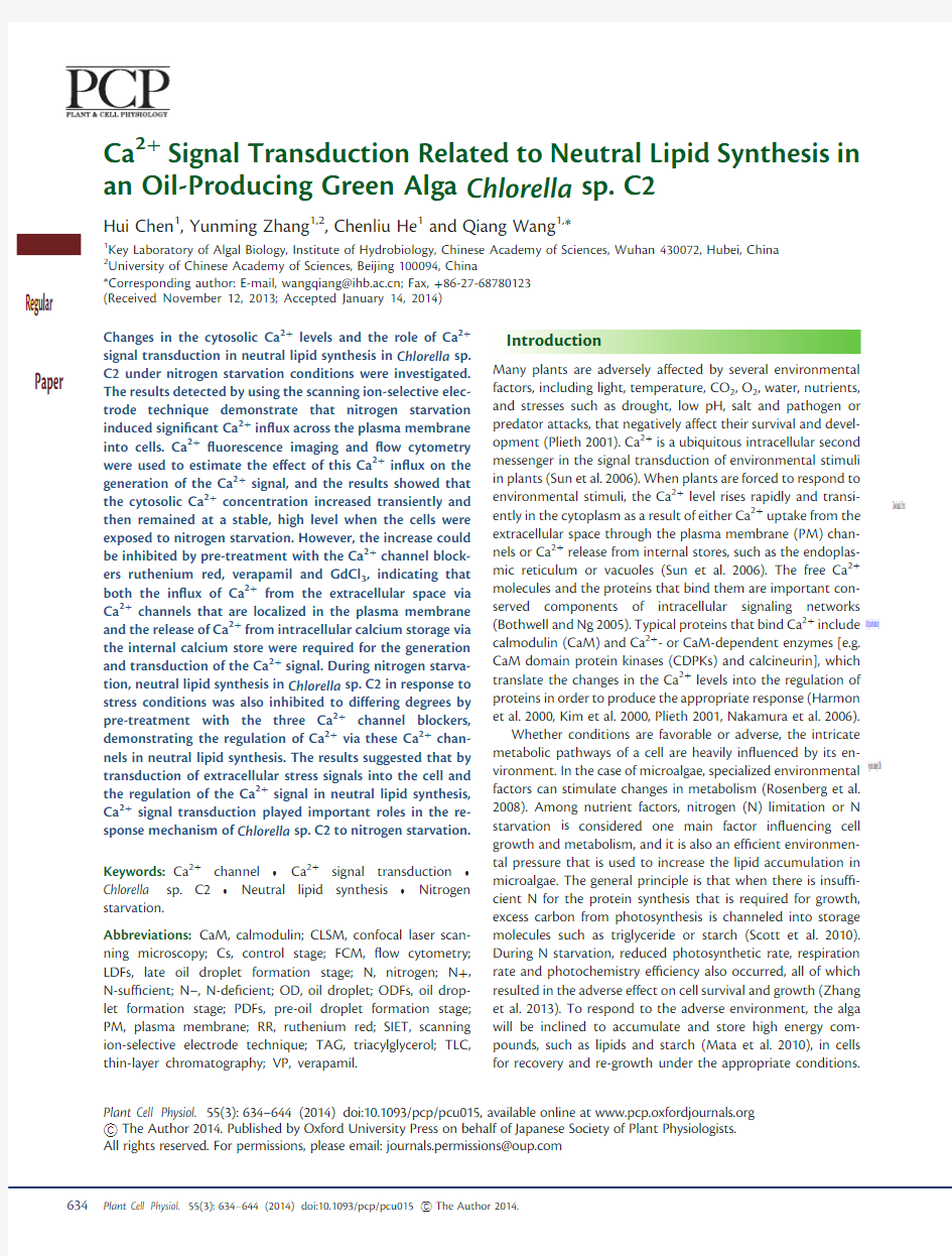
Ca2+?ux in Chlorella sp.C2under N–treatment To determine whether Ca2+mediates the signal transduction under N-de?cent(N–)treatment to regulate lipid biosynthesis for stress adaptation,the N–treatment-induced Ca2+?ux in Chlorella sp.C2was measured.Fig.1shows that the Ca2+ef?ux across the PM could be observed in Chlorella sp.C2cells that were cultured in N-suf?cient(N+)BG11medium(Control). After the N–treatment,the Chlorella sp.C2cells exhibited marked decreases in the Ca2+ef?ux,which even changed to
a signi?cant Ca2+in?ux across the PM8d after the N–treat-
ment(Fig.1).This result indicates that the PM Ca2+channel
activity was enhanced by the N–treatment and that numerous
Ca2+molecules?ooded into cells using these Ca2+channels.
The increased Ca2+in?ux might form a Ca2+signal in Chlorella
sp.C2cells,regulating the cellular response mechanism to N
starvation via Ca2+-mediated signal transduction pathways.
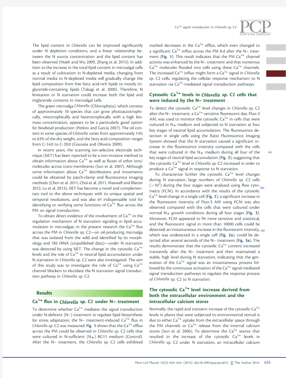
Cytosolic Ca2+levels in Chlorella sp.C2cells that
were induced by the N–treatment
To detect the cytosolic Ca2+level changes in Chlorella sp.C2
after the N–treatment,a Ca2+-sensitive?uorescent dye,Fluo-3
AM,was used to monitor the cytosolic Ca2+in cells that were
cultured in N+medium and subjected to N starvation at four
key stages of neutral lipid accumulation.The?uorescence de-
tection in single cells using the Ratio Fluorescence Imaging
System showed that the N starvation caused a signi?cant in-
crease in the?uorescence intensity compared with the cells
that were cultured in the N+medium during all four of the
key stages of neutral lipid accumulation(Fig.2),suggesting that
the cytosolic Ca2+level in Chlorella sp.C2increased in order to
produce a Ca2+signal in response to N starvation.
To characterize further the cytosolic Ca2+level changes
during N starvation,large numbers of Chlorella sp.C2cells
(>104)during the four stages were analyzed using?ow cyto-
metry(FCM).In accordance with the results of the cytosolic
Ca2+level change in a single cell(Fig.2),a signi?cant increase in
the?uorescent intensity of Fluo-3AM using FCM was also
observed compared with the cells that were cultured under
normal N+growth conditions during all four stages(Fig.3). Moreover,FCM appeared to be more sensitive and statistical,
and the?uorescent signal in more than10000cells could be detected;an instantaneous increase in the?uorescent intensity,
which was undetected in a single cell(Fig.2a),could be de-
tected after several seconds of the N–treatment(Fig.3a).The
results demonstrate that the cytosolic Ca2+content increased transiently after the N–treatment and then maintained a
stable,high level during N starvation,indicating that the gen-
eration of the Ca2+signal was an instantaneous process fol-
lowed by the continuous activation of the Ca2+signal-mediated
signal transduction pathways to regulate the response process
of Chlorella sp.C2to N starvation.
The cytosolic Ca2+level increase derived from
both the extracellular environment and the
intracellular calcium stores
Normally,the rapid and transient increase of the cytosolic Ca2+
levels in plants that were subjected to environmental stimuli is
due to either Ca2+uptake from the extracellular space through
the PM channels or Ca2+release from the internal calcium
stores(Sun et al.2006).To determine the Ca2+source that
resulted in the increase of the cytosolic Ca2+levels in
Chlorella sp.C2under N starvation,an intracellular calcium
Ca2+signal transduction in Chlorella sp.C2
by guest on January 6, 2016
https://www.360docs.net/doc/fc5787783.html,/
Downloaded from
store Ca 2+channel blocker [ruthenium red (RR)]and two PM Ca 2+channel blockers [verapamil (VP)and GdCl 3]were used.Fig.2shows the effect of the Ca 2+channel blockers on the cytosolic Ca 2+levels in single cells under N starvation.After the N–treatment,the cells that were pre-treated with RR,VP or GdCl 3exhibited lower cytosolic Ca 2+levels at all four stages compared with the cells that were not treated with a Ca 2+channel blocker (Fig.2),indicating that the N starvation-induced cytosolic Ca 2+level increase was blocked by RR,VP or GdCl 3and that either the Ca 2+channels in the PM or the internal calcium stores regulated the cytosolic Ca 2+https://www.360docs.net/doc/fc5787783.html,ing FCM,the blockage by RR,VP or GdCl 3of the increase in the cytosolic Ca 2+levels at all four stages in large numbers of cells under N starvation was also observed (Fig.3).These results from both the single cell and the multicellular level demon-strate that the increase in the cytosolic Ca 2+levels under N starvation was derived from both the extracellular environment and the intracellular calcium stores as a result of Ca 2+uptake via the PM Ca 2+channels and the Ca 2+release via the internal calcium stores.In addition,the cytosolic Ca 2+levels in Chlorella sp.C2cells that were pre-treated with RR were always less than those of the cells that were treated with VP or GdCl 3(Figs.2,3),suggesting the more marked role of the internal calcium stores in the regulation of the cytosolic Ca 2+levels for Ca 2+signal transduction.
In Figs.2and 3,the cytosolic Ca 2+levels in Chlorella sp.C2cells that were pre-treated with ionomycin did not increase signi?cantly at any of the four stages compared with the cells that were cultured in the N–medium.A tough cell wall is typical of some microalgae (e.g.Chlorella )(Running et al.1994,Ueno 2009).Therefore,it has been suggested that a small amount of ionomycin could permeate the cell wall and act as a Ca 2+ionophore in shuttling Ca 2+across the PM into the cytoplasm of Chlorella sp.C2.The increase in the cytosolic Ca 2+levels in the cells that were pre-treated with ionomycin might be independent of the Ca 2+ionophore but dependent on the Ca 2+channels,similar to the process in the cells that were cultured in the N–medium.
Ca 2+signal transduction-regulated neutral lipid synthesis during N starvation
In previous studies,four key stages of lipid accumulation,the control stage (Cs;0d),pre-oil droplet formation stage (PDFs;0–0.5d),oil droplet formation stage (ODFs;0.5–2d)and late oil droplet formation stage (LDFs;2–8d),were de?ned in Chlorella sorokiniana C3(Zhang et al.2013)and Chlorella sp.C2(unpub-lished data).To determine the regulation of Ca 2+signal trans-duction during neutral lipid accumulation and oil droplet (OD)formation in Chlorella sp.C2,the neutral lipid levels in the cells that were cultured under various conditions were examined at four stages of N starvation using thin-layer chromatography (TLC).During the ?rst two stages,no cells in any treatment accumulated detectable levels of neutral lipids (Fig.4a,b ).When the treatment was prolonged,the cells that were cul-tured in the N–medium began to accumulate neutral lipids at ODFs (Fig.4c ,lane 1),but the OD formation that was induced by the N starvation was blocked signi?cantly in the cells that were pre-treated with RR,VP or GdCl 3(Fig.4c ,lanes 2–4,Fig.4e ).In accordance with the cytosolic Ca 2+levels,although neutral lipid synthesis was beginning (Fig.4c ,lane 5,Fig.4e ),the cells that were pre-treated with ionomycin did not show a signi?cant increase in the neutral lipid content compared with the cells that were cultured in the N–medium (Fig.4c ,lane 1,Fig.4e ).Rising gradually with the duration of the stress,neutral lipids signi?cantly accumulated in the cells that were cultured in the N–medium at LDFs (Fig.4d ,lane 1),and the relative neutral lipid content reached 1.67times that of the reference substance (Fig.4f ).Similarly,the relative value of the neutral lipids in the cells that were pre-treated with ionomycin was 1.66times that of the reference substance (Fig.4f ).The neutral lipid accumulation in cells that were pre-treated with RR,VP or GdCl 3was also blocked to differing degrees,1.35,1.59or 1.55times that of the reference substance (Fig.4d ,lanes 2–4,Fig.4f ).All of these results indicate that the increase in the cytosolic Ca 2+level via these Ca 2+channels transmitted Ca 2+signals in order to regulate neutral lipid synthesis in Chlorella
sp.
Fig.1The total Ca 2+?ux rates over 5min in Chlorella sp.C2under N starvation.(a)Microphotographic examples of Ca 2+ion ?ux/voltage-clamp measurements.(b)Total ?ux rates of Ca 2+were detected at 0,0.5,2and 8d after N starvation.The columns represent the means of three replicated studies in each sample,with the SD of the means (t -test,P <0.05).The signi?cance of the differences between the control (0d)and other test values was tested using a one-way analysis of variance.*P <0.05vs.control.
H.Chen et al .
by guest on January 6, 2016
https://www.360docs.net/doc/fc5787783.html,/Downloaded from
C2in response to N starvation,and ionomycin had no effect on neutral lipid synthesis.Moreover,the blocking effect of the three Ca 2+blockers on the neutral lipid accumulation at LDFs was obviously less than the effect on the OD formation at ODFs,indicating that the regulation of Ca 2+signal transduc-tion during neutral lipid synthesis occurred during the initial stage of OD formation and that the blockers deferred the OD formation that was induced by N starvation.
To visualize the regulation of OD formation by Ca 2+signal transduction,Chlorella sp.C2cells that were stained with BODIPY 505/515were observed using confocal laser scanning microscopy (CLSM).In agreement with the TLC results (Fig.4),no BODIPY 505/515?uorescence (green)was detected at the Cs (Fig.5,0d)or PDFs (Fig.5,0.5d)in any treatment.At the ODFs (Fig.5,2d),no ?uorescence was detected in the cells that were cultured in the N +medium,but a weak
green
Fig.2Effect of Ca 2+channel blockers under N starvation on the cytosolic Ca 2+levels in a single cell of Chlorella sp.C2.The ?uorescence of Fluo-3AM (green)was detected at 0d (a),0.5d (b),2d (c)and 8d (d)after N starvation;.(e)Fluorescence image of cells in each treatment at 0,0.5,2and 8d.N–,N starvation;RR,ruthenium red;VP,verapamil;Gd,GdCl 3;Ion,ionomycin;N +,N suf?cient.The data points and ?gures represent the means of three replicated studies in each sample.The size of the scale bar is shown directly on the image.
Ca 2+signal transduction in Chlorella sp.C2
by guest on January 6, 2016
https://www.360docs.net/doc/fc5787783.html,/Downloaded from
?uorescence was detected in the cells that were cultured in the N–medium or pre-treated with ionomycin.However,the ?uor-escence intensity was weakened in the cells that were pre-trea-ted with RR,VP or GdCl 3.With prolonged N starvation,a strong green ?uorescence signal was observed at the LDFs in the cells that were cultured in the N–medium and that were pre-trea-ted with ionomycin (Fig.5,8d).The cells that were pre-treated with RR,VP or GdCl 3also exhibited a decrease in the ?uores-cence intensity.However,only a weak green ?uorescence was detected at LDFs in the cells that were cultured in the N +medium (Fig.5,8d).In addition,the Chl auto?uorescence (red)intensities in the cells in every treatment at each stage were nearly identical (Fig.2).
To characterize further the regulation of Ca 2+signal trans-duction during neutral lipid accumulation in response to N starvation,large numbers of Chlorella sp.C2cells (>104)at the four stages were analyzed using FCM.The ?uorescence intensity of BODIPY 505/515in the cell populations during N starvation increased with time,compared with that in the cells that were cultured in the N +medium,indicating a constant increase in the cellular neutral lipid content.The results from the FCM analysis also suggest that the Ca 2+ionophore iono-mycin did not play a role in the regulation of neutral lipid synthesis in Chlorella sp.C2(Fig.6).However,the constantly
increased ?uorescence intensity could be blocked in the cells that were pre-treated with RR,VP or GdCl 3during the OD formation (Fig.6),which corresponded to the ?ndings by CLSM (Fig.5)and TLC (Fig.4).
Total lipid contents in Chlorella sp.C2cells at ODFs and LDFs,the stages of OD preliminary formation and signi?cant accumulation,were also determined in order to evaluate dir-ectly the regulation of Ca 2+signal transduction.Similarly,the OD formation and accumulation in cells during N starvation could be blocked by pre-treatment with RR,VP or GdCl 3,and ionomycin did not have an effect (Fig.7),suggesting the regu-lation by Ca 2+signal transduction of lipid metabolism in Chlorella sp.C2under N starvation.All of the above results suggest that Chlorella sp.C2accumulated and stored lipids for recovery and re-growth under the appropriate conditions in the cells that were cultured under N starvation and that the N starvation response process was regulated by Ca 2+signal transduction pathways.
Discussion
As a key signal factor,speci?c changes in the cytosolic Ca 2+levels occur when plants or microalgae are exposed
to
Fig.3FCM analysis the cytosolic Ca 2+levels in Chlorella sp.C2under N starvation.The ?uorescence of Fluo-3AM was detected at 0d (a),0.5d (b),2d (c)and 8d (d)after N starvation.N–,N starvation;RR,ruthenium red;VP,verapamil;Gd,GdCl 3;Ion,ionomycin;N +,N suf?cient.The data points at each second represent the means of 2?103–3?103cells in three replicated studies with similar ?ndings.
H.Chen et al .
by guest on January 6, 2016
https://www.360docs.net/doc/fc5787783.html,/Downloaded from
various environmental stresses,and the generation and trans-duction of a Ca 2+signal contribute to the transfer of extra-cellular stimuli to cells in order to regulate the response mechanism to environmental stresses (Chinnusamy et al.2004).The mechanisms giving rise to the changes in the cytosolic Ca 2+levels under environmental stresses are only beginning to be identi?ed in microalgae,and recent studies using Ca 2+chelators or Ca 2+channel blockers have impli-cated Ca 2+channels in the regulation of cytosolic Ca 2+.For example,a study by Torrecilla et al.(2001)demonstrated that both salinity and osmotic stress trigger transient in-creases in the intracellular free Ca 2+concentration in the cells of the cyanobacterium Anabaena sp.PCC7120,and both of the Ca 2+transients were completely blocked by the calcium chelator EGTA and were partially inhibited by the calcium channel blocker VP.In the present study,a transient increase in the cytosolic Ca 2+levels that was induced by N starvation and a stable,high level of cytosolic Ca 2+during N starvation were detected.The Ca 2+increase could also be markedly blocked by RR and partially inhibited by VP or GdCl 3(Figs.2,3),indicating that all of the corres-ponding Ca 2+channels,in particular the channels in the intracellular calcium stores,played special roles in the regu-lation of the cytosolic Ca 2+levels under N starvation.
To tolerate or adapt to these environmental stresses,micro-algae possess a variety of special response mechanisms,and some response mechanisms are regulated by the Ca 2+-mediated signal transduction pathways.In the halotolerant green alga Dunaliella bardawil ,the increase in the intracellular glycerol contents under hypertonic shock was sharply decreased by low concentrations of Ca 2+(1and 5mM)but increased by high concentrations of Ca 2+(10mM)(Issa 1996),which demonstrates that Ca 2+could regulate the intra-cellular glycerol content under osmotic stress.In a previous study,N starvation resulted in neutral lipid accumulation,and OD formation and signi?cant neutral lipid accumulation occurred after 2and 8d of starvation,respectively,in Chlorella sorokiniana C3(Zhang et al.2013)and Chlorella sp.C2(unpub-lished data).However,the Ca 2+signal transduction pathway in microalgae under nitrogen starvation conditions has not been studied,and the regulation mechanism of Ca 2+signal transduc-tion in lipid accumulation in microalgae is not yet known.In the present study,intracellular lipid especially neutral lipid synthe-sis during N starvation was blocked to differing degrees
by
Fig.4TLC analysis of the neutral lipid accumulation in Chlorella sp.C2cells under N starvation.The neutral lipid accumulation was detected at 0d (a),0.5d (b),2d (c)and 8d (d)after N starvation.The relative value of the neutral lipid content in each sample compared with the reference substance was calculated at 2d (e)and 8d (f).1/N–,N starvation;2/RR,ruthenium red;3/VP,verapamil;4/Gd,GdCl 3;5/Ion,ionomycin;6/N +,N suf?cient.An asterisk indicates the reference substance glyceryl trioleate.All of the ?gures for TLC are representative of three replicated studies with similar ?ndings.The columns represent the means of three replicated studies in each sample,with the SD of the means (t -test,P <0.01).The signi?cance of the differences between the control (N–)and test values was tested using a one-way analysis of variance.*P <0.05vs.control.
Ca 2+signal transduction in Chlorella sp.C2
by guest on January 6, 2016
https://www.360docs.net/doc/fc5787783.html,/Downloaded from
pre-treatment with RR,VP or GdCl 3(Figs.4–7),indicating that Ca 2+signal transduction that is regulated by the corresponding Ca 2+channels signi?cantly contributed to the regulation of neutral lipid synthesis during N starvation.A similar ?nding has shown that the changes in the glycerol content and G3PDH activity in Dunaliella salina with respect to osmotic stress were partially inhibited by pre-treatment with the Ca 2+channel blockers LaCl 3,VP and RR,indicating that the in?ux of Ca 2+via Ca 2+channels is required for the transduction of the osmotic signal in order to regulate the osmotic responses of D.salina (Chen et al.2011).Therefore,Ca 2+channels are also involved in the Ca 2+signal transduction for the regulation of response mechanisms.
De Beer and Larkum (2001)found that the Ca 2+channel blockers VP and nifedipine had no effect on the Ca 2+dynamics or steady-state pro?les in the calcifying alga Halimeda discoidea .In the present study,the blocking effects of RR,VP and GdCl 3on the cytosolic Ca 2+levels (Figs.2,3)and neutral lipid
synthesis (Figs.4–6)were different,suggesting that the Ca 2+channel blockers acted on different channels or had different sites of action.This result also demonstrates various blocking effects and suggests an in?ux of Ca 2+from the extracellular space via the Ca 2+channels that are localized in the PM or from the intracellular calcium store via the Ca 2+channels that are localized in the calcium stores.In addition,no blocker could completely block the increase in the cytosolic Ca 2+levels (Figs.2,3)and neutral lipid synthesis (Figs.4–6)during N starvation.The tolerance and susceptibility to environmental stress are very complex phenomena,and Ca 2+signal transduc-tion is also a complex mechanism that might be regulated by the synergistic effect of the Ca 2+channels but not by the effect of any single channel.Furthermore,due to the diversity and complexity of microalgae,non-Ca 2+channel-regulated cyto-solic Ca 2+mechanisms might also exist.For example,Karimova et al.(2000)found that (i)the Ca 2+and Na +coun-ter-transporters span the membranes of two Dunaliella species (D.salina and Dunaliella maritima );(ii)the Ca 2+uptake de-pended on the intracellular Na +release;and (iii)the agents blocking the Ca 2+channels did not affect the transport of Ca 2+and Na +.
The Ca 2+ionophores A23187and ionomycin could enhance the Ca 2+in?ux by shuttling Ca 2+across biological membranes at stoichiometries of 2:1(A23187/Ca 2+)and 1:1(ionomycin/Ca 2+)(Liu and Hermann 1978,Hable et al.2001).For example,Hable et al.(2001)used A23187and ionomycin to increase drastically the cytosolic Ca 2+activity in an analysis of the effects of Ca 2+ionophores on the early development of fucoid algae.In another study,compared with the transient increases in the intracellular Ca 2+level that were induced by NaCl and sucrose,the Ca 2+ionophore A23187enhanced the Ca 2+in?ux further in Anabaena sp.PCC7120(Torrecilla et al.2001).However,in the present study,ionomycin did not have an effect as a Ca 2+ionophore to enhance the Ca 2+in?ux,possibly as a result of the impenetrable cell wall of Chlorella ;therefore,the Ca 2+iono-phore may be unsuitable to induce an extracellular Ca 2+in?ux into the cytoplasm in Chlorella sp.C2.
The comparison of the changes in the cytosolic Ca 2+level and intracellular neutral lipid content showed that the cytosolic Ca 2+level in each treatment nearly corresponded to the neutral lipid accumulation (Figs.2–6),which further demonstrates the regulation of Ca 2+signal transduction during neutral lipid syn-thesis in Chlorella sp.C2.However,the blocking effects on the cytosolic Ca 2+levels (VP
similar
Fig.5Representative confocal laser scanning micrographs of Chlorella sp.C2cells labeled in vivo with BODIPY 505/515.CLSM images of cells with BODIPY 505/515?uorescence (green)and Chl auto?uorescence (red)in each treatment were recorded at 0,0.5,2and 8d after N starvation.N–,N starvation;RR,ruthenium red;VP,verapamil;Gd,GdCl 3;Ion,ionomycin;N +,N suf?cient.All of the ?gures are repre-sentative of three replicated studies with similar ?ndings.The size of the scale bar is shown directly on the image.
H.Chen et al .
by guest on January 6, 2016
https://www.360docs.net/doc/fc5787783.html,/Downloaded from
mechanisms might occur in plants.Another study also revealed a copper-induced cross-talk among the signal factors calcium,H 2O 2and NO in Ulva compressa (Gonzalez et al.2012).
Perception of stress cues to excite Ca 2+signals and relay of the signals to switch on adaptive responses are the key steps leading to plant stress tolerance (Chinnusamy et al.2004).When Chlorella sp.C2was exposed to N starvation,the envir-onmental stimuli might be recognized by membrane sensors and then activate the Ca 2+channels in the PM and the mem-branes of intracellular calcium stores through a series of phos-phorylation reactions,all of which resulted in rapid rises in the Ca 2+level in the cytoplasm derive both from the extracellular space and from intracellular calcium stores to excite Ca 2+
signals (Figs.1–3).The Ca 2+signals might be translated to the downstream pathways via interactions with calcium-bind-ing proteins,and a series of biochemical reactions (e.g.neutral lipid synthesis)were induced and regulated to respond to N starvation (Figs.4–7).However,the N starvation stress sensors are not known and most of the signaling intermediates have not been identi?ed,so the studies on Ca 2+signal transduction pathway in microalgae under nitrogen starvation conditions are signi?cant and will be carried out step by step in the future.In summary,N starvation induced a transient increase in the cytosolic Ca 2+levels in Chlorella sp.C2,and the algal cells then maintained a stable,high cytosolic Ca 2+level during N starva-tion.Both the in?ux of Ca 2+from the extracellular space via
the
Fig.6FCM analysis of Chlorella sp.C2cells labeled in vivo with BODIPY 505/515.The ?uorescence of BODIPY 505/515in the cells (>104)was detected at 0d (a),0.5d (b),2d (c)and 8d (d)after N starvation.N–,N starvation;RR,ruthenium red;VP,verapamil;Gd,GdCl 3;Ion,ionomycin;N +,N suf?cient.All of the curves are representative of three replicated studies with similar
?ndings.
Fig.7Effect of Ca 2+channel blockers under N starvation on the total lipid contents in Chlorella sp.C2.The total lipid contents were detected,and the relative value of the lipid content in each sample compared with the control (N–)was calculated at 2d (a)and 8d (b)after N starvation.N–,N starvation;RR,ruthenium red;VP,verapamil;Gd,GdCl 3;Ion,ionomycin;N +,N suf?cient.The relative content of “1.0”represents the actual total lipid contents in cells under N starvation are 4.23%and 29.24%of dry cell weight at 2d (a)and 8d (b).The columns represent the means of three replicated studies in each sample,with the SD of the means (t -test,P <0.01).The signi?cance of the differences between the control (N–)and test values was tested using a one-way ANOVA.*P <0.05vs.control.
Ca 2+signal transduction in Chlorella sp.C2
by guest on January 6, 2016
https://www.360docs.net/doc/fc5787783.html,/Downloaded from
Ca 2+channels that are localized to the PM and the release of Ca 2+from the intracellular calcium stores via the internal cal-cium store Ca 2+channels resulted in the increase in the cyto-solic Ca 2+levels that was required for the generation and transduction of the Ca 2+signal.By regulating lipid,in particular neutral lipid,synthesis,Ca 2+signal transduction pathways played signi?cant roles in the regulation of the stress response mechanism of Chlorella sp.C2under N starvation.In addition,different Ca 2+channels produced different regulatory effects on the stress response,and there might be a synergistic effect of the Ca 2+in?ux via all of the Ca 2+channels or another Ca 2+in?ux mechanism of Ca 2+signal transductions.Another non-Ca 2+-mediated signal pathway may also exist in Chlorella sp.C2in response to N starvation.
Materials and Methods
Growth conditions and N–treatment
The N-suf?cient (N +)medium that was used was full-strength BG11medium (Stanier et al.1971).The N-de?cient (N–)medium was BG11medium without NaNO 3.Chlorella sp.C2in the exponential phase was used to inoculate at an initial OD 700of 0.05(about 1.1?106cells ml –1)into a 1liter Erlenmeyer ?ask containing 500ml of BG11medium at 20 C with continuous illumination of 70m mol m –2s –1,continuously bubbled with ?ltered air.The cells were harvested by centrifu-gation at 6,000?g for 3min at 20 C at the mid-logarithmic growth phase (OD 700approximately 0.8,about 1.1?107cells ml –1)and were then washed and resuspended in N–medium to an OD 700of approximately 0.7(about 9.3?106cells ml –1).
Ca 2+?ux measurements
The net ?ux of Ca 2+was measured non-invasively using the SIET (the SIET system,BIO-001A,Younger USA Sci.&Tech.Corp.,Applicable Electronics Inc.and Science Wares Inc.).The record-ings of the Ca 2+?ux were performed as described by Sun et al.(2010)with some modi?cations.For the Ca 2+?ux recordings,the control or N–Chlorella sp.C2cells were settled on the center of a poly-L -lysine-pre-treated cover slip and then placed in 4ml of measuring solution (0.18mM K 2HPO 4á3H 2O,0.3mM MgSO 4á7H 2O,0.24mM CaCl 2and 0.19mM NaCO 3,pH 7.1).The cell populations (approximately 10cells)of Chlorella sp.C2on the cover slip were selected for the Ca 2+?ux measure-ment,which was continuously recorded for 5min.Three-dimen-sional ionic ?ux signals were plotted with MageFlux software that was developed by Yue Xu (https://www.360docs.net/doc/fc5787783.html,/mage?ux).
Pre-treatment with Ca 2+channel blockers and a Ca 2+ionophore
Before the N–treatment,three Ca 2+channel blockers and a Ca 2+ionophore were added to some of the Chlorella sp.C2cells and pre-incubated for 1h.These channel blockers included a stretch-activated Ca 2+channel blocker GdCl 3,a voltage-
dependent Ca 2+channel blocker VP,and a putative mitochon-drial and endoplasmic reticulum Ca 2+channel blocker RR,whose ?nal concentrations were 0.2mM,20m M and 20m M,respectively.The ?nal concentration of the Ca 2+ionophore ionomycin was 2m M.
Fluorescence imaging of cytosolic Ca 2+
For the ?uorescence imaging of the cytosolic Ca 2+,the Chlorella sp.C2cells were loaded with a Ca 2+-sensitive ?uorescent dye,Fluo-3AM,according to Chen et al.(2011).The ?uorescence images of the cytosolic Ca 2+from the Chlorella sp.C2cells that were loaded with Fluo-3AM were obtained using the Ratio Fluorescence Imaging System (EasyRatioPro),and the change in the ?uorescence of the cytosolic Ca 2+in a single cell was also recorded.The excitation and emission wavelengths were 488and 525nm,respectively.The ?uorescence of the cytosolic Ca 2+was continuously recorded for 5min.
Thin-layer chromatography lipid analysis
A 10ml culture at OD 700=1(about 1.3?107cells ml –1)was harvested at 6,000?g for 3min,and the cell pellet was washed with fresh medium and centrifuged again.The har-vested cell pellet was resuspended in 400m l of a metha-nol :chloroform mixture (1:1,v/v).The mixture was shaken for 2min,followed by phase separation using 120m l of 1M potassium chloride in 0.2M phosphoric acid.The mixture was then centrifuged at 12,000?g at room tempera-ture for 5min,and the chloroform phase was transferred to a glass tube and dried under nitrogen.The residue was re-suspended in a volume of 20m l of chloroform to obtain the lipid extracts.A TLC analysis of the lipid extracts from whole cells was performed according to Reiser and Somerville (1997)with some modi?cations.The triacylglycerols (TAGs)were separated by developing the plates in hexane :ethyl ether (7.5:2.5,v/v).The samples were visualized by exposure to iodine vapor for approximately 10min.Aliquots of 3m l for each sample were extracted at different time points and used for the TLC analysis.Glyceryl trioleate (3m l,10mg ml –1)was used as a reference substance for the TAGs,and the neutral lipid content of Chlorella sp.C2was then determined using ImageJ (v1.41,NIH)(Tsihlis et al.2010)and calculated as a value relative to the reference substance.
Confocal laser scanning microscopy analysis
A microscopic analysis of the cells was performed using a confocal scanner (Zeiss LSM 710NLO).The transmission micrographs that were used for the visualization of the non-?uorescent protoplast structures were generated using the manufacturer’s ?lter settings.A lipophilic ?uorescent dye,BODIPY 505/515(4,4-di?uoro-1,3,5,7-tetramethyl-4-bora-3a,4a-diaza-sindacene;Invitrogen Molecular Probes),was used to stain the intracellular oil-containing organelles,known as lipid bodies,with a ?nal labeling concentration of 1m M to-gether with 0.1%dimethylsulfoxide (DMSO;v/v)according to
H.Chen et al .
by guest on January 6, 2016
https://www.360docs.net/doc/fc5787783.html,/Downloaded from
Cooper et al.(2010).The BODIPY?uorescence(green)was excited with an argon laser(488nm)and detected at 505–515nm.The auto?uorescence(red)of the algal chloro-plasts was detected simultaneously at650–700nm.
Flow cytometry analysis
The samples that were stained with Fluo-3AM or BODIPY 505/515were analyzed on a board using a FACSAria?ow cyt-ometer(Becton Dickinson)that was equipped with a laser emitting at488nm and an optical?lter FL1(530/30nm). The collected data were analyzed using FlowJo software(Tree Star).
Total lipid analysis
Cells at OD700=1(about1.3?107cells ml–1)were harvested by centrifugation at6,000?g for3min,and the cell pellet was washed with fresh medium and centrifuged again.Then the cell pellet was dried using a freeze dryer.Total lipid of micro-algae was extracted with methanol:chloroform(1:1,v/v)from 1g of lyophilized material and quanti?ed gravimetrically(Bligh and Dyer1959).
Statistical analyses
Each result that is shown is the mean of at least three biological replicates.A statistical analysis of the data was performed using the program SPSS-13,and the signi?cance was determined at the95%or99%con?dence limits.
Funding
This work was supported by the National Program on Key Basic Research Project[2011CB200902];the National Natural Science Foundation of China[31300030]. Acknowledgments
We thank Professor Xudong Xu for providing the Chlorella strain C2.
Disclosures
The authors have no con?icts of interest to declare.
References
Bligh,E.G.and Dyer,W.J.(1959)A rapid method of total lipid extrac-tion and puri?cation.Can.J.Biochem.Phys.37:911–917. Bothwell,J.H.F.and Ng,C.K.Y.(2005)The evolution of Ca2+signalling in photosynthetic eukaryotes.New Phytol.166:21–38.
Chen,H.,Chen,S.L.and Jiang,J.G.(2011)Effect of Ca2+channel block on glycerol metabolism in Dunaliella salina under hypoosmotic and hyperosmotic stresses.PLoS One6e28613.Chinnusamy,V.,Schumaker,K.and Zhu,J.K.(2004)Molecular genetic perspectives on cross-talk and speci?city in abiotic stress signalling
in plants.J.Exp.Bot.55:225–236.
Choi,K.H.,Song,C.,Cheong,C.S.and Rhim,H.(2011)Pharmacological
studies of Ca(v)3.1T-type calcium channels using automated
patch-clamp techniques.Gen.Physiol.Biophys.30:100–105.
Cooper,M.S.,Hardin,W.R.,Petersen,T.W.and Cattolico,R.A.(2010)
Visualizing‘green oil’in live algal cells.J.Biosci.Bioeng.109:198–201.
De Beer,D.and Larkum,A.W.D.(2001)Photosynthesis and calci?ca-
tion in the calcifying algae Halimeda discoidea studied with micro-
sensors.Plant Cell Environ.24:1209–1217.
de Montaigu,A.,Sanz-Luque,E.,Galvan,A.and Fernandez,E.(2010)A
soluble guanylate cyclase mediates negative signaling by ammo-
nium on expression of nitrate reductase in Chlamydomonas.
Plant Cell22:1532–1548.
Gonzalez, A.,Cabrera,M.D.,Henriquez,M.J.,Contreras,R.A.,
Morales,B.and Moenne,A.(2012)Cross talk among calcium,
hydrogen peroxide,and nitric oxide and activation of gene expres-
sion involving calmodulins and calcium-dependent protein kinases
in Ulva compressa exposed to copper excess.Plant Physiol.158:
1451–1462.
Gouveia,L.and Oliveira,A.C.(2009)Microalgae as a raw material for
biofuels production.J.Ind.Microbiol.Biotechnol.36:269–274.
Hable,W.E.,EerNisse,P.,Hoggan,M.and Kropf,D.L.(2001)Effect of
calcium ionophores on early development in fucoid algae.Phycol.
Res.49:145–154.
Harmon,A.C.,Gribskov,M.and Harper,J.F.(2000)CDPKs—a kinase
for every Ca2+signal?.Trends Plant Sci5:154–159.
Hsieh,C.H.and Wu,W.T.(2009)Cultivation of microalgae for oil
production with a cultivation strategy of urea limitation. Bioresource Technol.100:3921–3926.
Issa,A.A.(1996)The role of calcium in the stress response of the halotolerant green alga Dunaliella bardawil Ben-Amotz et Avron.
Phyton(Horn,Austria)36:295–302.
Karimova, F.G.,Kortchouganova, E.E.,Tarchevsky,I.A.and Iagoucheva,M.R.(2000)The oppositely directed Ca2+and Na+ transmembrane transport in algal cells.Protoplasma213:93–98.
Kim,K.N.,Cheong,Y.H.,Gupta,R.and Luan,S.(2000)Interaction
speci?city of Arabidopsis calcineurin B-like calcium sensors and
their target kinases.Plant Physiol.124:1844–1853.
Liu,C.M.and Hermann,T.E.(1978)Characterization of ionomycin as a
calcium ionophore.J.Biol.Chem.253:5892–5894.
Lu,L.L.,Tian,S.K.,Liao,H.B.,Zhang,J.,Yang,X.E.,Labavitch,J.M.et al.
(2013)Analysis of metal element distributions in rice(Oryza sativa
L.)seeds and relocation during germination based on X-ray?uor-
escence imaging of Zn,Fe,K,Ca,and Mn.PLoS One8:e57360.
Mata,T.M.,Martins,A.A.and Caetano,N.S.(2010)Microalgae for
biodiesel production and other applications:a review.Renew.
Sust.Energ.Rev.14:217–232.
Maxwell,J.T.and Blatter,L.A.(2012)Facilitation of cytosolic calcium
wave propagation by local calcium uptake into the sarcoplasmic
reticulum in cardiac myocytes.J.Physiol.590:6037–6045.
Nakamura,S.,Hayashi,H.and Chino,M.(2006)Detection of calmodu-
lin and calmodulin-binding proteins in pure phloem sap of rice
plants.Soil Sci.Plant Nutr.52:195–202.
Petkov,G.and Garcia,G.(2007)Which are fatty acids of the green alga Chlorella?Biochem.Syst.Ecol.35:281–285.
Plieth, C.(2001)Plant calcium signaling and monitoring:pros
and cons and recent experimental approaches.Protoplasma218:
1–23.
Ca2+signal transduction in Chlorella sp.C2
by guest on January 6, 2016
https://www.360docs.net/doc/fc5787783.html,/
Downloaded from
Reiser,S.and Somerville, C.(1997)Isolation of mutants of Acinetobacter calcoaceticus de?cient in wax ester synthesis and complementation of one mutation with a gene encoding a fatty acyl coenzyme A reductase.J.Bacteriol.179:2969–2975.
Rosenberg,J.N.,Oyler,G.A.,Wilkinson,L.and Betenbaugh,M.J.(2008)A green light for engineered algae:redirecting metabolism to fuel a biotechnology revolution.Curr.Opin.Biotechnol.19:430–436.
Running,J.A.,Huss,R.J.and Olson,P.T.(1994)Heterotrophic produc-tion of ascorbic-acid by microalgae.J.Appl.Phycol.6:99–104.Scott,S.A.,Davey,M.P.,Dennis,J.S.,Horst,I.,Howe,C.J.,Lea-Smith,D.J.et al.(2010)Biodiesel from algae:challenges and prospects.Curr.Opin.Biotechnol.21:277–286.
Stanier,R.Y.,Kunisawa,R.,Mandel,M.and Cohen-Bazire,G.(1971)Puri?cation and properties of unicellular blue-green algae (order Chroococcales ).Bacteriol.Rev.35:171–205.
Sun,J.,Wang,M.J.,Ding,M.Q.,Deng,S.R.,Liu,M.Q.,Lu,C.F.et al.(2010)H 2O 2and cytosolic Ca 2+signals triggered by the PM H plus-coupled transport system mediate K +/Na +homeostasis in NaCl-stressed Populus euphratica cells.Plant Cell Environ.33:943–958.
Sun,Q.P.,Guo,Y.,Sun,Y.,Sun,D.Y.and Wang,X.J.(2006)In?ux of extracellular Ca 2+involved in jasmonic-acid-induced elevation of
[Ca 2+](cyt)and JR1expression in Arabidopsis thaliana .J.Plant Res.119:343–350.
Sun,T.,Xu,Y.,Li,P.,Yu,S.G.and Yin,L.P.(2007)Non-invasive scanning ion-selective electrode technique and its applications to the re-search of higher plants.Prog.Nat.Sci.17:625–629.
Takagi,M.,Watanabe,K.,Yamaberi,K.and Yoshida,T.(2000)Limited feeding of potassium nitrate for intracellular lipid and triglyceride accumulation of Nannochloris sp UTEX LB1999.Appl.Microbiol.Biot.54:112–117.
Torrecilla,I.,Leganes,F.,Bonilla,I.and Fernandez-Pinas,E.(2001)Calcium transients in response to salinity and osmotic stress in the nitrogen-?xing cyanobacterium Anabaena sp PCC7120,ex-pressing cytosolic apoaequorin.Plant Cell Environ.24:641–648.Tsihlis,N.D.,Murar,J.,Kapadia,M.R.,Ahanchi,S.S.,Oustwani,C.S.,Saavedra,J.E.et al.(2010)Isopropylamine NONOate (IPA/NO)moderates neointimal hyperplasia following vascular injury.J.Vasc.Surg.51:1248–1259.
Ueno,R.(2009)Visualization of sporopollenin-containing pathogenic green micro-alga Prototheca wickerhamii by ?uorescent in situ hy-bridization (FISH).Can.J.Microbiol.55:465–472.
Zhang,Y.M.,Chen,H.,He,C.L.and Wang,Q.(2013)Nitrogen starva-tion induced oxidative stress in an oil-producing green alga Chlorella sorokiniana C3.PLoS One 8:e69225.
H.Chen et al .
by guest on January 6, 2016
https://www.360docs.net/doc/fc5787783.html,/Downloaded from
Thinkcell操作指引
PPT之Think-cell操作指引 一、Think-cell简介 Think-cell 是一款Office插件,可以帮助PowerPoint 快速便捷地创建图表。能够快速访问所有类型的图表,对所访问图表的字体、颜色、线条、数字和类型等格式,直接进行修改。 二、安装 1.安装方式 自行下载,注意兼容性。如Office 2010通常需要Think-cell5.2及以上版本,且需下载破解版。下面均以Office 2010,Think-cell 5.2为例进行演示。 2.安装完成后,打开任意PPT文件,插入一栏会出现Think-cell选项,这表示Think-cell 安装成功,此时可以进行调用。由于Think-cell没有汉化,所以内置图标均为英文。
三、常用图表介绍 1.簇状图(Clustered):主要用于同期对比。由一系列高度不等的纵向条纹或线段表示数 据分布的情况。一般用横轴表示数据类型,纵轴表示分布情况。
2.百分比柱状图(100%):与堆叠柱状图类似,在柱体内各部分显示其占整体的百分比; 3.瀑布图(Waterfall):用来解释从一个数值到另一个数值的变化过程具有的特殊效果。常 用于分析成本、利润等相关数据。 4.折线图(Line):主要显示随时间而变化的连续数据,因此非常适用于显示在相等时间 间隔下数据的变化趋势。
5.组合图(Combination):既包含柱状图相关信息,又包含折线图功能,可以复合表示 多项指标。非常常用,建议重点关注练习。 6.饼状图(Pie):用整个圆表示总体的数量或整体值“1”,用圆内各个扇形的大小表示 各部分数量或该部分占总体的百分比。
CSS表单设计
CSS表单设计 文章出处:https://www.360docs.net/doc/fc5787783.html, 今天我们开始学习《十天学会web标准(div+css)》的css表单设计,包含以下内容和知识点: 改变文本框和文本域样式 用图片美化按钮 改变下拉列表样式 用label标签提升用户体验 一、改变文本框和文本域样式 如果前边几章学习的比较扎实的话,本节教程就相当容易了。下边先说一下文本框,文本框和文本域都是可以用css进行美化的。比如改变边框精细,颜色,添加背景色、背景图像等。请看下边的实例: .text1 { border:1px solid #f60; color:#03C;} .text2 { border:2px solid #390; width:200px; height:24px; font-size:16px; font-weight:bold; line-height:1.6;} .text3 { border:2px solid #C3C; height:20px; background:url(icon9.gif) right 3px no-repeat;} .text4 { border:2px solid #F60; width:150px; height:29px;font-size:16px; line-height:1.6; background:url(bg_9.gif) 0 0 no-repeat;} 这四个样式表分别对应第2、3、4、5行表单,第一行是文本框的默认样式;第二行为设置边框和字体颜色的样式;第三行为设置边框、宽度、高度、字体大小、行高的样式;第四行设置边框和增加背景色和背景图片;第五行为增加一个gif动画的背景图片,看起来是不是生动许多,具体步骤不再赘述。下面我们看一下文本域的样式设置: .area { border:1px solid #F90; overflow:auto; background:#fff url(bg_9_1.gif) right bottom no-repeat; width:99%; height:100px;} 上图中第一个为默认的文本域样式,第二个为设置边框、宽度为百分比、高度和景图片。overflow:auto定义当内容不超过现在文本域高度时不出现滚动条。好了,下面运行一下代码看看两者的效果吧
Cell Line 方 式
浅析 Cell Line 方式运作与管理 随着社会的发展,市场需求呈多样性变化,为适应市场需求的不断变化,生产方式大致经历了如下演变: 手工生产方式大量流水线生产方式 JIT 生产方式; 制造业也经历了类似的演变,可细分为以下几个阶段: 手工作坊制造批量流水线生产模块化单元生产 JIT、精益方式敏捷制造。 1913 年福特创立了大批量流水线技术 ,加速了工业化进程. 但在上世纪中叶开始,市场需求呈现多样化,多变化,单元式生产应运而生,上世纪 70 年代,日本丰田公司平台单元式生产代表了这种生产方式的应用水准。 为进一步加强对市场的快速反应,降低生产成本,日本的 JIT 方式产生,丰田公司研究的“ U "字型单元流水线采用拉动式管理代表了这一时期制造方式的先进水平。 1990 年美国根据 JIT 方式,创造性的提出精益生产方式,世界上许多大公司根据自身情况,结合 JIT,精益思想,推出了许多单元式生产,如平台作业、模块生产、单元作业、 Cell Line 等。而我们公司正是借鉴了索尼的 Cell Line 单元式生产技术,这种兼备手工生产及大量生产二者的优点,又能克服二者缺点的高柔性、低成本、灵活、敏捷的单元生产正成为“整机制造"的最佳选择。 我们公司在推行索尼公司的单元流水线取得了极大的成功,但把 Cell Line 技术作为一种生产方式来研究还不成熟,因为它毕竟只是 单元作业、 JIT方式与精益生产思想指导下的一种体现形式,还谈不 上一种生产方式。目前还没有见到这方面的专着和论文,而业内人士称其为“ Cell Line 方式",在这里我也引用“ Cell Line 方式"以区别于其它 Cell 作业方式 ( 如 Cell 平台作业,模块化生产等 )。 Cell Line 方 式是众多技术、生产方式、思想的揉合,要全面了解、掌握这种 方式的运作与管理,必须掌握其核心技术与思想:单元生产方式、
fancyBox中文文档(教程) (1)
fancyBox 中文文档 fancyBox 为您的网页提供惊艳的缩放功能,同时支持图片、网页内容以及各种多媒体文件。 她基于目前最流行的jQuery 框架开发,不仅易于应用,而且可以定制。欢迎来到fancyBox 的世界~ ?更新日志 ?快速入门 ?常见用法 ?官方演示 ?使用许可&下载地址 ?完整API ?常见问题 更新日志 重大变更 ?可扩展的幻灯片导航工具 ?响应式(调整浏览器窗口大小时自适应窗口大小) ?集成幻灯片图片播放功能
?新增幻灯片过渡效果 ?引入CSS3阴影、圆角特效 ?新的插件选项(与之前版本不兼容) ?改用知识共享署名-非商业性使用3.0许可协议授权 即将上线 ?WordPress小插件 ?重构本站及API文档 ?完善缩略图浏览工具 ?fancyBox 手机版(待定) ?fancyBox 3.0 ↑回顶部 快速入门 1. 下载fancyBox,解压后根据需要将文件复制到网页文件夹中(建议不要更 改目录结构),并在网页源码中引入相应的css 样式和js 文件(如果更改了目录结构,引入的时候请调整相应代码,对应它们所在的路径)。 注意:别忘了还要先加载jQuery 库!推荐使用公共库:百度 | 新浪。示例代码: < !-- 引入 mousewheel 插件(可选,如果想通过鼠标滚轮控制图片播放则必须) --> 2. 给触发fancyBox 特效的元素(如一张缩略图或“点击我”三个字)加上a 链接,并将其href 属性值设置为遮罩层中需要展示的文件的链接(如刚刚那张缩略图的原图的网址)。示例代码: 
 3. 使页面加载完毕时初始化fancybox() 函数。如果你不懂jQuery但英语还行,请看官方推荐的新手教程。英语不好肿么办?请点右上角的红叉...因为暂时找不到中文版的帮助文档。示例代码:
让iframe自适应大小 解决方案
让iframe自适应大小解决方案 第一种: 例子: 1,创建页面test.html 。页面中含有一个iframe,name为ifrname ,id为ifrid,src 为iframe.html 页面。 2,创建iframe.html 页面,里面含有一些内容。 这是iframe页面,通过在父窗口页面或子页面添加JS来自动更改宽高,以适应内容的多少。 要想使iframe自动适应宽和高,可以在test.html 页面iframe onload处增加一些JS代码。如: 这样iframe即可以自动适应高度了。如果不在onload处增加js,那也可以放到页面上来调用。比如写个函数。 这样也可以达到自适应高宽的目的,在这里要注意的是,iframeElement参数必须是 document.getElementById("iframeid"),iframeWindow参数是window.frames[0] 或 document.frames["ifrname"]。这是因为通过name得到的对象是窗口(window)对象,非窗口里的iframe 对象。同时document.getElementById("iframeid)不能像window对象可以得到window.document。 所以这里最好给iframe分别加上name和id,id用来更改宽高样式属性,name用来执行window相关事件如location.href,document.write。bgColor例外,元素对象和窗口对象都可以得到,这是因为他们都有这个属性。
html特效代码大全精美免费下载
html特效代码大全精美免费下载.txt每天早上起床都要看一遍“福布斯”富翁排行榜,如果上面没有我的名字,我就去上班。谈钱不伤感情,谈感情最他妈伤钱。我诅咒你一辈子买方便面没有调料包。 HTML代码大全免费下载 HTML特效代码1。忽视右键
或 2。加入背景音乐 IE: