Interpretation of magnetic data in the Sinop area of Mid Black Sea
作文猜猜他是谁唱的英语

In the realm of music,there are songs that resonate with listeners on a profound level,evoking emotions and memories that are uniquely personal.One such song that has captured the hearts of many is Guess Whos Back,a track that has been covered by various artists over the years. This piece delves into the enigmatic nature of the songs original singer, exploring the various renditions and the impact they have had on the music industry and the hearts of listeners.The song Guess Whos Back has a history that is as intriguing as the melody itself.Its a tale of mystery and talent,where the identity of the original artist remains shrouded in a veil of speculation.The song has been performed by a multitude of artists,each bringing their own flavor and interpretation to the lyrics and melody.The question of who originally sang it is a topic of heated debate among music enthusiasts and scholars alike.The songs lyrics are a narrative of a return,a comeback story that speaks to the resilience and determination of an individual who has faced adversity and is now ready to reclaim their place in the spotlight.The powerful message of the song has resonated with audiences,making it a timeless piece that continues to be relevant and impactful.One of the most notable versions of Guess Whos Back comes from a legendary artist who has a reputation for delivering powerful performances with a magnetic stage presence.This artists rendition of the song is characterized by a deep,soulful voice that carries the weight of the lyrics,making listeners feel every word.The passion and emotion conveyedthrough this version have made it a favorite among fans,and it has become a benchmark for other artists attempting to cover the song.Another interpretation that stands out is by a contemporary artist known for their unique style and innovative approach to music.This rendition takes the original song and infuses it with a modern twist,blending elements of various genres to create a fresh and exciting sound.The artists distinct voice and innovative arrangement have garnered critical acclaim and have introduced the song to a new generation of listeners.The impact of Guess Whos Back extends beyond the music charts and into the cultural zeitgeist.The song has been featured in films,television shows, and commercials,further cementing its status as a cultural icon.Its universal themes of resilience and triumph have made it a goto anthem for those seeking inspiration and motivation.Moreover,the song has sparked numerous discussions and debates among music aficionados.The quest to uncover the original singer has led to the discovery of lesserknown artists and has shed light on the rich tapestry of the music industry.This pursuit has also highlighted the importance of recognizing and celebrating the contributions of all artists, regardless of their level of fame or notoriety.In conclusion,the song Guess Whos Back is more than just a catchy tune it is a testament to the power of music to inspire,connect,and endure.The various renditions by different artists have showcased the songs versatility and its ability to be reinterpreted and reinvented while still retaining itscore message.The ongoing quest to identify the original singer adds an element of intrigue and excitement to the songs legacy,making it a fascinating topic for music lovers and casual listeners alike.As the song continues to be covered and celebrated,its legacy will undoubtedly grow, and its impact will be felt for generations to come.。
The Trial That Rocked the World高级英语第三版第一册第四课翻译和词汇

Lesson 4 The Trial That Rocked the World震撼世界的审判A buzz ran through the crowd as I took my place in the packed court on that sweltering July day in 1925. The counsel for my defence was the famous criminal lawyer Clarence Darrow. Leading counsel for the prosecution was William Jennings Bryan, the silver-tongued orator , three times Democratic nominee for President of the United States, and leader of the fundamentalist movement that had brought about my trial.在一九二五年七月的那个酷热日子里,当我在挤得水泄不通的法庭里就位时,人群中响起一阵嘁嘁喳喳的议论声。
我的辩护人是著名刑事辩护律师克拉伦斯.达罗。
担任主控官的则是能说会道的演说家威廉.詹宁斯.布莱恩,他曾三次被民主党提名为美国总统候选人,而且还是导致我这次受审的基督教原教旨主义运动的领导人。
A few weeks before I had been an unknown school-teacher in Dayton, a little town in the mountains of Tennessee. Now I was involved in a trial reported the world over. Seated in court, ready to testify on my behalf, were a dozen distinguished professors and scientists, led by Professor Kirtley Mather of Harvard University. More than 100 reporters were on hand, and even radio announcer s, who for the first time in history were to broadcast a jury trial. "Don't worry, son, we'll show them a few tricks," Darrow had whispered throwing a reassuring arm round my shoulder as we were waiting for the court to open.几个星期之前,我还只是田纳西州山区小镇戴顿的一名默默无闻的中学教员,而现在我却成了一次举世瞩目的庭审活动的当事人。
基于Oasismontaj平台的磁法数据处理解释研究
科技广场2015.10 引言Geosoft软件主要包括Oasis Montaj平台及有关模块。
Oasis Montaj平台对大量的地球物理、地球化学和地质数据进行综合、视图和比较。
它能够对数据进行快速分析,以帮助用户及时解决问题并作出决定。
montaj 扩展模块有地球物理和地球化学分析、三维钻孔成图、重力和磁力滤波、水平校准、解释和其他功能。
另外,由Geosoft公司的技术合作伙伴开发的montaj+扩展模块,采用Oasis Montaj平台在montaj环境下提供专业的重力和磁力数据处理和正演。
本次数据处理采用克里格网格化,并对其化磁极,垂向一阶导数得出成果图再将其3D成图,结合RGIS经同样的方法得出的成果图进行比较。
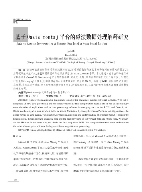
基于Oasis montaj平台的磁法数据处理解释研究Study on Accurate Interpretation of Magnetic Data Based on Oasis Montaj Platform汤乐峰Tang Lefeng(江西省煤田地质勘察研究院,江西南昌330001)(Jiangxi Research Institute of Coalfield Geological Survey,Jiangxi Nanchang 330001)摘要:高精度磁法勘探是常用的地球物理方法,随着新的数据处理的出现和资料解释技术的提高,其应用领域越来越广泛,并且数据处理软件也层出不穷,如RGIS、Geosoft等等。
本文通过对玉华山部分地区磁法数据利用Geosoft的Oasis montaj平台的数据存取、可视化、处理、成图及图形输出进行了解开发。
对其进行克里格(kringing)网格化、化磁极和垂向一阶导数成果图,并生成3D图。
再经过RGIS,用同样的方法得出成果图,并将其进行对比,突显出哪种软件效果更明显,对其解释更好,从而判断哪种软件在高精度磁法数据处理更有利。
关键词:Oasis montaj;化磁极;垂向一阶导数;3D中图分类号:P631 文献标识码:A 文章编号:1671-4792(2015)01-0076-05Abstract:High-precision magnetic exploration is one of the commonly used geophysical methods. With the e-mergence of new data processing and the improvement in data interpretation techniques, it has an increasingly more domains of application, and its data processing software is emerging, such as the RGIS, and Geosoft, etc. Based on the magnetic data of some areas in Yuhua Mountain, by using the Geosoft's Oasis montaj platform, the paper carries on data access, visualization, processing, mapping and understanding of graphics output. Through the kringing grid, the reduction to magnetic pole and the first derivative of the vertical obtained results map, we gener-ate the 3D map. In the same way, we obtain the final map from RGIS. We compare these two maps to determine the more advantageous software for high-precision magnetic data processing.Keywords:Oasis Montaj;Reduce to Magnetic Pole;First Derivative of the Vertical;3D76基于Oasis montaj 平台的磁法数据处理解释研究1 数据处理及3D 成图1.1克里格网格化对玉华山部分地区实测数据进行正常场校正和日变校正,得到该区域的每个测点的磁异常值,然后将这些点的异常值分别用Oasis motaj 和RGIS 软件进行克里格(Kringing )网格化处理后,分别得到该区域的磁异常等值线图,如图一和图二所示。
文献翻译(二次电流层)

激光等离子体相互作用中磁重联引起的等离子体与二次电流层生成的研究摘要:以尼尔逊[物理学家、列托人,97,255001,(2006)]为代表的科学家首次对等离子体相互作用引起的磁重联进行了研究,该研究在固体等离子体层上进行,在两个激光脉冲中间设置一定间隔,在两个激光斑点之间可以发现一条细长的电流层(CS),为了更加贴切的模拟磁重联过程,我们应该设置两个并列的目标薄层。
实验过程中发现,细长的电流层的一端出现一个折叠的电子流出区域,该区域中含有三条平行的电子喷射线,电子射线末端能量分布符合幂律法则。
电子主导磁重联区域强烈的感应电场增强了电子加速,当感应电场处于快速移动的等离子体状态时还会进一步加速,另外弹射过程会引起一个二级电流层。
正文:等离子体的磁重联与爆炸过程磁能量进入等离子体动能和热能能量的相互转换有关。
发生磁重联的薄层区域加速并释放等离子体[1-5]。
实验中磁重联速度与太阳能的观察结果大于Sweet-Parker与相关模型[4-6]的标准值,这是由霍尔电流和湍流[7-12]引起的。
二级磁岛以及该区域释放的等离子体可以提高磁重联速度,当伦德奎斯特数S﹥104[13]时二级磁导很不稳定。
这些理论预测值与近地磁尾离子扩散区域中心附近的二级磁岛观察值相符[14],激光束与物质的相互作用的过程中,正压机制激发兆高斯磁场(▽ne×▽Te)生成[15-16]。
以尼尔逊[17]为代表的科学家首次运用两个类似的的激光产生的等离子体模拟磁重联过程。
尼尔逊[17]与Li[18]等人实验测量数据为磁重联的存在提供了决定性的证据,他们运用了随时间推移的质子偏转技术来研究磁拓扑变化,除此之外尼尔逊[17]等人观察到高度平行双向等离子喷射线与预期的磁重联平面成40°夹角。
本次研究调查了自发磁场的无碰撞重联,激光等离子体相互作用产生等离子体,为了防止磁场与等离子体连接在一起实验过程使用了两个共面有一定间隔的等离子体。
两种新的长方体重力异常正演公式及其理论推导

+(
z) arctan ( z)R | 2 | 2 | 2 ( x)( y) 1 1 1
g(x, y, z) = G ||| ( x) ln{( y) + R} + ( y) ln{( x) + R}
( z) arctan ( x)( y) | 2 | 2 | 2 ( z)R 1 1 1
g(x, y, z) = G ||| ( x) ln{( y) + R} + ( y) ln{( x) + R}
两种新的长方体重力异常正演公式及其理论推导
骆遥 1, 2
1 中国科学院地质与地球物理研究所,北京(100029) 2 中国科学院研究生院,北京(100049) E-mail:geo@
摘 要: 在前人推导长方体重力场、磁场正演理论表达式工作的基础上,重新对长方体重 力场正演理论表达式进行理论推导,提出了两种全新的长方体重力异常正演公式形式,并给 出了全部的理论推导过程,对比模型正演计算结果表明,新导出长方体重力场正演理论表达 式的正确。 关键词:长方体,重力场,正演,积分 中图分类号:P631
线数据单位为 g.u.
Fig2. The cubic model gravity contour map
5. 结论
综合前人对长方体重力场正演理论表达式的推导过程,并借鉴长方体磁场及其梯度场理 论表达式的推导,推导出了两种新的长方体重力场正演理论表达式(11)式和(13)式,对 比模型正演计算结果表明,新导出长方体重力场正演理论表达式是完全正确的。
似积分的推导[15~17],对 2 的推导有:
2= 2( 1
z)2 ( {(
y) {( x) +
R}2
国外主要无损检测标准含中英文名称对照

ASTM A 754/A 754M-1996X射线荧光法测量涂层厚度的试验方法Test Method for Coating Thickness by X-Ray FluorescenceASTM B567-1998用β射线背散射法测量涂层厚度的试验方法Test Method for Measurement of Coating Thickness by the Beta Backscatter MethodASTM B568-1998χ射线光谱仪法测量涂层厚度的试验方法Test Method for Measurement of Coating Thickness by X-Ray SpectrometryASTM C637-1998辐射屏蔽混凝土用集料的标准规范Standard Specification for Aggregates for Radiation-Shielding ConcreteASTM C638-1992辐射屏蔽混凝土集料组分的描述术语Descriptive Nomenclature of Constituents of Aggregates for Radiation-Shielding ConcreteASTM C1455-2000用γ射线谱法无损检定仍然有效特殊核材料指南ASTM D2599-1987X射线光谱法测定汽油含铅量的试验方法(05.02)Test Method for Lead in Gasoline by X-Ray Spectrometry (05.02)ASTM D4294-1998用非色散X射线荧光光谱法测定石油产品中含硫量试验方法Sulfur in Petroleum Products by Non-Dispersive X-Ray Fluorescence Spectrometry, Method of Test for (05.02) ASTM D4452-1985土壤样品的X射线照相法X-Ray Radiography of Soil SamplesASTM D5059-1998X-射线光谱法测定汽油含铅量的试验方法Test Method for Lead in Gasoline by X-Ray Spectroscopy (05.03)ASTM D5187-1991X射线衍射法测定煅烧石油焦炭中结晶尺寸(LC)的试验方法Test Method for Crystallite Size (LC) of Calcined Petroleum Coke by X-Ray Diffraction (05.03)ASTM D6247-1998X射线荧光光谱法分析聚烯烃中元素含量的试验方法Test Method for Analysis of Elemental Content in Polyolefins by X-Ray Fluorescence SpectrometryASTM E94-2004(2010)射线照相检验标准指南Standard Guide for Radiographic ExaminationASTM E142-1996射线照相检测的质量控制方法Method for Controlling Quality of Radiographic Testing ASTM E155-2010铝镁合金铸件射线照相检验标准参考照片Standard Reference Radiographs for Inspection of Aluminum and Magnesium CastingsASTM E170-1999有关辐射测量和剂量测定的术语ASTM E181-1998放射性核素探测器的校准和分析的一般方法General Methods for Detector Calibration and Analysis of RadionuclidesASTM E186-2010厚壁(50.8-114mm)钢铸件标准参考射线照片Standard Reference Radiographs for Heavy-Walled (2 to 4 1/2-In./50.8 to 114-mm) Steel CastingsASTM E192-2004(2010)e1宇航用熔模钢铸件标准参考射线照片Standard Reference Radiographs for Investment Steel Castings of AerospaceApplicationsASTM E242-2001(2010)某些参数改变时射线照相图象显示的标准参考射线照片Standard Reference Radiographs for Appearances of Radiographic Images asCertain Parameters Are ChangedASTM E272-2010高强度铜基及镍铜合金铸件的标准参考射线照片Standard Reference Radiographs for High-Strength Copper-Base andNickel-Copper Alloy CastingsASTM E280-2010厚壁(114-305mm)铸钢件标准参考射线照片Standard Reference Radiographs for Heavy-Walled (4 1/2 to 12-in. (114 to305-mm)) Steel CastingsASTM E310-2010锡青铜铸件标准参考射线照片Standard Reference Radiographs for Tin Bronze CastingsASTM E390-2011钢熔焊焊缝标准参考射线照片Standard Reference Radiographs for Steel Fusion WeldsASTM E431-96(2011)半导体和相关器件射线照片判读指南Standard Guide to Interpretation of Radiographs of Semiconductors andRelated DevicesASTM E446-2010厚度至50.8mm钢铸件的标准参考射线照片Standard Reference Radiographs for Steel Castings up to 2 in. (50.8 mm) inThicknessASTM E505-2001(2011)铝和镁压铸件检验的标准参考射线照片Standard Reference Radiographs for Inspection of Aluminum and MagnesiumDie CastingsASTM E545-2005(2010)确定直接热中子射线照相检验成象质量的标准试验方法Standard Test Method for Determining Image Quality in Direct ThermalNeutron Radiographic ExaminationASTM E586-88γ与χ射线照相检测的术语定义ASTM E592-1999(2009)e16~51mm厚钢板X射线照相检验和25~152mm厚钢板钴60照相检验获得ASTM当量穿透灵敏度的标准指南Standard Guide to Obtainable ASTM Equivalent Penetrameter Sensitivity forRadiography of Steel Plates 1/4 to 2 in. (6 to 51 mm) Thick with X Raysand 1 to 6 in. (25 to 152 mm) Thick with Cobalt-60ASTM E665-1994测量暴露在X闪光射线机的X射线照射下的材料中相对深度的吸收剂量Determining Absorbed Dose Versus Depth in Materials Exposed to the X-RayOutput of Flash X-Ray MachinesASTM E666-1997γ或X射线剂量吸收的计算Calculating Absorbed Dose from Gamma or XRadiationASTM E689-2010球墨铸铁标准参考射线照片Standard Reference Radiographs for Ductile Iron CastingsASTM E746-2007测定工业射线照相成像系统相关图象质量响应的标准方法Standard Practice for Determining Relative Image Quality Response ofIndustrial Radiographic Imaging SystemsASTM E747-2004(2010)射线照相用线型象质计(IQI)的设计、制造及材料组分类的标准方法Standard Practice for Design, Manufacture, and Material GroupingClassification of Wire Image Quality Indicators (Iqi) Used for RadiologyASTM E748-2002(2008)材料热中子射线照相标准方法Standard Practices for Thermal Neutron Radiography of MaterialsASTM E801-2006(2011)电子装置射线照相检验的质量控制标准方法Standard Practice for Controlling Quality of Radiological Examination ofElectronic DevicesASTM E802-1995(2010)厚度至114mm的灰口铸铁标准参考射线照片Standard Reference Radiographs for Gray Iron Castings up to 4 1/2 in. (114mm) in ThicknessASTM E803-1991(2008)确定中子射线透照束长径比的标准方法Standard Method for Determining the L/D Ratio of Neutron Radiography BeamsASTM E915-1996残余应力测量用X射线衍射仪校准检定的试验方法Test Method for Verifying the Alignment of X-Ray DiffractionInstrumentation for Residual Stress MeasurementASTM E999-2010工业射线照相胶片处理的质量控制标准指南Standard Guide for Controlling the Quality of Industrial Radiographic FilmProcessingASTM E1000-98(2009)射线照相检测标准指南Standard Guide for RadioscopyASTM E1025-2011射线照相检测用孔型象质计设计、制造和材料组分类的标准方法Standard Practice for Design, Manufacture, and Material GroupingClassification of Hole-Type Image Quality Indicators(IQI) Used forRadiographyASTM E1030-2005(2011)金属铸件射线照相检验的标准试验方法Standard Test Method for Radiographic Examination of Metallic CastingsASTM E1032-2012焊缝射线照相检验的标准试验方法Standard Test Method for Radiographic Examination of WeldmentsASTM E1079-2010透射密度计校准的标准方法Standard Practice for Calibration of Transmission DensitometersASTM E1114-2009e1测定铱192工业射线照相源尺寸的标准试验方法Standard Test Method for Determining the Size of Iridium -192 IndustrialRadiopraphic SourcesASTM E1161-2009半导体和电子元件射线检验的标准方法Standard Practice for Radiologic Examination of Semiconductors andElectronic ComponentsASTM E1165-2004(2010)用针孔成象法测量工业X射线管焦点的标准试验方法Standard Test Method for Measurement of Focal Spots of Industrial X-RayTubes by Pinhole ImagingASTM E1168-1995核设施工人辐射防护训练Radiological Protection Training for Nuclear Facility WorkersASTM E1254-2008射线照片及未曝光工业射线照相胶片储藏的标准指南Standard Guide for Storage of Radiographs and Unexposed IndustrialRadiographic FilmsASTM E1255-2009射线透视检验标准方法Standard Practice for RadioscopyASTM E1320-2010钛铸件标准参考射线照片Standard Reference Radiographs for Titanium CastingsASTM E1390-2012工业射线照相观片灯标准规范Standard Specification for Illuminators Used for Viewing IndustrialRadiographsASTM E1400-1995高剂量辐射量测定校准实验室的特性和性能规程Characterization and Performance of a High-Dose Radiation DosimetryCalibration Laboratory, Practice for (12.02)ASTM E1411-2009射线照相系统鉴定的标准方法Standard Practice for Qualification of Radioscopic SystemsASTM E1416-2009焊缝射线检验的标准试验方法Standard Test Method for Radioscopic Examination of WeldmentsASTM E1441-2011计算机层析(CT)成像的标准指南Standard Guide for Computed Tomography (CT) ImagingASTM E1441-2000计算机层析成像(CT)指南Guide for Computed Tomography (CT) ImagingASTM E1453-2009含模拟或数字射线照相数据的磁带媒体存储标准指南Standard Guide for Storage of Magnetic Tape Media that Contains Analog orDigital Radioscopic DataASTM E1475-2002(2008)数字射线照相检验数据计算机化传输的数据区标准指南Standard Guide for Data Fields for Computerized Transfer of DigitalRadiological Examination DataASTM E1496-2005(2010)中子射线照相尺寸测量的标准试验方法Standard Test Method for Neutron Radiographic DimensionalMeasurements(With drawn 2012)ASTM E1570-2011计算机层析(CT)检验标准方法Standard Practice for Computed Tomographic (CT) ExaminationASTM E1647-2009确定射线照相检测对比度灵敏度的标准方法Standard Practice for Determining Contrast Sensitivity in RadiologyASTM E1648-1995(2011)铝熔焊焊缝检验标准参考射线照片Standard Reference Radiographs for Examination of Aluminum Fusion WeldsASTM E1672-2006选择计算机层析(CT)系统的标准指南Standard Guide for Computed Tomography (Ct) System SelectionASTM E1695-1995(2006)e1计算机层析(CT)系统性能测量的标准试验方法Standard Test Method for Measurement of Computed Tomography (Ct) SystemPerformanceASTM E1734-2009铸件射线照相检验标准方法Standard Practice for Radioscopic Examination of CastingsASTM E1735-2007确定经4-25MV X射线曝光的工业射线胶片相关成像质量的标准试验方法Standard Test Method for Determining Relative Image Quality of IndustrialRadiographic Film Exposed to X-Radiation from 4 to 25 MVASTM E1742/E1742M-2011射线照相检验标准方法Standard Practice for Radiographic ExaminationASTM E1814-1996(2007)铸件计算机层析(CT)检验标准方法Standard Practice for Computed Tomographic (CT) Examination of CastingsASTM E1815-2008工业射线照相胶片系统分类的标准试验方法Standard Test Method for Classification of Film Systems for IndustrialRadiographyASTM E1817-2008使用典型象质计(RQIs)控制射线检验质量的标准方法Standard Practice for Controlling Quality of Radiological Examination byUsing Representative Quality Indicators(RQI-s)ASTM E1894-1997选择脉冲X射线源用的剂量测定系统的标准指南Standard Guide for Selecting Dosimetry Systems for Application in PulsedX-Ray SourcesASTM E1931-2009X射线康普顿散射层析技术标准指南Standard Guide for X-ray Compton Scatter TomographyASTM E1935-1997(2008)校准和测量计算机层析(CT) 密度的标准试验方法Standard Test Method for Calibrating and Meausring CT DensityASTM E1936-2003(2011)评估射线照相数字化系统性能的标准参考射线照片Standard Reference Radiograph for Evaluating the Performance ofRadiographic Digitization SystemsASTM E1955-2004(2009)与美国材料与试验协会ASTM E 390 参考射线照片等级比较钢中焊缝完善性的标准射线检验Standard Radiographic Examination for Soundness of Welds in Steel byComparison to Graded ASTM E390 Reference RadiographsASTM E2002-1998(2009)测定射线照相图象总不清晰度的标准方法Standard Practice for Determining Total Image Unsharpness in RadiologyASTM E2003-2010中子射线照相波束纯度指示计制作的标准方法Standard Practice for Fabrication of the Neutron Radiographic Beam PurityIndicators [Metric]ASTM E2007-2010计算机射线照相标准指南(用于CR的标准指南)(可激射线发光[PSL]法)Standard Guide for Computed Radiology (Photostimulable Luminescence (PSL)Method)ASTM E2023-2010制作中子射线照相灵敏度指示计的标准方法Standard Practice for Fabrication of Neutron Radiographic SensitivityIndicatorASTM E2033-1999(2006)计算机射线照相的标准方法(用于CR的标准实施方法)(可激射线发光[PSL]法) Standard Practice for Computed Radiology (Photostimulable LuminescenceMethod)ASTM E2104-2009优质航空与涡轮材料和构件射线照相检验的标准方法Standard Practice for Radiographic Examination of Advanced Aero andTurbine Materials and ComponentsASTM E2120-2000便携式X射线荧光光谱仪测量涂膜中铅含量的性能评估规程Practice for the Performance Evaluation of the Portable X-Ray FluorescenceSpectrometer for the Measurement of Lead in Paint FilmsASTM E2339-2004无损评价中的数字成像和通讯Digital Imaging and Communication in NDE(DICONDE)ASTM E2422-2011铝铸件标准参考数字射线图像(钛和钢铸件也适用)Standard Digital Reference Images for Al. Casting(Titanium & steel Castingalso available)ASTM E2445-2005(2010)计算机射线照相系统的长期稳定性与鉴定的标准方法(用于CR系统的质量认定和长期稳定性的标准实施方法)Standard Practice for Qualification and Long-Term Stability of ComputedRadiology SystemsASTM E2446-2005(2010)计算机射线照相系统分类的标准方法(用于CR系统分类的标准实施方法)Standard Practice for Classification of Computed Radiology SystemsASTM E2597-2007e1数字探测器阵列制造特性的标准规程Standard Practice for Manufacturing Characterization of Digital DetectorArraysASTM E2660-2011航空用优质钢铸件标准参考数字射线图像Standard Digital Reference Images for Investment Steel Castings forAerospace ApplicationsASTM E2662-2009航空用平面与夹芯复合材料射线照相检验的标准方法Standard Practice for Radiologic Examination of Flat Panel Composites andSandwich Core Materials Used in Aerospace ApplicationsASTM E2669-2011数字射线照相(DR)检测方法的数字图像与通信无损评价(DICONDE)的标准方法Standard Practice for Digital Imaging and Communication in NondestructiveEvaluation (DICONDE) for Digital Radiographic (DR) Test MethodsASTM E2698-2010使用数字探测器阵列的射线照相检验标准方法Standard Practice for Radiological Examination Using Digital DetectorArraysASTM E2736-2010数字探测器阵列射线照相检测标准指南Standard Guide for Digital Detector Array RadiologyASTM E2737-2010评价数字探测器阵列性能和长期稳定性的标准方法Standard Practice for Digital Detector Array Performance Evaluation andLong-Term StabilityASTM E2738-2011使用计算机射线照相(CR)检测方法的数字图像与通讯无损评价(DICONDE)的标准方法Standard Practice for Digital Imaging and Communication NondestructiveEvaluation (DICONDE) for Computed Radiography (CR) Test MethodsASTM E2767-2011使用X射线计算机层析(CT)检测方法的数字图像与通讯无损评价(DICONDE)的标准方法Standard Practice for Digital Imaging and Communication in NondestructiveEvaluation (DICONDE) for X-ray Computed Tomography (CT) Test MethodsASTM E2861-2011测量中子辐射束发散与校准的标准试验方法Standard Test Method for Measurement of Beam Divergence and Alignment inNeutron Radiologic BeamsASTM F629-1997铸造金属外科手术植入物射线照相检查实施方法(F-4)ASTM F727-1981透明照相干版透光度测量的试验方法Test Method for Measuring Transmittance of See-Through PhotoplateASTM F784-1982校准放射性同位素密封测试仪的试验方法Test Method for Calibrating Radioisotope Hermetic Test ApparatusASTM F864-1984硬表面玻璃照相干板的检验Inspection of Hard-Surface Glass PhotoplatesASTM F947-1985测定照相胶片低能级X射线辐射灵敏度的试验方法Test Method for Determining Low-Level X-Radiation Sensitivity ofPhotographic FilmsASTM F1035-1991使用橡胶帘布圆盘验证轮胎X射线成象系统的辩别能力Use of Rubber-Cord Pie Disk to Demonstrate the Discernment Capability of aTire X-Ray Imaging SystemASTM F1039-1987X射线安全屏系统中测量低剂量X辐射的试验方法Test Method for Measurement of Low Level X-Radiation Used in X-RaySecurity Screening SystemsASTM F1467-1999微电子装置电离辐射效应中X射线测试仪(近似等于10keV辐射量子)的使用Use of an X-Ray Tester (is Approximately Equal to 10 keV Photons) inIonizing Radiation Effects Testing of Microelectronic DevicesASTM PS95-1998便携式X射线荧光(XRF)装置现场测定涂料或其它涂层含铅量的质量体系的标准临时操作规程Standard Provisional Practice for Quality Systems for Conducting In SituMeasurements of Lead Content in Paint or Other Coatings usingField-Portable X-Ray Fluorescence (XRF) DevicesASTM PS 116-1999测量涂膜含铅量用的便携式X射线荧光光谱仪性能评价的临时操作规程Provisional Practice for the Performance Evaluation of the Portable X-RayFluorescence Spectrometer for the Measurement of Lead in Paint FilmsFactorsANSI/IEEE 309-1970盖革-弥勒计数器的试验程序Geiger-Muller Counters, Test Procedure forANSI IT9.2-1991成象介质-已处理的照相胶片、平板和相纸-归档盒及储存箱Imaging Media - Photographic Processed Films, Plates and Papers - FilingEnclosures and Storage ContainersANSI IT9.8-1989成象介质-照相胶片耐折强度的测定Imaging Media - Photographic Film - Determination of Folding EnduranceANSI N13.2-1969辐射监测的管理规程指南Administrative Practices in RadiationMonitoring, Guide toANSI N13.5-1972直读和非直读式袖珍X和γ射线辐射剂量仪的性能Direct Reading and Indirect Reading Pocket Dosimeters for X- and GammaRadiation, PerformanceANSI N13.7-1983辐射防护照相胶片剂量仪性能标准Radiation Protection - Photographic Film Dosimeters - Criteria forPerformanceANSI N13.11-2001个人剂量测定的试验标准Personnel Dosimetry Performance, Criteria forTestingANSI N13.27-1981袖珍式报警辐射剂量仪和报警记数率计的性能要求Performance Requirements for Pocket-Sized Alarm Dosimeters and AlarmRatemetersANSI N15.36-1994核材料无损化验测量的控制和保证Nuclear materials - Nondestructive assay measurement control and assuranceANSI N15.37-1981核材料控制的自动无损化验系统指南Automation of Nondestructive Assay Systems for Nuclear Materials Control,Guide toANSI N42.16-1986用于液体闪烁计数器的密封放射检查源的规范Specifications for sealed radioactive check sources used inliquid-scintillation countersANSI N42.20-1995个人辐射监视仪的性能标准Performance criteria for active personnelradiation monitorsANSI N42.26-1995辐射防护仪器监测设备X和γ辐射个人报警装置Radiation Protection Instrumentation - Monitoring Equipment - PersonalWarning Devices for X and Gamma RadiationsANSI N43.3-1993通用辐射安全非医疗应用的X射线和密封γ射线源的安装能量达10MevGeneral radiation safety - Installations using non-medical X-ray andsealed gamma-ray sources, energies up to 10 MeVANSI N43.6-1997密封放射性源的分类Classification of Sealed Radioactive SourcesANSI N43.9-1991γ射线照相仪器的设计和试验规范Gamma Radiography - Specifications for Design and Testing of ApparatusANSI N322-1996直接和间接读取石英纤维袖珍剂量计的检验和试验规范Inspection and Test Specifications for Direct and Indirect Reading QuartzFiber Pocket DosimetersASME Boiler & Pressure VesselCode(ASME锅炉压力容器规范)第V卷《无损检测》2004版,第2篇“射线检测”,强制性附录-包含动态射线照相、实时射线成像检测内容ASME SE-1647确定射线照相对比灵敏度的推荐实施方法ASME Code Case 2476使用荧光成像板的射线照相Radiography using phosphor imaging platesMIL-HDBK-55-66射线照相无损检测手册(已由MIL-HDBK-728/5取代)MIL-STD-139A-65射线检测铝镁合金铸件的完好性要求MIL-STD-453C-88射线照相检测MIL-STD-746A-63铸造爆破器材的射线照相检测要求MIL-STD-779-68钢焊缝参考χ射线照片(由ASTM E390取代)MIL-STD-1257A-87钴铬合金枪管射线照相及目视检验MIL-R-11470A-71对射线检验设备,操作方法和操作人员的合格审查(由MIL-STD-453取代) MIL-I-36013B-72折迭式χ射线观片灯MIL-R-45226-62石墨的射线照相检测(已停用)MIL-R-45774A(92)铝,镁导弹零件熔焊完好性要求-射线照相检测MIL-STD-1948(91)中子射线照相检验的术语和定义汇编MIL-HDBK-728/5(92)射线照相检验MIL-HDBK-733(92)复合材料无损检验方法-射线照相法MIL-STD-1166A(91)固体火箭推进剂射线照相检验要求MIL-STD-1264B(93)钢焊缝完好性射线照相检验-与ASTM E390 各级参考底片比较MIL-STD-1265A(92)钢铸件射线照相检验分类和完好性要求MIL-STD-1894A(86)不完全焊透钢焊缝的射线照相参考标准及射线照相程序MIL-STD-1895A(86)不完全焊透铝焊缝的射线照相参考标准及射线照相程序BAC 5915(美国波音公司) 射线检验DPS 4.736(美国麦道公司) 射线检验API 1104(美国石油协会) 管道及有关设备的焊接AWS B 5.15射线照相评片资格技术条件。
医学影像学专业英语
医学影像学专业英语Title: Advancements in Medical Imaging TechnologyMedical imaging is a pivotal field within the broader domain of healthcare, providing critical insights into the diagnosis, treatment, and management of diseases. Over the years, this field has witnessed remarkable advancements, transforming the way medical professionals approach patient care. This essay delves into the evolution of medical imaging technology, highlighting its significance in modern medicine and discussing future prospects.The journey of medical imaging began with simple X-rays in the late 19th century, which allowed for the visualization of bones and other dense structures within the body. This groundbreaking discovery laid the foundation for more sophisticated imaging techniques that followed. The development of computed tomography (CT) scans in the 1970s marked a significant milestone, enabling detailed cross-sectional images of the body's internal structures. CT scans revolutionized the diagnosis and monitoring of various conditions, from traumatic injuries to cancerous growths.Magnetic Resonance Imaging (MRI) further advanced the capabilities of medical imaging by utilizing powerful magnetsand radio waves to produce high-resolution images of soft tissues. Unlike X-rays and CT scans, MRI does not involve ionizing radiation, making it a safer option for repeated use and particularly beneficial for imaging the brain, spinal cord, and joints. The introduction of functional MRI (fMRI) expanded the scope of MRI by allowing doctors to observe brain activity and understand how different regions of the brain communicate with each other.Ultrasound imaging, another non-invasive technique, employs high-frequency sound waves to create real-time images of organs and tissues. Its portability, safety, andcost-effectiveness make ultrasound an indispensable tool in prenatal care, cardiovascular assessments, and guiding invasive procedures.Positron Emission Tomography (PET) scans represent yet another leap forward in medical imaging. By measuring metabolic processes, glucose metabolism, and blood flow within the body, PET scans are invaluable for detecting cancerous tissues, assessing treatment efficacy, and studying brain disorders such as Alzheimer's disease.The integration of artificial intelligence (AI) into medical imaging represents the latest frontier in this field. AIalgorithms can analyze vast amounts of imaging data rapidly and with high accuracy, assisting radiologists in identifying abnormalities that might be overlooked by the human eye. Moreover, AI-powered tools are being developed to predict disease progression and personalize treatment plans based on individual patient data.Despite these advancements, challenges remain. Ensuring equitable access to advanced imaging technologies across different populations and geographies is a significant concern. Additionally, the interpretation of complex imaging data requires highly trained professionals, underscoring the need for continuous education and training in medical imaging.Looking ahead, the future of medical imaging holds immense promise. Innovations such as molecular imaging, which targets specific biological processes at the cellular level, and photoacoustic imaging, which combines laser excitation with ultrasound detection, are poised to further enhance our understanding of disease mechanisms. As these technologies continue to evolve, they will undoubtedly play a crucial role in advancing personalized medicine and improving patient outcomes.In conclusion, the field of medical imaging has come along way since its inception, driven by continuous innovation and technological advancements. These developments have not only transformed diagnostic capabilities but also paved the way for new therapeutic approaches. As we look to the future, the integration of emerging technologies like AI promises to usher in a new era of precision medicine, ultimately leading to better health outcomes for patients worldwide.。
高英the trail that rocked the world 翻译
A buzz ran through the crowd as I took my place in the packed court on that sweltering July day in 1925. The counsel for my defense was the famous criminal lawyer Clarence Darrow. Leading counsel for the prosecution was William Jennings Bryan, the silver-tongued orator, three times Democratic nominee for President of the United States, and leader of the fundamentalist movement that had brought about my trial.在一九二五年七月的那个酷热日子里,当我在挤得水泄不通的法庭里就位时,人群中响起一阵嘁嘁喳喳的议论声。
我的辩护人是著名刑事辩护律师克拉伦斯达罗。
担任主控官的则是能说会道的演说家威廉詹宁斯布莱恩,他曾三次被民主党提名为美国总统候选人,而且还是导致我这次受审的基督教原教旨主义运动的领导人。
A few weeks before I had been an unknown school-teacher in Dayton, a little town in the mountains of Tennessee. Now I was involved in a trial reported the world over. Seated in court, ready to testify on my behalf, were a dozen distinguished professors and scientists, led by Professor Kirtley Mather of Harvard University. More than 100 reporters were on hand, and even radio announcer s, who for the first time in history were to broadcast a jury trial. "Don't worry, son, we'll show them a few tricks," Darrow had whispered throwing a reassuring arm round my shoulder as we were waiting for the court to open.几个星期之前,我还只是田纳西州山区小镇戴顿的一名默默无闻的中学教员,而现在我却成了一次举世瞩目的庭审活动的当事人。
核磁共振 英语词汇
核磁共振英语词汇英文回答:Nuclear magnetic resonance (NMR) is a powerfulanalytical tool that utilizes magnetic fields and radio waves to investigate the properties of atoms and molecules. It offers a non-destructive and versatile technique for characterizing materials at the atomic and molecular level. NMR has various applications across multiple scientific disciplines, including chemistry, physics, biology, and medicine.The basic principle of NMR involves the interaction between atomic nuclei with a magnetic field. Certain nuclei, such as 1H (proton), 13C, 15N, and 31P, possess anintrinsic magnetic moment due to their nuclear spin. When placed in a magnetic field, these nuclei align with or against the field, resulting in two distinct energy states. By applying radio waves to the sample at specific frequencies, it is possible to induce transitions betweenthese energy states.The absorption of radio waves by the nuclei leads to the resonance phenomenon, which forms the basis of NMR. The resonant frequency for a particular nucleus depends on its chemical environment, including the electron density and surrounding atoms. By analyzing the resonance frequencies and patterns, NMR provides detailed information about the structure, dynamics, and interactions of molecules.NMR spectroscopy is a widely used technique for identifying and quantifying different atoms and functional groups within molecules. It plays a crucial role in determining the molecular structure of organic and inorganic compounds, as well as studying chemical reactions and reaction mechanisms. NMR also finds applications in drug discovery and development, protein structure determination, and metabolomics.In medical imaging, NMR is employed as a non-invasive tool for obtaining detailed anatomical and functional information about the human body. Magnetic resonanceimaging (MRI) utilizes NMR techniques to create high-resolution images of organs, tissues, and blood vessels. MRI is particularly valuable for diagnosing and monitoring a wide range of medical conditions, including brain disorders, cardiovascular diseases, and musculoskeletal injuries.NMR also has applications in other fields, such as materials science, polymer characterization, and geological studies. It is a versatile technique that provides valuable insights into the structure, dynamics, and properties of various materials and systems.In summary, nuclear magnetic resonance (NMR) is a powerful analytical tool that offers a non-destructive and versatile approach for investigating the properties of atoms and molecules. Its applications span multiple scientific disciplines, including chemistry, physics, biology, and medicine, providing insights into molecular structure, dynamics, and interactions.中文回答:核磁共振(NMR)是一种强大的分析工具,利用磁场和射频波来研究原子和分子的性质。
GJI-2007-fanke-Identification of magnetic Fe–Ti oxides in marine sediments by
Geophys.J.Int.(2007)170,545–555doi:10.1111/j.1365-246X.2007.03410.xG J I G e o m a g n e t i s m ,r o c k m a g n e t i s m a n d p a l a e o m a g n e t i s mIdentification of magnetic Fe–Ti oxides in marine sediments by electron backscatter diffraction in scanning electron microscopyC.Franke,1,2G.M.Pennock,3M.R.Drury,3R.Engelmann,4ttard,4J.F .L.Garming,1,5T.von Dobeneck 1and M.J.Dekkers 21Departmentof Geosciences,University of Bremen,PO Box 330440,D-28334Bremen,Germany.E-mail:cfranke@uni-bremen.de 2PaleomagneticLaboratory ‘Fort Hoofddijk’,Utrecht University,Budapestlaan 17,3584CD Utrecht,The Netherlands3Department of Earth Sciences,Utrecht University,PO Box 80021,3508TA Utrecht,The Netherlands 4Mineralogisches Institut,Ruprecht-Karls Universit¨a t,Im Neuenheimer Feld 236,69120Heidelberg,Germany5TNO Built Environment and Geosciences,Geological Survey of the Netherlands,PO BOX 80015,TA Utrecht,The NetherlandsAccepted 2007February 15.Received 2007February 8;in original form 2006November 3S U M M A R YIn paleomagnetic and environmental magnetic studies the magnetomineralogical identification is usually based on a set of rock magnetic parameters,complemented by crystallographic and chemical information retrieved from X-ray diffraction (XRD),(electron)microscopy or energy dispersive spectroscopy (EDS)of selected samples.While very useful,each of these supple-mentary techniques has its limitations when applied to natural sample material which are related to low particle concentrations (down to the ppm range in marine sediments)and very fine grain sizes (down to the nm scale).Therefore,meaningful application of such techniques depends on sample quality.Electron backscatter diffraction (EBSD)of individual grains in scanning elec-tron microscopy (SEM)enables mineralogical identification of grains down to ∼0.2micrometer and is particularly powerful when combined with EDS.In this study,we show the merits of EBSD for rock magnetic investigations by analyzing titanomagnetites and hemoilmenites of various compositions and submicron lamella of titanomagnetite–hemoilmenite intergrowths.Such particles often occur in natural marine sediments where EDS often has a semi-quantitative character and compositionally similar intergrowths may be difficult to distinguish.With the mineralogical information provided by EBSD unambiguous identification of spinel-type and trigonal oxides is obtained.Optimal EBSD patterns are gathered from smooth,polished sur-faces,but here we show that interpretable EBSD patterns can be obtained directly from the surface of unconsolidated,so called ‘non-embedded’particles from marine sediments.This information enhances the interpretative value of rock magnetic parameters.Key words:electron backscatter diffraction (EBSD),energy dispersive spectroscopy (EDS),hemoilmenite,rock magnetism,scanning electron microscopy (SEM),titanomagnetite.1I N T R O D U C T I O NIron–titanium oxide minerals are fundamental to paleo-,rock and environmental magnetic purposes because they constitute the most common magnetic particles on Earth.Two Fe–Ti oxide solid solution series are relevant for magnetic studies,the titanomagnetite (TM)and the titanohematite–hemoilmenite series.Titanomaghemites,with their large compositional field,also play an important role(Fig.1;cf.also O’Reilly 1984;Waychunas 1991;Dunlop &¨Ozdemir1997).TM is the cubic spinel series between the magnetite (Mt,Fe 3O 4)and ulv¨o spinel (Usp,Fe 2TiO 4)end-members.The general TM formula is Fe 3−x Ti x O 4,where x is the mole fraction of the ulv¨o spinel component.Hereafter,TM compositions will be given as ‘TM x %’,for example TM60for x =0.6.Phases of the trigonalα-oxide series between hematite (Hmt,Fe 2O 3)and ilmenite (Ilm,FeTiO 3)are called titanohematite or hemoilmenite,depending on their compositions,and will be here simply abbreviated as Hilm.Their general formula is Fe 2−y Ti y O 3,where y is the mole fraction of the ilmenite endmember.Titanomaghemites (Tmh),which are generally formed by oxidation of TM at low temperatures,have a cubic crystal structure related to that of the spinels,but are character-ized by variable concentrations of cationic vacancies ( )related to the charge-balanced substitution 3Fe 2+=2Fe 3++ .Consequently,their compositional field extends over a large range of Fe 2+/Fe 3+and Fe/Ti values (Fig.1).Although natural Fe–Ti oxides generally contain small amounts of other elements,such as Al,Mg or Cr,the general characteristics listed above are also applicable to naturally occurring compositions.C2007The Authors545Journal compilation C2007RAS546 C.Franke et al.TiO 25)2O 3hematite maghemite(Fe 3O 4)wüstite (Fe 1-x O)Figure 1Ternary system of TiO 2-FeO-1/2Fe 2O 3(modi fied afterDunlop &¨Ozdemir 1997)showing the titanomagnetite and titanohematite –hemoilmenite solid-solution lines and the titanomaghemite compositional field (grey).The mole fraction of the ulv ¨o spinel end member is given by the parameter x with respect to the Fe 3−x Ti x O 4formula of the titanomag-netite solid-solution,the mole fraction of the ilmenite end member by the parameter y with respect to the Fe 2−y Ti y O 3formula of the titanohematite solid-solution.The degree of oxidation is measured by the parameter z .Dur-ing high-temperature oxidation of titanomagnetites,their bulk compositions are shifted to the right,following the horizontal dashed lines and the result-ing products are not single-phase titanomagnetites,but instead mixtures of TM and Hilm.During low-temperature oxidation (<300◦C)single-phase titanomaghemites are formed,but the Fe/Ti ratio usually decreases and the compositions follows an upwards sloping line in the ternary diagram (e.g.Feitknecht &Gallagher 1970;Petersen et al.1979;Kr ´a sa et al.2005).In contrast to most hard rock samples,the identi fication of the magnetic carrier minerals in marine sediments is often dif ficult,in particular for regions in the vicinity of the continental slopes.Various rock magnetic techniques and parameters are available to identify the magnetic Fe –Ti oxide minerals (e.g.low-and high-temperature magnetic remanence and susceptibility measurements,magnetic hysteresis,first-order reversal curve (FORC),anhysteretic (ARM)and isothermal remanent magnetization (IRM)determina-tion).These magnetic techniques are very sensitive,comparatively rapid and in suitable combinations,particularly discriminatory for fine,submicron sized grains and magnetically contrasting minerals.Rock magnetic parameters are therefore extensively used in strati-graphic correlations and paleoenvironmental proxy studies.How-ever,the mixing of particles from various sources –which generally occurs in marine sediments –complicates the identi fication of the different magnetic components.After deposition,magnetic minerals may undergo a series of diagenetic reactions that alter the concen-trations of magnetic particles and add more complexity to the inter-pretation of the magnetic signal.Independent information provided by non-magnetic methods is essential to reduce some inherent am-biguities of rock magnetic data and to gain an understanding of the state and origin of the different types of magnetic particles.Powder X-ray diffraction (XRD)is not suited to identi fication of individual magnetic minerals in marine sediments because concentrations of the complete magnetic assemblage are typically <1‰by volume.Detailed mineralogical characterizations of magnetic particles by optical microscopic techniques have a long tradition (e.g.Lindley1926;Ramdohr 1955;Petersen et al.1979),but have limited spa-tial resolution.SEM with back scattered electron (BSE)imaging (Lloyd 1985)or energy dispersive spectroscopy (EDS)(Goldstein et al.1992)are conventional techniques in petrology (Prior et al.1999).Electron microprobe analysis (EMP)is able to yield the chemical composition of an excitation volume of about 5µm in diameter.Analytical transmission electron microscopic (TEM)techniques have been successfully applied in mineral magnetic studies to iden-tify very small magnetic particles in natural samples.For example,Xu et al.(1994,1996,1997a,b);Shau et al.(2000)and Zhou et al.(1997,1999a,b,2001a,b)performed combined rock magnetic and TEM investigations on Fe –Ti oxides of Mid Ocean Ridge Basalts (MORB)using selected area electron diffraction or convergent beam electron diffraction.TEM methods are time-consuming in analysis (Kumar et al.2001;Humphreys 2004)and TEM sample preparation is often not straightforward.Feinberg et al.(2004)applied the electron backscatter diffraction (EBSD)technique for rapid and precise determination of lattice orientations (Randle &Engler 2000)of clinopyroxene-hosted mag-netite inclusions.They describe EBSD as signi ficantly less cum-bersome and labour intensive compared to TEM and single-crystal XRD methods.EBSD is not yet well established in rock magnetic investigations,but offers a reasonable basis to identify the miner-alogy/crystallography of single grains or grain sections within the spatial resolution of the technique (Humphreys et al.1999;Kumar et al.2001;Humphreys 2001,2004).In principle,grains as small as ∼200nm can be analyzed by EBSD,as shown by Ohfuji et al.(2005).They performed a detailed crystallographic study on mi-crocrystals of natural pyrite framboids.Other work from El-Naggar et al.(2005)has even reported EBSD measurements on grains as small as 50nm.Here,we test the merits and limitations of the EBSD technique on examples of mineral phases from the Fe –Ti –O system.Several dif-ferent sample types of synthetic and natural magnetic Fe –Ti oxides have been investigated with EBSD and EDS.We aim to demon-strate the usefulness of EBSD for magnetic carrier identi fication of marine sediments.Therefore,most analyses were performed on polished section samples,but a novel application of EBSD was also performed on magnetic particles extracted from unconsolidated ma-rine sediments.2E L E C T R O N B A C K S C AT T E R D I F F R A C T I O N 2.1The EBSD techniqueThe EBSD technique is brie fly described below (for further infor-mation see e.g.Schwartz et al.2000;Baba-Kishi 2002).We fol-low the terminology of Prior et al.(1999)using the term EBSD to denote the diffraction technique,and the term EBSP to refer to the individual diffraction pattern.The interaction of the incident electron beam with the specimen causes electron scattering over a wide range of angles.This omni-directional population of scat-tered electrons,derived from a small interaction volume,acts as a very small source of electrons,some of which are subsequently Bragg diffracted.The low-loss electrons that backscatter out of the uppermost layer (10–50nm)of the sample give useful informa-tion for EBSD analysis.Diffraction cones of high intensity intersect with the phosphor screen producing a pattern of almost straight bands (Fig.2).The weak EBSD signal is processed to improveC2007The Authors,GJI ,170,545–555Journal compilation C2007RASIdenti fication of magnetic Fe –Ti oxides in marine sediments547Figure 2.Sketch of the EBSD hardware set up (modi fied after Prior et al.1999).The EBSD pattern is a projection of the spherical angles between the crystallographic directions onto the flat plane of the phosphor screen.Here,we show a schematic sample for non-embedded particles,position 1is favourable and position 2is not favourable for obtaining an EBSD signal of the particle.In standard EBSD,polished sections with smooth surfaces are used.pattern quality before indexing.Pattern processing involves removal of a background signal followed by contrast enhancement.The selection of the appropriate background signal is crucial for achiev-ing a reasonable EBSP (e.g.Baba-Kishi 2002).The geometry of the EBSD pattern is then calibrated.The geometry of the diffraction bands in the EBSP is used to index patterns (Lloyd et al.1991;Prior et al.1999).The bands are identi-fied using commercial software (Channel 5,HKL Technology)and compared to crystallographic information selected from the Amer-ican Mineralogist database (Table 1),which included magnetite,TM (TM75,TM100)and titanomaghemite (Tmh42),hematite and ilmenite (Blake et al.1966;Wechsler &Prewitt 1984;Wechsler et al.1984;Collyer et al.1988).In some cases,multiple solutions to an individual EBSP were possible and a best-fit solution was se-lected by inspection of the geometry,width and relative intensity of diffraction bands in the simulated and observed EBSP .In addition,the goodness of fit between the observed and the indexed EBSPs in the software was given by the mean angular deviation (MAD).A lower MAD value implies a better solution.Typically MAD valuesTable 1.Chemical and crystallographical data of the mineral phases used to generate the simulated EBSD pattern solutions for this study,originating from the ‘American Mineralogic database ’.References:(1)Blake et al.(1966);(2)Collyer et al.(1988);(3)Wechsler &Prewitt (1984);(4)Wechsler et al.(1984).Mineral phase Structural formula Space groupTi [at%]Fe [at%]O [at%]Ref.Mt Fe 3O 4Fd ¯3m 0.042.957.14Tmh42Fe 3+0.96 0.04Fe 2+0.23Fe 3+0.99Ti 4+0.42 0.37O 4P4332 6.333.060.72TM75Fe 2+1.75Fe 3+0.5Ti 4+0.75O 4Fd ¯3m 10.732.157.14TM100Fe 2+2.0Ti 4+1.0O 4Fd ¯3m 14.328.657.14Hmt αFe 2O 3R ¯3c 0.040.060.01IlmFeTiO 3R ¯3c20.020.060.03<1.3◦are considered acceptable for crystal orientation measure-ments.Details about software and pattern processing are given by Dingley (1984);Wilkinson &Hirsch (1997)and Day &Quested (1999).2.2EBSD sample preparationHighly polished sample surfaces are needed to optimize EBSD pat-tern quality.Furthermore,as the detector-sample geometry is highly directional,flat,smooth samples are required to improve the line-of-sight between the sample and the detector (Fig.2).Therefore,most samples were mechanically polished to a 1µm finish before chemical –mechanical polishing with colloidal silica (±0.03µm)(Fynn &Powell 1979).To reduce charging in the SEM,large non-conducting areas surrounding the sample were coated with silver paint.Where possible,carbon coating was avoided,as any coat-ing diminished the EBSD signal.Samples which contained many small magnetic particles (unconsolidated magnetic particles ex-tracted from marine sediments,GeoB 6229-6,655cm)were first embedded in epoxy resin before polishing and carbon coating.Pol-ished sections only succeeded when individual grains were numer-ous and large enough (≥100µm).For other unconsolidated material (GeoB 4313-2,130cm),the material of interest was only available in very limited quantities and the very small grain size (down to a few nm)created problems for embedding and polishing,especially as the particles were magnetic and very dif ficult to disperse.For this sample,the extracted material was dispersed in propanol and a drop of this dispersion was applied to a carbon sticker on a standard SEM stub.After fluid evaporation,the sample was carbon coated.EBSD was dif ficult on this sample,in part because of the variable back-ground scattering,but also because of the variable surface quality and limited line-of-sight for many of the particles (Fig.2).A FEI XL30SFEG scanning electron microscope was used,op-erating with beam currents between 2.11and 2.41nA and working distances between 15and 20mm.Acceleration voltages between 12and 30kV were used,balancing on the one hand weak pattern signals and on the other hand charging problems of the sample sur-face.For polished sections (see Section 3.2),lower voltages around ∼12kV gave reasonable EBSPs whereas higher voltages generally resulted in better EBSP signals for embedded and non-embedded particle samples.2.3Background signalThe normal approach for polished sections is to collect a background signal on the same sample over many grains.For the non-embedded particle sample (see Section 3.4)this method of background collec-tion was not appropriate,because the sample was not flat and the 3-D topography of the individual grains produced large variationsC2007The Authors,GJI ,170,545–555Journal compilation C2007RAS548 C.Franke et al.in the background scattering.Here,a more uniform background signal obtained from the polished synthetic sample was found to be suitable for background subtraction of EBSPs from individual grains of the non-embedded sample.3S A M P L E M AT E R I A L 3.1Sample selection strategyDifferent types of samples were selected for this study,ranging from coarse-grained well-constrained synthetic materials to extracts of unconsolidated,fine-grained magnetic particles from marine sedi-ments (Table 2).The coarse-grained synthetic samples were used to obtain high quality EBSPs from materials with a well-known struc-ture and composition.A variety of sample preparation techniques were assessed (Section 2.2).The two samples described in Sec-tion 3.3and 3.4represent natural magnetic mineral assemblages from marine environments of the Equatorial and South Atlantic.Their composition is highly complex and depends on the degree of reductive diagenesis:the proportions of the different Fe –Ti ox-ides within the mineral assemblage were therefore expected to vary dramatically.3.2Synthetic Fe –Ti oxides from the Fe –Ti –O system (polished sections)A polycrystalline sample consisting of a TM –Hilm assemblage with intermediate Fe-composition (sample 6F92×0.15;Tables 2and 3)was synthesized at 1300◦C in the Fe –Ti –O system.The starting materials for the synthesis were mixtures of Fe 2O 3(99.9%;Alpha Products)and TiO 2(99.9%;Aldrich Chemical Comp.Inc.)which were weighed,ground and mixed in an agate mortar under ace-tone and,after drying,pressed into a pellet.The pellet was fired for 24h at 1300◦C,at subsolidus conditions in a vertical furnace flushed with a CO/CO 2gas mixture to control the oxygen fugac-Table 2.Overview of the samples used in this study,their origin,mineral phases present and their sample preparation.Sample name Mineral phase Origin Samplepreparation Treatments 6F92×0.15TM16+Hilm42SyntheticPolished sectionNo coating,colloidal silica polishing (±0.03µm)GeoB 6229-6,655cm TM,HilmArgentine Basin,South AtlanticPolished section,embedded particles Mechanically polished to 1µm finish,carbon coating,GeoB 4313-2,130cm TM,Hilm,MtMid Atlantic Ridge,Central AtlanticNon-embedded loose particlesCarbon coating,no polishTable 3.Modal proportions and chemical compositions of the synthetic Fe –Ti oxide phases,results from electron microprobe analyses.V ol.%are means over n image analyses,x(Usp)and y(Ilm)are means over N analyses.Modal proportions Chemical compositionsTMHilm TM Hilm Sample Phases n vol.%σvol.%σN x(Usp)σN y(Ilm)σ6F92×0.15TM +Hilm6862142100.1610.002100.4190.002ity (cf.Deines et al.1974).The synthesis experiment was termi-nated by drop-quenching the sample into water.More details about the synthesis are given in Lattard et al.(2005).The experimental material was characterized by XRD,SEM and EMP investigation.It consisted of polycrystalline,roughly equigranular aggregates,with a mean grain size between 10and 50µm.The modal pro-portions and the chemical composition of the Fe –Ti oxide phases within the synthetic sample are given in Table 3.Both TM and Hilm have very homogeneous chemical compositions within the crystals and over the whole sample pellet.The EBSD specimen was made from blocks of the pellet,approximately 2to 3mm in diameter,which were embedded in epoxy resin,sectioned and polished.3.3Natural Fe –Ti oxides (unconsolidated magnetic particles,embedded and polished)Magnetic particles originating from hemipelagic sediment were chosen from sediment gravity core GeoB 6229-6,which was re-trieved from the Western Argentine Basin (South Atlantic)during the RV Meteor cruise M46/2(Schulz et al.2001).The sediment in 655cm core depth is characterized by high hydrogen sulphide concentrations in the pore water released by anaerobic oxidation of methane between 4and 6meters depth,which caused diagenetic alteration of reactive iron phases (Riedinger et al.2005).Due to this reducing chemical environment,Fe –Ti oxides,such as TM and Hilm,are dominant in the magnetic assemblage since they are more resistant to dissolution than pure Fe oxides (e.g.Karlin &Levi 1983;Can field &Berner 1987;Karlin 1990a,b;Can field et al.1992).Mag-netic mineral grains from this core depth were collected by mag-netic extraction (after Petersen et al.1986)and embedded in epoxy resin.After hardening,the section was ground down until grains were exposed at the polished surface.The resulting polished section (Table 2)was coated with carbon for SEM analysis (Garming et al.2005,in rev.)and EBSD.C2007The Authors,GJI ,170,545–555Journal compilation C2007RASIdenti fication of magnetic Fe –Ti oxides in marine sediments5493.4Natural non-embedded particle sample(unconsolidated magnetic particles,unpolished)The third sample originated from pelagic sediments of the central Equatorial Atlantic,from gravity core GeoB 4313-2(Fischer et al.1998).Magnetic extracts (after Dekkers 1988)(130cm core depth;Table 2)contained Fe-and Fe –Ti oxides from different sources,such as detritus of the Amazon river,submarine basalt weathering products of Mid-Atlantic Ridge material or eolian dust input from the Sahelian Zone (Funk et al.2004a,b;Franke 2006).We tested the EBSD method directly on the non-embedded magnetic grains dispersed on an SEM stub.4R E S U LT S4.1Discrimination between titanomagnetite and hemoilmeniteAs shown in Fig.3,the two mineral phases TM and Hilm present in the synthetic sample 6F92×0.15were discriminated with stan-dard BSE imaging (Fig.3a)or from their EDS element spectra (Figs.3c and d).The reason being that in the simple Fe –Ti –O sys-tem,TM always has much higher Fe/Ti ratios than the coexisting Hilm (e.g.Buddington &Lindsley 1964;Lattard et al.2005).Con-sequently,the intensity ratios of the characteristic Fe K αand Ti K αpeaks are signi ficantly different in the EDS spectra of the respective phases.In standard BSE images,the TM phases display a lighter grey level which re flects the higher mean atomic number of this phase compared to that of Hilm.Orientation contrast imagingwithFigure 3.SEM images and EDS element spectra of a two-phase synthetic sample 6F92×0.15containing 86vol.%of titanomagnetite (TM16)and a relative small amount (14vol.%)of Hilm.(A)BSE of part of the sample,obtained on the polished sample surface perpendicular to the electron beam.The grey tones re flect the different chemical compositions of the two phases,with lighter areas corresponding to TM (richer in Fe)and darker areas corresponding to Hilm (poorer in Fe).Black spots and lines are pores and cracks in the sample.(B)Orientation contrast image of forescatter electrons of another part of the same sample surface.The different grey levels show the different orientation of the individual crystals of TM or Hilm.(C)EDS spectrum of the TM phase from the spot marked in A.(D)EDS spectrum of the Hilm phase from the spot marked in A.a forescatter geometry (Prior et al.1996)shows the varying orien-tations of the grains,which have no preferred alignment.In these images,however,TM and Hilm cannot be discriminated (Fig.3b).Fig.4shows typical EBSPs acquired on both mineral phases of the synthetic sample 6F92×0.15.The observed EBSPs from TM grains clearly show a spinel pattern (Fig.4a),those acquired from Hilm grains have trigonal patterns (Fig.4b).Fig.4c shows that the TM solution matches the geometry and width of all observed diffraction bands in the EBSP of Fig.4a.There is no Hilm pattern solution that matches all of the bands in this EBSP .The same applies,vice versa,for Fig.4d.An indexed solution is acceptable when all observed bands are matched by the solution and the MAD of the detected and calculated bands is less than a few degrees.The EBSPs obtained from the coarse synthetic samples are of high quality,displaying many relatively low-intensity diffraction bands and some higher order Laue zone (HOLZ)diffraction rings (Fig.4a and b).Such HOLZ rings can be used to obtain data on lattice spacings (Michael &Eades 2000)that can assist in the discrimination of different phases with similar structures.Fig.5shows two magnetic Fe –Ti oxide particles,extracted from the marine sediment sample from core GeoB 6229-6,655cm,and their respective EDS spectra.The relatively large grains (20to 35µm in diameter)contain visible exsolution lamellae in the submi-cron scale.In the grains the TM matrix is either partly or completely dissolved by reductive diagenesis (compare particles 1and 2).The EDS spectrum of particle one (Fig.5c)was taken from the inner part of the grain,where the mineral matrix seems to be less dissolved.The element spectrum suggests TM as the matrix phase because of the high Fe/Ti intensity ratio (somewhat lower than that of TM16C2007The Authors,GJI ,170,545–555Journal compilation C2007RAS550 C.Franke et al.Figure4.(A)EBSP obtained from the TM phase in the two-phase synthetic sample6F92×0.15;white arrows show HOLZ rings,(B)EBSP obtained from the Hilm phase from the same sample.(C)EBSP solution of the TM phase.Zone axes are labelled using Miller indices.(D)EBSP solution of the HIlm phase. Zone axes are labelled using Weber numbers.Figure5.SEM images and EDS spectra of the natural sample GeoB6229-6,655cm,(A)BSE of a TM grain(particle1)with Hilm lamellae(dark background is due to the epoxy resin).The grain experienced partial,low-temperature dissolution in its reducing sedimentary environment,showing a porous region of thin lamellae and a solid matrix region in whichfine-scale lamellae are also present.Exsolution features within the grain are relicts from former high-temperature oxidation processes in the source rock.(B)BSE of Hilm lamellae(particle2)showing a more advanced stage of reductive diagenesis.(C)EDS spectrum of the spot marked in the TM matrix in particle1:note that the small amounts of Na,Al,Si and Ca might be due to inclusions,C is due to the carbon coating.(D) EDS spectrum of the selected Hilm area marked in particle2,C is due to the carbon coating.C 2007The Authors,GJI,170,545–555Journal compilation C 2007RASIdentification of magnetic Fe–Ti oxides in marine sediments551Figure6.(A)EBSP obtained from the TM matrix phase of particle1from the same location as the EDS spectrum(see Fig.5).(B)EBSP obtained from the lamellae of particle2from the same area as the EDS spectrum(see Fig.5).(C)Miller indexed EBSP solution for TM.(D)Weber indexed EBSP solution for Hilm.in sample6F92×0.15,see Fig.3c).It is clear,however,that the lamellae contribute to the EDS signal because they are present in the excitation volume of the electron beam.Since the TM matrix is completely dissolved in particle2,an EDS analysis could be performed on a small selected area on the residual lamellae,provid-ing an average element spectrum with a low Fe/Ti intensity ratio, which is typical for Hilm(Fig.5d).However,only the EBSD anal-ysis of both particles gave unambiguous crystallographic evidence of the presence of both TM and Hilm.Figure6shows the observed EBSPs of the Fe-rich matrix phase(Fig.6a)and the Ti-rich exsolu-tion lamellae(Fig.6b).Presumably due to the carbon coating and the lack of afinal polish with colloidal silica,the obtained EBSPs were weaker than from the non-coated synthetic samples.Index-ing of the observed EBSPs yielded a spinel(TM)pattern solution for the matrix phase and a trigonal(Hilm)pattern solution for the lamellae.4.2Non-embedded particle sampleDespite dispersing the particles,the individual magnetic grains in sample GeoB4313-2,130cm were randomly oriented and clumped together on the SEM stub(Fig.7a).Although the sample had sig-nificant topography,and many of the particles were not in direct line-of-sight for the detector(compare Fig.2,position2),it was still possible to obtain EBSPs from a few regions at the edges of grain surfaces(Figs.7a).This condition was fulfilled for only a few locations on grains(compare Fig.2,position1).Faceted grains gave a reasonable signal and information about the possible crystal-lography of the grain,but rounded grains,with less obvious shape information,also gave reasonable EBSPs.The sample was rotated to obtain EBSPs from other particles which were not in direct line-of-sight to the detector.The EBSPs from the non-embedded particles were generally weaker compared to those from the polished sections(Fig.7c). Weaker EBSPs were,however,also obtained from the polished sec-tion of sample GeoB6229-6,655cm,which although polished,was also carbon coated.The quality of the EBSPs obtained from both natural marine particle samples was comparable and we concluded that the weaker signal was mostly due to the carbon coating rather than due to the non-ideal sample surface of the non-embedded par-ticles.The EBSPs contained the main bands and were of sufficient quality for the mineral identification(Fig.7c and d).The best-fit for a pattern solution was found for a Ti-poor TM(Fig.7d).This result is consistent with the particle’s clear octahedral crystal morphology and its EDS spectra(Fig.7b).5D I S C U S S I O N A N D P E R S P E C T I V E SOur results show that EBSD is a reliable tool to discriminate between magnetic spinel and trigonal Fe–Ti oxides.This is particularly help-ful in combination with EDS analyses.In samples with submicron size particles,EDS information alone is often not sufficient to un-ambiguously identify the mineral phases,because of the very close chemical compositions of the mineral phases and the limited spatial resolution of the EDS.In this study we have obtained meaningful EBSPs from magnetite,TM and hemoilmenite grains in the range of50to1µm.The analyzed Hilm lamellae are submicron sizeC 2007The Authors,GJI,170,545–555 Journal compilation C 2007RAS。
- 1、下载文档前请自行甄别文档内容的完整性,平台不提供额外的编辑、内容补充、找答案等附加服务。
- 2、"仅部分预览"的文档,不可在线预览部分如存在完整性等问题,可反馈申请退款(可完整预览的文档不适用该条件!)。
- 3、如文档侵犯您的权益,请联系客服反馈,我们会尽快为您处理(人工客服工作时间:9:00-18:30)。
Interpretation of magnetic data in the Sinop area of Mid Black Sea,Turkey,using tilt derivative,Euler deconvolution,and discrete wavelet transformB.Oruça ,⁎,H.H.Selim ba Kocaeli University,Engineering Faculty,Department of Geophysical Engineering,Umuttepe Campus,41380İzmit/Kocaeli,Turkey bKocaeli University,Engineering Faculty,Department of Geological Engineering,Umuttepe Campus,41380İzmit/Kocaeli,Turkeya b s t r a c ta r t i c l e i n f o Article history:Received 8March 2011Accepted 25May 2011Available online 12June 2011Keywords:Magnetic anomalies Tilt derivativeEuler deconvolutionDiscrete wavelet transform Structural lineamentsThe identi fication of lineaments in magnetic anomaly data is an important step in interpretation.In this paper,three methods for locating magnetic lineaments are applied to total field magnetic anomaly data of the Sinop area of mid Black Sea,northern Turkey.Based upon the variation in magnetic lineaments,structural map of the study area was obtained.The tilt derivative (TDR),Euler deconvolution (ED)and discrete wavelet transform (DWT)have been of great utility in the interpretation of magnetic anomaly data.These three methods were applied to enhance magnetic anomalies due to anomalous sources in part of the basement complex of study area.The TDR obtained from the magnetic anomaly reduced to pole (RTP)was designed to map geologic contacts.The source boundaries and depths are determined from the zero contours,and the half distance between ±π/4contours or the distance between zero and +π/4or −π/4contour of TDR,respectively.The ED technique is used to estimate the source depth at the semi-in finite contact location.The DWT technique was carried out to infer the substructure orientation.The direction of maxima and minima coef ficients of the horizontal,vertical and diagonal decompositions of the DWT works in imaging the horizontal locations of the anomalous sources.The results allow us to image and characterize subsurface lineaments for the survey area.©2011Elsevier B.V.All rights reserved.Contents 1.Introduction ..............................................................1942.Geological setting............................................................1953.Magnetic data .............................................................1953.1.Data enhancement........................................................1953.1.1.TDR image .......................................................1963.1.2.Euler deconvolution solutions ..............................................1973.1.3.Two-dimensional DWT parison of ED with TDR andDWT images.......................................2003.2.Delineation of magnetic features .................................................2014.Conclusions ..............................................................202Acknowledgments ..............................................................203References ........................... (203)1.IntroductionAn important objective in the interpretation of potential field data is to improve the resolution of observed data.In magnetic prospecting,inareas of limited exposure,delineating lateral change in magnetic susceptibilities provides information not only on lithological changes but also on structural trends.Especially,mapping the edges of causative bodies is fundamental to the application of potential field data to geological mapping.The edge detection techniques are used to distinguish between different sizes and different depths of the geological discontinuous.The derivatives of magnetic data are used to enhance the edges of anomalies and improve signi ficantly the visibilityJournal of Applied Geophysics 74(2011)194–204⁎Corresponding author.Fax:+902623352812.E-mail addresses:bulent.oruc@.tr (B.Oruç),haluk.selim@.tr (H.H.Selim).0926-9851/$–see front matter ©2011Elsevier B.V.All rights reserved.doi:10.1016/j.jappgeo.2011.05.007Contents lists available at ScienceDirectJournal of Applied Geophysicsj o u r n a l h o me p a g e :w w w.e l sev i e r.c om /l oc a te /j a p p ge oof such features.Many techniques for mapping have been developed to delineate structural features from magnetic data.Cordell and Grauch (1985)suggested a method for the location of the horizontal extents of the sources from the maxima of the horizontal gradient of the pseudogravity computed from the magnetic anomalies.Finding the maxima is then efficiently done with the curve-fitting approach of Blakely and Simpson(1986).The wavelet transform has been introduced into the geophysical field(Foufoula-Georgiou and Kumar,1994)as a powerful analysis technique.In contrast to the large number of studies into wavelet methods for seismic applications,there appears to be a relatively small amount of published research into wavelet methods for potentialfields.Moreau et al.(1997)applied the DWT analysis to the case of gravity or magnetic anomalies,using wavelets built on the Poisson kernel.Fedi and Quarta(1998)used the wavelet transform for the separation of regional and residualfields.Hornby et al.(1999) have used the wavelet transform to the analysis of gravity data for the characterization of geological boundaries.Ridsdill-Smith and Dentith (1999)has separated aeromagnetic anomalies using wavelet matched filters.Moreau et al.(1999)have developed an efficient technique for identification of sources of potentialfields.The DWT can be employed to transform the potentialfield data in the space scale domain(Fedi and Quarta,1998;Moreau et al.,1997,1999)without any a priori information.Li and Oldenburg(2003)have demonstrated the application of the wavelet domain to be inversion of magnetic data. Albora et al.(2004)have applied the DWT to estimate the boundaries of an archeological site.In the last few years there have been a number of methods proposed to help normalize the signatures in images of magnetic data so that weak,small amplitude anomalies can be amplified relative to stronger,larger amplitude anomalies.Verduzco et al.(2004)have developed tilt derivative from gravity or magneticfield anomaly map and suggested using the HGM of the tilt derivative as an edge detector. Keating and Pilkington(2004)have showed that the HGM of the tilt derivative is related to the local frequency.Wijns et al.(2005)use the analytic signal amplitude to normalize the total horizontal derivative. Cooper and Cowan(2006)have used tilt angles which act as normalized horizontal derivatives to detect the edges of anomalous sources.Salem et al.(2008)have developed a new method for interpretation of gridded magnetic data based on the Tilt derivative, without specifying prior information about the nature of the source.The main goal of this work is to analyze magnetic anomaly data to identify possible lineaments in the Sinop basin area,mid Black Sea, Turkey.Our pattern recognition criteria are based on the TDR,ED,and DWT decompositions obtained from RTP anomaly data.All three methods agree closely in determining the horizontal locations of contacts;thefirst two also give similar results for source depths. 2.Geological settingThe Black Sea is a large marginal sea located within the Alpine orogenic belt,surrounded by compressive tectonic belts,the Pontides orogeny in the south,Caucasus in the northeast and Crimean Range in the north(Çiftçi et al.,2003).It is located on the westernflank of the active Arabia–Eurasia collision and north of the North Anatolian Fault (NAF)that permits the tectonic escape of Anatolia(Rangin et al., 2002).The region was under the effect of compressive forces in the direction of North Northeastern–South Southwestern between Upper Cretaceous–Upper Miocene and at the end of the Miocene,this activity was suppressed by the activity of North Anatolian Fault(NAF) system(Özhan,2004).As shown in Fig.1a,the Black Sea basin comprises western and eastern Black Sea subbasins,which are separated by a regional high,the mid Black Sea Ridge,which is divided into two parts,the Andrusov Ridge in the north and the Archangelsky Ridge in the south(Dondurur andÇiftçi,2008).Gökaşan(1996),Özhan(2004),and Yılmaz et al.(1997)have reported the geological evolution of Black Sea in land and sea areas. The Sinop Basin is located between the Archangelsky Ridge and the Turkish coastline(Fig.1a)and has been affected by late Miocene normal faults along the Turkish margin and the Archangelsky Ridge (Rangin et al.,2002).The overall compressional deformation of the Sinop Basin was superimposed onto the footwall block forming the Archangelsky Ridge(Çiftçi et al.,2003).In Sinop peninsula,Miocene aged sandy limestones overly Upper Crateceous agglomerates (Fig.1b).3.Magnetic dataThe total intensity magneticfield dataset provided by the Turkish Navy Department of Navigation,Hydrography and Oceanography were recorded using the cesium-vapor marine magnetometer with sensitivity at0.004nT.The acquisition of magnetic data in the southern part of the Sinop basin was carried out along a north–south-striking profile perpendicular to the main tectonic structures. The magnetic data was acquired to allow structural interpretations to be carried out in regions where little or any data had not previously existed.Fig.2a shows the totalfield anomaly map covering a portion of the Sinop basin(onshore partially).In the study area,the magnetic signature is generally smooth,with anomalies of different wave-lengths and amplitudes,denoting the different magnetic contents of the Sinop basin infill.A correlation is seen between linear and circular patterns on the anomaly map.The anomaly map shows high amplitude and high wavenumber anomalies in the region.The high gradient contours suggest fault like structures in the central part of the study area in WNW–ESE direction(Fig.2a).This major trend was observed along the border of prominent high-amplitude linear anomalies with contrasting magnetic properties.3.1.Data enhancementThe totalfield anomaly map was processed to prepare for analysis and interpretation.The reason for this pre-processing is that the major features in the data which may be defined by the edges and minor features that are not easily seen in the original image are to detect.Before applying the TDR,ED and DWT methods,the totalfield anomaly data were converted to RTP using a magnetic inclination of 60°and a declination of5.5°.Thus,the RTP reduces the effect of the Earth's ambient magneticfield and provides a more accurate determination of the position of anomalous sources.It is therefore understood that the total magnetization direction is equivalent to that of the current inducingfiled.As is well known,this assumption states that the direction of magnetization in the anomalous sources lies in the same direction as the local magneticfield.Unfortunately,we had to ignore the remanent magnetization because of a lack of relevant information.Thus the real data example demonstrated in this section could correspond to the case of zero remanent magnetization. However,in the case of the presence of strong remanent magnetiza-tion,this will adversely affect the interpretation of the magnetic anomaly data and lead to erroneous size or shape.The RTP signature of the Sinop Basin is marked by a rugged relief with positive and negative anomalies of several wavelengths and amplitudes,with values ranging between−900and1400nT (Fig.2b).Northward there is a low NE–SW gradient which appears on both magnetic anomaly maps(Fig.2a and b).The RTP anomaly,in the central portion of the study area,shows an important gradient zone elongated in approximate E–W direction,characterized by much longer wavelength,most likely reflecting variations in lateral basement magnetization contrasts.In the south of study area,other important positive anomalies in the NE–SW and E–W directions are caused probably by the volcanic structures.195B.Oruç,H.H.Selim/Journal of Applied Geophysics74(2011)194–2043.1.1.TDR imageThe magnetic images are used to indicate areas of considerable magnetic contrast and to visualize features such as faults and dykes, which are depicted as lineaments.Because the amplitude of magnetic anomalies depends on magneticfield strength and depth of anomalous sources,lower amplitude,anomalies may be suppressed at the expense of higher amplitudes.For this reason,the edge-detection filters are presented for delineating linear features without diminish-ing the long-wavelength information.One of the most important filters is TDR,an edge-detectionfilter.The tilt derivative or tilt angle wasfirst proposed by Miller and Singh(1994)as a tool for locating magnetic sources on magnetic profile data.The horizontal gradient magnitude(HGM)is given by the square root of the sum of the squares of the horizontal derivatives of the potentialfield f:HGM=ffiffiffiffiffiffiffiffiffiffiffiffiffiffiffiffiffiffiffiffiffiffiffiffiffiffiffiffiffiffiffiffiffiffiffiffi∂f∂x2+∂f∂y2s:ð1ÞFig.1.(a)Simplified tectonic map showing major tectonic elements of the Black Sea(after Robinson et al.,1996;Kazmin et al.,2000).(b)Geological sketch map of the Sinop area (Varol,2004)and the location of the study area.196 B.Oruç,H.H.Selim/Journal of Applied Geophysics74(2011)194–204where ∂f /∂x and ∂f /∂y are the first derivatives of the field f in the x and y directions.Verduzco et al.(2004)generalized the concept so that it could be applied to grid data by de fining a generalized tilt derivative asTDR =tan −1∂f ∂z HGM 0B B @1C C A ;ð2Þwhere ∂f /∂z is the first vertical derivative of the field f .The TDR values are restricted to values between −π/2and +π/ler and Singh (1994)have shown that the TDR crosses through zero at or near the edge of a vertical-sided source and is negative outside the source region.The TDR is therefore very effective in allowing anomalies to be traced out along strike.Verduzco et al.(2004)have shown that this derivative also performs an automatic-gain-control (AGC)filter which tends to equalize the response from both weak and strong anomalies.Salem et al.(2007)have shown that half-distance between ±π/4contours provides an estimate of the source depth for vertical contacts or the distance between zero and +π/4or −π/4contour obtained from the TDR corresponds to the depth to the top of the vertical contact model.Fig.3shows that the TDR map facilitates the recognition of the horizontal location and extent of edges of anomalous sources assuming vertical contact model.The zero contours estimate the horizontal location of abrupt lateral changes in susceptibility.The TDR map accentuates short wavelength and reveals the presence of magnetic lineaments.As shown in Fig.3,the structural elements are enhancedsimply by inspecting the alignment of anomalies and by distinguishing the noticeable abrupt change between positive and negative anomalies in Fig.2b,particularly at the location of sharp gradients.It is interesting to note that the zero contours of TDR images produce the fully elongated zone by nonclosed contours in an approximate E –W trending.Fig.3shows also the high frequency images of TDR from RTP anomaly data.This area is ornamented with zero contours of TDR,are characterized by four zones.The half distance between ±π/4contours is used to estimate the depth of the edge of the magnetized sources.Accordingly,the TDR indicated the anomalous sources began at a depth of 0.2km and extended to 1km with an average depth estimated at 0.6km (Fig.3).Thus,one can easily trace the lineaments corresponding to a zone along the southern edge of Sinop basin.In this gradient zone,the east part of basin edge is deepest edge and is obtained shallow depths towards the west.3.1.2.Euler deconvolution solutionsIn order to estimate the positions of structural lineaments in the study area from total field data,Euler deconvolution method was employed.The reason for this is that the method requires no prior information about the source magnetization direction,and its results are not affected by the presence of remanence (Ravat,1996).The method primarily responds to the gradients in the data and effectively traces the edges and de fines the depths of the source bodies.The 3D Euler equation is written as (Reid et al.,1990;Thompson,1982)x −x 0ðÞ∂f ∂x +y −y 0ðÞ∂f ∂y +z −z 0ðÞ∂f ∂z=N B −f ðÞð3Þwhere x ,y and z are the coordinates of a point of observation,x 0,y 0,and z 0are the coordinates of the source location,and B is a base level.The structural index (SI)N ,de fines the anomaly attenuation rate at the observation point and depends on the geometry of the source.The advantages of the derivative mode of the Euler method are actually derived from the general advantages of using the derivatives in the potential field applications.The combination,using the Euler method on derivatives,is a powerful tool to characterize shallow sources.As long as the signal-to-noise ratio is suf ficiently high,the method can be applied to higher-order derivatives to locate shallow sources.Accordingly,Hsu (2002)gave the general formula for Euler's equation as∂∂n f n x −x 0ðÞ+∂∂n f n y −y 0ðÞ+∂∂n f n z −z 0ðÞ=−N ∂n fn ;ð4ÞFig.3.TDR image obtained from Fig.2b.Dashed lines show the 0radian contour of the TDR.Solid lines are contours of TDR for +π/4and −π/4radians.Fig.2.(a)Total field anomaly map of survey area,(b)RTP anomaly data computed fromtotal field anomaly.197B.Oruç,H.H.Selim /Journal of Applied Geophysics 74(2011)194–204where n is the order of the gradient used.When the ED can be applied to the first vertical gradient (n =1)of the magnetic field (T =f ),Eq.(4)provides a solution:x −x 0ðÞ∂2T ∂x ∂z +y −y 0ðÞ∂2T ∂y ∂z +z −z 0ðÞ∂2T ∂z2=−N ∂T∂z :ð5ÞThe source points that are calculated as solutions by ED arepositioned at the estimated border of the susceptibility inhomogene-ities.Thus,the ED relies on the derivatives of the magnetic data,the resulting depth estimates relate primarily to the areas of basement heterogeneities identi fied as distinct sources of the field.The first vertical gradient of magnetic data is calculated by using the fast Fourier transform (FFT)technique (Gunn,1975).The horizontal derivatives and vertical derivative of the first vertical gradient,necessary for the calculation of Eq.(5),are also been calculated using the FFT technique.The horizontal source locations from ED solutions can be used for delineation of structural and lithological trends.A location where these solutions tend to cluster is considered to be the most likely location of the source.In our study,we seek to locate the edges of anomalous sources assuming magnetic contacts which may delineate sedimentary basin.As s well known,two important parameters of Euler deconvolution are the choice of window and N .The N value must be assumed as a priori information.Reid et al.(1990)and Thompson (1982)showed that the optimum structural index usually yields the tightest clustering of the solutions.For the first derivative field of magnetic data,the N values are 1for a contact,2for a semi-in finite thin dike (Stavrev,1997).However,Schmidt (2006)has showed the N value could also be solved and accuracy of the ED to the magnetic gradient tensor is improved.These are theoretical model-derived estimates of field fall-off rates and should be integer values.In practice,the Euler solutions provide non-integer values of N ,which are rounded to the nearest integer values for subsequent analysis since real geological bodies are more complex than simple model bodies,and thus the application of ED to real data is always an approximation.Ravat (1996)discussed the effect of the size of the moving window to estimate the source location using the ED.Generally,the choice of the window size should be selected to be large enough to incorporate substantial variation of the total field anomaly and their gradients (Ravat,1996).Therefore,the window size has to be adapted to the structural dimensions of the anomalous sources to obtain optimal results.We have adopted a window size of 5×5with a 0.78km grid cell size in the ED to the size of the observed features to mostly encompass the effects of edges of causative bodies.Fig.4a shows the good clustering solutions computed using N =1.2with depth uncertainty set to a maximum of 20%using the first vertical derivative field.The N value indicates that anomalous sources are close to contact-like structures.Thus the causative bodies are mainly contacts,but also the contacts itself show a slight contrast and N is slightly shifted towards the thin dyke value.Euler's depth estimates imaged well the source edges in central,NW and SE sectors of the study area (Fig.4).Note that the ED that yielded best results to resolve source depths,that mapped the source edges are as shown in Fig.3.The depth values from the ED are positioned at the boundaries of contrasting susceptibility and range from 0.27to 0.7km.3.1.3.Two-dimensional DWTIn this study,we use orthogonal wavelet transform,since it allows an input data to be decomposed into a set of independent coef ficients,corresponding to each orthogonal basis.The basic idea of wavelet transform (WT)is to analyze different frequencies of a signal using different scales.High frequencies are analyzed using low scales while low frequencies are analyzed in high scales and thus enable us to analyze both local and global features.In wavelet transform,all of thebasis functions,which are called wavelets,are derived from scaling and translation of a single function,called mother wavelet.Many types of mother wavelets and associated wavelets exist (Daubechies,1992).Mallat (1989)introduced an ef ficient algorithm known as DWT.The DWT of a signal f(x)may be achieved in different scales of the frequency domain by means of an orthogonal family of wavelet functions.Each wavelet,ψa ,b ,is de fined by the scaling and shifting of the mother wavelet ψas follows:ψa ;b x ðÞ=1ffiffiffiap ψx −bð6Þwhere a and b are scaling (or level)and shifting (or location)parameters,respectively.For certain mother functions,the set of wavelets ψa ,b forms a smooth and orthogonal basis.The wavelet transform is used to de fine the wavelets de fined in Eq.(6)as basis functions:w a ;b ðÞ=1ffiffiffia p ∫∞−∞f x ðÞψx −ba dx :ð7ÞEq.(7)is also called wavelet decomposition of the f (x )by means ofthe set of wavelets ψa ,b .Therefore,the set of all wavelet coef ficients w (a ,b )gives the wavelet domain representation of the f (x ).If the parameters a ,b in Eq.(7)take discrete values,this transform is called a discrete wavelet transform (DWT),otherwise it is called a continuous wavelet transform (CWT).The two-dimensional DWT can be implemented by performing the one-dimensional splitting algorithm to the horizontal (rows)and vertical lines (columns)of an image,successively.However,one-dimensional WT is performed sequentially level by level.After the first level wavelet transform operation,the input image can be divided into four:approximation,horizontal detail,vertical detail and diagonal detail where the size of each part is reduced by the factor of two compared to the input image as depicted by Fig.5a.The horizontal detail coef ficients,vertical detail coef ficients and diagonal detail coef ficients contain the horizontal,vertical and diagonal components of the input data.When the second level WT is applied,the approximation part of the first level will be furtherdecomposedFig.4.(a)Euler deconvolution solutions obtained from the first vertical derivative calculated by Fig.2b.The solutions are superimposed with first vertical derivative map.Euler's depth estimates are classi fied in two different classes represented by circles based on their depths.(b)Comparison of tilt images and ED results.The dashed lines represent the zero contours of the TDR.198 B.Oruç,H.H.Selim /Journal of Applied Geophysics 74(2011)194–204into four components as shown in Fig.4b.For the higher level,the iteration is done in the same way until the desired level is reached.Fig.4c shows the schematic representation of this process at level j +1.Accordingly,the two-dimensional DWT is illustrated from level j to level j +1,where the wavelet representation at level j +1consists of four sub-images.The first sub-image cA j +1is obtained by applying the horizontal low-pass filter and the vertical low-pass filter successively (approximation detail coef ficients).The second sub-image cD j +1h (horizontal detail coef ficients)is obtained by applying the horizontal low-pass filter followed by the vertical high-pass filter.The third sub-image cD j +1v (vertical detail coef ficients)is obtained by applying the horizontal high-pass filter followed by the vertical low-pass filter.Finally,the fourth sub-image cD j +1d (diagonal detail coef ficients)is obtained by applying the horizontal and vertical high-pass filters successively.The same process can be applied to every wavelet level.3.1.3.1.Theoretical example.In this section,the robustness of the DWT technique used for the edge enhancement is tested with wavelet decomposition of total field anomaly map (Fig.6)caused by two vertical-sided prisms at a depth to the top of 1.5km (labeled 1)and 2km (labeled 2).Fig.6shows the theoretical total field anomaly map due to the two vertical sided prisms in the case of induced magnetization with inclination and declination of 90°and 0°,respectively.The theoretical magnetic anomalies of the vertical sided prisms are calculated using the formula given by Rao and Babu (1993)on a regular grid with a spacing of 0.1km.Fig.7a and b shows that the DWT leads to a decomposition of approximation coef ficients at levels 1and 2in four components:the approximation coef ficients (A 1and A 2),and the details in three orientations (horizontal —H 1and H 2,vertical —V 1and V 2,and diagonal —D 1and D 2),respectively.It should be noted that A 1and A 2coef ficients well re flect the theoretical anomaly map in Fig.6.Thus,one can conclude that theoretical anomaly was successfully decomposed in three orientations.The resolution of the coef ficients (H 1,H 2,V 1,V 2,D 1and D 2)in three orientations is similar in that their maxima and minimawork well in imaging the edges of the prisms (Fig.7a and b).It is interesting to note that the maxima and minima of H 1,H 2,V 1and V 2locate the positions of all the edges of the tilted prism while imaging N –S and E –W edges of the other one.The D 1and D 2have imaged the corners of the prism models,as expected.As shown in Fig.7a and b,the advantage of wavelet transform is that it preserves most of the information of the original image in the coarse image.Consequently,we concluded that image decomposition process keeps maxima and minima coef ficients of the decomposed image in the first level and second level of decompo-sition using the Haarwavelets.Fig.5.The concept of wavelet transforms.(a)The first level wavelet transform.(b)The second level wavelet transform.(c)Two-dimensional wavelet transform leads to a decomposition of approximation coef ficients at level j in four components:the approximation at level j +1,and the details in three orientations (horizontal,vertical,and diagonal).The c and A ,D represent the wavelet coef ficients,approximation coef ficients and detail coef ficients,respectively.The arrows represent sub-sampling,a decimation of the coef ficients by a factor of2.Fig.6.Total field magnetic anomaly of vertical prism models at the depths of 1.5km (prism 1)and 2km (prism 2)for magnetization vector at 90°and magnetization strength of 470A/m.199B.Oruç,H.H.Selim /Journal of Applied Geophysics 74(2011)194–204。