大脑解剖及功能区
应该是“1234沟后前,5678再后前,九十一二C最前,十七八九枕向前,三七三八颞后前;;二十一二下中上,三九四十沟后上,四一二三颞中上;;四五六七额中下。”这样背。
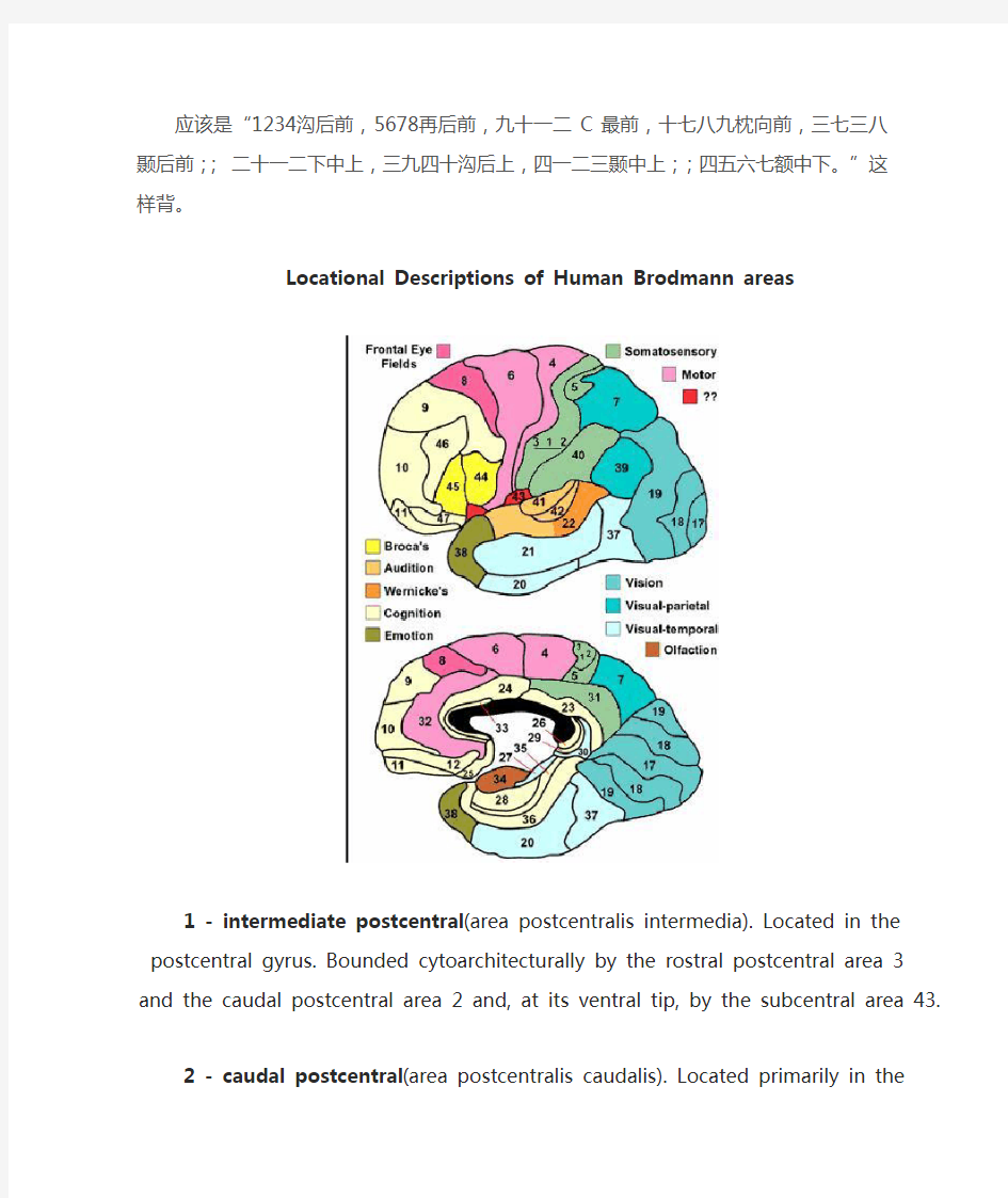
Locational Descriptions of Human Brodmann areas
1 - intermediate postcentral(area postcentralis intermedia). Located in the postcentral gyrus. Bounded cytoarchitecturally by the rostral postcentral area 3 and the caudal postcentral area
2 and, at its ventral tip, by the subcentral area 43.
2 - caudal postcentral(area postcentralis caudalis). Located primarily in the caudal portion of the postcentral gyrus and the rostral lip of the postcentral sulcus with a caudal extension along the intraparietal sulcus. Cytoarchitecturally bounded rostrally by the intermediate postcentral area 1 and caudally by the preparietal area 5, the superior parietal area 7 and the
supramarginal area 40.
3 - rostral postcentral(area postcentralis oralis). Located primarily in the rostral portion of
the postcentral gyrus including the caudal bank of the central sulcus. At either end of the sulcus it can extend beyond the depth of the sulcus into the precentral gyrus.
Cytoarchitecturally bounded rostrally by the gigantopyramidal area 4 and caudally by the
intermediate postcentral area 1.
4 - gigantopyramidal(area gigantopyramidalis). Located in the precentral gyrus. Cytoarchitecturally the caudal boundary with the rostral postcentral area 3 does not coincide precisely with the floor of the central sulcus but lies variably in the banks of the postcentral gyrus and the precentral gyrus. The area also does not extend in all cases to the cingulate sulcus medially or to the end of the central sulcus ventro-laterally. Bounded rostrally by the
frontal agranular area 6.
5 - preparietal(area praeparietalis). Occupies the superior parietal lobule and a portion of the
postcentral gyrus, particularly on the medial aspect of the hemisphere. Bounded approximately by the cingulate sulcus on the medial aspect of the hemisphere and by the superior postcentral sulcus on the lateral aspect. Cytoarchitecturally bounded by the caudal postcentral area 2, the superior parietal area 7 and on the medial bank of the hemisphere by the gigantopyramidal area 4 and the dorsal posterior cingulate area 31.
6 - agranular frontal(area frontalis agranularis). Located primarily in the caudal portions of the superior frontal gyrus and the middle frontal gyrus and the rostral portions of the precentral gyrus not occupied by the gigantopyramidal area 4. It extends from the cingulate sulcus on the medial aspect of the hemisphere to the lateral sulcus on the lateral aspect. Cytoarchitecturally bounded rostrally by the frontal region and caudally by the gigantopyramidal area 4.
7 - superior parietal(area parietalis superior). Occupies much of the superior parietal lobule
and some of the precuneus. Bounded approximately by the superior postcentral sulcus rostrally, the intraparietal sulcus laterally, the parieto-occipital sulcus caudally and, on the medial bank of the hemisphere, the subparietal sulcus. Cytoarchitecturally bounded rostrally by the preparietal area 5 and the caudal postcentral area 2; caudally by the peristriate area 19;
and medially by the dorsal posterior cingulate area 31.
8 - intermediate frontal(area frontalis intermedia). Located primarily in the superior frontal
gyrus extending from the cingulate sulcus on the medial surface over the margin of the hemisphere to the middle frontal gyrus. Cytoarchitecturally bounded caudally by the agranular frontal area 6 and ventrally by the granular frontal area 9.
9 - granular frontal(area frontalis granularis). Occupies portions of the superior frontal gyrus
and the middle frontal gyrus. Its approximate boundary on the medial aspect of the hemisphere is the cingulate sulcus and, on the lateral aspect, the inferior frontal sulcus. Cytoarchitecturally bounded dorsocaudally by the intermediate frontal area 8, caudally by the agranular frontal area 6, and ventrally by the frontopolar area 10, the middle frontal area 46
and the opercular area 44.
10 - frontopolar(area frontopolaris). Occupies the most rostral portions of the superior frontal
gyrus and the middle frontal gyrus. On the medial aspect of the hemisphere it is bounded ventrally by the superior rostral sulcus. It does not extend as far as the cingulate sulcus. Cytoarchitecturally bounded dorsally by the granular frontal area 9, caudally by the middle frontal area 46, and ventrally by the orbital area 47 and by the frontopolar area 12.
11 - prefrontal(area praefrontalis). Constitutes most of the orbital gyri, gyrus rectus and the
most rostral portion of the superior frontal gyrus. Bounded medially by the inferior rostral sulcus and laterally approximately by the frontomarginal sulcus. Cytoarchitecturally bounded on the rostral and lateral aspects of the hemisphere by the frontopolar area 10, the orbital area 47, and the triangular area 45; on the medial surface it is bounded dorsally by the area 12 and
caudally by the subgenual area 25.
12 - prefrontal(area praefrontalis). Occupies the area between the superior rostral sulcus and the inferior rostral sulcus. Cytoarchitecturally bounded dorsally by the frontopolar area 10 and the dorsal anterior cingulate area 32; caudally, ventrally and rostrally it is bounded by the prefrontal area 11. (Originally described as part of prefrontal area 11 but not shown in the map, subsequently it was labeled as an independent area 12.)
17 - striate(area striata). Part of the occipital lobe of the cerebral cortex that is defined on the
basis of cyto- and myeloarchitecture, primarily by the band/stripe of Gennari.
18 - parastriate(area parastriata). Located in parts of the cuneus, the lingual gyrus and the lateral occipital gyrus of the occipital lobe. Cytoarchitecturally bounded on one side by the striate area 17, from which it is distinguished by absence of a band/stripe of Gennari, and on
the other by the peristriate area 19.
19 - peristriate(area peristriata). Located in parts of the lingual gyrus, the cuneus, the lateral
occipital gyrus and the superior occipital gyrus of the occipital lobe where it is bounded
approximately by the parieto-occipital sulcus. Cytoarchitecturally bounded on one side by the parastriate area 18 which it surrounds. Rostrally it is bounded by the angular area 39 and the
occipitotemporal area 37.
20 - inferior temporal(area temporalis inferior). Corresponds approximately to the inferior temporal gyrus. Cytoarchitecturally bounded medially by the ectorhinal area 36, laterally by the middle temporal area 21, rostrally by the temporopolar area 38 and caudally by the
occipitotemporal area 37.
21 - middle temporal(area temporalis media). Corresponds approximately to the middle
temporal gyrus. Bounded rostrally by the temporopolar area 38, ventrally by the inferior temporal area 20, caudally by the occipitotemporal area 37, and dorsally by the superior
temporal area 22.
22 - superior temporal(area temporalis superior). Corresponds approximately to the lateral and caudal two thirds of the superior temporal gyrus. Bounded rostrally by the temporopolar area 38, medially by the posterior transverse temporal area 42, ventrocaudally by the middle temporal area 21 and dorsocaudally by the supramarginal area 39.
23 - ventral posterior cingulate(area cingularis posterior ventralis). Occupies most of the posterior cingulate gyrus adjacent to the corpus callosum. At the caudal extreme it is bounded approximately by the parieto-occipital sulcus. Cytoarchitecturally bounded dorsally by the dorsal posterior cingulate area 31, rostrally by the ventral anterior cingulate area 24, and ventrorostrally in its caudal half by the retrosplenial region.
24 - ventral anterior cingulate(area cingularis anterior ventralis). Occupies most of the
anterior cingulate gyrus in an arc around the genu of corpus callosum. Its outer border corresponds approximately to the cingulate sulcus. Cytoarchitecturally bounded internally by the pregenual area 33, externally by the dorsal anterior cingulate area 32, and caudally by the ventral posterior cingulate area 23 and the dorsal posterior cingulate area 31.
25 - subgenual(area subgenualis). A narrow band located in the caudal portion of the subcallosal area adjacent to the paraterminal gyrus from which it is separated by the posterior parolfactory sulcus. It is bounded by the prefrontal area 11rostrally and by the paraterminal
gyrus caudally.
26 - ectosplenial(area ectosplenialis). A narrow band located in the isthmus of cingulate
gyrus adjacent to the fasciolar gyrus internally. It is bounded externally by the granular
retrolimbic area 29.
28 - entorhinal(area entorhinalis). Located in the entorhinal area on the medial aspect of the
temporal lobe. It and the dorsal entorhinal area 34 together constitute approximately the
entorhinal area.
29 - granular retrolimbic(area retrolimbica granularis). A narrow band located in the isthmus of cingulate gyrus. Cytoarchitecturally bounded internally by the ectosplenial area 26 and externally by the agranular retrolimbic area 30.
30 - agranular retrolimbic(area retrolimbica agranularis). Located in the isthmus of cingulate gyrus. Cytoarchitecturally bounded internally by the granular retrolimbic area 29, dorsally by the ventral posterior cingulate area 23 and ventrolaterally by the ectorhinal area 36.
31 - dorsal posterior cingulate(area cingularis posterior dorsalis). Occupies portions of the posterior cingulate gyrus and medial aspect of the parietal lobe. Approximate boundaries are the cingulate sulcus dorsally and the parieto-occipital sulcus caudally. It partially surrounds the subparietal sulcus. Cytoarchitecturally bounded rostrally by the ventral anterior cingulate area 24, ventrally by the ventral posterior cingulate area 23, dorsally by the gigantopyramidal area 4 and preparietal area 5 and caudally by the superior parietal area 7.
32 - dorsal anterior cingulate(area cingularis anterior dorsalis). Forms an outer arc around the anterior cingulate gyrus. The cingulate sulcus defines approximately its inner boundary and the superior rostral sulcus its ventral boundary; rostrally it extends almost to the margin of the frontal lobe. Cytoarchitecturally bounded internally by the ventral anterior cingulate area 24, externally by medial margins of the agranular frontal area 6, intermediate frontal area 8, granular frontal area 9, frontopolar area 10, and prefrontal area 11.
33 - pregenual(area praegenualis). A narrow band located in the anterior cingulate gyrus
adjacent to the supracallosal gyrus in the depth of the callosal sulcus. Cytoarchitecturally bounded by the ventral anterior cingulate area 24 and the supracallosal gyrus.
34 - dorsal entorhinal(area entorhinalis dorsalis). Located in the entorhinal area on the
medial aspect of the temporal lobe. It and the entorhinal area 28 together constitute
approximately the entorhinal area.
35 - perirhinal(area perirhinalis). Located along the rhinal sulcus. Cytoarchitectually
bounded medially by the entorhinal area 28 and laterally by the ectorhinal area 36.
36 - ectorhinal(area ectorhinalis). Located primarily in the fusiform gyrus, with its medial
boundary corresponding approximately to the rhinal sulcus. Cytoarchitecturally bounded laterally and caudally by the inferior temporal area 20, medially by the perirhinal area 35 and
rostrally by the temporopolar area 38.
(四)大脑
大脑半球为中枢神经中最大的结构,左右各一。上面观略呈卵圆形。在大脑半球表面,呈现许多深浅不同的裂或沟,沟、裂之间有隆起的脑回。胎儿5个月发生沟、回,在出生后逐渐完成。沟、裂的产生是因为大脑皮质各部在发生上快慢不匀所致,发育快的露在表面而将生长的部分挤住深部。
1.大脑半球的外形每个半球分为背外侧面、内侧面和底面。大脑半球借三条大的沟裂分为五叶,即额叶、顶叶、枕叶、颞叶和岛叶(图10—35,10—36)。
外侧沟(sulcus lateralis)为背外侧面最明显的深裂。自前向后上方斜行,将大脑半球额叶、颞叶(lobus temporalis)分隔。外侧沟的深部隐藏着岛叶(insula)。
中央沟(sulcus centralis)在背外侧面近中间的稍后方。此沟自顶部向前向下,下端接近外侧沟,但未与其通连。中央沟顶端多半越背内侧缘至内侧面,向后下延伸1cm即终止。此沟前部为额叶(lobus frontalis),后部为顶叶(lobus parietalis)。
顶枕沟(sulcus parieto—occipitalis)位于半球内侧面的后部,略转至背外侧面,在顶叶和枕叶(lobus occipitalis)之间。枕叶、顶叶、颞叶之间的分界是假设的。在大脑下缘,自枕叶后端向前约4cm处有一枕前切迹,自顶枕裂至枕前切迹的连线为枕叶的前界,由此线的中点连至外侧构的后端是顶、颞二叶的分界。
1)半球背外侧面的沟和回
(1)额叶在中央沟的前方有与中央沟平行的中央前沟,中央沟与中央前沟之间为中央前回。自中央前沟向前走出上、下两条与半球上缘平行的沟,分别称额上、下沟。额上沟的上方为额上回,额上、下沟之间为额中回,额下沟的下方为额下回。
(2)顶叶在中央沟的后方有与中央沟平行的中央后沟。中央沟与中央后沟之间为中央后回,自中央后沟向后走出一条与半球上缘平行的顶内沟。顶内沟的上方为顶上叶、下方为顶下叶。顶下叶前部称缘上回,后部称角回。
(3)颞叶在大脑外侧沟下方有与之平行的颞上沟和颞下沟,颞上沟的上方为颞上回,下方为颞中回。颞下沟的下方为颞下回。此外,在大脑外侧沟内,颞上回的上面有几条短的横回,称颞横回。
2)半球内侧面的沟和回自中央前、后回背外侧面延续到内侧面部分称中央旁小叶。在枕叶有一深沟称距状沟。顶枕沟与距状沟之间为楔叶。距状沟的下方为舌回。舌回向前与颞叶的海马旁回相连。海马旁回的前端弯成钩形,称海马旁回钩。环抱胼胝体上方的为扣带回(图10—36)。
3)半球底面额叶底面有一条纵行的纤维束,称嗅束。嗅束的前端膨大,称嗅球,嗅球与嗅神经相连。嗅束向后扩大为嗅三角,连于海马旁回前部和海马旁回钩等嗅觉高级中枢(图10—37)。
2.大脑半球的内部结构大脑半球表层被覆一层灰质,为皮质,皮质的深部为髓质。髓质内含有神经纤维束与核团。
1)基底核(huclei basles)共有四对,因位置靠近脑底故名。
(1)尾状核(nucleus caudatus)呈“C”字形(图10—38),状如松鼠之尾。前端膨大称头,在侧脑室前角底部,内囊内侧,后部变为细长作弓形弯曲称体与尾,它沿着侧脑室壁自前向后行进,至侧脑室中央部分即位于下角的顶部,并随下角在颞叶向内向前行进而与杏仁体相连接。
(2)豆状核(nucleus lentiformis)在岛叶的深部(图10─38),介于内囊与外囊之间,其前下端与尾状核头相连接。豆状核在水平切面上呈三角形,核内有两个白质薄板层将它分为三部,外侧部最大称壳(putamen),其余二部称苍白球(globus pallidus)。
尾状核与豆状核合称纹状体(corpus striatum)。爬行类、鸟类已具有尾状核和壳,它们在种系发生上出现较晚,称新纹状体;苍白球出现较早,在鱼类已经存在,故称旧纹状体。
纹状体主要与肌肉运动活动有关,在进化阶梯上比控制肌肉进行随意运动的锥体系要古老得多。在哺乳类以下,它是调节躯体、内脏活动对环境作本能性适应的高级中枢,在高等哺乳类,它则退居次要地位,成为大脑皮质控制下的锥体外系中枢。
用荧光组织化学方法发现,纹状体内含有大量多巴胺能神经末梢,而其胞体主要位于中脑黑质。正常的纹状体活动,要求其内部的神经递质多巴胺和乙酰胆碱的含量保持平衡。已知多巴胺是抑制性递质,乙酰胆碱是兴奋性递质。
生理学的研究还表明,尾状核可以对各种感觉刺激(包括视、听、躯体、内脏)发生非特异性反应,刺激尾状核能影响感觉传入活动。在针刺镇痛原理研究中,我国学者证明了尾状核参与了针刺镇痛的过程,并在形态学上证明了尾状核与有关镇痛的核团具有广泛的联系。临床上用埋藏电极刺激尾状核,成功地解除了晚期癌症病员的顽痛。
(3)屏状核(claustrum)为岛叶和豆状核之间的一薄层灰质。
(4)杏仁体(corpus amygdaloi deum)与尾状核的末端相连,是边缘系统的皮质下中枢。
2)半球内的白质可分为三系:
(1)连合纤维(commissural fibers)是连合左、右两大脑半球的纤维(图10—39)。
①胼胝体(corpus callosum)是大脑半球中最大的连合纤维。它在两半球中间形成一个弧形板,其纤维向四面投射到大脑皮质。胼胝体在种系发生上出现较晚,它的发育和大脑皮质的发育是相平行的。脑重量大的动物,其胼胝体也大。人类的胼胝体最发达。
②前连合(commissura anterior)为前后两个弓形的纤维束组成,呈“H”形,中间彼此连接。主要连合两侧颞叶,小部分连合两侧嗅球(参见图10—36)。
③穹窿(fornix)是嗅脑的连合纤维,也是嗅脑的投射纤维。它起自海马,纤维先向后再弓形向上贴附在胼胝体的下面,然后再弯向前下,而终于下丘脑的乳头体(图10—39)。
(2)联合纤维(association fibers)是同侧半球皮质之间的联合纤维,纤维长短不一。
(3)投射纤维(Projection fibers)是进出大脑半球的神经纤维束。纤维有长短之分,短纤维如大脑与间脑之间的升降纤维。但以长纤维为主,长纤维主要为内囊(capsula interna),在水平切面上内囊呈“”形板,主要成分为投射纤维(其中也含有纹状体的横行交错纤维),内囊分三部。
内囊前脚:位于尾状核与豆状核之间,含有额桥束。
内囊膝:位于豆状核、尾状核与丘脑之间,含有皮质脑干束。
内囊后脚:位于豆状核与丘脑之间,由前向后排列为:皮质脊髓束、丘脑皮质束、枕颞桥束、听辐射与视辐射。
内囊是大脑半球内极重要的“关口”,在此有大量的上、下行纤维束紧密排列。若一侧内囊受损(如脑溢血),根据病变位置和被损传导束的性质及局部位置,可出现多种症状,如
对侧半身感觉丧失,对侧肢体偏瘫等等。
(4)侧脑室(ventriculus lateralis)是左右大脑半球内的腔隙,内含透明的脑脊液,通过室间孔与第三脑室相通。侧脑室有三个角:前角伸入室间孔以前的额叶,后角伸入枕叶,下角伸入颞叶(图10—40)。
3.大脑皮质及其组织结构从种系发生来说,大脑皮质可以分为:古皮质(archicortex)又称原始皮质,由海马和齿状回组成;旧皮质(paleocortex),由梨状区的嗅皮质及部分海马旁回组成;古皮质与旧皮质称异型皮质(allocortex),只显示三层构造。新皮质(neocortex),又称同型皮质(isocortex),一般均可分六层,大脑皮质的绝大部分属新皮质。
大脑皮质含有许多锥体形的神经细胞、其它各型神经细胞及神经纤维。显微镜观察,按细胞与纤维排列情况可以分为多层。一般以颞叶皮质为典型,大致分为六层(图10—41)。
Ⅰ)分子层在皮质的最浅层,内含少量水平细胞和星形细胞,有许多与表面平行的神经纤维。
Ⅱ)外粒层内含小型锥体细胞和少量星形细胞。
Ⅲ)外锥体层内含中、小型锥体细胞,以中型锥体细胞为主。
Ⅳ)内粒层细胞密集,多数是星形细胞。
Ⅴ)内锥体层内含大型锥体细胞,其轴突向髓质行进。在中央前回和中央旁小叶,此层的锥体细胞特大,称贝茨(Betz)细胞,高120μm,宽80μm。
Ⅵ)多形层内含梭形与三角形的神经细胞。
此外,还可根据皮质有髓纤维分布的特点分为六层,但名称不同(图10—41)。
来自丘脑外侧核的腹后核和内、外侧膝状体的特异性上行纤维主要进入第四层与星形细胞形成突触,而星形细胞又与其它细胞进行广泛的联系。第三层的锥体细胞发出纤维对皮质各层之间、各部位之间和左右半球之间进行联络及连合,经过错综复杂而广泛的联系,从而对进入皮质的各种冲动进行分析综合,然后作出反应。一般说来,第五、六层的锥体细胞和棱形细胞的轴突组成传出纤维,下行到脑干、脊髓、并通过脑神经、脊神经将冲动传到有关部位(图10—42)。
以上所述仅是最基本的模式,事实上,大脑皮质中约有140亿神经细胞,按照排列与组合推算神经元间可能的结合方式已多到天文数字。在理论和实际上,皮质的一个神经元可连接任何其它神经元。皮质的构造是如此复杂,而机能还要比这组合起来的方式繁复、灵活得多!由于人脑皮质极大量的神经元和它们之间互相联系的广泛性和复杂性,使得人类大脑皮质获得了完善的分析与综合的能力,构成了思维与语言的物质基础。
4.皮质的分区与功能定位大脑皮质的六层结构是基本型,而在皮质的不同部位,各层的厚薄,各种神经细胞的分布,纤维的疏密都有差异。一些学者根据皮质的这些不同特点,参考功能方面的资料把大脑分为若干区,其中Brodmann(1909)把大脑皮质分为52区,较为常用(图10—43)。
大脑皮质是统一机体的最高神经中枢。机体的各种功能在皮质的各个不同部位是有不同分工的,就是说,各种功能在皮质上具有定位关系,如运动区、感觉区等。但是这种分法是相对的,而不是绝对的,这些中枢只是执行这种功能的核心部位,皮质的其它区域也分散有类似的功能。如:中央前回(4区)主要管理全身骨骼肌的运动,称运动中枢,但中央前回也接受部分的感觉冲动。中央后回主要管理全身感觉,但刺激它也可产生少量运动。除了这些中枢以外,构成人类皮质的大部分区域称联合区。临床实践也证明,某一中枢的损伤,并不能使人永远完全丧失该中枢所管理的功能,经过适当的治疗和功能锻炼,常由于其它区域的代偿而恢复到一定程度。
1)运动中枢(第一运动区,)位于中央前回(4区),此区皮质极厚,约3.5—4.5mm,第三层锥体细胞多,第五层含有贝茨细胞。皮质运动中枢对于对侧骨骼肌运动的管理具有一定的顺序和局部关系:管理头部有关的肌肉在中央前回的下部;管理上肢与躯干的肌肉在中央前回的中部,管理下肢的肌肉则在中央前回的上部及旁中央小叶前半。身体各部在运动中枢的投影犹如一个倒置的人形,但比例并不相当,如在人体上,口和手比下肢要小得多,但
它们在皮质上所占的面积却很大,这是因为功能复杂所致。
此外,还有运动前区与补充运动区。运动前区位于6区内,在半球内侧可延至扣带沟处,结构上6区与4区相似,但6区缺乏贝茨细胞。补充运动区在半球内侧面4区的前方。
2)感觉中枢在躯体受刺激部分的对侧皮质,即在中央后回和中央旁小叶后半,相当于Brodmann的3、1、2区,它们的共同特点是内颗粒层较厚,它管理痛、温、触、压以及位置觉和运动觉等体躯感觉。身体各部分在感觉中枢上的投影和运动中枢相似。此外,还有第Ⅱ躯体感觉,位于中央后回的最下部。
3)视觉中枢在距状沟两旁的楔回与舌回上,相当于17区,此区皮质最薄,约为1.5—2.5mm,第Ⅱ、Ⅲ、Ⅴ层均窄,第Ⅳ层最厚,中间有一条白色细纹为视辐射投入的纤维。
4)听觉中枢位于颞横回(41、42区),皮质厚约3mm,第Ⅱ、Ⅲ、Ⅳ、Ⅵ层均较厚,它接受内侧膝状体发出的听辐射投入的纤维。
5)嗅觉中枢大概在海马回旁钩附近,因外侧嗅纹终止于此。
以上各中枢是高等动物和人所共有的,此外,人类有语言和思维,而与此有关,在人类的大脑皮质上也具有相应的中枢(图10—44),这种中枢在大脑皮质上偏于左侧,因此称之为优势半球。
6)运动性语言中枢在额下回后1/3处(44区),紧靠中央前回的下部。如果此区受损,咽、喉、舌、唇诸肌虽未瘫痪但却丧失了说话能力,此为运动性失语症。
7)听觉性语言中枢位于颞上回后部。如该区受损,听觉无障碍,有说话能力但失去理解能力,此为感觉性失语症。
8)视运动性语言中枢(书写中枢)位于额中回的后部,损伤该区则不能以书写方式表达意见,此为失写症。
9)视觉性语言中枢(阅读中枢)位于角回(39区),此区受损,视觉无障碍,但不能理解文字意义,不能阅读,此为视觉失语症。