Chromatin Prepattern and Histone Modifiers in a Fate Choice for Liver and Pancreas
4.M.J.Axtell,J.A.Snyder,D.P.Bartel,Plant Cell19,1750
(2007).
5.M.S.Barker,H.Vogel,M.E.Schranz,Genome Biol.Evol.
1,391(2009).
6.H.Tang et al.,Science320,486(2008).
7.H.Tang et al.,Genome Res.18,1944(2008).
8.M.Ghildiyal,P.D.Zamore,Nat.Rev.Genet.10,94
(2009).
9.D.Chen et al.,Bioinformatics26,1391(2010).
10.K.D.Kasschau et al.,PLoS Biol.5,e57(2007).
11.K.Nobuta et al.,Nat.Biotechnol.25,473(2007).
12.S.H.Cho et al.,PLoS Genet.4,e1000314(2008).
13.D.Bourc’his,O.Voinnet,Science330,617(2010).
14.D.H.Chitwood et al.,Genes Dev.23,549(2009).
15.A.Peragine,M.Yoshikawa,G.Wu,H.L.Albrecht,
R.S.Poethig,Genes Dev.18,2368(2004).
16.Z.Xie,E.Allen,A.Wilken,J.C.Carrington,Proc.Natl.
Acad.Sci.U.S.A.102,12984(2005).
17.S.K.Floyd,J.L.Bowman,Curr.Biol.16,1911(2006).
18.C.J.Harrison et al.,Nature434,509(2005).
19.S.Tsuji et al.,J.Plant Res.120,281(2007).
20.F.Grewe et al.,Nucleic Acids Res.39,2890(2011).
21.V.Knoop,Cell.Mol.Life Sci.68,567(2011).
22.T.Demura,H.Fukuda,Trends Plant Sci.12,64
(2007).
23.M.Bonke,S.Thitamadee,A.P.M?h?nen,M.T.Hauser,
Y.Helariutta,Nature426,181(2003).
24.K.Hirano et al.,Plant Cell19,3058(2007).25.Y.Yasumura,M.Crumpton-Taylor,S.Fuentes,N.P.Harberd,
Curr.Biol.17,1225(2007).
26.Y.Cao et al.,Fitoterapia81,253(2010).
Acknowledgments:Selaginella sequences were deposited at
GenBank with the accession numbers GL377566to
GL378322.1and HM173080.Genome sequencing and
analysis were performed by the U.S.Department of
Energy(DOE),Joint Genome Institute,supported by
the Office of Science of the U.S.DOE,Contract
DE‐AC02‐05CH11231(I.V.G.,U.H.,D.L.,E.L.,S.L.,T.M.,R.O.,
D.R.,A.S.,J.S.,H.V.S.).Support was provided by NSF
0844413(J.A.B.);Japan Society for the Promotion of
Science(M.H.,T.N.,T.F.,K.M.,T.M.,M.S.);Ministry of
Education,Culture,Sports,Science,and Technology,
Japan(M.H.,T.N.,T.F.);NSF0515435and Australian
Research Council FF0561326(J.L.B.);NSF0519970
(M.G.);NSF0638595(C.D.);NSF0922742(V.A.A.);The
Lewis B and Dorothy Cullman Program(B.A.A.,A.L.);NSF
1020443(B.A.A.);Natural Sciences and Engineering
Research Council of Canada(NSERC)2982(N.W.A.);
NIH GM84051(M.J.A.);NSERC(E.I.B.);NIH T32GM007757
and NSERC PGS-D(M.S.B.);NSF0607123(J.L.B.);Life
Sciences Research Foundation(NDB);National
Human Genome Research Institute(NHGRI)HG004164
(M.D.);German Science Foundation(Deutsche
Forschungsgemeinschaft,DFG)DR430/4-2(I.D.);Czech
Ministry of Education21620828(M.E.);Jeffress Memorial
Trust J-938(E.M.E.);NSF0744800(M.E.and M.P.);DOE
DE-FG02-04ER15542(W.B.F.and S.L.);The Danish
Council for Independent Research,Technology and
Production Sciences009-066624/274-09-0314(J.H.);
The Villum Kann Rasmussen Foundation(J.H.,I.S.,B.P.,
P.U.,W.W.);NIH T32-HG00035,NSF1020660,and NSF
1036466(K.G.K.);NSF0735191(E.L.);NHGRI
HG004164(G.M);U.S.Department of Agriculture(USDA)
DE-FG02-08ER64630(T.P.M.);NSF0228660(R.G.O.);
The Danish Council for Strategic Research09-063090
(B.L.P.and P.U.);Marie Curie FP6RTN ZOONET(B.P.);
DFG RE837/10-2,BMBF FRISYS0313921(S.A.R.);
Bundesministerium fuer Bildung und Forschung,
Germany,GABI-FUTURE grant0315046(D.M.R.);USDA
NRI2007-35318-18389(A.W.R.);NSF0421604
(C.S.);NIH GM065383(D.E.S.);Burgundy Regional
Council20100112095254682-1(D.W.);and DFG RE
837/10-2(A.D.Z.).K.Wall,D.Hurley,and S.Hentel
provided computational assistance.
Supporting Online Material
https://www.360docs.net/doc/df2925228.html,/cgi/content/full/science.1203810/DC1
SOM Text
Figs.S1to S14
Tables S1to S8
References
4February2011;accepted8April2011
Published online5May2011;
10.1126/science.1203810
Chromatin“Prepattern”and Histone Modifiers in a Fate Choice for Liver and Pancreas
Cheng-Ran Xu,1Philip A.Cole,2David J.Meyers,2Jay Kormish,1*Sharon Dent,3Kenneth S.Zaret1?
Transcriptionally silent genes can be marked by histone modifications and regulatory proteins that indicate the genes’potential to be activated.Such marks have been identified in pluripotent cells,but it is unknown how such marks occur in descendant,multipotent embryonic cells that have restricted cell fate choices.We isolated mouse embryonic endoderm cells and assessed histone modifications at regulatory elements of silent genes that are activated upon liver or pancreas fate choices.We found that the liver and pancreas elements have distinct chromatin patterns.Furthermore,the histone acetyltransferase P300,recruited via bone morphogenetic protein signaling,and the histone methyltransferase Ezh2have modulatory roles in the
fate choice.These studies reveal a functional“prepattern”of chromatin states within multipotent progenitors and potential targets to modulate cell fate induction.
E arly pluripotent cells of the mammalian
embryo develop into multipotent endo-
derm,ectoderm,and mesoderm germ layers.In pluripotent cells,silent genes that will be activated later in development often exist with histone modifications and/or bound transcription factors that reflect the chroma-tin being“poised”for activity(1–3).It is un-clear whether such poised states exist for silent
genes in germ layer cells and,if so,whether
genes poised for different tissue fates exhibit
different chromatin features.Furthermore,it is
not known whether enzymes that establish chro-
matin states can control germ layer fate choices.
Embryonic germ layer cells are few in number,
they have not been purified,and chromatin
analysis on small cell populations is challeng-
ing(4).Yet germ layer cells represent the first
lineage-restricted,multipotent progenitors of
the embryo and a paradigm for all subsequent
fate decisions.
V entral foregut endoderm cells undergo a fate
choice for liver or ventral pancreas progenitors
(5,6).FoxA1or FoxA2,GA TA4or GA TA6,
vHNF1,and Hnf6(also known as Oc1)are nec-
essary in the endoderm for both liver and ventral
pancreas induction(7).In the absence of any
set of the factors,the earliest liver marker genes
Alb1,Afp,and Ttr and the ventral pancreas tran-
scription factor gene Pdx1fail to be activated,or
expression is delayed,and tissue buds fail to form
(7).It is not clear how the same group of factors
can be necessary for both liver and ventral
pancreas and how signaling promotes the differ-
ent fates.We sought to map chromatin states at
silent liver-and pancreas-specific regulatory se-
quences in endoderm cells,to discover the factors
or relevant histone-modifying enzymes,and
test the enzymes’functions in the liver-versus-
pancreas decision.
We used fluorescence-activated cell sorting
(FACS)with the ENDM1antibody to isolate ven-
tral foregut endoderm cells from embryonic day
8.25(E8.25)mouse embryos with four to six
somite pairs(4-6S)(8)(fig.S1),just prior to the
induction of hepatic and pancreatic fates(5,9).
We also used the liver-specific antibody Liv2to
isolate nascent hepatoblasts expressing Alb1,Afp,
and Ttr from E9.5embryos(fig.S2)(10).Chro-
matin marks in ENDM1+and Liv2+populations
were identified with a low–cell number chroma-
tin immunoprecipitation(ChIP)protocol(4)for
H3K9acK14ac,H3K4me2,H3K4me3,H3K9me3,
H3R17me2a,H3K27me3,H3K36me2,H3K36me3,
H3K79me2,H4K20me3,H3T3ph,H3S10ph,
the histone variant H2A.Z,and the chromatin re-
modelers Brg1and SNF2.We assessed the liver-
specific promoter and enhancer of Alb1(11,12),
the liver-specific promoters of Afp and Ttr genes
(13,14),and the I,II,III,and IV upstream ele-
ments and local promoter of the pancreatic de-
termination gene Pdx1(fig.S3).The I,II,and III
upstream elements and promoter of Pdx1recon-
stitute pancreas-specific activation(15);the IV
element may function later(16,17).All of the
target genes are silent in endoderm cells,and only
the liver genes become activated in hepatoblasts.
1Institute for Regenerative Medicine,Epigenetics Program,Depart-ment of Cell and Developmental Biology,University of Pennsylvania School of Medicine,Philadelphia,PA19104,USA.2Department of Pharmacology and Molecular Sciences,Johns Hopkins University School of Medicine,Baltimore,MD21205,USA.3Department of Molecular Carcinogenesis,University of Texas M.D.Anderson Cancer Center,Smithville,TX78957,USA.
*Present address:Biochemistry and Molecular Biology, University of Calgary,Calgary,Alberta T2N4N1,Canada.
?To whom correspondence should be addressed.E-mail: zaret@https://www.360docs.net/doc/df2925228.html, o n M a y 1 9 , 2 0 1 1 w w w . s c i e n c e m a g . o r g D o w n l o a d e d f r o m
We also assessed the active Gapdh gene(encod-ing glyceraldehyde-3-phosphate dehydrogenase) as a control.For each mark and target,we per-formed at least quadruplicate ChIP assays and displayed quantitative polymerase chain reaction (qPCR)results over immunoglobulin G(IgG) ChIP controls.Low–cell number ChIP yields low-amplitude signals;with replicates,statistical sig-nificance can be reached(4).
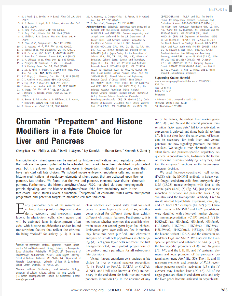
Two chromatin marks in undifferentiated en-doderm cells exhibited striking differences be-tween the liver and pancreas regulatory elements. H3K9acK14ac,which is associated with gene ac-tivity(18),was poorly represented at all of the liver regulatory elements,relative to the active Gapdh promoter,and enriched at all of the Pdx1reg-ulatory elements(Fig.1,open boxes).H3K27me3, which is associated with gene silencing(19),was also poorly represented at the liver elements and Gapdh,yet was enriched at the pancreas ele-ments,except at area IV(Fig.1,open boxes).
Only H3K9acK14ac showed a significant in-crease at the liver elements when the foregut endoderm cells differentiated into hepatoblasts (Fig.1,solid boxes,P<0.001).H3K4me2/3, H3K20me3,and H3K36me2exhibited variable changes,and none of the other chromatin marks changed significantly(fig.S4).Hyperacetylation persisted at the Pdx1elements in hepatoblasts, where the gene remained silent,as did H3K27me3 (Fig.1,solid boxes).By contrast,silent promot-ers that are active in adipocytes and T cells (20,21)lacked both marks in endoderm and hepatoblasts(fig.S5).We conclude that the silent liver and pancreas regulatory elements exhibit distinct chromatin states.
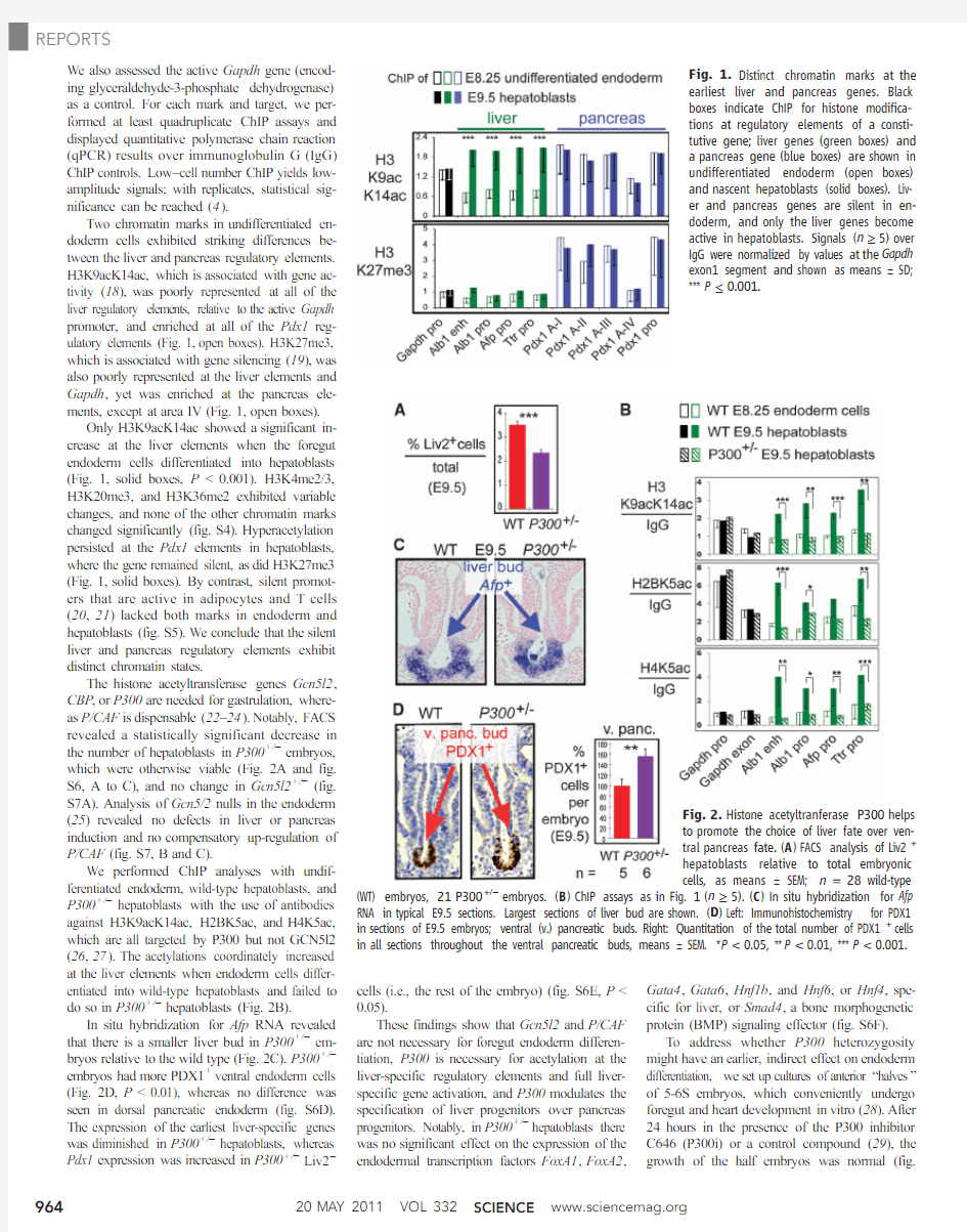
The histone acetyltransferase genes Gcn5l2, CBP,or P300are needed for gastrulation,where-as P/CAF is dispensable(22–24).Notably,FACS revealed a statistically significant decrease in the number of hepatoblasts in P300+/?embryos, which were otherwise viable(Fig.2A and fig. S6,A to C),and no change in Gcn5l2+/?(fig. S7A).Analysis of Gcn5/2nulls in the endoderm (25)revealed no defects in liver or pancreas induction and no compensatory up-regulation of P/CAF(fig.S7,B and C).
We performed ChIP analyses with undif-ferentiated endoderm,wild-type hepatoblasts,and P300+/?hepatoblasts with the use of antibodies against H3K9acK14ac,H2BK5ac,and H4K5ac, which are all targeted by P300but not GCN5l2 (26,27).The acetylations coordinately increased at the liver elements when endoderm cells differ-
entiated into wild-type hepatoblasts and failed to do so in P300+/?hepatoblasts(Fig.2B).
In situ hybridization for Afp RNA revealed that there is a smaller liver bud in P300+/?em-bryos relative to the wild type(Fig.2C).P300+/?embryos had more PDX1+ventral endoderm cells (Fig.2D,P<0.01),whereas no difference was seen in dorsal pancreatic endoderm(fig.S6D). The expression of the earliest liver-specific genes was diminished in P300+/?hepatoblasts,whereas Pdx1expression was increased in P300+/?Liv2?cells(i.e.,the rest of the embryo)(fig.S6E,P<
0.05).
These findings show that Gcn5l2and P/CAF
are not necessary for foregut endoderm differen-
tiation,P300is necessary for acetylation at the
liver-specific regulatory elements and full liver-
specific gene activation,and P300modulates the
specification of liver progenitors over pancreas
progenitors.Notably,in P300+/?hepatoblasts there
was no significant effect on the expression of the
endodermal transcription factors FoxA1,FoxA2,
Gata4,Gata6,Hnf1b,and Hnf6,or Hnf4,spe-
cific for liver,or Smad4,a bone morphogenetic
protein(BMP)signaling effector(fig.S6F).
To address whether P300heterozygosity
might have an earlier,indirect effect on endoderm
differentiation,we set up cultures of anterior“halves”
of5-6S embryos,which conveniently undergo
foregut and heart development in vitro(28).After
24hours in the presence of the P300inhibitor
C646(P300i)or a control compound(29),the
growth of the half embryos was normal(fig.
Fig.1.Distinct chromatin marks at the
earliest liver and pancreas genes.Black
boxes indicate ChIP for histone modifica-
tions at regulatory elements of a consti-
tutive gene;liver genes(green boxes)and
a pancreas gene(blue boxes)are shown in
undifferentiated endoderm(open boxes)
and nascent hepatoblasts(solid boxes).Liv-
er and pancreas genes are silent in en-
doderm,and only the liver genes become
active in hepatoblasts.Signals(n≥5)over
IgG were normalized by values at the Gapdh
exon1segment and shown as means T SD;
***P≤
0.001.
Fig.2.Histone acetyltranferase P300helps
to promote the choice of liver fate over ven-
tral pancreas fate.(A)FACS analysis of Liv2+
hepatoblasts relative to total embryonic
cells,as means T SEM;n=28wild-type (WT)embryos,21P300+/?embryos.(B)ChIP assays as in Fig.1(n≥5).(C)In situ hybridization for Afp
RNA in typical https://www.360docs.net/doc/df2925228.html,rgest sections of liver bud are shown.(D)Left:Immunohistochemistry for PDX1
in sections of E9.5embryos;ventral(v.)pancreatic buds.Right:Quantitation of the total number of PDX1+cells
in all sections throughout the ventral pancreatic buds,means T SEM.*P<0.05,**P<0.01,***P<0.001.
o
n
M
a
y
1
9
,
2
1
1
w
w
w
.
s
c
i
e
n
c
e
m
a
g
.
o
r
g
D
o
w
n
l
o
a
d
e
d
f
r
o
m
S8A).We observed a significant reduction in the number of hepatoblasts in P300i-treated embryos (fig.S8B,P <0.001).P300i also inhibited the induction of liver genes in hepatoblasts and stim-ulated Pdx1expression in Liv2?cells without affecting the expression of P300itself (fig.S8C).Hence,the genetic and pharmacologic data in-dicate that P300modulates the choice between liver and pancreas fates.
EZH2,a member of the PRC2Polycomb complex,is a methyltransferase for H3K27me3(30).ChIP in wild-type endoderm cells revealed that EZH2was enriched at the upstream elements of the Pdx1gene where H3K27me3was present,and that EZH2was absent from the liver reg-ulatory elements (Fig.3A,P <0.05).We used an Ezh2conditional allele (Ezh2CA )(31)and a FoxA3Cre transgene (32)to delete Ezh2in foregut endoderm cells.Strikingly ,the FoxA3Cr e/Cr e ;Ezh2CA/CA embryos,by E10,exhibited an expanded PDX1+ventral pancreatic progenitor domain,with mul-tiple bud-like structures (Fig.3B,arrows),but no change in the dorsal pancreatic domain (fig.S9A).By E11.5,the size of ventral,but not dorsal,pan-creas was markedly increased (Fig.3C and fig.S9B).There was no change in cell proliferation (fig.S9C).This occurred at the expense of liver development,as the liver bud was smaller and less extensively penetrated the mesenchyme (Fig.3D).Thus,Ezh2indirectly promotes the liver program by restraining the extent of ventral pan-creatic specification in the endoderm.
Previously,we found that BMP signaling pro-motes the liver fate over the pancreas fate in the ventral endoderm,and shortly afterward,BMP enhances pancreatic specification (28).SMAD pro-teins are BMP effectors and interact with P300(33,34).FACS analysis of half embryos (3-4S)revealed that the number of Liv2+hepatoblasts was slightly increased after BMP4treatment,as was the expression of liver genes in Liv2+cells,without affecting proliferation (fig.S10,A to D).BMP4treatment increased histone acetylation only at the liver regulatory elements,not at Pdx1elements (fig.S10E,P <0.05).
Deletion of Smad4in the foregut endoderm led to an expected (28,35)decrease in Smad4expression in Liv2+hepatoblasts as well as a de-crease in the number of hepatoblasts,but no ef-fect on P300was observed (Fig.4,A and B).ChIP of hepatoblasts from FoxA3Cre ;Smad4CA embryos revealed a loss of SMAD4from the liver regulatory elements,a loss of P300at the liver elements,and diminished H3acetylation
(Fig.4C,black bars).Taken together,these studies reveal a pathway by which the BMP signal is mediated by SMAD4,which recruits P300and results in histone acetylation at liver target ele-ments,enhanced liver gene activation,and en-hanced liver bud emergence (fig.S11).
By screening chromatin sites that were small in number and necessary for an impending cell choice,for many different chromatin marks in progenitor cells,we obtained clues about rel-evant histone modifiers.The modifiers function in conjunction with,rather than modify the ex-pression of,known endoderm transcription fac-tors.We suggest that spatially localized signaling to the endoderm with P300recruitment to par-ticular chromatin sites could help to initiate the liver program.Despite a failure to induce acet-ylation of liver-specific regulatory elements in P300+/?or Smad4?/?embryos,the induction of liver progenitors was reduced but not eliminated.We thus suggest that the histone modifications or P300itself,or both,play a modulatory role rather than one of absolute governance of liver in-duction.Further studies are required to deter-mine how these events at a specific moment in development link to the subsequent maintenance of the hepatic program.
Given that the H3K27me3mark is not seen at the Pdx1sites after the pancreas is induced (36),EZH2complexes in the endoderm restrain pan-creatic commitment.The persistence of both H3Ac and H3K27me3marks on the silent Pdx1gene in sorted hepatoblasts is consistent with their coexistence on individual genes.Such has been seen in embryonic stem cells (37)and may constitute a new kind of “bivalent ”mark at silent genes that are destined for activation in develop-ment.These results could explain the results of tissue explant studies indicating that the ventral endoderm is inherently set to express the pan-creatic program (5).We note that the regulatory elements at silent liver genes in the endoderm lacked the H3K4/H3K27bivalent marks seen in pluripotent cells (1).
The distinct histone modification states at the liver and pancreas regulatory elements in endo-derm indicate a chromatin “prepattern ”in un-differentiated,multipotent cells.The enzymes that elicit the prepattern play a modulatory role in
a
Fig.3.The H3K27methyltransferase gene Ezh2restricts the extent of ventral pancreas specification.(A )ChIP assays (n =4),means T SD.(B )Immuno-histochemistry for PDX1in cross sections of representative E10embryos.(C )Immunohistochemistry for NKX6.1in cross sections of representative E11.5embryos,means T SEM.(D )In situ hybridization for Afp RNA in cross sections of E10embryos.*P <0.05,**P <0.01.
Fig.4.BMP4signaling regulates hepatic specification via a pathway to SMAD4and P300.(A )Smad4and P300expression by quantitative reverse transcription PCR relative to levels in Liv2+WT cells,means T SEM.(B )FACS analysis of the percentage (%)of Liv2+hepatoblasts among total embryonic cells,means T SEM.(C )ChIP assays (n ≥4)in Liv2+cells from WT or FoxA3cre/+;Smad4CA embryos,means T SD.*P <0.05,**P <0.01,***P <0.001.
o n M a y 19, 2011
w w w .s c i e n c e m a g .o r g D o w n l o a d e d f r o m
cell fate decision.We suggest that identifying such prepatterns in other progenitors could help to predict lineage-specific developmental potential. Such information from native embryonic cells can be used to define benchmarks of proper pro-genitor cell programming from stem cells.In ad-dition,the relevant chromatin-modifying enzymes can serve as pharmacologic targets to enhance particular cell fate transitions from stem cells,as we did with P300from native endoderm.
References and Notes
1.B.E.Bernstein et al.,Cell125,315(2006).
2.A.Rada-Iglesias et al.,Nature470,279(2011).
3.J.Xu et al.,Genes Dev.23,2824(2009).
4.J.A.Dahl,P.Collas,Nat.Protoc.3,1032(2008).
5.G.Deutsch,J.Jung,M.Zheng,J.Lóra,K.S.Zaret,
Development128,871(2001).
6.R.Bort,J.P.Martinez-Barbera,R.S.Beddington,
K.S.Zaret,Development131,797(2004).
7.K.S.Zaret,Nat.Rev.Genet.9,329(2008).
8.P.Gadue et al.,Stem Cells27,2103(2009).
9.R.Gualdi et al.,Genes Dev.10,1670(1996).
10.T.Watanabe et al.,Dev.Biol.250,332(2002).
11.K.Gorski,M.Carneiro,U.Schibler,Cell47,767(1986).
12.J.K.Liu,C.M.DiPersio,K.S.Zaret,Mol.Cell.Biol.11,
773(1991).13.M.H.Feuerman,R.Godbout,R.S.Ingram,
S.M.Tilghman,Mol.Cell.Biol.9,4204(1989).
14.R.H.Costa,D.R.Grayson,J.E.Darnell Jr.,Mol.Cell.
Biol.9,1415(1989).
15.D.F.Boyer et al.,Dev.Biol.298,616(2006).
16.N.Gao et al.,Genes Dev.22,3435(2008).
17.K.Gerrish,J.C.Van Velkinburgh,R.Stein,Mol.Endocrinol.
18,533(2004).
18.S.Y.Roth,J.M.Denu,C.D.Allis,Annu.Rev.Biochem.
70,81(2001).
19.J.A.Simon,R.E.Kingston,Nat.Rev.Mol.Cell Biol.10,
697(2009).
20.L.Qiao,J.Shao,J.Biol.Chem.281,39915(2006).
21.F.Seydel et al.,J.Autoimmun.31,377(2008).
22.W.Xu et al.,Nat.Genet.26,229(2000).
23.T.P.Yao et al.,Cell93,361(1998).
24.T.Yamauchi et al.,Proc.Natl.Acad.Sci.U.S.A.97,
11303(2000).
25.W.Lin et al.,Dev.Dyn.237,928(2008).
26.R.L.Schiltz et al.,J.Biol.Chem.274,1189(1999).
27.T.Kouzarides,Cell128,693(2007).
28.E.Wandzioch,K.S.Zaret,Science324,1707(2009).
29.E.M.Bowers et al.,Chem.Biol.17,471(2010).
30.R.Cao et al.,Science298,1039(2002).
31.I.H.Su et al.,Nat.Immunol.4,124(2003).
32.C.S.Lee,J.R.Friedman,J.T.Fulmer,K.H.Kaestner,
Nature435,944(2005).
33.C.Pouponnot,L.Jayaraman,J.Massagué,J.Biol.Chem.
273,22865(1998).
34.M.P.de Caestecker et al.,J.Biol.Chem.275,2115(2000).
35.G.C.Chu,N.R.Dunn,D.C.Anderson,L.Oxburgh,
E.J.Robertson,Development131,3501(2004).
36.J.van Arensbergen et al.,Genome Res.20,722(2010).
37.V.Azuara et al.,Nat.Cell Biol.8,532(2006).
Acknowledgments:We thank T.Jiang,R.Hardy,
J.Oesterling,and W.DeMuth for assistance;Z.Zhang,
J.Xu,and S.Hua for advice;P.Streeter and K.Kaestner
for reagents;J.Epstein,E.Wandzioch,D.Metzger,
A.Wecker,A.Hines,and D.Freedman-Cass for comments;
and E.Pytko for preparing the manuscript.Supported by the
FAMRI Foundation and NIH grant U54MH084691(P.A.C.
and D.J.M.),NIH grant RO1GM067718(S.D.),and NIH
grants R37GM36477and U01DK072503(K.S.Z.).There
is a patent on P300i:“Methods and compositions for
modulating p300/CBP activity”(pub.WO/2008/157680;
international application PCT/US2008/067477to
D.J.M.and P.A.C.).P.A.C.is co-founder,owns equity,
and receives consulting fees as science advisor to Acylin
Therapeutics,which is attempting the development of
clinically useful HAT inhibitors and licenses from Johns
Hopkins some of our discoveries related to C646.
Supporting Online Material
https://www.360docs.net/doc/df2925228.html,/cgi/content/full/332/6032/963/DC1
Materials and Methods
Figs.S1to S12
Tables S1to S3
References
13January2011;accepted8April2011
10.1126/science.1202845
Spatial Coupling of mTOR
and Autophagy Augments Secretory Phenotypes
Masako Narita,1*Andrew R.J.Young,1*Satoko Arakawa,2Shamith A.Samarajiwa,1,3 Takayuki Nakashima,1?Sei Yoshida,4Sungki Hong,4Lorraine S.Berry,1Stefanie Reichelt,1 Manuela Ferreira,1?Simon Tavaré,1,3Ken Inoki,4Shigeomi Shimizu,2Masashi Narita1§
Protein synthesis and autophagic degradation are regulated in an opposite manner by mammalian target of rapamycin(mTOR),whereas under certain conditions it would be beneficial if they occurred in unison to handle rapid protein turnover.We observed a distinct cellular compartment at the trans side of the Golgi apparatus,the TOR-autophagy spatial coupling compartment(TASCC), where(auto)lysosomes and mTOR accumulated during Ras-induced senescence.mTOR recruitment to the TASCC was amino acid–and Rag guanosine triphosphatase–dependent,and disruption of mTOR localization to the TASCC suppressed interleukin-6/8synthesis.TASCC formation was observed during macrophage differentiation and in glomerular podocytes;both displayed increased protein secretion.The spatial coupling of cells’catabolic and anabolic machinery could augment their respective functions and facilitate the mass synthesis of secretory proteins.
D uring oncogene-induced senescence(OIS),
a dynamic transition in phenotype is me-
diated by multiple effector mechanisms, including the secretory phenotype(1,2)and autophagy(3).Senescent cells continue to grow in size and produce large amounts of secretory proteins;therefore,protein synthesis also seems to be activated.The active protein turnover caused by coupling protein degradation and synthesis may facilitate acute phenotypic regeneration.To understand how these processes are coregulated during OIS,we examined the spatial relation be-tween mammalian target of rapamycin(mTOR) (4,5)and autophagy(6,7)in IMR90cells stably expressing4-hydroxytamoxifen(4OHT)–inducible H-Ras V12(ER:Ras)(3,8).
Autophagy markers,p62and LC3,formed
prominent puncta and colocalized throughout the
cytoplasm upon amino acid(AA)starvation in
IMR90cells,whereas growing cells only formed
occasional puncta(Fig.1A)(9–11).In contrast,
during Ras-induced senescence(RIS),cells ex-
hibited distinct cytoplasmic areas that were en-
riched for p62and LC3(Fig.1A).This autophagic
area overlapped with senescence-associated–b-
galactosidase activity(a lysosomal enzyme)and
LAMP2(a lysosomal protein),suggesting auto-
lysosome enrichment(Fig.1A and fig.S1,A and
B).Locally concentrated p62can form detergent-
insoluble aggresomes.However,they typically
do not colocalize to lysosomes(11,12),and there
was no increase in p62or LC3in the Triton
X-insoluble fraction during RIS(Fig.1B),in-
dicating active autophagy(fig.S1C).
We next determined the subcellular localiza-
tion of mTOR,which also congregated in distinct
cytoplasmic areas in RIS cells,but not in growing
cells(Fig.1C).The mTOR-,LAMP2-,and p62-
positive areas overlapped(Fig.1,C and D,and
fig.S1D).Furthermore,the mTOR and LAMP2
signals were spatially associated with each other
more frequently inside than outside these areas
(fig.S2).Thus,protein degradation and synthesis
may be spatially coupled during RIS in an area
we call the TOR-autophagy spatial coupling com-
partment(TASCC).
mTOR complex1(mTORC1)negatively reg-
ulates autophagy upstream of the Atg1/ULK com-
plex,which plays an essential role at the early
stage of autophagosome formation(13,14).To
test whether autophagosomes are formed outside
the TASCC and then move into the TASCC to
form autolysosomes,we examined the location
of ULK1and A TG12,which only localize tran-
siently to developing autophagosomes(15–17).
The developing autophagosomes containing
1Cancer Research UK Cambridge Research Institute(CRI),Li Ka
Shing Centre,Robinson Way,Cambridge CB20RE,UK.2De-
partment of Pathological Cell Biology,Medical Research
Institute,Tokyo Medical and Dental University,1-5-45
Yushima,Bunkyo-ku,Tokyo113-8510,Japan.3Department
of Oncology,University of Cambridge,Cambridge CB20RE,
UK.4Life Sciences Institute,Department of Molecular and
Integrative Physiology and Internal Medicine,University of
Michigan,210Washtenaw Avenue,Ann Arbor,MI48109,USA.
*These authors contributed equally to this work.
?Present address:Kyowa Hakko Kirin,Shizuoka411-8731,
Japan.
?Present address:Instituto de Medicina Molecular,Faculdade
de Medicina de Lisboa,1649-028Lisbon,Portugal.
§To whom correspondence should be addressed.E-mail:
masashi.narita@https://www.360docs.net/doc/df2925228.html,
o
n
M
a
y
1
9
,
2
1
1
w
w
w
.
s
c
i
e
n
c
e
m
a
g
.
o
r
g
D
o
w
n
l
o
a
d
e
d
f
r
o
m