Stromal Fibroblasts Present in Invasive Human Breast Carcinomas
Cell,Vol.121,335–348,May6,2005,Copyright?2005by Elsevier Inc.DOI10.1016/j.cell.2005.02.034
Stromal Fibroblasts Present in Invasive Human Breast Carcinomas Promote Tumor Growth and Angiogenesis through Elevated SDF-1/CXCL12Secretion
Akira Orimo,1Piyush B.Gupta,1,2Dennis C.Sgroi,4Introduction
Fernando Arenzana-Seisdedos,7Thierry Delaunay,8
Rizwan Naeem,6Vincent J.Carey,5Neoplastic epithelial cells coexist in carcinomas with Andrea L.Richardson,3and Robert A.Weinberg1,2,*
several distinct stromal cell types that together create 1Whitehead Institute for Biomedical Research the microenvironment of the cancer cells.The contribu-
tion of the stromal microenvironment to the develop-Cambridge,Massachusetts02142
2Department of Biology ment of a wide variety of tumors has been supported by Massachusetts Institute of Technology
extensive clinical evidence(Coussens and Werb,2002; Cambridge,Massachusetts02139Jacobs et al.,1999)and by the use of experimental 3Department of Pathology
mouse models of cancer pathogenesis(Barcellos-Hoff The Brigham and Women’s Hospital and Ravani,2000;Bhowmick et al.,2004a;Elenbaas et Harvard Medical School
al.,2001;Sieweke et al.,1990).The accumulated evi-Boston,Massachusetts02115dence indicates that tumor cells actively recruit stromal 4Department of Pathology
cells,such as inflammatory cells,vascular cells,and Harvard Medical School fibroblasts(Bhowmick et al.,2004b;Cunha et al.,2003;
Olumi et al.,1999;Tlsty,2001),into the tumor,and that Molecular Pathology Research Unit
Massachusetts General Hospital this recruitment is essential for the generation of a mi-Boston,Massachusetts02129
croenvironment that actively fosters tumor growth.
5Channing Laboratory The presence of large numbers of myofibroblasts, Harvard Medical School
which expressα-smooth muscle actin,is apparent in Boston,Massachusetts02115the stromal compartment of most invasive human 6Texas Children’s Cancer Center
breast cancers(Sappino et al.,1988).Such myofi-Department of Pediatrics and Pathology broblasts,sometimes termed“activated fibroblasts,”Baylor College of Medicine
are also known to be present in areas of inflammation Houston,Texas77030and in tissues undergoing the remodeling seen during 7Unitéd’Immunologie Virale
wound healing(Mueller and Fusenig,2004;Serini and Institute Pasteur Gabbiani,1999).Various types of“reactive”or“des-Paris75724
moplastic”tumor stroma generate granulation tissue, France which is composed of myofibroblasts as well as large 8INRA
amounts of accumulated extracellular matrix(Bissell Villenave d’Ornon33883and Radisky,2001).Desmoplastic stroma is often found France
in commonly occurring epithelial malignancies,includ-
ing those of the breast,prostate,colon,lung,and
uterus(Orimo et al.,2001).
Summary Previous reports indicate poorer prognoses associ-
ated with carcinomas bearing desmoplastic stroma Fibroblasts often constitute the majority of the stro-(Cardone et al.,1997;Maeshima et al.,2002).Stromal mal cells within a breast carcinoma,yet the functional myofibroblasts extracted from human breast carcino-contributions of these cells to tumorigenesis are mas also exhibit distinctive gene-expression profiles poorly https://www.360docs.net/doc/066463194.html,ing a coimplantation tumor xe-(Allinen et al.,2004),and fibroblasts explanted from hu-nograft model,we demonstrate that carcinoma-asso-man prostate carcinomas can assist tumor formation ciated fibroblasts(CAFs)extracted from human breast(Olumi et al.,1999).These reports suggest that fibro-carcinomas promote the growth of admixed breast car-blasts in tumor masses possess biological characteris-cinoma cells significantly more than do normal mam-tics distinct from those of normal fibroblasts.However, mary fibroblasts derived from the same patients.The the precise functional contributions of the fibroblasts CAFs,which exhibit the traits of myofibroblasts,play within stroma to carcinoma growth and progression re-a central role in promoting the growth of tumor cells main poorly understood at the biochemical and cellular through their ability to secrete stromal cell-derived level.In the present study,we sought to elucidate the factor1(SDF-1);CAFs promote angiogenesis by re-properties of fibroblasts isolated from invasive human cruiting endothelial progenitor cells(EPCs)into carci-mammary carcinomas that facilitate tumorigenesis. nomas,an effect mediated in part by SDF-1.CAF-
secreted SDF-1also stimulates tumor growth directly,Results
acting through the cognate receptor,CXCR4,which is
expressed by carcinoma cells.Our findings indicate Isolation of Primary Fibroblastic Population
that fibroblasts within invasive breast carcinomas from Invasive Human Breast Cancers
contribute to tumor promotion in large part through We extracted fibroblasts from six human invasive mam-the secretion of SDF-1.mary ductal carcinomas obtained from mastectomies.
The tumor masses were dissociated,and various cell
types were separated to obtain populations of carci-*Correspondence:weinberg@https://www.360docs.net/doc/066463194.html,
Cell
336
noma-associated fibroblasts(CAFs;see the Supple-fold(p=0.02)when compared to those of tumors co-mental Experimental Procedures,Isolation of Human
mingled with corresponding counterpart fibroblasts or Breast Fibroblasts and Cell Culture,in the Supplemen-normal fibroblasts,respectively(Table1).
tal Data available with this article online).We also iso-
We also confirmed that the initially injected GFP-lated from each of the same six patients a second pop-labeled MCF-7-ras cells,when comingled with CAF1 ulation of fibroblasts,taken from a noncancerous
cells,contributed prominently to the resulting recon-region of the breast at least2cm from the outer tumor structed tumors by demonstrating strong immunoreac-margin.We termed these cells“counterpart fibro-
tivity against GFP in essentially all cancer cells within blasts.”All experiments were performed by comparing the tumors(Figures1Ca and1Cb).This observation ex-these pairs of CAFs and corresponding counterpart fi-
cludes the possibility that contaminating breast cancer broblasts,thereby avoiding bias due to interindividual cells,originally present in the tumors from which the differences.In addition,we extracted fibroblasts from
fibroblast populations were prepared,were responsible a sample obtained from a reduction mammoplasty,in for growth of the observed tumors.In addition,the co-which only normal mammary tissue was detectable.
inoculated CAF1cells(Figure1Cc),as well as the coun-We then verified the purity of the various fibroblast terpart and normal fibroblasts(data not shown),survived populations by immunostaining.These fibroblast pop-
in large numbers in tumors together with carcinoma ulations strongly expressed fibroblastic markers such cells for periods of up to9weeks after injection,as as vimentin(Figures1Aa and1Ab),prolyl4-hydroxylase
determined by immunohistochemistry using an anti-(Figures1Ad and1Ae),fibronectin,and fibroblast sur-body specific for human vimentin,which MCF-7-ras face protein(data not shown),whereas these cells were
cells fail to express.
negative for cytokeratin(Figures1Ag and1Ah).We also These initial experiments were extended using pairs found that no more than0.1%of the cells in each fibro-
of CAF and counterpart fibroblast populations from five blast population were positive for CD31,CD45,CD3e,other patients,using,as before,admixed normal fibro-CD11b,CD45R/B220,Ly6G,Ly-6C,and TER-119using
blast populations as controls.We observed that four flow cytometry(data not shown).Taken together,these out of six CAF populations were more competent in en-observations indicate that these fibroblast populations
hancing MCF-7-ras tumor growth than were the pa-were prepared with minimal contamination by epithe-tient-specific counterpart fibroblasts,or the normal fi-lial,endothelial,or hematopoietic cells,such as leuko-
broblasts populations(Table1).These observations cytes and erythrocytes.echo findings of others demonstrating that stromal fi-
broblasts isolated from human prostate carcinomas
have an increased ability to foster tumor formation CAFs Are More Competent in Enhancing
when compared to normal prostatic fibroblasts(Olumi Tumor Growth
et al.,1999).
In order to assess the contribution of various fibroblast
We wished to assess the stability of the tumor-populations to tumor growth in vivo,we developed a
enhancing phenotype of the CAFs.To do so,we pas-human tumor xenograft model that enabled us to spec-
saged CAF1cells for up to10population doublings ify the majority of cells constituting both the stromal
(PDs)as pure cultures and then coinjected these cells and epithelial compartments in the engrafted tumors.
with tumor cells into nude mice.These CAFs largely Thus,we mixed CAFs,counterpart fibroblasts,or nor-
retained the tumor-enhancing phenotype,indicating mal fibroblasts with MCF-7-ras human breast cancer
that these cells can maintain this trait in the absence cells in a3:1ratio and inoculated these mixtures subcu-
of ongoing contact with carcinoma cells(Figure1D). taneously in immunodeficient nude mice.The MCF-7-
ras cell line carries an activated ras oncogene allele,
allowing these cells to form tumors in the absence of Cultured CAFs Express Traits of Activated exogenous estrogen stimulation(Kasid et al.,1985).We
Fibroblasts(Myofibroblasts)
also introduced the GFP reporter gene into these cells Expression ofα-smooth muscle actin(α-SMA)is a de-to allow their detection in vivo.
fining characteristic of myofibroblasts(Serini and Gab-In an initial set of experiments,MCF-7-ras cells co-biani,1999).Using anti-α-SMA antibody,we observed mixed with CAFs(CAF1cells in Table1)generated tu-
that the human mammary carcinomas from which we mors of greater volume(Figure1Ba)and weight(Figure had extracted CAFs carried large numbers of myofi-1Bb)than did MCF-7-ras cells that had been mixed
broblasts in their stroma(Figure S1).An increased pro-with other types of fibroblasts prior to injection into portion ofα-SMA-positive myofibroblasts also existed host animals.The tumors that developed in the pres-
in three isolated CAF populations(Figures2Ac and2B) ence of CAF1cells also contained far more GFP-when compared to their counterpart fibroblasts(Fig-labeled carcinoma cells when compared to tumors
ures2Ab and2B)and to normal fibroblasts(Figures2Aa developing in the absence of CAF1cells(Figure1Bc),and2B).This increasedα-SMA expression was largely indicating increased tumor cell proliferation.To further
maintained in the initially characterized CAF1cells for quantify tumor growth rate,we determined the averaged up to nine PDs in vitro(Figure2B),indicating that iso-slope of the tumor growth curves for each tumor type
lated CAFs contain a high proportion of myofibroblasts. using mixed effects model estimation(Pinheiro and Myofibroblasts also exhibit an increased ability to in-Bates,2000;Supplemental Experimental Procedures,
duce collagen gel contraction upon growth in such gels Data Analysis).This statistical estimation indicated that(Hinz et al.,2001).Indeed,we observed that CAF1cells the growth rate of tumors(increase of tumor volume/
contracted collagen gels to a much greater extent than day)admixed with CAF1cells increased by2.8-or1.6-did their corresponding counterpart fibroblasts and two
Fibroblast-Secreted SDF-1Enhances Tumor Growth
337
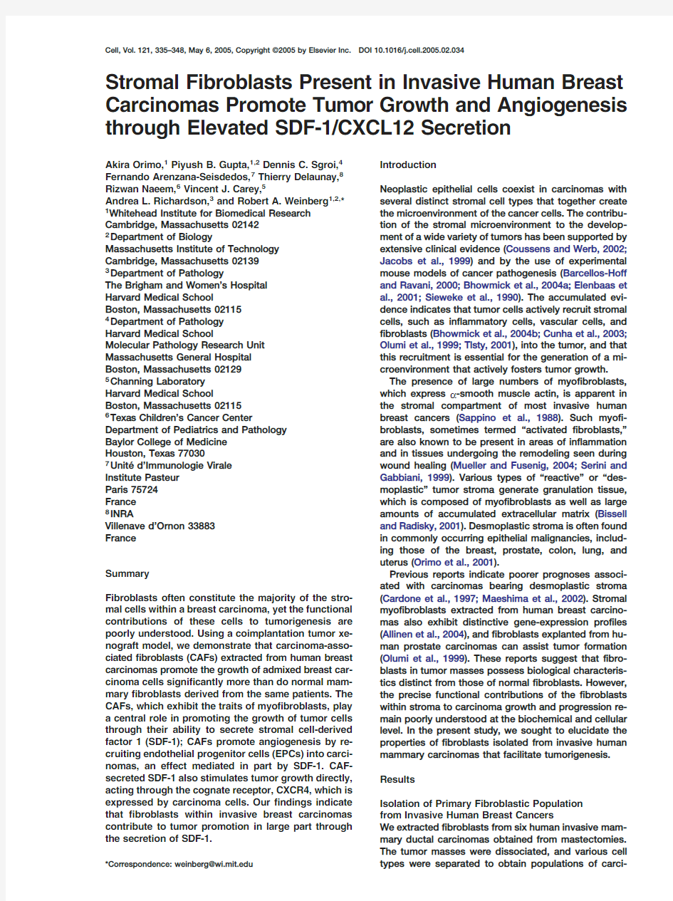
Figure1.Enhanced Tumor Growth Kinetics of MCF-7-ras Breast Cancer Cells Comingled with CAFs
(A)Fibroblastic properties of extracted primary human fibroblasts.CAFs(b,e,and h);Counterpart fibroblasts(a,d,and g);MCF-7-ras cells (c,f,and i)Sections were immunostained by anti-vimentin(a,b,and c),anti-prolyl4-hydroxylase(d,e,and f),or anti-cytokeratin antibodies (g,h,and i).Scale bar,50?m.
(B)MCF-7-ras cells were injected alone or coinjected with various fibroblasts subcutanously into nude mice.Tumor volume(a)was ploted in indicated days.Tumor weight(b)and the number of GFP-labeled MCF-ras cells(c)in each tumor were evaluataed at62days after injection. Error bars depict the standard error around the mean.*p<0.05by Student’s t test.
(C)Sections of GFP-labeled MCF-7-ras tumors comingled with CAFs at60days after injection were immunostained using an anti-GFP antibody(b)or stained by hematoxylin and eosin(H&E)(a).A different section(c)from a CAF-containing tumor was also immunostained using both anti-GFP(pink)and human-specific anti-vimentin(brown)antibodies.Scale bar,100?m.
(D)MCF-7-ras cells were coinjected with CAF1(5or10PDs),counterpart(5PDs),normal fibroblasts(5PDs),or no fibroblasts subcutaneously into nude mice.Error bars depict the standard error of the mean.
independent normal fibroblasts(Figure2C).Interest-tions of activated fibroblasts also possess proportion-
ately greater tumor-enhancing ability.
ingly,this increased contractile ability was observed
in all six independently isolated CAFs when compared
to corresponding counterpart fibroblasts(data not CAFs Enhance Tumor Angiogenesis
shown).CAF1cells passaged for10.5PDs in vitro as Initial cursory examinations of tumor sections contain-pure cultures also contracted collagen gels to nearly ing various types of admixed fibroblasts suggested that the same extent as CAF1cells passaged for five PDs CAFs show an increased ability to stimulate tumor an-(Figure2C).Taken together,these results confirmed giogenesis.We assessed formation of tumor-associ-that the CAFs possessed the properties of myofi-ated microvasculature at55–58days after injection by broblasts and maintained these traits without the con-examining serial sections from xenograft tumors ad-tinued presence of carcinoma cells.Moreover,we mixed with various fibroblasts.Both Masson’s tri-found a statistically positive correlation(p<0.001)be-chrome stain(Figure3Aa)and anti-CD31immuno-tween the contractile abilities of the various CAF pop-staining(Figure3Ad)revealed extensive vascular ulations and their tumor-enhancing powers(Figure S2;formation in tumors containing CAF1cells.In contrast, Supplemental Experimental Procedures,Data Analy-capillaries in tumors containing counterpart fibroblasts sis).Hence,CAF populations having greater propor-
(Figures3Ab and3Ae),normal fibroblasts(Figures3Ac
Cell
338
Table1.Summary of the Tumor-Promoting Ability in Six Different CAF Populations
CAFs Versus Normal Fibroblasts Versus Counterpart Fibroblasts
Increased
Increased Tumor Increased Increased
Tumor Growth Tumor Tumor Tumor Weight(g)Weight Tumor Weight(g)Rate Weight Tumor Weight(g)Growth Rate CAF10.384(n=10)+++*0.176(n=12)++*++*0.234(n=12)+++* (Std=0.145)(Std=0.054)(p=0.02)(Std=0.083)(p=0.02)
(62days)
CAF20.344(n=13)++*0.207(n=12)++*++*0.225(n=15)++* (Std=0.105)(Std=0.076)(p<0.01)(Std=0.070)(p<0.01)
(68days)
CAF30.381(n=13)++*0.235(n=15)+++*+*0.288(n=12)+* (Std=0.082)(Std=0.110)(p<0.01)(Std=0.079)(p<0.01)
(77days)
CAF40.443(n=15)?0.455(n=13)??0.528(n=11)?(Std=0.184)(Std=0.195)(p=0.7)(Std=0.188)(p=0.7)
(98days)
CAF50.337(n=11)+0.306(n=14)+?0.448(n=11)?(Std=0.144)(Std=0.112)(p=0.8)(Std=0.176)(p=0.8)
(103days)
CAF60.486(n=15)++*0.315(n=15)++*+0.445(n=13)+* (Std=0.143)(Std=0.147)(p<0.01)(Std=0.136)(p<0.01)
(83days)
CAFs and patient-matched,control counterpart fibroblasts were extracted from breast tissues obtained from six human breast cancer patients.In addition,normal breast fibroblasts were isolated from a healthy patient.Either set of described three types of fibroblasts was coinjected with MCF-7-ras cells subcutaneously into nude mice.Weight(g)and growth rate(increase of tumor volume/day)of tumors comingled CAFs were compared to those of tumors containing corresponding counterpart fibroblasts or normal fibroblasts.Thus,relative increased ratios of the averaged weight and growth rate in tumors containing CAFs are represented using an arbitrary scale.Averaged tumor weight(g)measured at the indicated day,the standard deviations of the tumor weights,and the numbers of examined tumors are also shown. *p<0.05by Student’s t test for tumor weight and by mixed effects models for tumor growth rate.Arbitrary scale;+++,more than2-fold up in CAF-containing tumors;++,1.5-to w2-fold up;+,1-to w1.5-fold up;?,not increased;Std,standard deviation.
and3Af),or no fibroblasts(data not shown)were far gle-cell suspensions at60–63days after subcutaneous less developed.Our assessment of the averaged
injection and then used flow cytometry to quantify the microvascular density per tumor type indicated a4.7-Sca1+cell population representing hematopoietic pro-fold greater level in CAF-containing tumors(Figure3Ba)
genitor cells and the Sca1+CD31+EPC population out when compared with tumors containing admixed coun-of the total viable cells in tumors.We found a far higher terpart fibroblasts.Moreover,Chalkley point counting
percentage of Sca1+cells(2.9-fold)and Sca1+CD31+ (numbers of grids covered on each vessel)indicated a EPCs(4.2-fold)in tumors containing CAF1cells when
compared to tumors containing counterpart fibroblasts 7.6-fold greater degree of vascularization in the same
tumors compared to tumors carrying admixed counter-(Figure3Cb).
part fibroblasts(Figure3Bb).Taken together,these ob-
Such EPCs likely arrive in tumors via the circulation, servations indicated that in the presence of CAFs,tu-having been mobilized in the bone marrow and there-mors became more vascularized,suggesting that CAFs
after introduced into the peripheral circulation(Heissig enhance tumor growth,at least in part,by promoting et al.,2002).To examine this possibility,we evaluated tumor neovascularization.
the extent of EPC mobilization into the peripheral blood
of mice harboring either CAF1-,counterpart fibroblast-,
or normal fibroblast-containing tumors.We observed a CAFs Induce Mobilization and Recruitment
higher level of Sca1+(2.1-fold)and Sca1+CD31+(2.0-of Endothelial Progenitor Cells
fold)mononuclear cells in the circulation of mice bear-Tumor angiogenesis has previously been demonstrated
ing CAF1-containing tumors compared to those circu-to occur in part through the recruitment of circulating
lating in mice bearing counterpart fibroblast-containing EPCs(Bertolini et al.,2003;Lyden et al.,2001).We
tumors(Figure3Cc).Moreover,we failed to observe a therefore wished to determine whether the ability of
significant difference in ratios of circulating EPCs in CAFs to promote tumor angiogenesis could be attrib-
mice bearing tumors developing in the absence of uted to an increased ability to recruit EPCs into the tu-
CAFs compared to those in non-tumor-bearing mice. mor masses.In these experiments,we considered
These observations confirmed that Sca1+CD31+cell Sca1+CD31+cells to represent the EPC-enriched cell
populations were indeed mobilized into the circulation fraction(Asahara et al.,1997;Rafii and Lyden,2003);
by the CAF-containing MCF-7-ras tumors in vivo. these cells also showed a20-fold higher ratio of VE-
To address whether these circulating cells could in cadherin-positive cells(an additional specific marker of
fact contribute to angiogenesis,we prepared Sca1+ endothelial cell lineages)when compared to Sca1?
CD31+EPCs or Sca1?CD31?cell fraction from the bone CD31?cells(Figure3Ca).
We dissociated the whole tumor xenografts into sin-marrow of RAG-1-deficient mice(having an H2k-b ma-
Fibroblast-Secreted SDF-1Enhances Tumor Growth
339
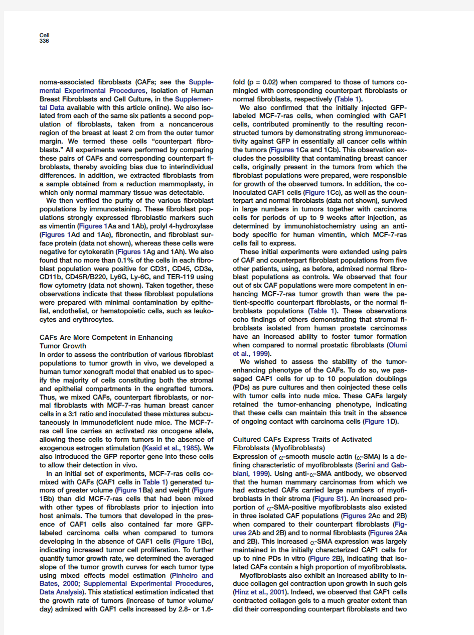
Figure2.CAFs Exhibit Characteristics of“Myofibroblasts”
(A)CAF1cells(c and d),cognate counterpart(b),or normal fibroblasts(a)were cultured in DMEM with2%FCS.Immunostaining with an anti-α-SMA antibody(a,b,and c)or control IgG(d).Scale bar,50?m.
(B)α-SMA-positive cell counts as a fraction of total cell numbers(>100counted cells)were evaluated in nine independent fields from three different wells of each fibroblast type under a fluorescence microscope.*p<0.05.
(C)The area of the contracted gels was measured at the indicated times.The appearance of the contracted gels at72hr is also shown. Error bars depict the standard error of the mean.
jor histocompatibility haplotype)by flow cytometry.three sets of CAFs and the respective patient-specific Subsequently,we injected5×105of these bone mar-
counterpart fibroblasts.These analyses showed an ele-row cells intravenously into haplotype mismatched vation of SDF-1expression in CAFs(data not shown). NOD-SCID(nonobese diabetic,severe combined im-
We therefore quantified expression levels of both munodeficient)mice(of an H2k-d haplotype)bearing SDF-1a and b mRNAs,which are alternative splicing 20-day-old MCF-7-ras tumors.As shown in Figure3Cd,
forms of a common pre-mRNA,in the various stromal at21days after injection of either bone marrow-derived fibroblast https://www.360docs.net/doc/066463194.html,ing real time RT-PCR,We cell preparation,we found that many tumor-associated
found increased mRNA levels of both SDF-1a(Figure capillaries(revealed by their display of CD31antigen)4Aa)and b(Figure4Ab)in CAF1(2.8-and 5.0-fold,
respectively),CAF2(2.3-and2.0-fold),and CAF3(5.6-were composed largely of cells staining positively for
the H2k-b haplotype(Figure3Cd3)by immunofluores-and2.3-fold)cell populations when compared to the cence.This demonstrates that circulating Sca1+CD31+
cognate counterpart fibroblasts of each set of CAFs. EPCs were,in fact,capable of differentiating into vas-In addition,we performed an ELISA assay on the me-cular endothelial cells in tumor-associated capillaries.
dium conditioned by each CAF population using an an-In contrast,in mice in which control Sca1?CD31?cells tibody that reacts with both SDF-1αandβforms.This had been injected intravenously,the tumor-associated
assay indicated elevated levels of SDF-1protein in the vasculature failed to incorporate cells of the H2k-b medium conditioned by the CAF1(1.8-fold),CAF2(2.6-
fold),and CAF3(3.1-fold)cells when compared to the haplotype(Figure3Cd4).These observations indicated
that one of the tumor-enhancing functions of CAFs de-levels produced by the respective counterpart fibro-rives from their ability to mobilize EPCs and to recruit
blast populations(Figure4Ac).We failed to observe them into the tumor mass.Once localized within tu-higher levels of SDF-1in CAF4and CAF5,CAFs that mors,such EPCs are capable of differentiating into tu-
had previously not shown any tumor-enhancing ability mor-associated vascular endothelial cells.in vivo(data not shown).Finally,MCF-7-ras cells
showed a far lower level of SDF-1secretion when com-CAFs Express High Levels of Stromal Cell-Derived
pared to CAFs(Figure4Ac).
Factor-1(SDF-1)We then performed immunostaining using both anti-Several secreted factors,such as vascular endothelial
SDF-1and anti-α-SMA antibodies on sections of inva-growth factor(VEGF),stromal cell-derived factor-1sive breast carcinomas obtained from human patients. (SDF-1),soluble c-Kit ligand,and matrix metalloprotei-
We observed that the subset of fibroblast-like cells nase9(MMP9),have been implicated as possible regu-positive forα-SMA(as indicated by arrows in Figure lators of EPC proliferation,mobilization,and migration
4Bb)also were positive for SDF-1(as indicated by ar-(Heissig et al.,2002).In order to examine factors in-rows,Figure4Ba)in tumor stroma.Notably,theα-SMA
protein present on myofibroblasts in tumor stroma was volved in the mobilization and recruitment of EPCs,we
performed DNA microarray expression analysis on largely colocalized with the SDF-1protein(Figure4Bf).
Cell
340
Figure3.MCF-7-ras Tumors Developing in the Presence of CAFs are Highly Angiogenic and Recruit Increased Numbers of EPCs
(A)Sections from MCF-7-ras tumors containing various fibroblasts were stained by anti-CD31antibody(d,e,and f)or by Masson’s trichrome (a,b,and c).Scale bar,100?m.
(B)Increased microvascular density(a)and Chalkley counts(b)in tumors comingled with CAFs**p<0.01.Error bars depict the standard error of the mean.
(C)(a)Mouse bone marrow cells were stained by anti-Sca1,-CD31,and-VE-cadherin antibodies.(b)MCF-7-ras tumors developing in the presence of various fibroblasts were dissociated into single cells at60–63days after injection.(c)Peripheral blood cells were also harvested from mice,and the positive cells out of the total viable cells were quantified using anti-Sca1and anti-CD31antibodies.*p<0.05.(d)Frozen sections were generated from tumors in mice injected intravenously with Sca1+CD31+EPCs(1,2,and3)or control Sca1?CD31?bone marrow cells(4).The sections were double-immunostained using both anti-H2k-b(1)and anti-CD31(2)antibodies and double-positive cells are shown in a merged view(3).Scale bar,50?m.
In contrast,we failed to detect any fibroblast-like cells To this end,we developed an in vitro transwell chemo-
taxis assay.GFP-labeled bone marrow fractions were positive for SDF-1in noncancer stroma(as shown by
arrowheads,Figure4Bc).As previously reported(Pab-sorted by flow cytometry to enrich for EPCs using anti-los et al.,1999),we detected the SDF-1protein to some
Sca1and anti-CD31antibodies,as before.Either Sca1+ extent in normal epithelial cells(as indicated by arrows,CD31+EPCs or control Sca?CD31?bone marrow cells Figure4Bc),breast carcinoma cells(depicted by an ar-
were then seeded into the upper wells of Boyden rowhead,Figures4Bd and4Bf)and endothelial cells chambers,and the lower wells of these chambers were
seeded with confluent layers of various stromal fibro-(data not shown).Together,these findings indicate that
the myofibroblasts present within breast carcinomas blast populations.
produce higher levels of SDF-1in vivo than do stromal
GFP-positive cells that migrated into the lower well fibroblasts present in noncancer stroma.were counted under a microscope18hr after initial co-
culturing.We found a3.3-fold increase in the chemo-SDF-1Released by CAFs Mediates the Chemotaxis
taxis index of the Sca1+CD31+EPCs when cocultured and Recruitment of EPCs with CAF1cells in the lower well(Figure5A)when com-We wished to test whether SDF-1released from CAFs
pared with cognate counterpart fibroblasts.Importantly, plays a functional role in stimulating EPC recruitment.treatment with an anti-SDF-1neutralizing antibody(10or
Fibroblast-Secreted SDF-1Enhances Tumor Growth
341
Figure4.CAFs Produce Increased SDF-1at Both mRNA and Protein Levels
(A)SDF-1a(a)and b(b)mRNA levels in various fibroblasts were quantified by real time PCR.SDF-1protein concentration(c)in the medium conditioned by these cells was measured by ELISA.Error bars represent the standard errors of the mean in three independent experiments. *p<0.05
(B)Invasive human breast carcinoma sections were immunostained with anti-SDF-1(a,c,d,and f)and anti-α-SMA(b,e,and f)antibodies. SDF-1+α-SMA+myofibroblasts are shown in a merged view(f).Scale bar,75?m.
20?g/ml)inhibited the increased chemotaxis toward8.6-fold higher in the Sca1+CD31+fraction when com-CAFs in a dose-dependent manner,whereas a control
pared to the Sca1?CD31?fraction(Figure5B),explain-mouse IgG failed to show this effect(Figure5A).More-ing the far higher SDF-1-dependent migration of the over,chemotaxis toward counterpart or normal fibro-
Sca1+CD31+fraction.
blasts was not affected by the anti-SDF-1neutralizing As an additional control,we verified that the migrated antibody(Figure5A).In contrast,the Sca1?CD31?frac-
Sca1+CD31+cells in these chamber assays were in-tion of the bone marrow cells showed a lower degree deed EPCs by assessing their colony-forming ability of chemotaxis that was largely SDF-1-independent and
upon long-term culture.After our transwell chemotaxis unaffected by the type of fibroblasts cultured in the migration assay,the mixed cell populations in the lower
wells were cultured in the absence of the upper wells lower well(Figure5A).Furthermore,the percentage of
cells expressing CXCR4—the receptor for SDF-1—was for an additional5weeks.CAF-containing cultures de-
Cell
342
Figure5.SDF-1Mediates the Increased Migration of EPC-Enriched Cells toward CAFs
(A)In vitro transwell chemotaxis assays were performed using four to five independent wells of each fibroblast type.The chemotaxis index was calculated as the ratio of the number of either Scal+CD31+EPCs or Scal?CD31?cells that migrated toward a particular fibroblast population to the number of these cells that migrated toward medium-only wells.**p<0.01.GFP-positive EPCs migrated on CAFs(d), counterpart(c)-,normal(b)-or nonfibroblast(a)layers.Scale bar,100?m.
(B)Bone marrow cells were stained with anti-CXCR4,anti-Sca1,and anti-CD31antibodies.Error bars represent the standard error around the mean in three independent experiments.
(C)Colonies formed on CAF1cells were evaluated by FITC-conjugated Ulex europaeus agglutinin I(lectin)binding(a)and DiI-labeled acet-ylated-LDL uptake(b).Merged images are also shown(c).Scale bar,50?m.
veloped EPC colonies(Figure5Cc)that were double-Petit et al.,2002)into nude mice bearing MCF-7-ras positive for DiI-labeled acetylated-LDL uptake and tumors admixed with either CAF1cells or counterpart FITC-conjugated Ulex europaeus agglutinin I(lectin)fibroblasts.Mice received intraperitoneal injections of binding,both of which are markers of EPCs(Kalka et antibodies twice a week for a period of68days follow-al.,2000).These observations,when taken together,in-ing the initial injection of cells.
dicate that CAFs have an enhanced ability in recruiting Significantly,the resulting MCF-7-ras tumors con-EPCs via SDF-1release in vitro.taining CAFs in mice treated with an anti-SDF-1anti-
body showed greatly attenuated tumor growth(Figure
6Aa),reduced tumor volume,and decreased tumor SDF-1Released by CAFs Contributes
weight(Figure6Ab)compared to those of CAF-contain-to the Recruitment of EPCs,
ing tumors in mice treated with control IgG.The CAF-Angiogenesis,and Tumor Growth
containing tumors in mice treated with an anti-SDF-1 We next examined whether SDF-1released by CAFs
antibody also exhibited a53%reduction in microvascu-did indeed play an essential role in enhancing tumor
lar density(Figure6Ac).Moreover,the proportion of growth and angiogenesis through EPC recruitment into
Sca1+CD31+EPCs out of the total viable cells in the the tumors.To do so,we administrated an anti-SDF-1
neutralizing antibody(Coulomb-L’Hermin et al.,1999;tumors was decreased by36%compared to CAF-con-
Fibroblast-Secreted SDF-1Enhances Tumor Growth
343
Figure6.An Anti-SDF-1Neutralizing Antibody Attenuated the Increased Growth of MCF-7-ras Tumor Developing in the Presence of CAFs, Decreased Angiogenesis,and Reduced Recruitment of EPCs into Tumors
(A)Mice bearing MCF-7-ras tumors comixed with CAF1cells or control counterpart fibroblasts were intraperitoneally treated with either anti-SDF-1antibody or control mouse IgG twice a week for a period of68days after initial injection of the tumor cells.We examined tumor growth kinetics(a),tumor weight(b),angiogenesis(c)and the percentage of Sca1+CD31+EPCs(d)within the tumor masses.*p<0.05.
(B)(a)Recombinant SDF-1αprotein was added to cultured MCF-7-ras cells in the presence of either control IgG or anti-CXCR4neutralizing antibody(20?g/ml).Total numbers of MCF-7-ras cells in each of four independent wells were counted at days3and5.(b)Two independent CXCR4-siRNA or control GFP-siRNA lentivirus vectors were introduced into MCF-7-ras cells.CXCR4protein expressed on the surface of MCF-7-ras cells was then analyzed using an anti-CXCR4antibody by flow cytometry.(c)MCF-7-ras cells expressing various siRNAs or non-siRNA were cultured in the presence or absence of SDF-1(100ng/ml).Total cell numbers in each of four independent wells were counted at day5.
Error bars depict the standard error of the mean.
taining tumors treated with control IgG(Figure6Ad),CAFs in vivo plays a crucial role in enhancing tumor whereas the anti-SDF-1antibody failed to affect signifi-
growth and angiogenesis by recruiting EPCs into tu-cantly any of the above parameters in those tumors mor masses.
formed by MCF-7-ras cells to which control counterpart
fibroblasts had been admixed(Figure6A).Stromal SDF-1Enhances Tumor Growth Significantly,as gauged by an ELISA assay,cultured
by Paracrine Stimulation of Tumor Cells
CAFs secreted SDF-1at a far higher level(33-fold Recent reports have indicated that SDF-1boosts the greater in the case of CAF1cells)than did MCF-7-ras
proliferation of several cancer cell lines in culture,in-cells,as shown in Figure4Ac.These findings argue that cluding breast carcinoma cells(Allinen et al.,2004;Hall
and Korach,2003).For this reason,we tested whether the inhibitory effect of the anti-SDF-1neutralizing anti-
body used in our xenograft model acted largely by in-CAF-secreted SDF-1could also directly stimulate MCF-hibiting SDF-1secreted by stromal CAFs,rather than
7-ras cell proliferation.Thus,we first treated MCF-7-by inhibiting SDF-1released by the MCF-7-ras cancer ras cells with recombinant SDF-1αprotein in culture. cells.These observations,together with those de-
Indeed,SDF-1(applied at1,10,or100ng/ml)stim-scribed earlier,indicate that the SDF-1released by ulated MCF-7-ras cell proliferation in a dose-dependent
Cell
344
Figure7.Schematic Representation of Tumor-Promoting Effects Provoked by Stromal Fibroblasts within Invasive Human Mammary Carci-nomas
(A)MCF-7-ras tumor cells expressing either control GFP-siRNA,CXCR4-siRNA-1,or-2vectors were admixed to SDF-1α-or control GFP-expressing normal human breast fibroblasts.These mixtures were then injected subcutaneously into nude mice.In each group,statistically significant difference of tumor volume is indicated by a bucket line.*p<0.05.Error bars depict the standard error of the mean.
(B)Two mechanisms by which SDF-1released by stromal fibroblasts in invasive human mammary carcinomas facilitates tumorigenesis. Stromal fibroblast-derived SDF-1enhances tumor growth both by stimulating angiogenesis through recruiting circulating EPCs into the tumor mass(endocrine effect)and by direct paracrine stimulation of tumor cells through CXCR4expressed on carcinoma cells(paracrine effect).
manner,generating1.2-,1.37-or1.5-fold increases in We found that MCF-7-ras tumors expressing either cell numbers,respectively,when compared to non-
CXCR4-siRNA vector showed more attenuated tumor SDF-1-treated MCF-7-ras cells(Figure6Ba).Moreover,growth kinetics and significantly reduced tumor volume an anti-CXCR4neutralizing antibody(20?g/ml),which
(p<0.05)in the presence of SDF-1α-expressing fibro-should block the ability of the CXCR4receptor to bind blasts compared to MCF-7-ras tumors expressing a
control GFP-siRNA vector(Figure7A).This observation its SDF-1ligand,largely inhibited the SDF-1-induced
proliferation(Figure6Ba),implicating CXCR4displayed demonstrates that stromal SDF-1enhances tumor by the MCF-7-ras cells as the mediator of this prolifera-
growth in vivo in part by direct paracrine stimulation tion effect.through CXCR4on MCF-7-ras breast cancer cells.
We then determined whether direct paracrine stimu-
We also found that MCF-7-ras tumors carrying either lation of tumor cells by stromal SDF-1occurs in vivo.CXCR4-siRNA vector admixed with control GFP-Furthermore,we sought to confirm that SDF-1acts
expressing fibroblasts grew more slowly and had a sig-through the CXCR4on the tumor cells themselves in nificantly decreased tumor volume(p<0.05)than did addition to other CXCR4-expressing cells such as
tumors arising from these same carcinoma cells that EPCs.To do so,we constructed MCF-7-ras cells un-had been commingled with SDF-1α-expressing fibro-able to respond to SDF-1by introducing lentiviral vec-
blasts(Figure7A).This result shows that stromal SDF-1,as tors expressing CXCR4-siRNA into these cells.The re-proposed above,also boosts tumorigenesis by affect-sulting significant inhibition of CXCR4receptor protein
ing other CXCR4-expressing cells besides the MCF-7-expression(by73%–78%)in the MCF-7-ras cells by ras tumor cells;these other targets might include the
EPCs studied in the earlier experiments.Together, either of two CXCR4-siRNA lentivirus vectors(Figure
6Bb)completely abrogated the ability of SDF-1to stim-these findings strongly suggest that CAF-secreted ulate MCF-7-ras cancer cell proliferation(Figure6Bc).
SDF-1promotes carcinoma growth in vivo by direct In contrast,a control GFP-siRNA construct failed to paracrine stimulation through CXCR4present on breast have this effect.Importantly,the loss of CXCR4did not
carcinoma cells in addition to its endocrine effects that affect MCF-7-ras cell proliferation in the absence of allow EPC recruitment into tumors(Figures7A and7B). SDF-1(Figure6Bc),indicating that the CXCR4-siRNA
vectors did not retard MCF-7-ras cell proliferation Discussion
through nonspecific effects.
In addition,we generated normal human mammary While the presence of fibroblasts in the stroma of inva-
sive mammary carcinomas is well documented,the stromal fibroblasts expressing retroviral SDF-1α-or
control GFP-expression vectors.SDF-1α-expressing specific contributions of these fibroblasts to tumor fibroblasts exhibited an18-fold higher level of SDF-1
growth have been unclear.We have shown here that protein,as measured by ELISA,when compared to fibroblasts present in invasive human mammary carci-control GFP-expressing fibroblasts(Figure S3).We ad-
noma masses are biologically very different from their mixed MCF-7-ras cells carrying CXCR4-or GFP-siRNAs counterparts located outside of the tumor masses in
several important functional respects.(1)Fibroblasts with SDF-1α-or control GFP-expressing fibroblasts
before injecting them subcutaneously into nude mice.extracted from within invasive human breast cancer
Fibroblast-Secreted SDF-1Enhances Tumor Growth
345
masses are more competent than their counterparts in Our work takes these other observations still further, enhancing tumor growth by comingled breast cancer by demonstrating that carcinoma cells can utilize the cells.(2)These fibroblasts exhibit increasedα-SMA ex-normal host stromal response by converting recruited pression as well as increased contractility,both indica-cells to CAFs,which in turn recruit EPCs into the tumor tive of myofibroblasts.(3)When comingled with breast via SDF-1(Figure7B).While CAFs contribute in a major cancer cells,these fibroblasts give rise to highly vascu-way to the recruitment of EPCs,it remains to be seen larized tumors.(4)These fibroblasts produce increased whether CAF-derived SDF-1is also involved in recruit-levels of SDF-1.(5)The SDF-1released by these fibro-ing leukocytes into tumors.Fibroblast-secreted SDF-1, blasts is responsible for recruiting EPCs into tumor for example,plays a key role in maintaining chronic in-masses,thereby boosting tumor angiogenesis.In addi-flammation by regulating the mobilization,recruitment, tion,the SDF-1produced by these CAFs enhances tu-and immigration of leukocytes into inflammatory tis-mor growth by direct paracrine stimulation via CXCR4sues(Douglas et al.,2002;Suratt et al.,2004).More-displayed by human breast carcinoma cells.over,chronic inflammation aids carcinoma growth by Significantly,the tumor-enhancing capability of CAFs generating a tumor-prone microenvironment(Coussens is largely maintained,at least during10PDs in vitro,in and Werb,2002).Furthermore,the recruitment of the absence of continuous interaction with carcinoma CD11b+Gr-1+leukocytes contributes to tumor angio-cells.The present experiments do not address how genesis by producing angiogenic factors,such as CAFs acquire these unique phenotypes and how these MMP9,and by directly differentiating into endothelial cells maintain the myofibroblastic phenotypes.Thus,cells(Yang et al.,2004).These lines of evidence support during tumor formation,preexisting mammary stromal the possibility that CAF-secreted SDF-1assists tumor fibroblasts or recruited progenitors(Ishii et al.,2003)progression in part through an inflammatory response. may be induced by nearby carcinoma cells to convert Importantly,endogenous CXCR4expression on car-into the myofibroblasts.Alternatively,myofibroblasts or cinoma cells is known to correlate with a poor progno-their progenitors may be recruited from outside of the sis for several types of carcinomas(Balkwill,2004; tumor mass.Staller et al.,2003),although relatively little is known Stromal regions microdissected from human breast about the role of its ligand,SDF-1,in primary tumor cancers show a high frequency of genetic alterations,growth.It is also known that CXCR4ectopically ex-such as loss of heterozygosity(LOH)and somatic mu-pressed on carcinoma cells enhances primary tumor tations(Fukino et al.,2004;Kurose et al.,2002;Moinfar growth in a mouse xenograft model(Darash-Yahana et et al.,2000).These observations raise the possibility al.,2004)and that the knockdown of CXCR4expression that an accumulation of genetic alterations may con-in breast carcinoma cells abrogates the tumor growth tribute to the CAFs’activated,tumor-enhancing pheno-(Lapteva et al.,2005;Smith et al.,2004).Moreover,a types.We note,however,that our CAFs show no detecta-small-molecule inhibitor of CXCR4reduces primary ble aneuploidy as determined by karyotype analysis,no brain tumor growth(Rubin et al.,2003).Thus,it is likely anchorage-independent growth in culture,and no tu-that SDF-1secreted by stromal myofibroblasts signifi-morigenicity in vivo(data not shown).Moreover,some cantly affects CXCR4-expressing human breast carci-of our CAFs begin to senesce after15PDs in culture.nomas through direct paracrine stimulation.Inhibition It is also quite likely that CAFs carry epigenetic alter-of the activated SDF-1/CXCR4signal pathway in a pri-ations,such as the generation of an autocrine TGF-βmary tumor environment may therefore attenuate carci-loop that can convert normal fibroblasts into myofi-noma growth in breast cancer patients.
broblasts(Ronnov-Jessen et al.,1996;Serini and Gab-
biani,1999).Importantly,CAFs exhibit activated pheno-
Experimental Procedures
types(e.g.,increased contractility)very similar to those
of myofibroblasts within wound sites.Such non-carci-Immunostaining of the Human Fibroblasts
noma-derived myofibroblasts may also have the poten-and Breast Cancer Tissues
tial to aid tumor growth.If such capability is indeed
For double immunostaining of vimentin and GFP in paraffin sec-
tions of MCF-7-ras tumors,the DAKO Envision Doublestain System observed,this would indicate that CAFs,such as those
was used.Primary cultured fibroblasts or frozen sections from hu-characterized here,are not unique to the stroma of car-
man breast carcinomas were also examined by immunofluores-cinomas and that once myofibroblasts are formed,
cence using the antibodies indicated below.
such cells may already be fully competent to exert tu-
mor-enhancing effects.
Antibodies
We have demonstrated that SDF-1secreted from
Primary antibodies used for staining included anti-pan-cytokeratin CAFs recruits EPCs into carcinomas,enhancing angio-(Sigma),human specific anti-vimentin(V9;Novocastra Laborato-genesis and thus tumor growth.This observation mir-ries,Ltd.,United Kingdom),anti-fibronectin(Sigma),anti-prolyl rors the observation that the tumor stroma resembles
4-hydroxylase(5B5;Dako,Denmark),anti-fibroblast surface pro-
tein(1B10;Sigma),anti-human CD31(Dako,Denmark;Santa Cruz, that present in sites of tissue injury(Dvorak,1986).
California),α-SMA(1A4;Dako,Denmark),FITC-conjugated anti-Thus,SDF-1released from injured tissues recruits
H2k-b,phycoerythrin(PE)-conjugated anti-CD31(BD PharMingen), EPCs into injured sites,inducing tissue regeneration
anti-SDF-1(K15C),and anti-GFP(Abcam,United Kingdom). (Askari et al.,2003;Ceradini et al.,2004;De Falco et
al.,2004;Schober et al.,2003).Moreover,our finding
Measurement of Collagen Gel Contraction
that CAFs release higher levels of SDF-1is supported
by Human Breast Fibroblasts
by recent work indicating that myofibroblasts extracted2×105fibroblasts were suspended in300?l of collagen lattices from human mammary tumors express increased levels prepared in24-well plates,and4–5independent collagen gels in-of the SDF-1mRNA(Allinen et al.,2004).
cluding various fibroblasts were cultured in DMEM with2%FCS.
Cell
346
The assay was performed as previously described(Bogatkevich et munostained by FITC-conjugated anti-H2k-b and PE-conjugated al.,2001).
anti-CD31antibodies.
Subcutaneous Tumorigenicity Assays Flow Cytometry
One million MCF-7-ras cells and three million fibroblasts of various Extracted human fibroblasts were stained by biotin-conjugated types were coinjected subcutaneously into nude mice.Tumorige-CD45,CD3e,CD11b,CD45R/B220,Ly6G,Ly-6C,and TER-119anti-nicity assays were performed as previously described(Elenbaas et bodies(BD PharMingen).Streptavidin(SA)-allophycocyanin(APC) al.,2001).was then used for detection of biotin-positive cells.106mononu-
clear cells from mouse peripheral blood or bone marrow,106cul-
tured MCF-7-ras cells,or106cells dissociated from xenograft tu-Plasmid Construction
mors were incubated for20min at4°C with the following The GFP gene was cloned into the pWZL-Blasticidin retroviral vec-
antibodies:Anti-FITC-or PE-conjugated Sca1,anti-PE-or APC-tor.The human SDF-1a cDNA(Shirozu et al.,1995)was also cloned
conjugated CD31,VE-cadherin,anti-FITC-conjugated mouse CXCR4, into the pBabe-puro retroviral vector.For inhibition of endogenous
or anti-PE-conjugated human CXCR4(12G5)(BD PharMingen). CXCR4expression in MCF-7-ras cells,oligonucleotides against hu-
Cells incubated with7-amino-actinomycin D(7-AAD;BD Phar-man CXCR4or GFP genes were generated and cloned into a lenti-
Mingen)were analyzed using flow cytometry(Becton Dickinson). virus-derived pLKO-puro-RNAi vector(Stewart et al.,2003).The fol-
lowing oligomers were used:CXCR4-siRNA-1,5#-TGGAGGGGAT
CAGTATATACA-3#;CXCR4-siRNA-2,5#-GTTTTCACTCCAGCTAAC In Vitro Transmigration Assay
ACA-3#as previously described(Anderson et al.,2003;Doench et1×105fibroblasts were seeded in the lower well of a24-well cham-al.,2003);GFP-siRNA,5#-GCAAGCTGACCCTGAAGTTCA-3#.ber two days prior to coculture.GFP-labeled Sca1+CD31+or
Sca1?CD31?cell populations were sorted from whole bone marrow
cells from GFP-transgenic mice(Ikawa et al.,1995)using flow cyto-Retroviral and Lentiviral Infections
metry.4×104of these bone marrow cells were then seeded in Retrovirus infection was performed as previously described(Elen-
DMEM+2%FCS in the upper well of a transwell chamber with a baas et al.,2001).pBabe-puro-SDF-1a or-GFP vectors were used
5?m pore size(Costar,Massachusetts).Cells were incubated with to infect normal human breast tissue-derived fibroblasts immortal-
either anti-SDF-1neutralizing antibody or control IgG one day prior ized by hTERT,the catalytic subunit of telomerase.A pWZL-Blas-
to coculture.
ticidin-GFP vector was also introduced by infection into MCF-7-
ras cells.Lentivirus infection of either siRNA-CXCR4or siRNA-GFP
vector was performed as previously described(Stewart et al.,MCF-7-ras Cell Proliferation Assay in Culture
2003).After infection,MCF-7-ras cells were selected by2?g/ml1×104MCF-7-ras cells were cultured in DMEM with2%FCS in puromycin for3.5days and used for experiments on the following the presence or absence of SDF-1α(1–100ng/ml).An anti-CXCR4 day.neutralizing antibody(12G5;20?g/ml)or control mouse IgG was
added12hr prior to treatment with SDF-1α.4×104MCF-7-ras Real Time PCR cells carrying different siRNAs were also cultured in the presence Quantitative real time RT-PCR analysis was performed as pre-or absence of SDF-1α(100ng/ml).Total cell numbers of MCF-7-viously described(Brisken et al.,2002).Data were normalized rela-ras cells in each of four independent wells in24-well plates were tive to the expression level ofβ2-microglobin for each sample.counted at the indicated days.
Primers used for RT-PCR were human SDF-1a,5#-TGAGAGCTC
GCTTTGAGTGA-3#(sense)and5#-CACCAGGACCTTCTGTGGAT-3#Treatment with Anti-SDF-1Neutralizing Antibody(K15C) (antisense);SDF-1b,5#-CTAGTCAAGTGCGTCCACGA-3#(sense)in Mice Bearing Tumors
and5#-GGACACACCACAGCACAAAC-3#(antisense);and b2-micro-MCF-7-ras cells admixed with either CAFs or control counterpart globin,5#-TGAGTGCTGTCTCCATGTTTGA-3#(sense)and5#-TCTG fibroblasts were injected subcutaneously into nude mice.K15C(32 CTCCCCACCTCTAAGTTG-3#(antisense).?g/mouse)or control isotype IgG2(32?g/mouse)antibodies were
intraperitoneally injected starting one day prior to cell injection and Immunoassay for Human SDF-1subsequently injected twice weekly at the same dose(32?g/ 1.5×10⁶fibroblasts were cultured in DMEM with2%FCS on a10mouse)for a period of68days.
cm culture plate for2days.The supernatants were measured using
a commercially available SDF-1ELISA kit(R&D Systems).
Supplemental Data
Evaluation of Angiogenesis in Xenograft MCF-7-ras Tumors Supplemental Data include three figures and Supplemental Experi-Serial sections(taken at2mm intervals)were prepared from MCF-mental Procedures and can be found with this article online at 7-ras tumors(comingled with various fibroblasts or no fibroblasts).https://www.360docs.net/doc/066463194.html,/cgi/content/full/121/3/335/DC1/.
A total of30sections from six independent tumors of each group
were then immunostained using an anti-CD31antibody.Microves-
sel density was assessed as described previously(Weidner et al.,Acknowledgments
1991).After taking these counts,a49-point Chalkley point eyepiece
graticule(Klarmann Rulings,Inc.,New Hampshire)was employed We would like to thank Dr.Todd R.Golub for help of DNA microar-over the same tissue section and the number of graticular points ray experiment and critical reading of this manuscript,Drs.Ittai (at×400magnification)that fell within the lumen of vessels was Ben-Porath,Kimberly Hartwell,Christina Scheel,Nir Hacohen, counted(Fox et al.,1995).Konrad Hochedlinger,and Chengcheng Zhang for critical reading
of this manuscript and Drs.Paul Matsudaira,S.A.Mani,and Sridhar Colony Formation Assay of Endothelial Progenitor Cells Ramaswamy for helpful experimental suggestions and data analy-After5weeks of coculture in DMEM+2%FCS,uptake of DiI-sis and members of R.A.W.’s laboratory for helpful comments and labeled acetylated-LDL and staining of FITC-conjugated Ulex euro-discussion.We appreciate Dr.Charlotte Kuperwasser’s assistance paeus agglutinin I(lectin)were examined according to a method in the isolation of fibroblasts from human breast tissues,Dr.Tasuku previously described(Hatzopoulos et al.,1998).Honjo for gift of the human SDF-1cDNA,and Dr.Ittai Ben-Porath
for a pLKO1-GFP-siRNA vector.This work was supported by Incorporation of EPCs into MCF-7-ras Xenograft Tumors
Merck/MIT(R.A.W.),NIH/NCI grant R21CA87081-02(R.A.W.),the NOD-SCID(H2k-d haplotype+)and RAG-1-deficient mice(H2k-b Ludwig Trust(R.A.W.),the Breast Cancer Research Foundation haplotype+;Jackson Laboratory,)were used.Sca1+CD31+or
(R.A.W.),Uehara Memorial Foundation(A.O.),Sankyo Foundation Sca1?CD31?bone marrow fractions from the RAG-1-deficient mice of Life Science(A.O.),and a US Army Pre-doctoral Breast Cancer were intravenously injected into the NOD-SCID mice bearing MCF-
Fellowship DAMD17-02-1-0468(P.B.G).R.A.W.is an American Can-7-ras tumors.Frozen sections from the resulting tumors were im-cer Society Research Professor and a Daniel K.Ludwig Cancer
Fibroblast-Secreted SDF-1Enhances Tumor Growth
347
Research Professor.This work was conducted utilizing the W.M.Coussens,L.M.,and Werb,Z.(2002).Inflammation and cancer.Na-Keck Foundation Biological imaging facility at the Whitehead In-
ture420,860–867.
stitute.Cunha,G.R.,Hayward,S.W.,Wang,Y.Z.,and Ricke,W.A.(2003).
Role of the stromal microenvironment in carcinogenesis of the
prostate.Int.J.Cancer107,1–10.
Received:February26,2004
Revised:November30,2004Darash-Yahana,M.,Pikarsky,E.,Abramovitch,R.,Zeira,E.,Pal,B., Accepted:February25,2005Karplus,R.,Beider,K.,Avniel,S.,Kasem,S.,Galun,E.,and Peled, Published:May5,2005 A.(2004).Role of high expression levels of CXCR4in tumor growth,
vascularization,and metastasis.FASEB J.18,1240–1242. References
De Falco,E.,Porcelli,D.,Torella,A.R.,Straino,S.,Iachininoto,M.G.,
Orlandi,A.,Truffa,S.,Biglioli,P.,Napolitano,M.,Capogrossi,M.C., Allinen,M.,Beroukhim,R.,Cai,L.,Brennan,C.,Lahti-Domenici,J.,
and Pesce,M.(2004).SDF-1involvement in endothelial phenotype Huang,H.,Porter,D.,Hu,M.,Chin,L.,Richardson,A.,et al.(2004).
and ischemia-induced recruitment of bone marrow progenitor cells. Molecular characterization of the tumor microenvironment in breast
Blood104,3472–3482.
cancer.Cancer Cell6,17–32.
Doench,J.G.,Petersen,C.P.,and Sharp,P.A.(2003).siRNAs can Anderson,J.,Banerjea,A.,Planelles,V.,and Akkina,R.(2003).Po-
function as miRNAs.Genes Dev.17,438–442.
tent suppression of HIV type1infection by a short hairpin anti-
Douglas,M.R.,Morrison,K.E.,Salmon,M.,and Buckley, C.D. CXCR4siRNA.AIDS Res.Hum.Retroviruses19,699–706.
(2002).Why does inflammation persist:a dominant role for the stro-Asahara,T.,Murohara,T.,Sullivan,A.,Silver,M.,van der Zee,R.,
mal microenvironment?Expert Rev.Mol.Med.2002,1–18.
Li,T.,Witzenbichler,B.,Schatteman,G.,and Isner,J.M.(1997).Iso-
lation of putative progenitor endothelial cells for angiogenesis.Sci-Dvorak,H.F.(1986).Tumors:wounds that do not heal.Similarities ence275,964–967.
between tumor stroma generation and wound healing.N.Engl.J.
Med.315,1650–1659.
Askari,A.T.,Unzek,S.,Popovic,Z.B.,Goldman,C.K.,Forudi,F.,
Kiedrowski,M.,Rovner,A.,Ellis,S.G.,Thomas,J.D.,DiCorleto,P.E.,Elenbaas,B.,Spirio,L.,Koerner,F.,Fleming,M.D.,Zimonjic,D.B., et al.(2003).Effect of stromal-cell-derived factor1on stem-cell Donaher,J.L.,Popescu,N.C.,Hahn,W.C.,and Weinberg,R.A. homing and tissue regeneration in ischaemic https://www.360docs.net/doc/066463194.html,n-(2001).Human breast cancer cells generated by oncogenic trans-cet362,697–703.formation of primary mammary epithelial cells.Genes Dev.15,50–
65.
Balkwill,F.(2004).Cancer and the chemokine network.Nat.Rev.
Cancer4,540–550.Fox,S.B.,Leek,R.D.,Weekes,M.P.,Whitehouse,R.M.,Gatter,K.C.,
and Harris,A.L.(1995).Quantitation and prognostic value of breast Barcellos-Hoff,M.H.,and Ravani,S.A.(2000).Irradiated mammary
cancer angiogenesis:comparison of microvessel density,Chalkley gland stroma promotes the expression of tumorigenic potential by
count,and computer image analysis.J.Pathol.177,275–283. unirradiated epithelial cells.Cancer Res.60,1254–1260.
Bertolini,F.,Paul,S.,Mancuso,P.,Monestiroli,S.,Gobbi, A.,Fukino,K.,Shen,L.,Matsumoto,S.,Morrison,C.D.,Mutter,G.L., Shaked,Y.,and Kerbel,R.S.(2003).Maximum tolerable dose and
and Eng,C.(2004).Combined total genome loss of heterozygosity low-dose metronomic chemotherapy have opposite effects on the scan of breast cancer stroma and epithelium reveals multiplicity of mobilization and viability of circulating endothelial progenitor cells.
stromal targets.Cancer Res.64,7231–7236.
Cancer Res.63,4342–4346.Hall,J.M.,and Korach,K.S.(2003).Stromal cell-derived factor1,a Bhowmick,N.A.,Chytil,A.,Plieth,D.,Gorska,A.E.,Dumont,N.,novel target of estrogen receptor action,mediates the mitogenic Shappell,S.,Washington,M.K.,Neilson, E.G.,and Moses,H.L.effects of estradiol in ovarian and breast cancer cells.Mol.Endocri-(2004a).TGF-beta signaling in fibroblasts modulates the oncogenic nol.17,792–803.
potential of adjacent epithelia.Science303,848–851.Hatzopoulos,A.K.,Folkman,J.,Vasile,E.,Eiselen,G.K.,and Ro-Bhowmick,N.A.,Neilson,E.G.,and Moses,H.L.(2004b).Stromal senberg,R.D.(1998).Isolation and characterization of endothelial fibroblasts in cancer initiation and progression.Nature432,332–progenitor cells from mouse embryos.Development125,1457–337.1468.
Bissell,M.J.,and Radisky,D.(2001).Putting tumours in context.Heissig,B.,Hattori,K.,Dias,S.,Friedrich,M.,Ferris,B.,Hackett, Nat.Rev.Cancer1,46–54.N.R.,Crystal,R.G.,Besmer,P.,Lyden,D.,Moore,M.A.,et al.(2002). Bogatkevich,G.S.,Tourkina,E.,Silver,R.M.,and Ludwicka-Bradley,
Recruitment of stem and progenitor cells from the bone marrow A.(2001).Thrombin differentiates normal lung fibroblasts to a myo-niche requires MMP-9mediated release of kit-ligand.Cell109, fibroblast phenotype via the proteolytically activated receptor-1
625–637.
and a protein kinase C-dependent pathway.J.Biol.Chem.276,Hinz,B.,Celetta,G.,Tomasek,J.J.,Gabbiani,G.,and Chaponnier, 45184–45192. C.(2001).Alpha-smooth muscle actin expression upregulates fibro-Brisken,C.,Ayyannan,A.,Nguyen,C.,Heineman,A.,Reinhardt,F.,blast contractile activity.Mol.Biol.Cell12,2730–2741.
Tan,J.,Dey,S.K.,Dotto,G.P.,Weinberg,R.A.,and Jan,T.(2002).
Ikawa,M.,Kominami,K.,Yoshimura,Y.,Tanaka,K.,Nishimune,Y., IGF-2is a mediator of prolactin-induced morphogenesis in the
and Okabe,M.(1995).A rapid and non-invasive selection of trans-breast.Dev.Cell3,877–887.
genic embryos before implantation using green fluorescent protein Cardone,A.,Tolino,A.,Zarcone,R.,Borruto Caracciolo,G.,and(GFP).FEBS Lett.375,125–128.
Tartaglia,E.(1997).Prognostic value of desmoplastic reaction and
Ishii,G.,Sangai,T.,Oda,T.,Aoyagi,Y.,Hasebe,T.,Kanomata,N., lymphocytic infiltration in the management of breast cancer.Pan-
Endoh,Y.,Okumura,C.,Okuhara,Y.,Magae,J.,et al.(2003).Bone-minerva Med.39,174–177.
marrow-derived myofibroblasts contribute to the cancer-induced Ceradini,D.J.,Kulkarni,A.R.,Callaghan,M.J.,Tepper,O.M.,Basti-stromal https://www.360docs.net/doc/066463194.html,mun.309,232–240. das,N.,Kleinman,M.E.,Capla,J.M.,Galiano,R.D.,Levine,J.P.,
Jacobs,T.W.,Byrne,C.,Colditz,G.,Connolly,J.L.,and Schnitt,S.J. and Gurtner,G.C.(2004).Progenitor cell trafficking is regulated by
(1999).Radial scars in benign breast-biopsy specimens and the hypoxic gradients through HIF-1induction of SDF-1.Nat.Med.10,
risk of breast cancer.N.Engl.J.Med.340,430–436.
858–864.
Kalka,C.,Masuda,H.,Takahashi,T.,Kalka-Moll,W.M.,Silver,M., Coulomb-L’Hermin,A.,Amara,A.,Schiff,C.,Durand-Gasselin,I.,
Kearney,M.,Li,T.,Isner,J.M.,and Asahara,T.(2000).Transplanta-Foussat,A.,Delaunay,T.,Chaouat,G.,Capron,F.,Ledee,N.,Gala-
tion of ex vivo expanded endothelial progenitor cells for therapeu-naud,P.,et al.(1999).Stromal cell-derived factor1(SDF-1)and
tic https://www.360docs.net/doc/066463194.html,A97,3422–3427. antenatal human B cell lymphopoiesis:expression of SDF-1by
mesothelial cells and biliary ductal plate epithelial cells.Proc.Natl.Kasid,A.,Lippman,M.E.,Papageorge,A.G.,Lowy,D.R.,and https://www.360docs.net/doc/066463194.html,A96,8585–8590.
mann,E.P.(1985).Transfection of v-rasH DNA into MCF-7human
Cell
348
breast cancer cells bypasses dependence on estrogen for tumori-(1990).Mediation of wound-related Rous sarcoma virus tumorigen-genicity.Science228,725–728.
esis by TGF-beta.Science248,1656–1660.
Smith,M.C.,Luker,K.E.,Garbow,J.R.,Prior,J.L.,Jackson,E.,Piw-Kurose,K.,Gilley,K.,Matsumoto,S.,Watson,P.H.,Zhou,X.P.,and
nica-Worms,D.,and Luker,G.D.(2004).CXCR4regulates growth Eng,C.(2002).Frequent somatic mutations in PTEN and TP53are
of both primary and metastatic breast cancer.Cancer Res.64, mutually exclusive in the stroma of breast carcinomas.Nat.Genet.
8604–8612.
32,355–357.
Staller,P.,Sulitkova,J.,Lisztwan,J.,Moch,H.,Oakeley,E.J.,and Lapteva,N.,Yang,A.G.,Sanders,D.E.,Strube,R.W.,and Chen,
Krek,W.(2003).Chemokine receptor CXCR4downregulated by von S.Y.(2005).CXCR4knockdown by small interfering RNA abrogates
Hippel-Lindau tumour suppressor pVHL.Nature425,307–311. breast tumor growth in vivo.Cancer Gene Ther.12,84–89.
Stewart,S.A.,Dykxhoorn,D.M.,Palliser,D.,Mizuno,H.,Yu,E.Y., Lyden,D.,Hattori,K.,Dias,S.,Costa,C.,Blaikie,P.,Butros,L.,
An,D.S.,Sabatini,D.M.,Chen,I.S.,Hahn,W.C.,Sharp,P.A.,et al. Chadburn,A.,Heissig,B.,Marks,W.,Witte,L.,et al.(2001).Im-
(2003).Lentivirus-delivered stable gene silencing by RNAi in pri-paired recruitment of bone-marrow-derived endothelial and
mary cells.RNA9,493–501.
hematopoietic precursor cells blocks tumor angiogenesis and
growth.Nat.Med.7,1194–1201.Suratt,B.T.,Petty,J.M.,Young,S.K.,Malcolm,K.C.,Lieber,J.G.,
Nick,J.A.,Gonzalo,J.A.,Henson,P.M.,and Worthen,G.S.(2004). Maeshima,A.M.,Niki,T.,Maeshima,A.,Yamada,T.,Kondo,H.,and
Role of the CXCR4/SDF-1chemokine axis in circulating neutrophil Matsuno,Y.(2002).Modified scar grade:a prognostic indicator in
homeostasis.Blood104,565–571.
small peripheral lung adenocarcinoma.Cancer95,2546–2554.
Tlsty,T.D.(2001).Stromal cells can contribute oncogenic signals. Moinfar,F.,Man,Y.G.,Arnould,L.,Bratthauer,G.L.,Ratschek,M.,
Semin.Cancer Biol.11,97–104.
and Tavassoli,F.A.(2000).Concurrent and independent genetic al-
Weidner,N.,Semple,J.P.,Welch,W.R.,and Folkman,J.(1991).Tu-terations in the stromal and epithelial cells of mammary carcinoma:
mor angiogenesis and metastasis—correlation in invasive breast implications for tumorigenesis.Cancer Res.60,2562–2566.
carcinoma.N.Engl.J.Med.324,1–8.
Mueller,M.M.,and Fusenig,N.E.(2004).Friends or foes—bipolar
Yang,L.,Debusk,L.M.,Fukuda,K.,Fingleton,B.,Green-Jarvis,B., effects of the tumour stroma in cancer.Nat.Rev.Cancer4,839–
Shyr,Y.,Matrisian,L.M.,Carbone,D.P.,and Lin,P.C.(2004).Expan-849.
sion of myeloid immune suppressor Gr+CD11b+cells in tumor-Olumi,A.F.,Grossfeld,G.D.,Hayward,S.W.,Carroll,P.R.,Tlsty,T.D.,
bearing host directly promotes tumor angiogenesis.Cancer Cell6, and Cunha,G.R.(1999).Carcinoma-associated fibroblasts direct
409–421.
tumor progression of initiated human prostatic epithelium.Cancer
Res.59,5002–5011.
Orimo,A.,Tomioka,Y.,Shimizu,Y.,Sato,M.,Oigawa,S.,Kamata,
K.,Nogi,Y.,Inoue,S.,Takahashi,M.,Hata,T.,and Muramatsu,M.
(2001).Cancer-associated myofibroblasts possess various factors
to promote endometrial tumor progression.Clin.Cancer Res.7,
3097–3105.
Pablos,J.L.,Amara,A.,Bouloc,A.,Santiago,B.,Caruz,A.,Galindo,
M.,Delaunay,T.,Virelizier,J.L.,and Arenzana-Seisdedos,F.(1999).
Stromal-cell derived factor is expressed by dendritic cells and en-
dothelium in human skin.Am.J.Pathol.155,1577–1586.
Petit,I.,Szyper-Kravitz,M.,Nagler,A.,Lahav,M.,Peled,A.,Habler,
L.,Ponomaryov,T.,Taichman,R.S.,Arenzana-Seisdedos,F.,Fujii,
N.,et al.(2002).G-CSF induces stem cell mobilization by decreas-
ing bone marrow SDF-1and up-regulating CXCR4.Nat.Immunol.
3,687–694.
Pinheiro,J.,and Bates,D.(2000).Mixed Effects Models in S and
S-Plus(New York:Springer).
Rafii,S.,and Lyden,D.(2003).Therapeutic stem and progenitor
cell transplantation for organ vascularization and regeneration.Nat.
Med.9,702–712.
Ronnov-Jessen,L.,Petersen,O.W.,and Bissell,M.J.(1996).Cellu-
lar changes involved in conversion of normal to malignant breast:
importance of the stromal reaction.Physiol.Rev.76,69–125.
Rubin,J.B.,Kung,A.L.,Klein,R.S.,Chan,J.A.,Sun,Y.,Schmidt,K.,
Kieran,M.W.,Luster,A.D.,and Segal,R.A.(2003).A small-molecule
antagonist of CXCR4inhibits intracranial growth of primary brain
https://www.360docs.net/doc/066463194.html,A100,13513–13518.
Sappino,A.P.,Skalli,O.,Jackson,B.,Schurch,W.,and Gabbiani,
G.(1988).Smooth-muscle differentiation in stromal cells of malig-
nant and non-malignant breast tissues.Int.J.Cancer41,707–712.
Schober,A.,Knarren,S.,Lietz,M.,Lin,E.A.,and Weber,C.(2003).
Crucial role of stromal cell-derived factor-1alpha in neointima for-
mation after vascular injury in apolipoprotein E-deficient mice.Cir-
culation108,2491–2497.
Serini,G.,and Gabbiani,G.(1999).Mechanisms of myofibroblast
activity and phenotypic modulation.Exp.Cell Res.250,273–283.
Shirozu,M.,Nakano,T.,Inazawa,J.,Tashiro,K.,Tada,H.,Shino-
hara,T.,and Honjo,T.(1995).Structure and chromosomal localiza-
tion of the human stromal cell-derived factor1(SDF1)gene.Ge-
nomics28,495–500.
Sieweke,M.H.,Thompson,N.L.,Sporn,M.B.,and Bissell,M.J.
情态动词can的用法及一般疑问句whatquestions
情态动词"can“的用法 情态动词不能独立作谓语,不以人称和数量的变化而变化,后接动词原形,也就是说情态动词和动词原形一起构成谓语。我们常见的情态动词有:can,must,would等。接下来,我们就来学习一下情态动词"can"的用法。 ①表示能力,"can"译为"能;会" 例句:I can speak English. 译:我会说英语。 She can dance. 译:她会跳舞。 ②表示可能性,"can"译为"可能;会" 例句:Can you sing 译:你会唱歌吗 Can you swim 译:你会游泳吗 ③"can"用在肯定句中:主语+can+动词原形 例句:I can play the guitar. 译:我会弹吉他。 I can join the music club. 译:我可以加入音乐俱乐部。 ④"can"用在一般疑问句中:Can+主语+动词原形 肯定回答:Yes,主语+can 否定回答:No,主语+can't 例句:Can he play chess 译:他会下国际象棋吗 Yes,he can. 译:是的,他会。 NO,he can't. 译:不,她不会。 ⑤"can"用在否定句中:主语+can't+动词原形(can't 是 can not 的缩写) 例句:She can't play the piano well. 译:她弹不好钢琴。 I can't sing. 译:我不会唱歌。 ⑥"can"用在特殊疑问句中:特殊疑问词+can+主语+动词原形 例句:What can you see in the room 译:在房间里你能看见什么 ⑦当特殊疑问词是句子的主语时:特殊疑问词+can+动词原形 例句:Who can answer my question 译:谁能回答我的问题 注意事项 "can"后一定要接动词原形。 "can" 不以人称和数量的变化而变化。
人教版英语八年级下册试题since 和 for 的用法(用于现在完成时)
初中英语学习材料 madeofjingetieji since 和for 的用法(用于现在完成时) 表示过去已经开始持续到现在的动作或状态常用的时间状语有:for, since, how long, so far, these days等。 1. since : a).since +时间点①年代②时刻数③一段时间+ago b).现在完成时:主句(主语+have/has +延续性动词的过去分词)+ since +从句一般过去时 c) It’s +时间段+since +短暂性动词的过去式 d).时间段+has passed +since +短暂性动词的过去式 for +一段时间 练习: 用since和for填空 1) ______ two years 2) _______ two years ago 3) _______ last month 4) ______ 1999 5) _______ yesterday 6) _______ 4 o’clock 7) ______ 4 hours 8) _______ an hour ago 9) _______ we were children 10) _____ lunch time 11) ______ she left here 12). He has lived in Nanjing ________ the year before last. 13). I’ve known him __________ we were children. 14). Our teacher has studied Japanese _________ three years. 15). She has been away from the city ___________ about ten years. 16). It’s about ten years __________ she left the city. 2. for: for +一段时间= since +一段时间+ ago 3. 与时间段连用时,短暂性动词应改为相应的延续性动词。 1) come/go to ------ be at /in 2)leave ----be away from 3)buy ----have 4) borrow /lend -----keep 4)open ---be open 5)close---be closed 6) die---be dead 7) start/begin ----be on 8) join—--be in /be a member of /be a soldier 9) become –be 10) fall asleep ---be asleep 11)catch a cold – have a cold 12) have/has gone to → have been in 13) put on → wear14) finish/end → be over 15) marry → be married 练习:.短暂性转换延续性 1) His grandfather died two years ago . His grandfather has ____ ____ for two years. _____two years _____ his grandfather ____. Two years ____ ____ ____ his grandfather _____. 2) I became a teacher in 2000. I ________ __________ a teacher for _________ _________. 3) The shop closed two hours ago. The shop ________ _________ _________ for _________ _________. 4) The door opened at six in the morning. The door ________ ________ ________ for six hours. 5)He left Fuzhou just now. He _______ ________ ________ _________ Fuzhou for five minutes. 6)The film began two minutes ago. The film ____ ____ ____ for ____ ____.How time flies! 7)They borrowed it last week. They _________ _________ it since __________ __________. 8)I bought a pen two hours ago. I _________ _________ a pen for ________ __________. 9)They married in 1990. They ________ _________ __________since _________. 10)The meeting finished at six. The meeting ________ ______ ______ for six hours. 11)My brother joined the army two years ago.
情态动词的基本用法及其区别
情态动词的基本用法及其区别 最近几年高考试题中常常借助语境来考查情态动词的基本用法及其区别,因此在平时学习时准确理解和掌握情态动词的基本用法十分重要。情态动词的用法复杂多变,在高考试题中,命题者常常利用语境和句子之间意义上的细微差别来考查学生对情态动词的理解和掌握。对于情态动词,除了要求考生能够准确掌握它们的基本用法外,还要充分利用高考试题所设置的语境来分析句子之间所体现的特殊关系。下面就近几年来高考试题中出现的情态动词的考点进行归纳分析,以便同学们复习掌握。 一、用“情态动词+have +done”结构表示对过去动作的推测,高考试题中常用过去时态或过去的时间状语给以暗示。情态动词的这一用法可以用“对立统一”来概括。 1.当试题的前句和后句在动作和意义上相互补充说明,且整个句意在动作和时间上是一个整体时,我们可用“统一”关系来解决这样的试题。常见的结构有: must have done: 表示对过去动作的肯定推测,常译作“一定做了……”,只能用于肯定句中。其否定形式为can’t/couldn’t have done 疑问式为Can/Could...have done﹖。 could /might have done:表示对过去发生的动作的可能性推测,常译作“可能做了……”。如: 1) My sister met him at the Grand Theater yesterday afternoon, so he _____your lecture. A.couldn’t have attended B.needn’t have attended C.mustn’t have attended D.shouldn’t have attended 本题选A。 2) Jack ____yet, otherwise he would have telephoned me. A.mustn’t have arrived B.shouldn’t have arrived C.can’t have arrived D.need not have arrived (C) 2.当试题的前后句在动作和意义上构成转折关系时,常借助“but, however, instead”等词来表示过去的动作与客观事实不符,这时我们就可以用“对立”关系来解决这样的试题。这种结构常见的有: should have done /ought to have done:表示过去本应该做某事而实际上没有做。 should not have done /ought not to have done:表示过去本不应该做某事但事实上却做了。 need have done:表示过去本来有必要去做某事,但事实上没有做。 need not have done:表示过去本来没有必要做某事,但事实上却做了。如: 3) I was really anxious about you.You _____home without a word.(NMET2001) A.mustn’t leave B.shouldn’t have left C.couldn’t have left D.needn’t leave “本不应该离家出走却走了”,故本题选B。 4) I told Sally how to get here, but perhaps I _____for her.
动词过去分词及独立主格结构的用法
动词过去分词及独立主格结构的用法 一、过去分词的定义及其基本形式 1.过去分词是动词的另一种非限定形式,过去分词具有动词、形容词和副词的特性,可以带宾语或受状语修饰。过去分词和宾语或状语一起构成过去分词短语。如: (1)spoken English 英语口语(过去分词具有形容词的特性,作定语) (2)Given more time, we could do it much better. =If we were given more time, we could do it much better. 多给点时间,我们会做得更好些。 (过去分词具有动词的特性,有自己的宾语more time,构成分词短语;过去分词短语在句中作状语,具有副词的特性。) 2.过去分词的基本形式 (1)规则动词的过去分词由动词+ed构成 (2)不规则动词的过去分词须逐个记忆。 (3)不规则动词的过去分词表示完成,如: fallen leaves =leaves which have fallen落叶 (4)及物动词的过去分词表示被动或完成,如: the machines used in the workshop =the machines which are used in the workshop 车间用的机器(及物动词的过去分词表示被动) I heard the door opened. =I heard the door has been opened. 我听见门被打开了。(及物动词的过去分词表示被动和完成) 二、过去分词的句法功能 过去分词是一种非谓语动词,在句中不能单独做谓语。但它具有形容词和副词的特性,故在句中可以作定语、表语、宾语补足语和状语。
情态动词表推测用法总结及专项练习
情态动词表推测用法总结及专项练习 1.can / could用于表推测的用法 (1) 从使用句型上看,can 通常只用于否定句或疑问句,一般不用于肯定句,而could 可用于肯定句、否定句和疑问句。两者没有时间上的差别,只是could 比can 更委婉,更不确定。如:It can’t [couldn’t] be true. 那不可能是真的。 What can [could] they be doing? 他们会在干什么呢? We could go there this summer. 今年夏天我们可能要去那儿。 注:can 有时也用于肯定句中表示推测,主要用于表示理论上的可能性(即从理论上看是可能的,但实际未必会发生),或表示“有时”之意。如: Even experienced teachers can make mistakes. 即使是有经验的教师也可能出错。 She can be very unpleasant. 她有时很令人讨厌。 (2) 从时间关系看,对现在或将来情况作推测,后接动词原形;对正在进行的情况作推测,后接be doing 结构;对过去情况作推测,后接动词完成式。如: He could have gone home. 他可能已经回家了。 He can’t [couldn’t] have understood. 他不可能理解了。
Why does he know this? Can [Could] someone have told him about it? 他怎么知道? 会是哪个人告诉他了吗? (3) “could+完成式”除表示对过去的推测外,还有以下重要用法: ①表示过去没有实现的可能性,常译为“本来可以”。如: I could have lent you the money.Why didn’t you ask me? 我本来可以借这笔钱给你的。你为什么不向我提出? ②用来委婉地责备某人过去应该做某事而没有去做,常译为“本来应该”。如: You could have helped him. 你本来应该帮助他的。 ③表示“差点儿就要”。如: I could have died laughing. 我差点儿笑死了。 2. may / might用于表推测的用法 表示推测,两者都可用,只是might 比may 语气更不确定,表示的可能性更小。 (1) 在句型使用方面:两者均可用于肯定句和否定句,但用于疑问句时,may通常不用于句首,但可用于疑问句的句中(如特殊疑问句等),而might尽管可以用于疑问句的句首,但不算普通,通常会改用其他句式(如用could等)。如: He may [might] know the answer. 他可能知道答案。
情态动词的意义和用法
一、考点回顾 1、情态动词的基本用法 (1)can、be able to 和could ①can和be able to都表示能力,意思上没多大区别。 can只有现在和过去时,而be able to则有更多的形式。 但当成功地完成某一具体动作时,通常不用could而用was/were able to来表示。 这时was/were able to 相当于managed to,表示经过一番努力,终于能够完成某事。 ②can和could can和could都可以表示能力、技能、许可、建议或请求和可能性。但比较委婉客气地提出问题或陈述看法,一般用could,回答时则用can。 (2)may/might ①may/might表示可能,但may比might可能性大。 ②may/might表示“允许”,may用于现在时或将来时,might常用在间接引语中表过去时,但might也可用于现在时间,表示比较委婉的语气,回答用may。如: ③may / might 表示建议或请求,但might比may 更客气,意思更肯定而无过去时态的含义 用may表示推测一般不用于疑问句,在疑问句中通常用can来代替。 May I ... 问句常见的肯定回答和否定回答。 肯定回答 Yes, please.Certainly.Yes, of course.Sure.Go ahead, please. 否定回答 No, you can't. (最常见)No, you mustn't. (具有强烈禁止的意思) Please don't. You'd better not.I don't think you can.I'm sorry it's not allowed. (3)must ①must表示必须,应该,没有时态变化。 ②must表示肯定的推测。如: ③mustn’t 表示禁止做某事。如: (4)have to have to 表示“必须、不得不”,是由于某种外界(客观)原因而“必须”,“不得不”做某事,也可表示经常的或习惯性的事“必须”做。have to的否定形式表示不必。have to可用于多种时态中。如: (5)should / ought to ①should和ought to表示应当、应该,前者比后者语气轻。 ②should / ought to 表推测。 ③should / ought to的否定形式表示禁止之意。如: ④should可表示陈述意见,推出建议或请求;而ought to可以表示劝告之意。如: (6)will / would ①will 用于各种人称表示“意志”、“意愿”或“决心”等,否定式won’t + 动词。如: ②will用于疑问句中,常用在第二称时表示说话人向对方提出“请求”或“询问”如: ③will 表示习惯性的动作,有“总是”、“惯于”的含义。如: ④would 表示客气的请求、建议或意愿。如: ⑤would 表示过去反复发生的动作,总是会。 (7)need need 作“必要”讲,既可作情态动词,也可作实义动词。作实义动词时后面的动词不定式要
不规则动词过去式与过去分词详细整理
不规则动词过去式与过去分词分类记忆表一、AAA. (原形,过去式和过去分词一致) 二、ABB(过去式和过去分词一致) 过去式、过去分词改为-elt 三、ABA (过去式与原形一致) ABC (原形、过去式、过去分词各不一样). 过去分词在过去式或原形后加-n或-en
2) 三种形式都有变化 1. 是be( is, am, are ) was/were been 2. 开始begin began begun 3. 做do did done 4. 喝drink drank drunk 5. 飞fly flew flown 6. 忘记forget forgot forgotten(forgot) 7. 躺lie lay lain 8. 骑ride rode ridden 9. 打电话ring rang rung 10. 唱sing sang sung 11. 穿wear wore worn 12. 游泳swim swam swum 13. 写write wrote written 14. 去go went gone Grammar 一、现在完成时的构成 现在完成时的构成:助动词have (has)+动词的过去分词 过去分词的构成方法如下 : A:1.一般情况下,直接在动词原形后面加–ed. worked answered obeyed wanted 2.以不发音的 -e 结尾的动词只加–d. moved hoped divided 3.字尾是辅音+y的动词。则将y改i加ed. studied tidied satisfied 4.以重读闭音节结尾的词,这些词的末尾只有一个辅音字母时,双写这个辅音字母,再加–ed. stopped regretted dropped B: Learn the verb list on P122. 二、现在完成时的用法 1、现在完成时的"完成用法" 现在完成时的"完成用法" 指的是动作发生在过去某一时刻并已结束,但该动作对现在产生了影响,与现在情况具有因果关系。 例如:He has turned off the light. 他已把灯关了。 ( 动作结束于过去,但说明的是现在的情况-- 灯现在不亮了。) 现在完成时"完成用法" 的特点是动作不延续,因此,该时态只能与表示不定的过去时间状语( 如:already ,yet ,before ,recently 等) 、频度时间状语( 如:never ,ever ,once 等) 、包括现在时刻在内的时间状语( 如:this morning / month / year... ,today 等) 连用。 例如:Have you found your pen yet 你已找到你的钢笔了吗 2、现在完成时的" 未完成用法" 现在完成时的" 未完成用法" 指的是动作开始于过去某一时刻,一直延续到现在,或可能还要继续下去。
情态动词的一些特殊用法
情态动词的一些特殊用法 情态动词本身有语义, 表示说话人的语气或情态, 但语义不完整, 不能单独作谓语动词, 一般只能与动词原形一起构成谓语动词。现就考试中出现频率高的情态动词及其用法作些说明: 一、must 与must有关的题型经常与其后面应跟什么时态有关. 在有must的句中, 常表示说话人在进行依据较充分的推测。至于用哪些时态, 则有两个参考标准: 句中的时间状语和句间的关系。 Y ou look so sleepy. Y ou must have sat up late last night. 你看上去这么困倦, 昨晚一定睡得很晚。 I mailed that letter a week ago. He must have received it. 我一周前就发出了那封信, 想必他一定收到了。 must + have + -ed分词: 用于肯定句, 表示对过去情况的推测, 意为“肯定, 一定”。 Y ou had only a little egg at breakfast today. Y ou must be hungry now. Must 加动词原形的进行式, 表示对现在发生动作和事物的猜测, 有“一定, 准是”的意思。 其它情态动词如may (might), should (ought to), can(could), need的用法分别如下: 二、may(might) may(might) + have + -ed 分词: 用于肯定句和其它否定句, 表示对已发生事情的不肯定的推测, 意为“可能, 大概”, 其中might较may语气更弱, 把握更小。 They may have derived inspiration from these words. 他们可能从这些话语中获得了灵感。 Our manager may (might) have gone to Guangzhuo attending Spring Fair there last week. 我们经理可能已于上周去广州参加春交会了。
since通常和什么时态连用
since(自从)通常连用什么时态 一、since通常通常搭配的时态 当since表示“自从”时,不管它是用作介词、连词还是副词,它通常都要与现在完成时连用。如: I’ve worn glasses since my childhood.我从小就戴眼镜。 The works have been closed since January. 一月份以来这些厂就关闭了。 He’s put on a lot of weight since he gave up smoking.他戒烟后体重增加了许多。 Nothing has happened since. 从那以后未发生什么事。 She moved to London last May and has since got a job on a newspaper. 她去年五月到伦敦此后一直在报社工作。 有时连用现在完成进行时。如: He has been working since noon. 他从中午就开始工作了。 二、since搭配时态的特殊情况 虽然since通常要与现在完成时连用,但在以下情况,它也可以连用除完成时态以外的其他时态: 1. 当主句表示“多长时间”时,动词可用一般现在时(当然也可用现在完成时)。如:It’s a long time since I met you last.好久不见了。 It’s just a week since we arrived here.我们到这里才一个星期。 It’s a long time since I met you last.从上次见到你,已有很长时间了。 以上各句的it is也可换成 it has been,不过在口语或非正式文体中,用一般现在时的情形比较普遍。 2. 当主句谓语动词为seem等连系动词时。如: It seems like years since we last met. 我们似乎几年未见面了。 It seems like years since I last saw you. 从上次见到你以来好像已经有许多年了。 3. 当主句和从句表示情况“变化”时。如: She doesn’t come round to see us so much since her marriag e. 自从结了婚,她不怎么过来看我们了。(原来经常来,结婚后就不怎么来了)
不定式、动名词、过去分词 作定语的用法
不定式作定语的用法 不定式及其短语作定语,与其他短语作定语一样,一般都放在被修饰的词之后,通常表示一个将来的动作,有时也可以表示某一过去的特定动作。 一些名词后常用不定式作定语,如:chance(机会),way(方法),time(时间)等;另外,the first,the second,the last,the only等作名词或者它们作形容词用来修饰代词,它们或它们所修饰的代词后,也常用不定式作定语。 不定式作定语时,不定式与被修饰词之间存在的关系复杂,有的是主谓关系;有的是动宾关系;有的是动状关系,还有的不存在主谓、动宾、动状关系。 1、主谓关系的 (1)We must find a person to do the work. (2) There is no one to take care of her. (3) In my family, my mother is always the first one to get up. (4) He is always the first to come and the last to leave. (5) We need someone to go and get a doctor. 2、动宾关系的 如果不定式与被修饰的词在逻辑上存在动宾关系,不定式里的动词必须是及物动词。注意:是及物动词的,后面不要再加宾语,因为前面的被修饰词是它的宾语;是不及物动词的,要加上适当的介词或副词让它变成及物动词,只有这样,它才能带上前面的宾语。 (1) He has a lot of books to read. (2) I would like a magazine to look at. (3) Here is some advice for you to follow. (4) Do you have anything else to say? (5) There is no one to take care of. (6) She has nothing to worry about.
情态动词,特殊疑问句,一般疑问句
情态动词(Modal [‘modl:] verbs) 不能独立作谓语,只能和行为动词一起构成谓语。情态动词用在行为动词前,表示说话人对这一动作或状态的看法或主观设想。 情态动词主要有下列:can (could), may (might), must, need, ought to, dare (dared), shall (should), will (would) must not. 情态动词的特点: 情态动词无人称和数的变化,情态动词后面跟的动词须用原形,否定式构成是在情态动词后面加"not"。 个别情态动词有现在式和过去式两种形式,过去式用来表达更加客气,委婉的语气,时态性不强,可用于过去,现在或将来。 情态动词属非及物动词,故没有被动语态。
一般疑问句General ['d?enr?l] Questions 1.基本结构: ①谓语动词是助动词(do, have和be)、情态动词时,只要直接把这些词置于句首,句末改成问号。★be + 主语+ 其它部分 ★情态动词+ 主语+ 动词原形+ 其它部分 ★肯定回答用“Yes,主语+be\情态动词.”,否定回答用“No,主语+be\情态动词+not.”。
②谓语动词是行为动词时,必须在句首加上助动词Do、Does、Did,句子中谓语动词必须用原形。 ★助动词+ 主语+ 动词原形+ 其它 Do your parents like English Yes,they do. / No,they don’t ③注意:在把肯定句改成否定句或一般疑问句的时候, already要改成yet,some、something、somebody等分别改成any、anything、anybody等。在改成否定句的时候注意把too改成either, both改成neither, all改成none等.把第一人称I、we改成第二人称you。 Are you from Japan﹖Yes, I am./ No, I'm not. Is her sister doing her homework now﹖Yes, she is./ No, she isn't. Does he work in a bank﹖Yes, he does./ No, he doesn't. 2.将陈述句变为一般疑问句 ①含be动词或情态动词的句子(秘诀:一调二改三问号) 一调:即把句中的be或情态动词调到主语前; 二改:改换主语称谓,即将句中的主语I\my \mines\we\our\ours等第一人称分别改为相应的第二人称you\your\ yours等; 三问号:句末的句号改为问号。如: I am an English teacher. →Are you an English teacher We can speak English fluently. →Can you speak English fluently ②含行为动词(或称为实义动词)的句子(秘诀:一加二改三问号) 一加:即在句首加助动词Do或Does; 二改:①把谓语动词改为原形;②、改换主语称谓(同第一组); 三问号:句末的句号改为问号。 We read English every morning. → Do you read English every morning Tom’s father listens to English on the radio every evening. → Does Tom’s father listen to English on the radio every evening 特殊疑问句The special interrogative sentence 以疑问词开头,对句中某一成分提问的句子叫特殊疑问句。 疑问代词:what(什么)who(谁,作主语) which(哪个,在一定范围内选择) whose(谁的,指附属关系)whom(谁,作宾语) ②疑问副词:when(何时,询问时间)where(何地,询问地点) why(为什么,询问原因) how(如何,询问手段,方式,工具以及程度) 2.语序:
(完整版)现在完成时sincefor练习
练习 一.用since和for填空 1.______two years 2.____two years ago 3.___last month 4.______ 1999 5._______yesterday 6._______ 4 o’clock 7 4 hours 8._____an hour ago 9.___we were children 10._____ lunch time 11.____ she left here 12. He has lived in Nanjing ___ the year before last. 13. I’ve known him ______ we were children. 14. Our teacher has studied Japanese ______ three years. 15. She has been away from the city ______ about ten years. 16. It’s about ten y ears __________ she left the city. 二.单项选择。 1、Both his parents look sad . Maybe they ______what's happened to him . A. knew B. have known C. must know D.will know 2、He has _______ been to Shanghai , has he ? A. already B.never C.ever D.still 3、Have you met Mr Li ______? A. just B. ago C.before D. a moment ago 4、The famous writer _____ one new book in the past two year . A. is writing B.was writing C.wrote D.has written 5、—Our country ______ a lot so far . —Yes . I hope it will be even ______ . A.has changed ; well B.changed ; good C.has changed ; better D.changed ; better 6、Zhao Lan ______already ______in this school for two years . A. was ; studying B. will ; study C. has ; studied D. are ; studying 7、We ______ Xiao Li since she was a little girl . A. know B. had known C. have known D. knew 8、Harry Potter is a very nice film .I______ it twice . A.will see B.have seen C.saw D.see 9、—These farmers have been to the United States . —Really ? When _____ there ? A. will they go B. did they go C. do they go D. have they gone
动词-ed分词的用法讲解
动词-ed分词的用法 一、基本概念 1.分词的定义 1.动词的-ed 分词即过去分词,是由动词的过去分词构成,一般只有一种形式。 2.过去分词的语法作用:过去分词一方面具有动词的性质,另一方面也相当于一个形容词或副词(相当于被动语),在句中可以作表语、定语、状语和补足语。 1)过去分词作表语,主要表示主语的心理感觉或所处的状态。如: Don't touch the glass because it is broken. 不要碰那个杯子,它是坏的(它是被打坏的)。He is quite pleased with the design of the dress. 她很喜欢那礼服的式样。 2)过去分词做定语:单个的过去分词作定语一般放在名词的前面,相当于一个定语从句。 如:The excited people rushed into the building. 激动的人们奔进了大楼。 (excited people 被激动了的人) We need more qualified teachers. 我们需要更多合格的教师。(被资质认证过的老师,或经过资质认证的老师。)过去分词短语作定语通常放在被修饰的词后面,相当于一个定语从句。如: Is there anything planned for tomorrow? 明天有什么活动吗? The suggestion made by the foreign expert was adopted by the manager. 外国专家提出来的建议被经理采纳了。 过去分词作定语也可用作非限制性定语,前后用逗号隔开。如: The books, written by Lu Xun, are popular with many Chinese people. 这些书是鲁迅写的,受到了许多中国人民的喜爱。 The meeting, attended by one thousand students, was a success. 这次会议获得很大的成功,共有一千个学生出席了。 3)过去分词做状语: -ed 分词和-ing 分词作状语一样,也可以表示时间、原因、条件、让步、方式或伴随情况等。 ①表时间,相当于一个时间状语从句,有时过去分词前可加连词when 或while 来强调时 间概念。如: Seen from the top of the hill, the city looked like a big garden. 从山顶上看,这个城市
情态动词can的基本用法
情态动词can的基本用法: 情态动词can有一定的词义,但不能独立存在,它必须与动词原形一起构成谓语。情态动词can没有人称和数的变化。其具体用法如下: 1. 表示"能、会",指脑力或体力方面的"能力"。例如: I can speak En glish. 我会讲英语。 Jim can swim but I can't. 吉姆会游泳,但我不会。 2. 表示"可能",常用于否定句或疑问句中,指某种可能性,此时can 't译为“不可能”。例如: Han Mei ca n't be in the classroom. 韩梅不可能在教室里。 Can he come here today, please? 请问他今天能到这里来吗? 3. 表示"可以",常用于口语中,指许可或请求做某事,可以代替 may。例如: Can I have a cup of tea, please? 请问我可以喝一杯茶吗? You can go out. 你可以出去了。 补充:can的过去式could,意为“能、会”,表示过去的能力 女口: He could write poems when he was 10. 他十岁时就会写诗。 could在疑问句中,表示委婉的语气,此时could没有过去式的意思。 如:Could you do me a favour? 你能帮我个忙吗?一 Could I use your pen?我能用一下你的钢笔吗?一Yes, you can.可以。(注意回答) 情态动词can的基本句型:
1. 肯定句型为:主语+can+动词原形+其它。例如: They can play basketball. 他们能打篮球。 She can dance.她会跳舞。 You can go to watch TV. 你可以去看电视了。 2. ........................... 否定句型为:主语+can not(can't/cannot)+ 动词原形+其它。表示"某人不能(不会。不可能)做其中can't是can not的缩略式,英国多写成cannot。例如: You cann ot pass the ball like this. 你不能像这样传球。 I ca n't ride a motorbike. 我不会骑摩托车。 3. 疑问句句型分为:一般疑问句句型和特殊疑问句句型两种类型。 ⑴一般疑问句句型为:Can+主语+动词原形+其它。表示"某人会(能。可以)做吗?",用于口语时,常表示请求或许可。其肯定答语用""Yes,主语+can."作答; 否定答语用"No,主语+can't."作答。注意答语中做主语的人称代词,应根据问句中的主语作相应的变化。其变化规则为:第一人称问,则第二人称答;第二人称问,则第一人称答;第三人称问,第三人称答。例如: ①-Can you sing an En glish song for us? 你可以为我们大家唱一首英语歌吗?
现在完成时Since和for的用法
现在完成时Since和for的用法 Since 和for 的用法 表示过去已经开始持续到现在的动作或状态常用的时间状语有:for, since, how long, so far, these days等。 Since+过去点的时间,for+一段时间(数词+量词),此划线部分用how long提问。 一、since短语或从句表示过去的动作延续至今,since之后的时间为一点。如: Mr. Smith has worked here since 1984. 1984年以来,史密斯先生一直在这工作。 He’s learned about 5,000 English words since he went to college. 他上大学以来大约学了五千个英语单词。 二、for短语表示动作延续多长时间,for的宾语为时间段。如: We have known each other for twenty years. 我们认识有二十年了。 I haven’t seen her for a long time. 我好久没有见到她了。 练习:用since和for填空 1) ______ two years 2) _______ two years ago 3) _______ last month 4) ______ 1999 5) _______ yesterday 6) _______ 4 o’clock 7) ______ 4 hours 8) _______ an hour ago 9) _______ we were children 10) _____ lunch time 11) ______ she left here 1. He has lived in Nanjing ________ the year before last. 2. I’ve known him __________ we were children. 3. Our teacher has studied Japanese _________ three years. 4. She has been away from the city ___________ about ten years. 5. It’s about ten years __________she left the city. 2.短暂性转换延续性