sigma-aldrichh
SIGMA-ALDRICH
H&H Informational Primer
‘HEMATOXYLIN & EOSIN’
(The Routine Stain)
by H. Skip Brown, BA, HT(ASCP)
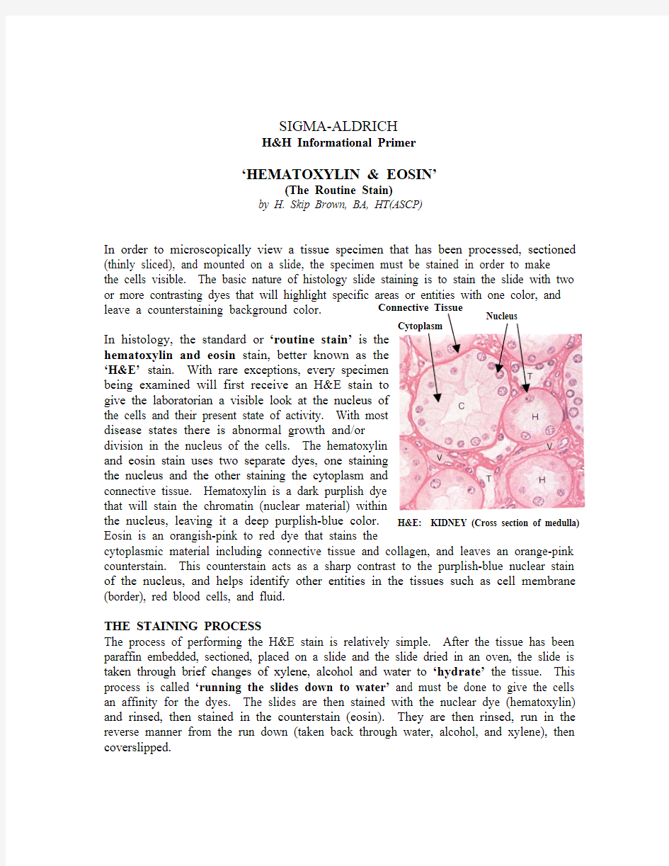
In order to microscopically view a tissue specimen that has been processed, sectioned (thinly sliced), and mounted on a slide, the specimen must be stained in order to make
the cells visible. The basic nature of histology slide staining is to stain the slide with two leave a counterstaining background color. In histology, the standard or ‘routine stain’hematoxylin and eosin ‘H&E’ the cells and their present state of activity. With most
disease states there is abnormal growth and/or
division in the nucleus of the cells. The hematoxylin
and eosin stain uses two separate dyes, one staining
the nucleus and the other staining the cytoplasm and
connective tissue. Hematoxylin is a dark purplish dye
the nucleus, leaving it a deep purplish-blue color. Eosin is an orangish-pink to red dye that stains the
cytoplasmic material including connective tissue and collagen, and leaves an orange-pink counterstain. This counterstain acts as a sharp contrast to the purplish-blue nuclear stain of the nucleus, and helps identify other entities in the tissues such as cell membrane (border), red blood cells, and fluid.
THE STAINING PROCESS
The process of performing the H&E stain is relatively simple. After the tissue has been paraffin embedded, sectioned, placed on a slide and the slide dried in an oven, the slide is taken through brief changes of xylene, alcohol and water to ‘hydrate’ the tissue. This process is called ‘running the slides down to water’ and must be done to give the cells an affinity for the dyes. The slides are then stained with the nuclear dye (hematoxylin) and rinsed, then stained in the counterstain (eosin). They are then rinsed, run in the reverse manner from the run down (taken back through water, alcohol, and xylene), then coverslipped.
Progressive/Regressive Staining
There are two methods for performing the H&E stain: Progressive in which the slides are placed in hematoxylin then rinsed and placed in eosin; and the regressive method in which the slides are placed in a stronger type of hematoxylin, then differentiated in acid alcohol to take the hematoxylin back out of everything except the nucleus, and then placed in eosin. In both types of staining, a bluing solution (Scott’s Tap Water or ammonia water) is often used to cause the nucleus to turn a deep purplish blue color.
In progressive staining, a milder form of hematoxylin is used that will only stain the nucleus of the cell and cause the nuclear material to turn a deeper blue when rinsed in water. With this method the technician can simply stain, rinse and move on to the next step. Its advantage is simplicity, fewer steps, and avoids the possibility of over/under differentiation in acid alcohols. The level or color of staining is standardized and consistent. However, many feel that the depth of staining color is not as rich as that which can be attained with the stronger form of hematoxylin. Most laboratories, in particular those who have automated their H&E staining, perform a progressive method of staining. Sigma key hematoxylin products for progressive staining are: Gill’s 1, Gill’s 2, Gill’s 3, and Mayer’s hematoxylin. The difference in the three Gill stains is the hematoxylin strength. Gill’s 1 is used primarily for cytology staining where a weaker hematoxylin is adequate because you are staining individual cells from a fluid suspension, not tissue. Gill’s 2 and 3 are stronger and generally used for histology staining. They are developed for tissue structure. The choice of whether to use Gill’s 2 or 3 is a matter of preference from technologist to technologist.
In regressive staining, a stronger form of hematoxylin is used called Harris hematoxylin. Harris hematoxylin will stain everything on the slide and hold fast to the tissue when rinsed. Therefore after staining and rinsing with water, the next step is to differentiate or take out the excess hematoxylin from everything except the nucleus. The slides are agitated in a mild acid alcohol solution that slowly removes the excess hematoxylin. Over or under differentiation of hematoxylin can be a problem, but this is generally addressed by standardizing a set procedure that will remove the optimal level of dye. After differentiating the slides are rinsed and placed in a bluing solution (Scott’s Tap Water or ammonia water), which will cause the nucleus to turn a deep purplish blue color. Sigma has Harris hematoxylin in their product line for laboratories that prefer regressive staining.
After hematoxylin staining the slides are rinsed, stained in eosin, dehydrated with graded strengths of alcohols, cleared in xylene and finally coverslipped using a permanent mounting media. Sigma has three premier eosin products to choose from, Eosin Y, Eosin Y Alcoholic, and Eosin Y with Phloxine. Similar to the three types of Gill’s stain, the eosins are differentiated by their strength and depth in color. Eosin Y is the weakest of the three and gives a pink stain to the cytoplasm and collagen. Eosin Y Alcoholic is a stronger stain and gives a more brilliant orangish red color due to its alcohol ingredient.
Eosin Y with Phloxine is the strongest stain and has an overwhelmingly red color due to the addition of phloxine. While the selection of eosin is preferentially dependent on the technician and/or pathologist, Eosin Y with Phloxine is generally considered too red for standard histology. Sigma’s preferred eosin, and the eosin that our standard procedure recommends is the Eosin Y Alcoholic.
Please consult the Sigma-Aldrich catalog or website for the complete line of H&E stain products. For technical service questions please contact Sigma-Aldrich technical service at (800) 325-0250.
2002 copyright Sigma-Aldrich Corporation