ONCOLOGY REPORTS-colon cancer
Abstract. Many chemotherapeutic agents induce apoptosis via a p53-dependent pathway. However, up to 50% of human cancers have p53 mutation and loss of p53 function. Histone deacetylase inhibitors (HDACIs) are emerging as a potentially important new class of anticancer agents. Here, we report that, Trichostatin A (TSA), a pan-HDAC inhibitor, could induce G2/M cell cycle arrest and apoptosis in both colorectal cancer cell lines with wild-type p53 (HT116 cells) and mutant p53 (HT29 cells), although HCT116 cells had more apoptotic cells than HT29 cells. TSA induces apoptosis in both cell lines via the mitochondrial pathway as indicated by decrease of the mitochondrial membrane potential (MMP) and activa-tion of caspase-3. Additionally, TSA induces expression of the pro-apoptotic protein Bax and decreases the expression of the anti-apoptotic proteins Bcl-2 and Bcl-xL in both cell lines. Bax knockdown by siRNA significantly impaired TSA-induced apoptosis in both cell lines. These data suggest that TSA induces G2/M cell cycle arrest and Bax-dependent apoptosis in colorectal cancer cells (HCT116 cells and HT29 cells) by both p53-dependent and -independent mechanisms. However, cells with normal p53 function are more sensitive to TSA-induced apoptosis.Introduction
Colorectal cancer (CRC) is the second leading cause of cancer-related mortality with 1,000,000 cases of all over the world in 2009 (1). Despite the fact that early-stage colorectal cancer is amenable and may be cured by surgical resection, recurrence rates remain high. Hence, there is an urgent need to develop novel treatment strategies.
Histone deacetylase inhibitors (HDACIs) have been extensively studied in basic biological research to gain an understanding of chromatin structure and transcriptional control and have recently been introduced as a potentially important new class of anticancer agents (2-4). Trichostatin A (TSA), a pan-HDAC inhibitor, has been shown to induce cell cycle arrest, promote cell differentiation and apoptosis, and inhibit metastasis (5) in various types of tumors (6). It is well known that p53 may play an important role in the anti-cancer effects induced by TSA. p53 stimulates the promoter activities of p21Waf1/Cip1 (p21) and Bax genes to enhance their expression as a transcriptional factor (7). The downstream events include cell cycle arrest and activation of apoptosis, as evidenced by changes in cell cycle kinetics and induction of caspase activity (8-10). However, the mechanisms by which TSA induces cell growth arrest and cell death remain to be fully defined.
Many chemotherapeutic agents induce apoptosis via p53-dependent pathway. However, there are up to 50% of human cancers that have p53 mutation and loss of p53 function (10). It is not clear whether TSA-induced anticancer effects are dependent on normal p53 function in CRC cells. In the present study, we treated CRC cells which have normal p53 function (HCT116 cells) and mutant p53 (HT29 cells), with HDAC inhibitor TSA and investigate its anticancer effects. We have demonstrated that TSA is effective in inducing G2/M cell cycle arrest and apoptosis in both CRC cell lines by both p53-dependent and -independent mechanisms. Cells with normal p53 function are more sensitive to TSA-induced apop-tosis, which is via Bax-dependent mitochondrial pathway. This study may provide evidence for the use of HDAC inhibitor in clinical treatment of colorectal cancers with wild-type or mutant p53.
The histone deacetylase inhibitor trichostatin A induces
cell cycle arrest and apoptosis in colorectal cancer cells via p53-dependent and -independent pathways JIN MENG1,4, HAI-HUA ZHANG2, CHUN-XIANG ZHOU3, CHEN LI1, FENG ZHANG1 and QI-BING MEI1 1Department of Pharmacology, School of Pharmacy, 2School of Clinical Medicine and Stomatology,
3Department of Pharmacy, Xijing Hospital, The Fourth Military Medical University, Xi'an 710032;
4Department of Pharmacy, The 309th Hospital of PLA, Beijing 100091, P.R. China
Received January 3, 2012; Accepted March 13, 2012
DOI: 10.3892/or.2012.1793
Correspondence to: Dr Qi-Bing Mei or Dr Feng Zhang, Department
of Pharmacology, School of Pharmacy, The Fourth Military Medical
University, 17 Changlexi Road, Xi'an 710032, P.R. China
E-mail: meiqb@https://www.360docs.net/doc/3918196165.html,
E-mail: zhangf37@https://www.360docs.net/doc/3918196165.html,
Abbreviations: TSA, trichostatin A; CRC, colorectal cancer; MMP,
mitochondrial transmembrane potential; HDACIs, histone deacetyl-
ase inhibitors
Key words: trichostatin A, colorectal cancer cells, apoptosis, cell
cycle arrest, p53
Materials and methods
Cell culture and treatments. Colorectal cancer cell lines HCT116 and HT29 cells were grown in Dulbecco's modified Eagle's medium (DMEM, Gibco, USA) supplemented with 10% fetal bovine serum, 10 mg/ml antibiotics (penicillin and streptomycin) and 2 mmol/l L-glutamine at 37?C under 5% CO2 and saturated moisture. Trichostatin A (TSA) (Sigma, St. Louis, MO, USA) was dissolved in dimethyl sulfoxide (DMSO, Sigma), final concentrations of 0.1, 1 and 5 μM were used to treat the cells and proper amount of DMSO was used as vehicle control.
Cell cycle analysis. Cells were treated with varying concen-trations of TSA for 24 h. At the time of harvesting, cells were digested with 0.25% trypsin (Gibco) and re-suspended in phosphate-buffered saline (PBS), fixed in 70% ethanol at 4?C overnight. When analyzing, cells were washed with PBS and treated with 20 μg/ml ribonuclease A (RNase A, Sigma) at 37?C for 30 min. Cells were then stained with 50 μg/ml propidium iodide (PI, Sigma) for 30 min and DNA content was analyzed by flow cytometry with FACScan (Becton-Dickinson, Mountain View, CA, USA) using the CELLQuest program (Becton-Dickinson). Cell cycle distribution was analyzed by WinMDI software.
Annexin V-FITC and PI staining. Cells were treated as above and were digested with trypsin, stained with 5 μl A nnexin V-FITC (BD Pharmingen) and 5 μl propidium iodide (PI, BD Pharmingen) staining solution in the dark at room temperature (RT) for 15 min. The cell samples were analyzed by flow cytometry on a FACScan station with CellQuest soft-ware using the FL1 and FL2 range for Annexin V FITC and PI, respectively.
Flow cytometry assessment of the changes in mitochondrial transmembrane potential (MMP). MMP was measured by flow cytometry with MitoTracker Red (Invitrogen) probe
which is a mitochondrion-selective stain concentrated by active mitochondria. The probe accumulates in normal mito-chondria and the reduction of MMP leads to the release of the stain. The probe was dissolved in DMSO and diluted in PBS before use. Cells were treated with MitoTracker for 45 min before trypsinization. Cells were then washed twice with PBS and analyzed by flow cytometry on a FACScan station with CellQuest software using the FL2 for MitoTracker Red staining.
Bax siRNA transfection. Bax siRNA and scramble siRNA were purchased from Santa Cruz Biotechnology Inc. siPORT NeoFX Transfection Agent (Ambion) was used for the siRNA transfection. Twenty-four hours after transfection, cells were treated with TSA for another 24 h. Knock-down of Bax expres-sion was confirmed by western blotting.
Western blotting. Cells were lysed in lysis buffer containing 150 mM NaCl, 1% NP40, 0.5% deoxycholic acid, 0.1% SDS, 50 mM Tris (pH 8.0), and 1:25 protease inhibitor cocktail. Protein concentrations of the lysates were determined by the Bradford protein assay system (Bio-Rad Laboratories, Hercules, CA). Equal amounts of protein (30 μg protein each lane) were separated by SDS-PAGE, transferred to nitrocel-lulose membranes (Hybond C, Amersham, UK). Immunoblots were blocked with 5% skim milk in TBS/Tween-20 (0.05%, v/v) for 1 h at RT. The membrane was incubated with primary antibody overnight at 4?C. Antibody against polyadenylribosyl polymerase (PARP) were from BD Pharmingen, antibodies against p21, Bcl-2, Bcl-xL and MCL-1 were purchased from Santa Cruz Biotechnology Inc., antibodies against Bax and active caspase-3 were purchased from Abcam, and actin antibody was purchased from Sigma. The membrane was incu-bated with corresponding secondary antibody conjugated with horseradish peroxidase (Sigma) (1:5000) at RT for 1 h. The blots were developed using an enhanced chemiluminescence western blotting detection system (Amersham Biosciences, UK).
Statistical analysis. The data represent at least three indepen-dent experiments. Statistical comparisons were made using Student's t-test. P<0.05 was considered to represent a statisti-
cally significant difference.
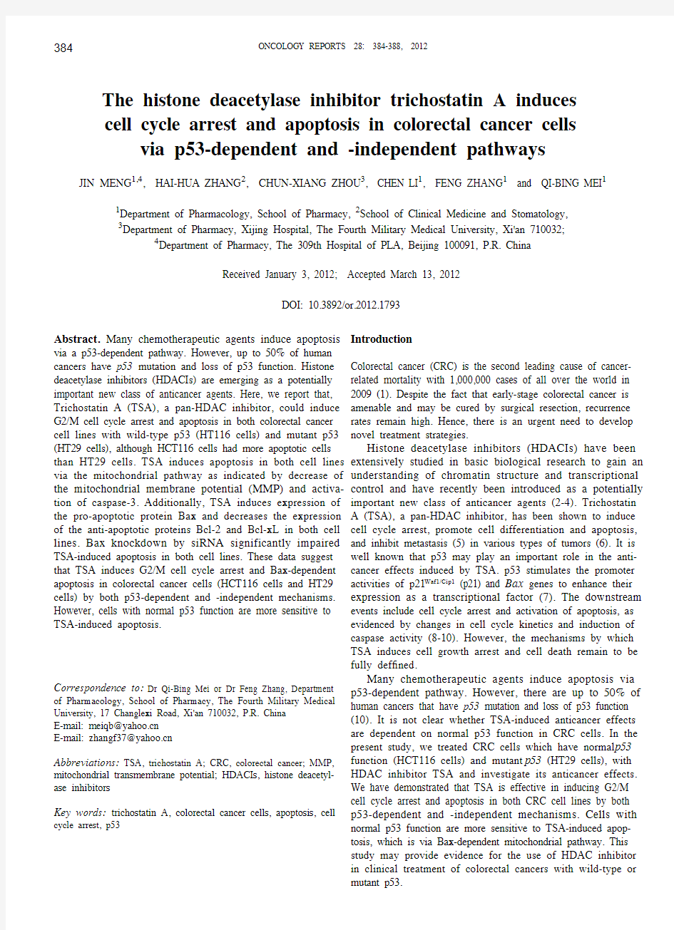
Figure 1. TSA induces G2/M arrest in CRC HCT116 and HT29 cells in a dose-dependent way. Cells were exposed to different concentrations of Trichostatin A (TSA) for 24 h. Cell cycle was measured by flow cytometry after PI staining. The percentage of cells in different cell cycle phases was analyzed using WinMDI software. Data are presented as means plus SD from
3 independent experiments and shown in the bar graph. (A) HCT116 cells.
a P<0.01 vs. corresponding control.
b P<0.05 vs. corresponding control. (B) HT29 cells. a P<0.01 vs. corresponding control. b P<0.05 vs. corresponding control. (C) p21 expression was detected by western blotting. Actin blot was included to show equal protein loading for all the samples.
Results
TSA induces G2/M arrest in CRC HCT116 and HT29 cells. Histone deacetylase (HDAC) inhibitors have been reported to cause growth arrest at the G1 and/or G2/M phases in a wide variety of tumour cells (11). We measured cell cycle distribution 24 h after varying concentrations of TSA (0.1, 1 and 5 μM) treatment by PI staining in both HCT116 cells with wild-type p53 and HT29 cells with mutant p53. As shown in Fig. 1A and B, TSA induced dose-dependent G2/M phase arrest in both cell lines, with decrease of S phase cells, indicating the inhibition of DNA replication. Since p21 is the principle cyclin dependent kinase inhibitor (CDKI) which is involved in cell cycle arrest upon DNA damage (12), we analyzed p21 expression by western blotting after TSA treat-ments. As shown in Fig. 1C, p21 expression was upregulated after TSA treatment in a dose-dependent manner in HCT116 cells, while in HT29 cells, p21 level was much lower, which
could be detected only when treated with TSA 1 and 5 μM (Fig. 1B).
TSA induces apoptosis in CRC cell lines with normal or mutant p53 function. We measured TSA-induced apoptosis with Annexin V-PI staining in both HCT116 cells and HT29 cells. Both types of cells were exposed to varying concentra-tions of TSA (0.1, 1 and 5 μM) for 24 h. As shown in Fig. 2A and B, TSA caused dose-dependent apoptosis in both cell lines, suggesting that TSA could induce apoptosis in CRC cells through both p53-dependent and -independent way. However, in HCT116 cells which have normal p53 function, cells were more sensitive to TSA-induced apoptosis than HT29 cells with mutant p53, indicating that normal p53 function could promote TSA-induced apoptosis in CRC cells. Apoptosis was further confirmed by PARP cleavage detected with western blotting (Fig. 2C). The cleaved PARP 85 kDa band was constantly detected in TSA treated CRC cells in a similar pattern with Annexin V-positive cells.
TSA induces apoptosis in CRC cells via mitochondrial
pathway by causing dissipation of MMP and caspase-3
Figure 2. TSA induces apoptosis in CRC cell lines. (A) Apoptosis was ana-lyzed by Annexin V-FITC and PI staining with flow cytometry. Representative plots of one set of triplicate experiments. Early apoptotic cells (Annexin V + and PI -) are displayed in the lower right quadrant and late apoptotic cells (Annexin V + and PI +) are shown in the upper right quadrant. (B) The percent-ages of apoptotic cells are indicated by Annexin V + cells shown as means plus SD from 3 independent experiments. Data are shown as means plus SD from 3 independent experiments with representative plots of one set of the experiments. a P<0.01 vs. corresponding control. b P<0.05 vs. corresponding HCT116 cells. (C) PARP cleavage was detected by western blotting. Actin
blot was included to show equal protein loading for all the samples.
Figure 3. TSA induces apoptosis in CRC cells via mitochondrial pathway by causing dissipation of MMP and caspase-3 activation. (A) MMP changes was measured by flow cytometry. Representative plots of one set of tripli -cate experiments. (B) Data are shown as means plus SD from 3 independent experiments. a P<0.01 vs. corresponding control. b P<0.05 vs. corresponding HCT116 cells. (C) Protein from each group of cells was extracted and ana-lyzed by western blotting using anti-active caspase-3 antibody. Actin blot was included to show equal protein loading for all the samples.
activation. We then asked whether TSA induced apoptosis in CRC cells via different pathways. Mitochondrial membrane injury and caspase-3 activation are key steps in the progres-sion of apoptosis. We first measured MMP dissipation, which plays a pivotal role in the initiation of apoptosis. The results showed that TSA caused increased loss of MMP in a dose-dependent way in both cell lines (Fig. 3A and B), there was not much difference between the two cell lines. We next detected caspase-3 activation by western blotting with antibody which could recognize the cleaved active form of caspase-3. Our results showed that TSA induced caspase-3 activation in a similar manner with MMP dissipation (Fig. 3C). These results demonstrated that TSA induced apoptosis in both CRC cell lines via mitochondrial pathway and dependent on caspase-3 activation.
TSA induces changes of apoptosis-related proteins. It is known that the ratio of pro-apoptotic protein (Bax) and anti-apoptotic proteins (Bcl-2 and Bcl-xL) of Bcl-2 family determines whether cancer cells respond to an apoptotic signal by mediating the disruption of the mitochondrial membrane and the release of caspase activators (13). By western blotting, we showed that (Fig. 4) Bax was upregulated after TSA treat-ment in both HCT116 and HT29 cells in a dose-dependent manner. On the contrary, the anti-apoptotic proteins Bcl-2 and Bcl-xL were downregulated in both cell lines, while Mcl-1 level did not change by TSA. The expression level of all three proteins were different in the cell lines, which might explain the difference of apoptosis induced by TSA in those cells.
TSA induces apoptosis in CRC cells in a Bax-dependent pathway. Bax is an important pro-apoptotic protein in trig-gering mitochondria-mediated apoptosis by permeabilizing mitochondrial outer membranes. Since TSA induces apoptosis in both cell lines via mitochondrial pathway, we examined whether Bax is essential in both p53-dependent and -inde-pendent apoptosis. As shown in Fig. 5, Bax siRNA impaired TSA-induced apoptosis in both HCT116 and HT29 cells, as indicated by the weakened cleaved bands of PARP. These results suggest that TSA induces apoptosis in CRC cells in a Bax-dependent way.Discussion
Acetylation and deacetylation of histones, are known to be involved in gene expression regulation, catalyzed by histone acetyl transferases (HATs) and histone deacetylases (HDACs), respectively (14). HDAC inhibitors (HDACIs) are introduced as potential clinical treatments for cancer that inhibit deacety-lase activity, thereby increasing acetylation of many proteins, including histones (15-17). TSA, a pan-HDAC inhibitor, has shown anti-neoplasia effects in many kinds of tumor cells. It is known that conventional chemotherapeutic agents induce apoptosis of cancer cells mostly through p53-dependent pathway. However, p53 mutations and inactivation have been found in more than half of all human carcinoma cells (10). We therefore observed whether TSA was effective in inducing cell cycle arrest and apoptosis in CRC cell lines with wild-type p53 (HCT116 cells) and mutant p53 (HT29 cells).
p21, a member of the cyclin-dependent kinase (CDK) inhibitor protein family, acts by binding cyclin-CDK complexes, inhibiting their kinase activity and effectively blocking DNA synthesis and cell cycle progression (18-20). The growth arrest at G1 phase by HDAC inhibitors is thought to be highly dependent on the upregulation of p21 (11). It is well established that p21 induced G1/S phase arrest through its interactions with cyclin E/CDK2 (21). p21 is also involved in G2/M arrest via inhibiting the CDK-activating kinase (CAK) and consequently inactivation of CDK1 (8-10). Some studies have reported that p21 induction in several p53 null cells also could induce G2/M arrest (22). In the current study, TSA was shown to induce cell cycle arrest at G2/M phase in both cells lines, with S phase cells reduced, indicating the inhibition of DNA replication in S phase. By western blotting, we showed that TSA increased p21 expression in HCT116 cells with wild-type p53, suggesting that HDACIs may activate the transactivation of p53. While the expression of p21 in p53 mutant HT29 cells was only slightly induced. The mechanisms by which TSA induces G2/M arrest in HT29 cells are not known. TSA has been shown to induce G2/M arrest in p53 (-/-) MG63 osteosarcoma cells through the induction of Gadd45 (23). The above results suggest that TSA induce cell cycle arrest in CRC cells through p53-dependent and -independent pathway.
Some HDAC inhibitors, including TSA, have been reported to induce apoptosis in cancer cells through activation of p53 by phosphorylation or acetylation (24,25), suggesting a p53-dependent apoptotic pathway by TSA. Here, we showed that TSA was effective in both p53 wild-type HCT116 cells and p53
mutant HT29 cells, indicating that TSA could induce
Figure 4. TSA induces changes of apoptosis related proteins. Protein from each group of cells was extracted and analyzed by western blotting using antibodies against Bax, Bcl-2, Bcl-xL and MCL1. Actin blot was included to
show equal protein loading for all the samples.
Figure 5. TSA induces apoptosis in CRC cells in a Bax-dependent pathway. Cells were transfected with Bax siRNA or scramble siRNA as control and then treated with TSA 1 μM. Apoptosis was detected by western blotting using anti-PARP antibody which could recognize the cleaved band. Actin blot was included to show equal protein loading for all the samples.
apoptosis in CRC cells via both p53-dependent and -indepen-dent mechanisms. However, apoptotic rates in HCT116 cells treated with TSA was higher than that in HT29 cells, demon-strating that the susceptibility to TSA-induced apoptosis in CRC cells might be regulated by p53 status.
Bax plays an important role in apoptosis, which is downstream of the p53 pathway and a direct target of p53. Exogenously expressed p53 increases Bax expression in several cancer cell types, which correlates with the induction of apoptosis (26). Direct activation of Bax by p53 mediates mitochondrial membrane permeabilization, allowing cyto-chrome c to be released from mitochondria, which associates with the 47 kDa procaspase-9/Apaf 1 and activates caspase cascade, leading to apoptosis (27). Bax and Bcl-2 are homolo-gous proteins that have opposite effects on cell fate (28). In the present study, the increase in apoptosis was associated with upregulation of pro-apoptotic protein Bax, and downregula-tion of anti-apoptotic proteins Bcl-2 and Bcl-xL. Since mutant p53 gene is unable to transactivate downstream target genes, such as Bax (10), the expression of Bax might be induced by other transcription factors (29). We also showed that TSA dose-dependently decreased MMP in both cell lines, suggesting the involvement of mitochondrial pathway. Caspase-3, the key executor of apoptosis, has been described to be essential for drug-induced apoptosis. We have detected the activation of caspase-3 after TSA treatment in both p53 wild-type and mutant CRC cell lines. These results demonstrate that TSA induces apoptosis in both CRC cell lines via mitochondrial pathway and dependent on caspase-3 activation, regardless of p53 status.
Bax is an initiator in mitochondrial apoptotic pathway. In the current study, Bax knock-down by siRNA significantly impaired TSA-induced apoptosis in both HCT116 and HT29 CRC cells. We concluded that Bax is an essential mediator for both p53-dependent and -independent apoptosis induced by TSA in CRC cells. TSA might induce cell death in CRC cells by increasing the acetylation of Ku70, a Bax-binding protein, which causes Bax release and become free to translocate from the cytoplasm to mitochondria to stimulate apoptosis (30).
In summary, based on these findings, we concluded that TSA induces G2/M cell cycle arrest and Bax-dependent apop-tosis in colorectal cancer cells (HT29 cells and HT29 cells) by both p53-dependent and -independent mechanisms. This study may provide useful information for the use of HDAC inhibitors to treat clinical patients with colorectal cancer. Acknowledgements
This study was supported by the National 973 project of China (2010CB535002).
References
1. Jernal A, Siegel R, Ward E, et al: Cancer statistics, 2009. CA Cancer J Clin 59: 225-249, 2009.
2. Johnstone RW: Histone-deacetylase inhibitors: novel drugs for the treatment of cancer. Nat Rev Drug Discov 1: 287-299, 2002.
3. Yu X, Guo ZS, Marcu MG, et al: Modulation of p53, ErbB1, ErbB2, and Raf-1 expression in lung cancer cells by depsipeptide FR901228. J Natl Cancer Inst 94: 504-513, 2002.
4. Zhu WG and Otterson GA: The interaction of histone deacetylase inhibitors and DNA methyltransferase inhibitors in the treatment of human cancer cells. Curr Med Chem AntiCancer Agents 3: 187-199, 2003.
5. Meinke PT and Liberator P: Histone deacetylase: a target for antiproliferative and antiprotozoal agents. Curr Med Chem 8: 211-235, 2001.
6. Yoshida M, Kijima M, Akita M and Beppu T: Potent and specific inhibition of mammalian histone deacetylase both in vivo and in vitro by trichostatin A. J Biol Chem 265: 17174-17179, 1990.
7. Lagger G, Doetzlhofer A, Schuettengruber B, et al: The tumor suppressor p53 and histone deacetylase 1 are antagonistic regu-lators of the cyclin-dependent kinase inhibitor p21/WAF1/CIP1 gene. Mol Cell Biol 23: 2669-2679, 2003.
8. Levine AJ: p53, the cellular gatekeeper for growth and division. Cell 88: 323-331, 1997.
9. Prives C and Hall PA: The p53 pathway. J Pathol 187: 112-126, 1999.
10. Vogelstein B, Lane D and Levine AJ: Surfing the p53 network. Nature 408: 307-310, 2000.
11. Hirose T, Sowa Y, Takahashi S, et al: p53-independent induction of Gadd45 by histone deacetylase inhibitor:coordinate regula-tion by transcription factors Oct-1 and NF-Y. Oncogene 22: 7762-7773, 2003.
12. Rich T, Allen RL and Wyllie AH: Defying death after DNA damage. Nature 407: 777-783, 2000.
13. Pommier Y, Sordet O, Antony S, Hayward RL and Kohn KW: Apoptosis defects and chemotherapy resistance: molecular inter-action maps and networks. Oncogene 23: 2934-2949, 2004. 14. Emiliani S, Fischle W, Van Lint C, Al-Abed Y and Verdin E: Characterization of a human RPD3 ortholog, HDAC3. Proc Natl Acad Sci USA 95: 2795-2800, 1998.
15. Kr?mer OH, Gottlicher M and Heinzel T: Histone deacetylase as
a therapeutic target. Trends Endocrinol Meta
b 12: 294-300, 2001.
16. Marks PA, Richon VM, Breslow R and Rifkind RA: Histone deacetylase inhibitors as new cancer drugs. Curr Opin Oncol 13: 477-483, 2001.
17. Melnick A and Licht JD: Histone deacetylases as therapeutic targets in hematologic malignancies. Curr Opin Hematol 9: 322-332, 2002.
18. Janicke RU, Sohn D, Essmann F and Schulze-Osthoff K: The multiple battles fought by anti-apoptotic p21. Cell Cycle 6: 407-413, 2007.
19. Le HV, Minn AJ and Massague J: Cyclin-dependent kinase inhibitors uncouple cell cycle progression from mitochondrial apoptotic functions in DNA-damaged cancer cells. J Biol Chem 280: 32018-32025, 2005.
20. Seoane J, Le HV and Massague J: Myc suppression of the p21(Cip1) Cdk inhibitor influences the outcome of the p53 response to DNA damage. Nature 419: 729-734, 2002.
21. Bartek J and Lukas J: Pathways governing G1/S transition and their response to DNA damage. FEBS Lett 490: 117-122, 2001.
22. Choi YH, Lee WH, Park KY and Zhang L: p53-independent induction of p21 (WAF1/CIP1), reduction of cyclin B1 and G2/M arrest by the isoflavone genistein in human prostate carcinoma cells. Jpn J Cancer Res 91: 164-173, 2000.
23. Hirose T, Sowa Y, Takahashi S, et al: p53-independent induction of Gadd45 by histone deacetylase inhibitor: coordinate regula-tion by transcription factors Oct-1 and NF-Y. Oncogene 22: 7762-7773, 2003.
24. Hsu YF, Sheu JR, Hsiao G, et al: p53 in trichostatin A induced C6 glioma cell death. Biochim Biophys Acta 1810: 504-513, 2011.
25. Oh ET, Park MT, Choi BH, et al: Novel histone deacetylase inhibitor CG200745 induces clonogenic cell death by modulating acetylation of p53 in cancer cells. Invest New Drugs 30: 435-442, 2010.
26. Miyashita T and Reed JC: Tumor suppressor p53 is a direct tran-scriptional activator of the human Bax gene. Cell 80: 293-299, 1995.
27. Li P, Nijhawan D, Budihardjo I, et al: Cytochrome c and dATP-dependent formation of Apaf-1/caspase-9 complex initiates an apoptotic protease cascade. Cell 91: 479-489, 1997.
28. Sugita K, Koizumi K and Yoshida H: Morphological reversion of sis-transformed NIH3T3 cells by trichostatin A. Cancer Res 52: 168-172, 1992.
29. Levrero M, De Laurenzi V, Costanzo A, et al: The p53/p63/ p73 family of transcription factors: overlapping and distinct functions. J Cell Sci 113: 1661-1670, 2000.
30. Cohen HY, Lavu S, Bitterman KJ, et al: Acetylation of the C terminus of Ku70 by CBP and PCAF controls Bax-mediated apoptosis. Mol Cell 13: 627-638, 2004.
《胃癌诊疗规范(2018年版)》要点
《胃癌诊疗规范(2018年版)》要点 一、概述 胃癌是指原发于胃的上皮源性恶性肿瘤。在我国胃癌发病率仅次于肺癌居第二位,死亡率排第三位。全球每年新发胃癌病例约120万,中国约占其中的40%。我国早期胃癌占比很低,仅约20%,大多数发现时已是进展期,总体5年生存率不足50%。近年来随着胃镜检查的普及,早期胃癌比例逐年增高。 胃癌治疗的总体策略是以外科为主的综合治疗。 二、诊断 应当结合患者的临床表现、内镜及组织病理学、影像学检查等进行胃癌的诊断和鉴别诊断。 (一)临床表现 早期胃癌患者常无特异的症状,随着病情的进展可出现类似胃炎、溃疡病的症状,主要有:①上腹饱胀不适或隐痛,以饭后为重;②食欲减退、嗳气、返酸、恶心、呕吐、黑便等。进展期胃癌除上述症状外,常出现:①体重减轻、贫血、乏力。②胃部疼痛,如疼痛持续加重且向腰背放射,则提示可能存在胰腺和腹腔神经丛受侵。胃癌一旦穿孔,可出现剧烈腹痛的胃穿孔症状。 ③恶心、呕吐,常为肿瘤引起梗阻或胃功能紊乱所致。贲门部癌可出现进行性加重的吞咽困难及反流症状,胃窦部癌引起幽门梗阻时可呕吐宿食。④出血和黑便,肿瘤侵犯血管,可引起消化道
出血。小量出血时仅有大便潜血阳性,当出血量较大时可表现为呕血及黑便。⑤其他症状如腹泻(患者因胃酸缺乏、胃排空加快)、转移灶的症状等。晚期患者可出现严重消瘦、贫血、水肿、发热、黄疸和恶病质。 (二)体征 一般胃癌尤其是早期胃癌,常无明显的体征,进展期乃至晚期胃癌患者可出现下列体征:①上腹部深压痛,有时伴有轻度肌抵抗感,常是体检可获得的唯一体征。②上腹部肿块,位于幽门窦或胃体的进展期胃癌,有时可扪及上腹部肿块;女性患者于下腹部扪及可推动的肿块,应考虑Krukenberg瘤的可能。③胃肠梗阻的表现:幽门梗阻时可有胃型及震水音,小肠或系膜转移使肠腔狭窄可导致部分或完全性肠梗阻;④腹水征,有腹膜转移时可出现血性腹水;⑤锁骨上淋巴结肿大;⑥直肠前窝肿物;⑦脐部肿块等。其中,锁骨上窝淋巴结肿大、腹水征、下腹部盆腔包块、脐部肿物、直肠前窝种植结节、肠梗阻表现均为提示胃癌晚期的重要体征。因此,仔细检查这些体征,不但具有重要的诊断价值,同时也为诊治策略的制订提供了充分的临床依据。 (三)影像检查 1. X线气钡双重对比造影:定位诊断优于常规CT或MRI,对临床医师手术方式及胃切除范围的选择有指导意义。 2. 超声检查(US):因简便易行、灵活直观、无创无辐射等特点,可作为胃癌患者的常规影像学检查。
心情不好说说发朋友圈,适合心情不好发的朋友圈
心情不好说说发朋友圈,适合心情不好发的朋友圈 1、如果一个人真的爱你,距离不是一个问题,它只会成为一种滋长爱情的力量。 2、曾经以为,伤心是会流很多眼泪的,原来,真正的伤心,是流不出一滴眼泪。什么事情都会过去,我就是这样活过来的。 3、人人都是自顾不暇的泥菩萨,别指望谁能帮你度过现实这条河。 4、你可能也不爱我,只是刚好遇见我。 5、烟灭酒半杯,往后日子多笑少流泪。 6、再也不幻想,再也不乱想,再也不会想,再也不用想。 7、听到你的消息还是会心头一震,不过这些都不重要了,孤独至少比爱你舒服。 8、所有回不去的良辰美景,都是举世无双的好时光。感谢过去,珍惜现在,憧憬未来。哭给自己听,笑给别人看,这就是所谓的人生。 9、我一直走一直走,直到走到回忆的尽头,才发现时光与你,都没有等我。 10、付出和接受都是种债都无法还清。
11、自以为是刻骨铭心的回忆,别人早已已经忘记了。 12、成熟就是自己吞下苦难、眼泪、委屈、转脸还能给别人一个笑容。 13、我也经常觉得冷可我不会随便抱别人。 14、这世上真的没有感同身受只能冷暖自知。 15、心情不好就少听悲伤的歌。 16、有的时候连自己都不知道自己心里想什么,只知道自己心好累。 17、心情不好的时候,音乐必须大声,这样才听不到心碎的声音。 18、我们就像仙人掌,防备了别人,孤单了自己。 19、不要在流眼泪的时候做任何决定,情绪负面的时候说话越少越好。 20、说出口的伤痛都已平复,绝口不提的才触及心底。 21、不该看的东西就别去看,很多时候我们心情不好是因为我们手贱。 22、说什么待我长发及腰,心情不好全剪了,叫他想一辈子去吧。 23、心情不好的时候,做什么事都那么力不从心。 24、很多人,因为寂寞而错爱了一人,但更多的人,因为错爱一
疲劳驾驶预警系统
DSD行车安全电脑(四合一版本) 产品介绍 DSD行车安全电脑是结合车载智能电脑 和车辆辅助驾驶安全电脑功能的全新一代创新 产品,其包括疲劳检测与防瞌睡系统、视频行 车记录仪、GPS定位导航以及全面的车载3G 平板电脑的功能。 DSD行车安全电脑的防瞌睡检测系统,利 用面部生物特征模式检测技术,通过对驾驶人 员视频图像的获取、跟踪和分析,对驾驶过程 中常见的注意力涣散、驾驶姿态异常、驾驶反应迟钝、疲劳瞌睡等非正常工作状态进行提示告警和记录;不仅如此,同步结合产品的视频行驶记录、GPS定位导航服务、3G实时信息推送等功能,DSD行车安全电脑可为行车安全提供最全面有效的保护。 DSD行车安全电脑将智能视频分析技术、生物模式识别技术与无线通讯及信息传递技术相结合,可全面应用于车辆主动安全驾驶及行车监察管理等关键环节,最终为行车安全提供功能完善、简便实用、可靠安全、能够全天候实时运行的创新科技产品。 产品功能 1、驾驶疲劳及防瞌睡预警 ■完成驾驶员的状态及姿态等异常驾驶状态 预警; ■完成驾驶员的多级疲劳检测及防瞌睡告警; ■完成驾驶员各类异常驾驶事件的主动分析 和记录; 2、GPS定位导航 ■正版GPS导航3D软件; ■全面的更新及扩展能力; DSD行车安全电脑提供功能全面的GPS定位导航服务,不仅如此,结合产品本身完善的处理能力和3G通讯能力,相应的导航软件可以做到实时更新,并为车辆加入更完善的车辆在线导航服务,预留了设备功能接口的链接扩展能力。 3、行车记录黑匣子 ■无论何时何地,DSD为你的合法权益提供行车保障。 DSD行车安全电脑提供完善的行车视频记录仪功能,通过广角视频获取和超大容量的自动存储,行车过程的全视频信息,可以在DSD设备中实时重现和清晰记录,并且叠加时间标签,为你的事后过程查询、责任
心情不好的时候怎么发朋友圈 形容心情不好的句子
1.手掌就那么大,握不住的东西太多了。 2.怎么可以对淋在雨里的小孩说要乖 3.还以为你不一样呢。 4.再大大咧咧的人也会觉得难过啊,就像下了很大的雨,别人在等伞,而我在等雨停。 5.不等了,也等不到了。 6.哭,是解决不了问题的。可是,就是解决不了才哭啊。 7.我也曾对你心动过,只是赶路要紧,我忘了说 8.都会走的,无一例外 9.我可以恢复出厂设置吗? 10.我也不想一个人,但是我就擅长一个人待着。
11.要离开的人,你不妨推他一把。 12.一哄就好的人活该受尽委屈。 13.我表达不满的方式是晚一点回消息。 14.今天天气很好,好像也就一般。 15.连不开心都要暗示,你就应该知道他不在意。 16.我不会怪你,但我不会忘记每一次难过的原因。 17.我还以为这次我真能好好谈一个恋爱了呢 18.这场自救的仗我不想打了 19.生活中的糟糕小事都在消磨我对世界的兴趣。 20.心情不好说话就喜欢加句号。
21.下雨了,我说的不是天气。 22.成为遗憾,或许会被记住的久一点。 23.去吹吹风吧,能醒的话,感冒也没关系。 24.不管你承不承认,人确实是经历了一些事情后,就偷偷 换了一种性格。 25.今天还好吗,被人左右情绪了吗。 26.庆幸的是我一直很理性的看待所有的事情,我可悲的是 我是个很感性的人,所以所有的情绪我一样都没有逃过去。 27.不用考虑我,我没有感受,不用对不起,反正下次还是 会对不起。 28.我又把头发剪短了,好像变温柔了,好像过得比以前好,好像又不怎么样,我不清楚。 29.你剥开一个很酸的橘子,而感到后悔了,可对于橘子来说,那是它的一切。 30.这不就是你梦寐以求的长大吗,你怎么不笑了。
司机疲劳驾驶检测系统设计
司机疲劳驾驶检测系统设计
司机疲劳驾驶检测系统设计 摘要:随着社会经济的发展,商用长途运输车越来越多,司机为了追求经济效益,经常罔顾交通法的规定疲劳驾驶,而一些私家车也因为各种各样的原因经常铤而走险疲劳驾驶,酿成很多人间惨剧。为了减少减轻司机的精神压力并对疲劳及时提示预警,本论文以计算机视觉技术为主体,设计实用操作简单的疲劳驾驶检测系统,辅助驾驶员安全驾驶。 司机疲劳驾驶实时检测系统在实际应用中有很重要的意义。设计了一个利用图像分析的方法,通过测量PERCLOS指标值来进行疲劳判断的该类系统。系统首先对图像进行预处理,然后采用基于YCbCr颜色空间肤色模型进行人脸粗定位,根据人脸特征,逐次进行人眼区域缩小;最后通过对边缘信息进行先验知识结合积分投影的方法进行人眼定位和闭合度测量。考虑到视频图像序列帧与帧之间的相关性,采用线性运动预测的方法对人眼进行跟踪,减少了系统的运算量。实验结果表明系统能实时、准确地反映司机的疲劳状态。 关键词:疲劳驾驶人脸检测肤色检测交通安全疲劳判断
目录 摘要 Abstract 1.疲劳驾驶检测系统研究背景与意义............................ 2.疲劳驾驶检测系统研究与实现 2.1国内外疲劳驾驶检测系统研究现状 2.1.1国外疲劳驾驶检测系统的研究成果...................... 2.1.2国内疲劳驾驶检测系统的研究现状...................... 2.2疲劳驾驶检测系统浅析............................................. 2.3驾驶员疲劳检测系统的研究..................................... 2.3.1人脸检测 2.3.2人眼定位 2.3.3疲劳程度的综合判定........................................................................................... 3.基于人脸特征的列车司机疲劳驾驶检测与识 别系统研究....................................................................... 3.1研究内容及目标......................................................... 3.1.1基于人脸特征的疲劳驾驶检测与识别算法 开发................................................................................... 3.1.2疲劳驾驶检测与识别算法OSP移植 3.2基于Adaboost算法的人脸检测
心情不好适合发的句子,心情不好朋友圈句子
心情不好适合发的句子,心情不好朋友圈句子 1、难过的时候别说话,因为一张口眼泪就停不下。 2、原来除了记忆外,什么也不能永久。 3、虽然知道自己是个普通人但还是会希望在特别的人心里能有特别的存在。 4、放开彼此的手,当爱已经无法挽留,终于看透幸福的背后,是一道道伤口。 5、从有你真好到没你也行,这中间的心酸与艰难,你怎么会知道。 6、在最后的时光里我决定不再哭泣,在剩下的路里我也决定放弃。 7、深夜总是那么多无奈。挣不脱从前,怕极了以后。 8、心情不好的时候删东西就有一种快感。 9、有时候,莫名的心情不好,不想和任何人说话,只想一个人静静的发呆。有时候,想一个人躲起来脆弱,不愿别人看到自己的伤口。 10、心情不好,微微抬起头,看看湛蓝的天,看看悠悠的云,也是一种舒心的幸福。 11、某些人,某些事,久而久之就忘了,久而久之就不那么在意了。 12、有人总说:已经晚了。实际上,现在就是最好的时光。对于一个真正有所追求的人来说,生命的每个时期都是年轻的、及时的。13、被特别在乎的人忽略,会很难过,而更难过的是你还要装作你不在乎。 14、你伤我如此之深,我心里却全是你的甜言蜜语。
15、我心情不好没关系,你开心就好。 16、我承认我在发脾气的时候最喜欢说很极端的话。 17、再深的记忆,也有淡忘的一天。 18、这几天我心情不好阿!全世界都烦死人,只有你是死烦人。 19、从相遇到离开,我欠自己良多,不欠你分毫。 20、太多心酸无处诉说,太多难过如何洒脱。 21、曾经无话不说,如今的无话可说。 22、当初我们那么不甘心,最后还不是成了陌生人。 23、人老的唯一好处就是:能失去的东西越来越少了。 24、你看的,你听的,你都相信。我用心说的话你却从不相信。 25、我的难过无人知晓,我的心情无人过问。 26、任何瞬间的心动都不容易,不要怠慢了它。 27、在一瞬间曾经所有的梦都幻灭,剩下回忆湿了我的眼。 28、悉数记忆的流沙,那些逝去的年华,洗尽了我的尘沙。 29、我心情不好的时候你不在你知道我多想你安慰我吗? 30、时间不是让人忘了痛,它只是让人习惯痛。 31、一个人吃饭也没什么不好,不过是空出来一个座,邀请了寂寞。 32、心情不好的时候,看看大海,大海那么大,足以包容你的一切。没有大海就望望天空吧,望着望着心就不痛了,脖子就会痛了。 33、嗯!生理性心情不好谢谢大哥没打死我。 34、以前觉得,人只要一天不洗澡就会长蛆。。。昨天心情不好没有洗澡就睡了,今天发现也没夸张到长蛆。。。
心情不好时发朋友圈的伤感心情说说100句
心情不好时发朋友圈的伤感心情说说100句 1、爱上等于哀伤。 2、离网络越近,离现实越远。 3、你不安定,注定我不安宁。 4、幻光留不住时光的时间。 5、你说的坚强,始终都学不会。 6、我爱你,爱了整整一个曾经。 7、有心则会累,无心者无所谓。 8、你的心不是能读懂我眼神的料。 9、他说爱你又没说只爱你一个。 10、一切都已结束,一切都将开始。 11、害怕背叛,所以不敢为爱承诺。 12、如果坦白是伤害,情愿选择谎言。 13、相爱的人,刚分开,却又心生思念。 14、女人从来不争气,男人从来不珍惜。 15、听你说他的一举一动我心如刀割。 16、不在恋爱中失败,就在恋爱中变态。 17、我就只有这么一颗心,你看着伤吧。 18、格式化我们的过去,一切重新开始。 19、有时爱情一眨眼,就把回忆当留言。 20、你说的太少或太多,都会让人很惶恐。 21、也许只有可悲,才能填补寂寞的空位。
22、不知道什么时候开始,变得如此狼狈。 23、如果说,你的忧伤是我最痛的伤口。 24、丢失的曼陀罗,我知道,不会太远。 25、我希望有个人懂我,即使我什么也不说。 26、明明说忘记,却总是不经意的想起。 27、我们都只是孩子,何必什么都懂。 28、我们在原地转了无数次,无法解脱。 29、有那么一瞬间,我以为我们会一辈子。 30、曾经的那些勇气,全都变成了回忆。 31、未知的下一秒才更容易让人刻骨铭心。 32、我尽量减少了难过,过平静的生活。 33、心不知下落,我早己找不回单纯的我。 34、宁可高傲的发霉,也不低调的凑合。 35、伤痛复合不了叻,心里永远有伤疤。 36、我捂着心脏,傻傻的痛到撕心裂肺。 37、习惯了伤感,竟然忘了什么是幸福? 38、只希望你能聆听我的世界,仅此而已。 39、看起来百毒不侵,其实早已百毒侵心。 40、给你自由的爱,冻结我们美好的回忆。 41、那种华丽旳颓废,有种令人心惊旳美丽。 42、我假装坚强,只是不想告诉自己我想哭。 43、没有人值得你放弃自己的卑微去讨好。 44、你的笑容,是我今生无法忘记的眷念。 45、明知是陌路,却还追逐,缠绵一生的毒。
司机疲劳驾驶检测系统设计
司机疲劳驾驶检测系统设计 摘要:随着社会经济的发展,商用长途运输车越来越多,司机为了追求经济效益,经常罔顾交通法的规定疲劳驾驶,而一些私家车也因为各种各样的原因经常铤而走险疲劳驾驶,酿成很多人间惨剧。为了减少减轻司机的精神压力并对疲劳及时提示预警,本论文以计算机视觉技术为主体,设计实用操作简单的疲劳驾驶检测系统,辅助驾驶员安全驾驶。 司机疲劳驾驶实时检测系统在实际应用中有很重要的意义。设计了一个利用图像分析的方法,通过测量PERCLOS指标值来进行疲劳判断的该类系统。系统首先对图像进行预处理,然后采用基于YCbCr颜色空间肤色模型进行人脸粗定位,根据人脸特征,逐次进行人眼区域缩小;最后通过对边缘信息进行先验知识结合积分投影的方法进行人眼定位和闭合度测量。考虑到视频图像序列帧与帧之间的相关性,采用线性运动预测的方法对人眼进行跟踪,减少了系统的运算量。实验结果表明系统能实时、准确地反映司机的疲劳状态。 关键词:疲劳驾驶人脸检测肤色检测交通安全疲劳判断
目录 摘要 Abstract 1.疲劳驾驶检测系统研究背景与意义............................................................................................................... 2.疲劳驾驶检测系统研究与实现 2.1国内外疲劳驾驶检测系统研究现状 2.1.1国外疲劳驾驶检测系统的研究成果......................................................................................................... 2.1.2国内疲劳驾驶检测系统的研究现状......................................................................................................... 2.2疲劳驾驶检测系统浅析................................................................................................................................ 2.3驾驶员疲劳检测系统的研究........................................................................................................................ 2.3.1人脸检测 2.3.2人眼定位 2.3.3疲劳程度的综合判定 ............................................................................................................................................................................. 3.基于人脸特征的列车司机疲劳驾驶检测与识别系统研究........................................................................... 3.1研究内容及目标............................................................................................................................................ 3.1.1基于人脸特征的疲劳驾驶检测与识别算法开发..................................................................................... 3.1.2疲劳驾驶检测与识别算法OSP移植 3.2基于Adaboost算法的人脸检测 3.2.1人脸检测技术概述 3.2.2Adaboost人脸检测算法 3.3基于Adaboost算法的人脸检测软件实现 3.3.1.样本训练过程 3.3.2人脸检测程序 3.4人眼检测与人眼状态分析算法 3.4.1基于Adaboost的人眼检测算法 3.4.2人眼级联分类器效果分析 3.4.3人眼状态分析算法 4.基于贝叶斯网络的驾驶疲劳程度识别模型 4.1基于贝叶斯网络模型的驾驶疲劳程度识别 4.2驾驶疲劳程度识别模型 4.2.1驾驶疲劳贝叶斯网络结构 4.2.2贝叶斯网络条件概率表的确定 4.2.3驾驶疲劳程度贝叶斯网络识别模型 4.3模型有效性验证 5.基于FPGA的疲劳驾驶检测系统设计 5.1疲劳驾驶检测系统总体设计方案 5.1.1系统红外光源原理 5.1.2系统总体设计 5.2系统硬件设计与实现 5.2.1系统硬件总体架构 5.2.2图像采集电路设计
病理标本取材的注意事项
1、活检组织包括:子宫内膜诊刮、浅表或深部组织穿刺物、皮肤组织、内镜钳取粘膜、微创手术切去不完整组织等 ?应描述标本的数量、大小(mm、cm、多量时聚堆测量)、形状、色泽、和质地等 ?小量小标本,应全部取材 ?多量小标本,取材后剩余者应保管好备用 ?粘膜和皮肤应“立埋”以便观察各层结构 2、大标本取材 记录标本的手术类型 描述和记录标本大小(三维长度、形状、色泽、表面、质地等)球形标本可测直径 切面:沿大面切开,描述和记录其特点,如实性或囊性,各占比例,色泽、质地、出血坏死,囊壁厚度,内容物颜色等 前列腺、甲状腺、胰腺等脏器应间隔一定距离做多个平行破面,以免漏掉微小肿瘤. 取代表性区域,并与正常组织交界处取材 完整瘤体要带包膜,如甲状腺、乳腺 取材块的大小2cmx1.5cmx0.3cm 编号:数量、剩余组织记录“留”,全部取材时记录(全包)一包等,微小组织试做 重取材要记录 描述和记录标本大小(三维长度、形状、色泽、表面、质地等)球形标本可测直径 切面:沿大面切开,描述和记录其特点,如实性或囊性,各占比例,色泽、质地、出血坏死,囊壁厚度,内容物颜色等 前列腺、甲状腺、胰腺等脏器应间隔一定距离做多个平行破面,以免漏掉微小肿瘤. 取代表性区域,并与正常组织交界处取材 完整瘤体要带包膜,如甲状腺、乳腺 取材块的大小2cmx1.5cmx0.3cm 编号:数量、剩余组织记录“留”,全部取材时记录(全包)一包等,微小组织试做 重取材要记录 3皮肤和皮下组织 肉眼观察:形状、大小、测量、三维 皮肤表面:平滑、粗糙、水肿、水疱、毛发附着、色素、白斑、疣状物、乳头、结节状隆起、皮下结节、硬度等全部描述 取材的组织块大小、方向、切缘等 4肿瘤 整体观察:肿瘤位置、数量、大小、形状、色泽、质地、活动度、生长方式等 溃疡病变:数目、部位、直径、形状、色泽、边缘、深度、底部、穿孔等 切面:最大径破面观察囊性、实性、囊实性
水产病理切片组织标本选择和固定注意事项
组织标本选择和固定的注意事项 组织标本的选择非常重要,不能随意切取组织来制作组织切片,否则病理检验的结果是不会令人满意的。切取组织块时必须注意下列几点: 一愈新鲜愈好: 为了使组织切片的结构清楚,组织块必须争取时间及时固 定,组织的固定以愈新鲜愈好。水产动物最好选择病重但还 没死的样品。 较大的标本不能及时检查时,可暂放在冰箱内,以减低 死后变化的速度,但应注意发生冰冻的变化。水产动物上一 般不采用暂放在冰箱内的方法。 二切勿挤压或损伤: 在切取各组织块时,切勿挤压或损伤,以免造成人为的 “病变”,肠粘膜组织上沾有少量粪便,亦不应以手拭去或以 水洗去。 产生压力的原因甚多: 1.所用的刀过钝或过短,致使切块时不得不依靠下压的力量, 这样难免把组织挤压。故作切块的刀要长而快,切时勿将 刀下压,应从刀根起向后拉,在拉的同时徐徐向下切,直 至切到刀尖时,组织的全后方被切开。 2.亦应避免其它形式的挤压,如镊子的钳夹,剪刀的剪切等。 3.避免摩擦、固定前不要用水过多地冲洗。 三切下的组织块,应尽量防止其弯曲扭转 如胃肠等组织,应先平展于草纸上,粘着以后,慢慢地放于 固定液中(故应在动手剖验之前做好准备工作)。为了携带方 便、可备带50毫升的广口瓶,瓶装置适量10%甲醛液。固定 液的量要充足,应为固定组织体积的10倍。其他固定液如纯酒 精或Zenker氏液等亦需准备齐全,以便需要时即可应用。瓶上 应先贴上一个标签,记录水产动物种类、组织名称、地点等情 况。 四组织切块的选择和切法: 1.若标本各部的病变不同,则应从各个不同的部位以及从病 变部与正常部相接处采取切块。 2.凡有层次结构的组织,如皮肤、胃、肠壁、主动脉壁等, 切块的切面应包括组织的所有各层,而且与各层的平面相垂直。 3.若标本为细的管状物,如血管等,则切块应为该管状组织的 横切面。 4.如为肌肉、神经等组织,则横切纵切都需要。 组织块的厚度以0.3厘米为宜,最厚不应超过0.5厘米,这样较易固定;至于长度和宽度,则无硬性规定,一般为0.3—1.0厘米(人蓄上,其大小不应小于1—3平方厘米,以便使病变可以看得全面些,又便于以后切取镜检组织块时修削)。在典型病变部分不妨多切数块,以备日后研究的需要。 五各组织块应包括各脏器的重要结构: 如肾脏组织应包括前肾、中肾(也可分开固定)。在有浆膜的脏器组织块中,要有一块带有浆膜。
精品-心情特烦想发个朋友圈_心情不好发朋友圈的句子短句
心情特烦想发个朋友圈_心情不好发朋友圈的句 子短句 心情不好发朋友圈的句子短句 1、突如其来的委屈,连笑都带着僵硬。 2、没有值不值得,只有愿不愿意。 3、一个人最伤心的事情无过于良心的死灭。 4、偶尔被需要,从来不重要。 5、走完同一条街,回到两个世界。 6、青春的抛物线,把将来始于相遇的地点。 7、别在没我的地方哭,我怕没人给你擦眼泪。 8、撕心裂肺的挽留,不过是心有不甘的表现。 9、我还是那么没出息,处处留意你的消息。 10、他依旧是我的软肋,却不再是我的盔甲。 11、我今天只做了两件事,呼吸和想你! 12、曾以为他生性冷淡,直到他对另一个人嘘寒问暖。 13、向日葵也知道,该面向太阳那头的希望。 14、决口不提不是因为忘记,而是因为铭记。
15、能动手尽量别吵吵,能整死尽量别留活口。 16、别对我笑,我怕以后得不到,还忘不掉。 17、有没有这样一个人,无论多么想念,却不曾再见面。 18、自古多情空余恨,此恨绵绵无绝期。 19、人生,是一场盛大的遇见。若你懂得,就请珍惜。 20、我的心好冷,等着你来疼。 21、过去的不再回来,回来的不再完美。 22、从现在起,我将不再期待,只珍惜我所拥有的。 23、是否在你们的生命划过,留下清晰可见的痕迹。 24、我在你眼里找不到出路,我倒在回忆里脱不开身。 25、我的倔强就是,宁愿笑着流泪,也不愿哭着说后悔。 26、你给我一滴眼泪,我就看到了你心中全部的海洋。 27、没有你,就算把世界给我,我还是一无所有。 28、这个不知所措的年龄,似乎一切都不尽人意。 29、最难过的喜欢,就是分开后的喜欢。 30、有时候突然不说话,回过神来才知道,自己在想他。 31、我想给他看最好看的我,可最好看的我却已经死了。 32、有一种单身,只是为了等待一个人。 33、你不会懂我的沉默,又怎么会懂我的难过。
司机疲劳驾驶检测系统设计
司机疲劳驾驶检测系统 设计 LG GROUP system office room 【LGA16H-LGYY-LGUA8Q8-LGA162】
司机疲劳驾驶检测系统设计 摘要:随着社会经济的发展,商用长途运输车越来越多,司机为了追求经济效益,经常罔顾交通法的规定疲劳驾驶,而一些私家车也因为各种各样的原因经常铤而走险疲劳驾驶,酿成很多人间惨剧。为了减少减轻司机的精神压力并对疲劳及时提示预警,本论文以计算机视觉技术为主体,设计实用操作简单的疲劳驾驶检测系统,辅助驾驶员安全驾驶。 司机疲劳驾驶实时检测系统在实际应用中有很重要的意义。设计了一个利用图像分析的方法,通过测量PERCLOS指标值来进行疲劳判断的该类系统。系统首先对图像进行预处理,然后采用基于YCbCr颜色空间肤色模型进行人脸粗定位,根据人脸特征,逐次进行人眼区域缩小;最后通过对边缘信息进行先验知识结合积分投影的方法进行人眼定位和闭合度测量。考虑到视频图像序列帧与帧之间的相关性,采用线性运动预测的方法对人眼进行跟踪,减少了系统的运算量。实验结果表明系统能实时、准确地反映司机的疲劳状态。 关键词:疲劳驾驶
目录 摘要 Abstract 3. 预警系统的组成及工作原理 典型的疲劳驾驶预警系统 疲劳驾驶预警系统比较 发展趋势 8.新型多功能驾驶员状态监测系统设计 无线脑电信号采集和分析 酒精监测 9.多源信息融合在驾驶疲劳检测中的应用 驾驶疲劳特征 模糊神经网络疲劳识别 智能控制技术在汽车疲劳驾驶监控中的应用研究 1.研究背景与意义 驾驶疲劳川是指驾驶员由于睡眠不足或长时间持续驾驶造成的反应能力下降,这种下降表现在驾驶员困倦、打磕睡、驾驶操作失误或完全丧失驾驶能力。美国印第安那大学对交通事故原因的调查研究发现85%的事故与驾驶员有关,车辆和环境因素只占15%。驾驶员在事故发生前一瞬间的行为和故障直接导致了事故的发生,这些行为包括知觉的延迟、对环境的决策错误、对危险情况的处理不当等。在所有的驾驶员错误中,最常见的是知觉延迟和决策错误,这些错误会产生注意力不集中、反映迟钝、操作不当等,产生这些错误的根本原因就是驾驶疲劳。 随着我国生活水平的提高,人们的衣食住行等方面有了很大的改善,在交通方面更是有了质的飞跃。四通八达的道路、便捷的交通工具大大地缩短了人与人的距离,其中汽车保有量更是与日俱增,一个家庭拥有两辆以上的小车已经不是什么新鲜的事
胃癌诊疗规范(2011年版)
胃癌诊疗规范(2011年版) 一、概述 胃癌是我国最常见的恶性肿瘤之一,2010年卫生统计年鉴显示,2005年,胃癌死亡率占我国恶性肿瘤死亡率的第3位。胃癌的发生是多因素长期作用的结果。我国胃癌发病率存在明显地区差异,环境因素在胃癌的发生中居支配地位,而宿主因素则居从属地位。有研究显示,幽门螺旋杆菌(Helicobacter pylori, H.pylori)感染、饮食、吸烟及宿主的遗传易感性是影响胃癌发生的重要因素。 为进一步规范我国胃癌诊疗行为,提高医疗机构胃癌诊疗水平,改善胃癌患者预后,保障医疗质量和医疗安全,特制定本规范。本规范所称的胃癌是指胃腺癌(以下简称胃癌),包括胃食管结合部癌。 二、诊断 应当结合患者的临床表现、内镜及组织病理学、影像学
检查等进行胃癌的诊断和鉴别诊断。 (一)临床表现。胃癌缺少特异性临床症状,早期胃癌常无症状。常见的临床症状有上腹部不适或疼痛、食欲减退、消瘦、乏力、恶心、呕吐、呕血或黑便、腹泻、便秘、发热等。 (二)体征。早期或部分局部进展期胃癌常无明显体征。晚期胃癌患者可扪及上腹部包块,发生远处转移时,根据转移部位,可出现相应的体征。出现上消化道穿孔、出血或消化道梗阻等情况时,可出现相应体征。 (三)辅助检查。 1.内镜检查。 (1)胃镜检查:确诊胃癌的必须检查手段,可确定肿瘤位置,获得组织标本以行病理检查。必要时可酌情选用色素内镜或放大内镜。 (2)超声胃镜检查:有助于评价胃癌浸润深度、判断胃周淋巴结转移状况,推荐用于胃癌的术前分期。对拟施行内镜下粘膜切除(EMR)、内镜下粘膜下层切除(ESD)等
微创手术者必须进行此项检查。 (3)腹腔镜:对怀疑腹膜转移或腹腔内播散者,可考虑腹腔镜检查。 2.组织病理学诊断。 组织病理学诊断是胃癌的确诊和治疗依据。活检确诊为浸润性癌的患者进行规范化治疗。如因活检取材的限制,活检病理不能确定浸润深度,报告为癌前病变或可疑性浸润的患者,建议重复活检或结合影像学检查结果,进一步确诊后选择治疗方案。 (1)胃镜活检标本处理。 ①标本前期处置:活检标本离体后,立即将标本展平,使粘膜的基底层面贴附在滤纸上。 ②标本固定:置于10%-13%福尔马林缓冲液中。包埋前固定时间须大于6小时,小于48小时。 ③石蜡包埋:去除滤纸,将组织垂直定向包埋。 ④HE制片标准:修整蜡块,要求连续切6~8个组织面,捞取在同一张载玻片上。常规HE染色,封片。
心情不好发朋友圈的说说 内心压抑憋屈的心情短语
心情不好发朋友圈的说说内心压抑憋屈的心情短语 1.与你无关的事,别问,别想,别多嘴。 2.多少事,从来急;天地转,光阴迫。一万年太久,只争朝夕。 3.有些事藏在心里是莫大的委屈,话到嘴边又觉得无足挂齿不值一提。 4.最怕的不是说出秘密,而是两个人的秘密,你却在第三个人那里听见。 5.放弃并不是心血来潮,各种失望累积在一起,最终在沉默中爆发。没有声音,没有吵闹,就这么静悄悄的放弃了。 6.人都会有心情不好的时候,说不上来的失落,道不明白的难过,理不清楚的交错。 7.活得累是因为心里装了多余的东西,跟吃饱了撑的是一个道理。 8.不要在人前哭,也不要在深夜做任何决定。 9.突如其来的脾气大概是积攒了很久的委屈。 10.这世界上根本不存在“不会做”这回事,当你失去了所有的依靠的时候,自然就什么都会了。 11.无论你活成什么样子,背地里都会有人对你说三道四。一笑了之,给自己一个灿烂的阳光,一片自由的海洋,让自己不断变强,其实就是最好的蔑视。 12.不卑不亢,落落大方,但行好事,莫问前程。 13.想得开,也就那么一回事。想不开,什么都是事。
14.我心里一直有你,只是比例变了而已。 15.爱到收不住,才是真的输。 16.要没点自我安慰的本事,还真活不到现在。 17.去找一个像太阳一样的人,帮你晒晒所有不值一提的迷茫。 18.奉劝各位:除了灾难、病痛,时时刻刻要快乐。 19.幸福如人饮水,冷暖自知,你的幸福,不在别人眼里,而在自己心里。 20.我是一个经常笑的人,可我不是经常开心的人 21.生活没那么多剧情,靠谱的人花样不多却能陪你过平淡生活。 22.有时候,明明心如刀割,却要灿烂的微笑,明明很脆弱,却表现的如此坚强,眼泪在眼里打转,却告诉每个人我很好。 23.世界再大还是遇见你,世界再小还是丢了你。 24.有些人,有些事,该忘就忘了吧,人家从没把你放心里过,你又何必自作多情。 25.有些事,现在看来不过如此,但在当时,真的就是一个人一秒一秒熬过来的。 26.愿我们,都有能力爱自己,有余力爱别人。 27.你真的很奇怪,烟对你不好你喜欢,酒对你不好你喜欢,我对你这么好,你却不喜欢。在我青春岁月里,唯有你最深得我意,也只有你最不识抬举。
组织标本采取及处理的正确方法
组织标本采取及处理的正确方法 近年来从国外引进许多先进的实验室检测技术和诊断方法,但是临床病理诊断仍然是最重要的“定性诊断”方法,是临床制定治疗措施和判断预后的重要依据。由于医学的发展使临床医生和病理医生有时对某些问题的理解有很大的差别,从而影响了对患者的治疗,甚至产生不良结果,也可因标本的采取部位和处理不当,直接影响病理医生做出准确的诊断。 1标本的固定 正确的病理诊断需要有高质量的切片,正确的标本固定是制成高质量切片的第一步。如果组织自溶、结构破坏、细胞变形,就无法诊断,而且这是无法补救的一步。 常用的固定液有10%福尔马林,80%酒精、丙酮、戊二醛、醋酸。10%福尔马林的渗透组织能力为1 mm/h,所以一般标本均需固定数小时,较大标本(直径大于6 cm)沿最大径切开后应固定24 h,腔状器官剪开后铺平(粘膜面向上),在大的容器内固定,表面覆以侵有固定液的湿纱布或湿棉花。小组织(直径小于2 cm)装在小瓶内固定以防丢失,囊性标本勿剖开以防上皮脱落。盛标本的容器口径应大于标本的直径。固定液的体积应10倍于标本的体积。 较大标本的固定方法:消化道沿系膜和胃大弯剖开;肺沿支气管树的冠状面剖开;子宫应从前面剖开宫腔固定内膜,以最大径剖开子宫的肿物;从外缘向肾门方向剖开肾脏;肝脾可沿最长径平行切开;眼球应开窗固定,在巩膜处剪开一小口,使固定液流入眼球内。 10%福尔马林配制:市销36%福尔马林1份加9份蒸馏水。用于分子生物学和免疫组化研究的标本应用中性福尔马林固定。 2关于快速冰冻问题 2.1快速冰冻的用途决定病变的性质;确定切除肿瘤边缘是否有残留的癌组织;辨认组织;确定有无淋巴结转移。 2.2快速冰冻有下列局限性①切片质量差,准确性可达97%〔1〕。②不适用于骨、脂肪和淋巴结非转移性病变的诊断。前两者因为组织坚硬和难以冷冻不能切片。淋巴瘤在石蜡切片上诊断都很困难。③疑难病变和变界性病变在石蜡切片上都很难诊断,需借助于免疫组化和电镜来诊断,因此要延缓诊断。 2.3临床医生在送检标本时注意事项①组织忌放在生理盐水、福尔马林或水中,应该用干纱布包裹组织,尤其脑组织,否则在制片中形成冰晶使切片上有大量空泡和网状结构无法诊断。②尽可能提供全面的临床资料,尤其是肿瘤的确切部位。如脑肿瘤位于髓内还是髓外,病理诊断大不相同。③认真填写手术所见。因为临床医生手术所见实际上就是病理医生的大体检查。需要描写的内容有:病灶的大小、形状、质地、颜色、有无包膜、包膜是否完整,与周围组织是否粘连,是否有出血、坏死、渗出、钙化、囊性变等。④手术送检组织尽可能取靠近病变中心的组织。例如,临床疑为乳腺癌送检冰冻时,病变中心可能是典型的癌,而周围的组织可能是导管上皮高度伴不典型增生。有些器官或组织的肿物尽可能将肿物全部切除送检。 2.4病理医生延迟冰冻诊断报告的原则一些疑难病例当缺乏足够的组织学改变依据时;病理诊断与临床诊断有原则性分歧时;病理医生们存在原则性分歧时;发现有特殊疑点时,应与临床医生讨论,待石蜡切片再做出诊断。
关于发在朋友圈表示心情抑郁,很不开心,心情压抑的伤感说说100句
关于发在朋友圈表示心情抑郁,很不开心,心情压抑的伤感说说100句 寂寞的人总是会用心的记住他生命中出现过的每一个人,于是我总是意犹未尽地想起你在每个星光陨落的晚上一遍一遍数我的寂寞。这份关于心情抑郁,很不开心,心情压抑的伤感说说希望大家会喜欢。 1、我总是躲在梦与季节的深处,听花与黑夜唱尽梦魇,唱尽繁华,唱断所有记忆的来路。 2、逃避,不一定躲得过;面对,不一定最难过。孤独,不一定不快乐;得到,不一定能长久。失去不一定不再拥有,可能因为某个理由而伤心难过。 3、足够真心的人经得起等待,随口说说的人转身就牵了别人的手。 4、后来我才知道,那些真正要走的人,吝啬得连说再见都觉得是浪费时间。 5、曾经,我想和你分享我的所有秘密,但现在,你成了我心底的秘密。 6、想你的滋味,就象喝了一杯冰冷的水,然后凝成了滴滴热泪。 7、这世上没有真正的公主!除非你老爸家资亿万,能引来一帮图你油水的男生女生常绕在左右虚意奉承,否则,你别指望全世界的人都能宠你若宝!
8、我忘记了哪年哪月的哪一天,我在哪面墙上刻下了一张微笑着,忧伤着,凝望着我的脸。 9、想你的时候有些幸福,幸福得有些难过。幸福对我说,你还太小。 10、人,不怕渺小,只怕卑微;人,可以无傲气,但不可无傲骨,无论别人怎样看你,你要自己看得起自己。 11、在这个城市里,我不断地迷路,不断地坐错车,并一再下错车,常常不知道自己在哪里,要去什么地方。 12、总有一些文字,触动心灵。总有一段心语,痛彻心扉。总有一些句子,你看着看着就哭了。 13、我耗尽了热情,丢失了自己,伤痛处还含着淡淡的甜蜜,原来只是场奇迹。 14、有些歌,深入人心,有时候我不知道我是在听歌,还是在听自己? 15、如果时间不可以令你忘记那些不该记住的人,我们失去的岁月又有什么意义。 16、有些话,说与不说都是伤害;有些人,留与不留都会离开。 17、遗忘是我们不可更改的宿命,所有的一切都像是没有对齐的图纸,从前的一切回不到过去就这样慢慢延伸一点一点的错开来。也许错开了的东西我们真的应该遗忘了。 18、绾青丝,挽情思,任风雨飘摇,人生不惧。浮生一梦醉眼看,海如波,心如皓月,雪如天赐。你自妖娆,我自伴。 19、我不是真的想踩着你的头往上爬,我也没有办法,他踩我,我踩你,踩来踩去大家也就习惯了。真高兴往上爬的人是我,
心情不好发朋友圈的说说,心情不好时发的朋友圈
心情不好发朋友圈的说说,心情不好时发的朋友圈 1、心情不好,也不想好。 2、你可能也不爱我,只是刚好遇见我。 3、其实我很难过,只是骄傲不让我哭,宁愿坚强转身,也不委屈留下。 4、唯你最深得我心,也只有你最不识抬举。 5、起码我不后悔爱过你,永远也不会忘记你。 6、怀孕时要担心心情不好哭泣会影响宝宝发育,坐月子了还要担心心情不好分泌乳汁对宝贝不好,为什么我总要担心这样的破问题…… 7、心情不好的时候,我只想一个人安安安静的待着。 8、明明不是陌生人,却装旳比陌生人还陌生。 9、有些事情好像在冥冥中早已注定,譬如遇见,比如感觉,比如离开。 10、最近吃的都很少,虽然吃的少会心情不好,不过无所谓啦,少吃起码还能瘦一瘦,毕竟吃的多了心情也没好过。 11、莫名其妙心情不好。差不多见到谁都想打一架。
12、不开心就不要想,对自己好些,因为没有谁会真的在乎你。 13、寂静的夜里,难以入眠。有一种情不自禁的感觉在撩拨着我的心弦。 14、有时候,没有下一次,没有机会重来,没有暂停继续。有时候,错过了现在,就永远永远的没机会了。 15、走过每个场景,都是回忆,你要我怎么忘记。 16、是否你也像我一样,用言不由衷的话语,逼走最爱的人,然后独自心痛。 17、以朋友的名义爱着一个人,连吃醋的资格都没有,有多喜欢,就有多心酸。 18、其实我还好,只是突然回首,看到了个陌生的自己和一条回不去的路。 19、我想我的思念是种病,久久不能痊愈。 20、没有回应,再深的感情也得憋回去。 21、我在努力的变成你喜欢的样子,可是你却告诉我你爱的是她。 22、世界上最残忍的事,不是没遇到爱的人,而是遇到了却是最终错过。 23、你本来很爱一个人,可是,当所有的失望累积到了一个临界
朋友圈说说心情不好短语,心情不好的说说发朋友
朋友圈说说心情不好短语,心情不好的说说发朋友 1、最荒芜的不是这个世界,而是人心。 2、不要轻易说爱,许下的承诺就是欠下的债! 3、时间不是让人忘了痛,它只是让人习惯痛。 4、我的硬伤不过是你的名字而已。总是一千次的忘记你,但又一千零一次的想起你。 5、天空下起了绵绵细雨,不知是否太过悲伤,连上帝也忍不住哭泣。 6、终于你只是一个名字而不是一段往事。 7、我们似乎都忘了那段感情,我忘得刻意,你忘得随意。 8、寂寞是听见某个熟悉名字,不小心想起某些故事;孤独是路过我身边的影子,笑着对我说似曾相识。 9、头发越剪越短,穿的越来越简单,睡的也越来越早,在乎的越来越少。 10、我会莫名其妙的心情不好,然后不惜得罪任何人。 11、有时候,没有下一次,没有机会重来,没有暂停继续。有时候,错过了现在,就永远永远的没机会了。
12、我没有朋友又怎样,这之后让我变得更加坚强。 13、一个人自以为刻骨铭心的回忆。别人也许早已经忘记了。 14、一停下来就迷失了方向,心情不好就是自己在这里坐坐,不管什么情绪都不会有人看见!想哭就哭吧! 15、我说我喜欢猫,可到现在为止,我也没拥有过猫。 16、懂我的人知道我一发笑脸就是心情不好。 17、当初我们那么不甘心,最后还不是成了陌生人。 18、看不见的时候以为忘记了,重逢时只需一眼还是会溃不成军。 19、你的长相,影响了我滴健康成长,我看到你,心情比上坟还要纠结。 20、真正疼的要命的哭泣。不是有太多的情绪。而是面无表情的留下一滴一滴的苦泪。 21、一个人值不值得你穷极一生去喜欢,不是看他能对你有多好,而是看他心情不好的时候能对你有多差。 22、不需要有谁能够看穿我的笑容,因为我知道,那里面藏着一个真实的自己。 23、我想我的思念是种病,久久不能痊愈。 24、每次看到你的背影,我都拼命去追。