Upregulation of transcription fact or NRF2-me diated
ORIGINAL PAPER
Upregulation of transcription factor NRF2-mediated oxidative stress response pathway in rat brain under short-term chronic hypobaric hypoxia
Niroj Kumar Sethy &Manjulata Singh &
Rajesh Kumar &Govindasamy Ilavazhagan &Kalpana Bhargava
Received:10May 2010/Revised:6August 2010/Accepted:13September 2010/Published online:5October 2010#Springer-Verlag 2010
Abstract Exposure to high altitude (and thus hypobaric hypoxia)induces electrophysiological,metabolic,and morphological modifications in the brain leading to several neurological clinical syndromes.Despite the known fact that hypoxia episodes in brain are a common factor for many neuropathologies,limited information is available on the underlying cellular and molecular mechanisms.In this study,we investigated the temporal effect of short-term (0–12h)chronic hypobaric hypoxia on global gene expression of rat brain followed by detailed canonical pathway analysis and regulatory network identification.Our analysis revealed significant alteration of 33,17,53,81,and 296genes (p <0.05,<1.5-fold)after 0.5,1,3,6,and 12h of hypoxia,respectively.Biological processes like regulation,metabolic,and transport pathways are temporally activated along with anti-and proinflammatory signaling networks like PI3K/AKT,NF-κB,ERK/MAPK,IL-6and IL-8sig-naling.Irrespective of exposure durations,nuclear factor (erythroid-derived 2)-like 2(NRF2)-mediated oxidative stress response pathway and genes were detected at all time points suggesting activation of NRF2-ARE antioxidant defense system.The results were further validated by
assessing the expression levels of selected genes in temporal as well as brain regions with quantitative RT-PCR and western blot.In conclusion,our whole brain approach with temporal monitoring of gene expression patterns during hypobaric hypoxia has resulted in (1)deciphering sequence of pathways and signaling networks activated during onset of hypoxia,and (2)elucidation of NRF2-orchestrated antioxidant response as a major intrinsic defense mechanism.The results of this study will aid in better understanding and management of hypoxia-induced brain pathologies.
Keywords Brain .Hypobaric hypoxia .Expression profile .Gene expression .NRF2-ARE
Introduction
Mammals have evolved sophisticated physiological net-works to sense and respond to altered oxygen conditions and to maintain oxygen homeostasis at the tissue level.The partial pressure of oxygen in the inspired air falls down with the increase in altitude resulting in compromised supply of oxygen to the tissues.This situation of hypobaric hypoxia is a common sickness of high-altitude sojourners like skiers,trekkers,soldiers,astronomers,miners,guides,tourists,and porters.The brain is exquisitely sensitive to hypoxia as it consumes 20%of body ’s oxygen and is the first organ to be compromised in response to low oxygen levels (Sharp and Bernaudin 2004;Wilson et al.2009).Within second to minutes of reduced oxygen supply to brain,a drastic reorganization of channel activities (Ballanyi 2004;Lopez-Barneo et al.2004),rearrangement of metabolic pathways (Pettersen et al.1986;Hochachka et
Electronic supplementary material The online version of this article (doi:10.1007/s10142-010-0195-y)contains supplementary material,which is available to authorized users.
N.K.Sethy (*):M.Singh :R.Kumar :G.Ilavazhagan :K.Bhargava (*)
Peptide and Proteomics Division,
Defence Institute of Physiology and Allied Sciences,Lucknow Road,
Timarpur,Delhi 110054,India e-mail:nksethy@https://www.360docs.net/doc/a76017976.html, K.Bhargava
e-mail:kalpanab@dipas.drdo.in
Funct Integr Genomics (2011)11:119–137DOI 10.1007/s10142-010-0195-y
al.1996;Harding et al.2003;Liu et al.2006),massive neurotransmitter release(Richter et al.1999),and overall suppression of energy-consuming processes(Pettersen et al. 1986;Hochachka et al.1996;Hochachka and Lutz2001) has been observed.While on a longer term,modulation of the expression of genes and proteins implicated in metabolism,apoptosis,inflammation and maintenance of structure under the transcriptional control of HIF-1α,AP1, CREB,and nuclear factor kappa-light-chain-enhancer of activated B cells(NF-κB)has been observed (Lopez-Barneo et al.2001;Sharp and Bernaudin2004;Seta and Millhorn2004).
Assessment of alterations in the gene expression profile has become an established strategy for the genome-wide correlation of gene transcript levels with hypoxia-induced physiological responses in various cells and tissues(Seta and Millhorn2004;Zhou et al.2008).In recent years, several groups have used gene expression analysis to understand brain genomic response to hypoxia using in vitro and in vivo experimental models(Bernaudin et al. 2002a;Ganfornina et al.2005;Gustavsson et al.2007; Zhou et al.2008).Majority of the studies have been focused to identify molecular mechanisms of oxygen sensing and response in neuronal cell models,such as PC12,RN46A cells,and astrocytes while,limited gene expression studies have been conducted in animal models (Rupert2008).The animal studies have been conducted on hypoxia sensitive tissues like hippocampus(Zhou et al. 2008),cortex(Gustavsson et al.2007;Zhou et al.2008), and carotid body(Ganfornina et al.2005)revealing biased information.Moreover,information for temporal gene expression patterns of brain during short-term chronic hypoxia is lacking limiting the understanding about early events.Hence,there is an urgent need to understand the brain gene expression patterns,underlying signaling path-ways,and molecular networks for better understanding and management of altitude-induced brain pathologies.
In this study,we conducted a systematic temporal(0–12h)search for hypoxia responsive early genes in rat brain using Illumina Bead-array platform followed by detailed canonical pathway analysis and regulatory net-work identification.Significant alterations in the expres-sion of a number of genes involved in functions like neuroprotection,angiogenesis,metabolism,transport,and inflammation were observed.Further,the expression levels of12differentially expressed genes were confirmed by quantitative real-time polymerase chain reaction(RT-PCR)along with region-specific expression analysis of nine genes.Our study identified significant activation of transcription factor nuclear factor(erythroid-derived2)-like2(NRF2)-orchestrated antioxidant response in brain during short-term chronic hypobaric hypoxia which was further validated.Materials and methods
Animals and hypoxia exposure
Male Sprague–Dawley rats,weighing180–200g(10week old)were used in this study.The animals were housed in the Experimental Animal Facility of the institute under 12/12h dark/light cycle and were fed standard food,and distilled water ad libitum.Animals were exposed to hypobaric hypoxia,at an altitude of25,000ft(310mm Hg)in a specially fabricated animal decompression cham-ber.The normoxic control group was maintained at normobaric conditions(760mmHg).The temperature of the simulation chamber was maintained at32°C and humidity at60±5%.The rate of ascent was about 300m/min and so was the rate of descent to sea level.A continuous air flow of8L/min was maintained in the chamber during the period of exposure to prevent accumu-lation of exhaled air.All experimental protocols were approved by the Animal Use and Care Committee of Defence Institute of Physiology and Allied Sciences and were in accordance with the principles and guidelines of the American Physiological Society’s“Guiding Principles in the Care and Use of Animals”.
RNA isolation
Whole brain as well as brain regions(n=3)like cortex, hippocampus,amygdala,and cerebellum was collected from 0.5,1,3,6,and12h hypoxia exposed as well as control rats and were immediately frozen in liquid nitrogen and stored at ?80°C until required.Total RNA from the whole brain and brain regions was extracted using TRI reagent(Sigma, USA),treated with RQ1DNase(Promega,USA),and later purified with RNeasy Mini kit(Qiagen,USA).RNA quality was determined using2100Bioanalyzer(Agilent,USA)and was quantified using a spectrophotometer(NanoDrop, USA).
Microarray hybridization
Microarray expression profiles were obtained using Illumi-na’s high-performance BeadArray technology.As per manufacturer’s instruction,Illumina TotalPrep RNA ampli-fication/in vitro transcription kit(Ambion,USA)was used to amplify and generate biotinylated cRNA from500ng total RNA for hybridization with Illumina BeadChip array. After purification,biotinylated cRNA was quantified with NanoDrop1000spectrophotometer(Thermo Scientific, USA).Of the biotinylated cRNA from each brain sample, 750ng was individually hybridized on the RatRef-12 Expression BeadChip.After incubation at58°C for20h, the BeadChip was washed,blocked,and incubated at room
temperature with streptavidin-Cy3-containing solution according to the manufacturer’s instruction.Fluorescent signals were scanned with BeadStation500GX Genetic Analysis System(Illumina Inc.,USA).Image registration and data extraction were automatic with BeadStudio3.1.1 (Illumina Inc.,USA).
Data analysis
Microarray data were analyzed by BeadStudio Gene Expression Module 3.2.6and Genome Viewer 3.1.7 (Illumina Inc.,USA).False discovery checks were per-formed with the“compute false discovery rate”function of the BeadStudio.The Illumina’s Cubic-Spline normalization algorithm was applied to adjust sample signals to minimize the effects of variation arising from non-biological factors. Comparison between groups of samples was done with the t test differential expression algorithm(detection p value< 0.05).Gene Ontology(GO)analysis for characterizing the biological properties of gene products was performed with Expression Analysis Systematic Explorer to statistically test for significant overrepresentation of the GO Consortium category Biological Process.
Network identification and canonical pathway analysis
The genes significantly regulated by hypoxia at all time points(<0.05FDR)were individually analyzed using Ingenuity Pathways Analysis(IPA)software(Ingenuity Systems,https://www.360docs.net/doc/a76017976.html,)to generate networks,ca-nonical pathways and functions.A dataset containing gene identifiers and their corresponding expression values was uploaded into the software.Each gene identifier was mapped to its corresponding gene object in the Ingenuity Pathways Knowledge Base.A0.05-FDR threshold cutoff was set to identify genes whose expression was signifi-cantly differentially regulated.These genes,called focus genes,were overlaid onto a global molecular network developed from information contained in the database. Networks of these focus genes were then algorithmically generated based on their connectivity and assigned a score.
Quantitative RT-PCR analysis
Aliquots of total RNA used for microarray hybridization along with total RNA from brain regions were used for quantitative RT-PCR to confirm the results obtained with microarray.Individual total RNA(1μg)samples from three rats for each time-point and six rats for each brain regions were reverse transcribed into cDNA using high-capacity cDNA Reverse Trancription Kit(Applied Biosystems, USA).The TaqMan probes(Applied Biosystems,USA) used for qRT-PCR were as follows:Lipocalin2(Rn00590612_m1),serum/glucocorticoid regulated kinase 1(Rn00570285_m1),adrenomedullin(Rn00562327_m1), vascular endothelial growth factor A(Rn00582935_m1), heme oxygenase(decycling)1(Rn00561387_m1),nitric oxide synthase3(Rn02132634_s1),solute carrier family16 (monocarboxylic acid transporters),member3 (Rn00578115_m1),EGF-like-domain,multiple7 (Rn00594758_m1),arachidonate15-lipoxygenase (Rn00696151_m1),eukaryotic elongation factor-2kinase (Rn00564087_m1),epoxide hydrolase2,cytoplasmic (Rn00576023_m1),prominin1(Rn00572720_m1),and n u c l e a r f a c t o r,e r y t h r o i d d e r i v e d2,l i k e2 (Rn00477784_m1)along with the endogenous control rat GAPDH and tubulin.The qRT-PCR was performed in a96-well format,under standard cycling conditions and reagents for TaqMan Gene Expression Assays(Applied Biosystems, USA)using StepOnePlus real time PCR system(Applied Biosystems,USA).All samples were run in triplicate with both primer set and endogenous control.Differences in gene expression,expressed as fold change,were calculated using the2àCΔΔt method.
Western blotting
After each hypoxic exposure,the animals(n=3)were sacrificed by cervical dislocation and total brain samples were collected.Ten percent tissue homogenate was pre-pared in ice-cold lysis buffer[10mM Tris–HCl,100mM NaCl,0.1mM Dithiothreitol,1mM EDTA,0.1%NaN3, 100μg/ml PMSF and protease inhibitor cocktail (Sigma-Aldrich,USA),pH7.6].The homogenate was centrifuged at10,000rpm for10min at4°C and the supernatant was collected.The concentrations of proteins samples were measured with Bradford reagent(Sigma-Aldrich,USA).Equal amount of protein samples(50μg) were separated by sodium dodecyl sulfate-polyacrylamide gel electrophoresis(10%SDS-PAGE)using a miniVE vertical gel electrophoresis apparatus(Amersham Bioscien-ces,USA).Protein was transferred to a polyvinylidene difluoride membrane(Millipore,USA)using a semi-dry Trans-Blot system(Amersham Biosciences,USA)and probed with anti-NRF2(1:500,Santa Cruz Biotechnology, USA),anti-HO-1(1:500,Santa Cruz Biotechnology,USA) and anti-β-ACTIN(1:5,000,Sigma-Aldrich,USA)primary antibodies.After an overnight incubation,the blots were washed for30min three times with wash buffer(PBS, 0.5%Tween-20)at room temperature and further incubated for2h with appropriate horseradish peroxidase-conjugated secondary antibodies(1:5,000,Santacruz Biotechnology). The membranes were washed thrice as mentioned above and visualized on X-ray films using chemiluminiscent peroxidase substrate(Sigma-Aldrich,USA).Autoradio-gram signals were captured using a gel documentation
system(Model Omega,Ultra Lum Inc.,USA)and quantitative densitometric analysis was done with the default software.Statistical analysis was performed using GraphPad Prism3(GraphPad Software,Inc.,USA). Student’s t test was used to evaluate statistical significance and a p value<0.05was considered as significant.Results were expressed as mean±D of three replicates.
Results
Identification of temporal gene expression patterns
in hypobaric hypoxia and ontologies
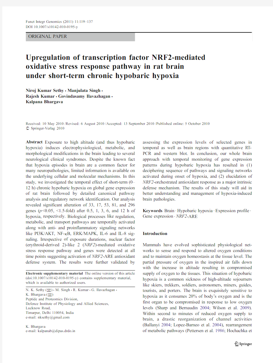
The primary aim of this study was to identify the hypoxia-regulated early genes in rat brain under chronic hypobaric hypoxia(25,000ft,310mmHg).To examine the effect of chronic hypoxia,we examined the global gene expression profiles of RNA samples from0.5,1,3,6,and12h hypoxia along with normobaric control brain samples on Illumina platform.Stringent data analysis was performed in order to ensure the genes identified through microarray analysis were truly differentially expressed.Among the22,523genes present on BeadChips,microarray analysis detected a total of108,286,1,020,1,800,and3,740differentially regulated genes at0.5,1,3,6,and12h of hypoxia exposure respectively (Fig.1a,Supplementary material1).Among the differential-ly regulated genes,74were upregulated and34were downregulated at0.5h of exposure,162were upregulated and124were downregulated at1h,538were upregulated and482were downregulated at3h,1,027were upregulated and773were down regulated at6h,1,856were upregulated and1,884were down regulated at12h of exposure.Of the differentially expressed genes26,12,37,67,and234genes were upregulated and7,5,16,14,and62genes were downregulated by1.5-folds after0.5,1,3,6,and12h of hypobaric hypoxia respectively(Fig.1b,Table1).Analysis of expression profiles of all the time points revealed that22 genes(17upregulated and five downregulated)are common differentially regulated(Fig.2).The genes that were upregulated at all the time points were LCN2,VERGE, NR1D1,ADAMTS1,NFKBIA,SGK,DDIT4,NDRG1,and PLAT while genes like DNMT3A and SERPINB6A were downregulated(Fig.2).
Biological processes affected by hypoxia were investi-gated with GO analysis of significantly regulated genes at all the time points.Table2shows the20most significant temporal regulated GO biological processes in the brain ordered by p value across all the time points.The GO processes like biological regulation,regulation of biological process,regulation of cellular process,negative regulation of biological process,and developmental process were regulated by onset of the hypoxia exposure(0.5h).Similarly,the GO processes like primary metabolic process, metabolic process,macromolecule metabolic process,cel-lular component organization and biogenesis,and estab-lishment of cellular localization were regulated only after 3h of hypoxia exposure.The6h exposure regulated GO biological processes like vesicle-mediated transport and secretory pathway(Table2).Taken together,the GO biological processes were differentially regulated depend-ing on the duration of hypoxia.
Signaling pathways and associated functions altered
by hypobaric hypoxia
To gain an understanding of functional cellular pathways and processes that may be coordinately regulated during early phases of chronic hypobaric hypoxia,all the significant differentially regulated genes at each time point were uploaded to IPA and visualized.The functions of associated networks detected at0.5h of exposure are immunological disease,gene expression,cellular growth,and proliferation. Similarly,the major functions for1h hypoxia are cellular assembly and organization,cell morphology,nervous system development and function followed by cellular assembly and organization,cellular movement,nervous system develop-ment and function,lipid metabolism,molecular transport,and small molecule biochemistry at3h.The functions of associated networks at6h of exposure are DNA replication, recombination,and repair,neurological disease,behavior and at12h are cellular compromise,gene expression,inflamma-tory disease,metabolic disease,genetic disorder,and neuro-logical disease(Supplementary material3).
From the significant functions at each time point,the molecular networks were visualized with the Ingenuity tool. The networks assembled with IPA profiling of genes differentially expressed at0.5,1,3,6,and12h of hypoxia exposure,including interacting partners are depicted (Fig.3a;Supplementary material4).Genes demonstrated to be differentially upregulated or downregulated after microarray analyses are colored in red and green respec-tively.Genes that are uncolored were not found to be significantly altered with the hypoxia exposure.The class function of each of the genes,including enzymes,growth factor,and receptors are depicted by nodal shape.The fold change in gene expression compared with control animals is shown below each color node in respective figures. Genes like JUN,FOS,ACTB represent the central nodes in the pathways detected after0–3h of exposure while genes like NFE2L2,STAT3,AGT,CAPS3,and CCND1represent the central nodes of pathway detected after6–12h of exposure along with JUN and FOS(Fig.3a,Supplementary material2).These data suggested that prolonged hypoxia exposure activates more number of signaling molecules and interacting pathways in brain.
We next assessed the canonical pathways after chronic hypobaric hypoxia exposure.The top three canonical path-ways at 0.5h of hypoxia exposure were IL-10signaling,tight junction signaling,and arachidonic acid metabolism;at 1h were dendritic cell maturation,VDR/RXR maturation,nico-tine,and nicotinamide metabolism;at 3h were notch signaling,NRF2-mediated oxidative stress,IL-8signaling;at 6h were notch signaling,MIF regulation of innate immunity,semaphoring signaling in neurons while NRF2-mediated oxidative stress,antigen presentation pathway,and semaphoring signaling in neurons were the most significant pathway at 12h (Table 3,Supplementary material 1).Temporal analysis of canonical pathways revealed that PI3K/AKT and NF κB signaling pathways were detected during 0.5–6h of exposure,glycolysis/gluconeogenesis and GABA receptor signaling pathways were detected after 1h,VEGF signaling after 3h and ERK/MAPK signaling after 6h of hypoxia exposure (Supplementary material 2).Several pathways like NRF2-mediated oxidative stress response,IL-6and IL-17signaling,acute phase response signaling,angiopoitein signaling,thrombin signaling were detected at all the time points.
Expression analysis of hypoxia regulated genes
To validate results obtained with the microarray data and to further asses the temporal and tissue-specific expression of the differentially regulated genes,we assessed mRNA
expression of 12genes by real-time PCR.Figure 4shows average mRNA expression of LCN2,SGK1,ADM ,VEGFA ,HO-1,NOS3,SLC16A3,EGFL7,ALOX15,EEF2K ,EPHX2,and PROM1after 0.5,1,3,6,and 12h of hypoxia exposure.The expression data was in good agreement with the microarray data for all the genes further validating the hypoxia-induced expression levels.From the microarray data followed by real-time validation,it was apparent that maximum expression was observed at 12h of hypoxia exposure for most of the genes (Table 1;Fig.4).So we assessed the expression of nine genes (LCN2,SGK1,ADM ,VEGFA ,NRF2,HO-1,NOS3,SLC16A3,EGFL7)exposed to 12h hypoxia in various brain parts viz.amygdala,hippocampus,cortex,and cerebellum.Hypobaric hypoxia exposure increased expression of all the studied genes in the four regions and the highest expression levels were observed for hippocampus and cortex regions (Fig.5).Protein expression analysis of NRF2and downstream targets
To validate the NRF2-mediated antioxidant response path-way (Fig.3b ),the protein expressio levels of NRF2and its downstream target HO -1was evaluated by western blot analysis..Figure 6depicts the western blot results of the two selected genes after 0.5,1,3,6,and 12h of hypoxia exposure along with β-ACTIN as loading control.The maximum expression of NRF2was observed after 3h of
A.
200400600800100012001400160018002000Exposure time (h)
N u m b e r o f g e n e s
B.
50100150200250
0.5h
1h
3h
6h
12h
0.5h
1h
3h
6h
12h
Exposure time (h)
N u m b e r o f g e n e s
Fig.1Hypobaric hypoxia ex-posure (25,000ft,310mm Hg)temporally regulates gene ex-pression in rat brain.a The total number of upregulated and downregulated genes detected after 0.5,1,3,6,and 12h of hypobaric hypoxia exposure (n =3,p <0.05).b The number of significant differential regulated genes (±1.5-fold,p <0.05)detected after 0.5,1,3,6,and 12h of hypobaric hypoxia ex-posure.The highest number of hypoxia regulated genes and significant differential genes were detected after 12h of exposure
Table1Genes differentially expressed by chronic hypoxia arranged in functional classes
Functional class/Gene name GenBank no Symbol Fold change(h)
0.501030612
Neurofunction and neuroprotective genes
Adrenomedullin NM_012715.1Adm 1.36 1.24 2.11 2.80 2.19 Plasminogen activator,tissue NM_013151.2tPA0.810.820.69 1.61 1.20 Stanniocalcin1NM_031123.2Stc1–0.75 1.36 2.14 1.69 TIMP metallopeptidase inhibitor1NM_053819.1Timp1–0.380.340.82 1.75 Lectin,galactose binding,soluble3NM_031832.1Lgals3–0.380.54 1.01 1.58 Annexin A2NM_019905.1Anxa2–0.380.55 1.12 1.86 Epithelial membrane protein1NM_012843.2Emp1–0.92 1.40 2.20 2.20 Nestin NM_012987.1Nes–– 2.23 1.92–Aspartoacylase NM_024399.1Aspa–– 1.21 1.57–Serine(or cysteine)peptidase inhibitor,clade E,member1NM_012620.1Serpine1–– 1.18 1.78–RNA binding motif(RNP1,RRM)protein3NM_053696.1Rbm3––– 1.53 1.86 Carboxypeptidase E NM_013128.1Cpe–––0.58 1.86 Neuronal regeneration related protein NM_178096.2Nrep–––– 2.18 Chemokine(C-X-C motif)receptor4NM_022205.3Cxcr4–––– 2.03 Ribosomal protein S9NM_031108.2Rps9?0.92?0.65?5.67–?5.53 Activity-regulated cytoskeleton-associated protein NM_019361.1Arc–?1.66?2.08?3.06?3.74 MAS1oncogene NM_012757.2Mas1–?0.47?0.55?0.78?1.69 Angiogenesis,blood vessel development,and blood pressure regulation
EGF-like-domain,multiple7NM_139104.1Egfl7 1.070.830.92 1.58 1.62 Fibronectin1NM_019143.2Fn10.57–0.89 1.84 1.80 Angiopoietin2NM_134454.1Angpt2–– 1.09 1.99 4.07 Vascular endothelial growth factor A NM_031836.2Vegfa–– 1.08 1.75 3.10 Nitric oxide synthase3,endothelial cell NM_021838.2Nos3–– 1.57 2.42 2.48 Placental growth factor NM_053595.2Pgf–– 1.14 2.12 2.42 Thrombomodulin NM_031771.2Thbd–– 1.20 1.560.96 Heparanase NM_022605.1Hpse–– 1.30 2.30 1.20 Collagen,type IV,alpha NM_001135009.1Col4a1––0.89 1.59 1.66 Epoxide hydrolase2,cytoplasmic NM_022936.1Ephx2––?0.30?0.39?4.65 Metabolism
Solute carrier family16,member3(monocarboxylic acid
NM_030834.1Slc16a3– 1.04 1.93 3.02 3.05 transporter4)
Pyruvate dehydrogenase kinase,isozyme4NM_053551.1Pdk4– 2.28 2.68 3.32 3.34 Glycerol-3-phosphate dehydrogenase1(soluble)NM_022215.2Gpd1–0.68 1.21 2.10 1.55 Lipolysis stimulated lipoprotein receptor NM_032616.1Lsr– 1.28 1.55 1.36–Phospholipase A1member A NM_138882.1Pspla1–– 1.10 2.03 2.83 Hexokinase2NM_012735.1Hk2––– 1.49 1.76 Dipeptidylpeptidase6NM_022850.1Dpp6––– 1.22 1.64 Caveolin2NM_131914.2Cav2–––0.74 1.74 Prominin1NM_021751.2Prom1––?1.44?2.43?2.75 Thyroid stimulating hormone,beta NM_013116.1Tshb?1.98–––?1.08 Transport
FXYD domain-containing ion transport regulator5NM_021909.1Fxyd5––0.85 1.570.86 Chloride intracellular channel4NM_031818.1Clic4––0.65 1.02 1.85 Solute carrier family6(neurotransmitter transporter,
NM_013034.3Slc6a4––– 2.99 1.86 serotonin),member4
Chloride intracellular channel2NM_001009651.1Clic2 1.85––––Solute carrier family4(anion exchanger),member1NM_012651.2Slc4a1 1.87––––
0.501030612 Solute carrier family6(neurotransmitter transporter,glycine),
NM_203334.1Slc6a5–––– 2.10 member5
Solute carrier family3,member1NM_017216.1Slc3a1–––– 2.76 Solute carrier family14(urea transporter),member1NM_019346.2Slc14a1–––– 3.90 Solute carrier family39(iron-regulated transporter),member1NM_133315.2Slc40a1–?0.60?1.26?1.71?1.17 Solute carrier organic anion transporter family,member1c1NM_053441.1Slco1c1––?0.81?1.77?1.83 Transcription factors and effectors
Activating transcription factor3NM_012912.1Atf3 1.75 2.14 1.75––Nuclear receptor subfamily4,group A,member3NM_017352.1Nr4a3––?0.52?1.20?2.22 Eukaryotic elongation factor-2kinase NM_012947.2Eef2k?3.34?3.12?2.50–?0.95 Early growth response1NM_012551.2Egr1–––?1.51?2.07 FBJ osteosarcoma oncogene NM_022197.2Fos–––?1.56?1.01 Early growth response2NM_053633.1Egr2––––?2.28 Antioxidant Response
Metallothionein1a NM_138826.4Mt1a– 1.80 2.84 3.89 4.24 Heme oxygenase(decycling)1NM_012580.2HO-1–– 1.13 1.96 3.07 DNA-damage-inducible transcript4NM_080906.1Ddit40.88 1.16 1.62 1.64 1.10 Transferrin receptor protein1NM_022712.1Tfrc–––– 2.13 Apoptosis
TIMP metallopeptidase inhibitor3NM_012886.2Timp3––– 1.61 1.72 Caspase4,apoptosis-related cysteine peptidase NM_053736.2Casp11–––– 2.66 Arachidonate15-lipoxygenase NM_031010.2Alox15 4.41––?0.56?0.77 BCL2/adenovirus E1B19kDa-interacting protein3NM_053420.2Bnip3–?0.58?0.49–?2.57 B-cell leukemia/lymphoma2related protein A1d NM_133416.1Bcl2a1––?1.72?1.99–Immunogenic response
FK506binding protein5NM_001012174.1Fkbp5?0.76– 1.00 1.590.82 RT1class Ia,locus A1NM_001008827.1RT1-A1–––0.29 1.53 RT1class I,locus T24,gene4NM_001008826.3RT1-149–––0.72 1.60 CD93molecule NM_053383.1C1qr1––– 1.18 2.25 Integrin,beta2NM_001037780.2Itgb2–––– 2.90 Defensin NP-4precursor NM_173299.1Np4––––7.33 Defensin beta1NM_031810.1Defb1––?0.56?1.52?0.91 Inflammation
Tumor necrosis factor receptor superfamily,member12a NM_181086.2Tnfrsf12a– 1.55 1.330.92 1.11 Zinc finger protein36NM_133290.3Zfp36–0.76 1.41 1.670.74 Lectin,galactose binding,soluble3NM_031832.1Lgals3–0.380.54 1.01 Haptoglobin NM_012582.2Hp––– 2.39 2.09 Secreted phosphoprotein1NM_012881.2Spp1–––– 2.22 Prostaglandin-endoperoxide synthase2NM_017232.3Ptgs2––?0.68?0.68?1.51 Novel genes
Lipocalin2NM_130741.1Lcn27.37 6.87 5.490.3411.94 Vascular early response gene protein NM_001003403.1Verge 2.61 1.82 2.52 2.58 1.56 Cathepsin E NM_012938.1Ctse 2.51– 1.34 1.36 1.65 A disintegrin-like and metallopeptidse(reprolysin type)with
NM_024400.2Adamts1 1.270.73 1.55 2.05 2.72 thrombospondin type1motif,1
Retinal pigment epithelium65NM_053562.1Rpe650.750.82 1.18 1.76 1.66 Serine(or cysteine)proteinase inhibitor,clade B,member1a NM_001031642.1Serpinb1a 1.000.900.81– 1.68 Interferon induced transmembrane protein3NM_001136124.1Ifitm3–0.320.460.94 1.75
0.501030612
Kinesin family member22NM_001009645.1Kif22–0.320.460.94 1.75 Transgelin NM_001013127.1Tagln2––0.74 1.26 1.81 C-type lectin domain family4,member a3NM_001005891.1Dcir3–– 1.16 1.13 1.63 Carbonic anyhydrase12NM_001080756.1Car12––– 2.02 2.44 RAB31,member RAS oncogene family NM_145094.2Rab31––– 1.35 2.09 Phosphoglycerate mutase family member5NM_001025272.1Pgam5––– 1.27 1.85 Proopiomelanocortin NM_139326.2Pomc–––– 2.07 Abhydrolase domain containing5NM_212524.1Abhd5–––– 1.94 DNA(cytosine-5-)-methyltransferase3alpha NM_001003957.1Dnmt3a?1.52?0.91?1.39?0.74?1.99 Serine(or cysteine)peptidase inhibitor,clade B,member6a NM_199085.1Serpinb6a?0.54?0.65?0.69?0.48?1.68 Hematopoietically expressed homeobox NM_024385.1Hhex–?1.71?1.06?0.57–
The respective GenBank number and the expression level at each time point of exposure is mentioned
Fig.2Heatmap indicating tem-
poral gene expression patterns.
The22common genes between
all the five hypoxia exposure
time points(0.5,1,3,6,and
12h)were depicted along with
symbol and name.The expres-
sion levels were shown in the
bar(red indicates upregulation,
green indicates downregulation,
and black indicates no change).
Comparison between groups of
samples was done with the t test
differential expression algorithm
(detection p value<0.05).The
gene symbol and name were
mentioned
exposure(6.2-fold,p<0.01)as compared to normobaric control rats after which a steady expression level was observed till12h(Fig.6b).Similarly,a significant increase in the expression of HO-1was observed only after6h(5.8-fold,p<0.01)which was further increased after12h(8.0 fold,p<0.001)of exposure(Fig.6b).These results along with IPA analysis and qRT-PCR results suggest that the NRF2-antioxidant responsive element(ARE)pathway is significantly upregulated in response to short-term chronic hypobaric hypoxia in rat brain.
Discussion
Hypobaric hypoxia induces a number of neurological effects on the brain and has a high social and economic impact.As increasing number of people visiting high altitude for work, leisure,and adventure,an understanding of altitude-induced cerebral adaptation,malfunction,management,and treat-ments are becoming increasingly important(Wilson et al. 2009).In an attempt to understand the brain response to hypobaric/environmental hypoxia,we conducted a system-atic expression profiling to identify the genes and molecular events associated with high altitude-exposure.We observed up-or downregulation of108,286,1,020,1,800,and3,740genes(p<0.05)after0.5,1,3,6,and12h of hypoxia exposure respectively(Fig.1a).Higher numbers of upregu-lated genes were detected at0.5,1,3,and6h,while higher numbers of downregulated genes were detected at12h of exposure(Fig.1a).Previous gene expression studies monitoring the brain response to environmental hypoxia have reported regulation of lesser number of genes (Bernaudin et al.2002a;Ganfornina et al.2005;Gustavsson et al.2007;Zhou et al.2008).This could be attributed either by the region specific gene expression like carotid body (Ganfornina et al.2005),cerebral cortex(Gustavsson et al. 2007;Zhou et al.2008)and hippocampus(Zhou et al.2008) or the lower number of analyzed genes(Bernaudin et al. 2002a).In this regard,our whole brain approach combined with temporal monitoring of gene expression,resulting in higher number of identified differential genes represents the most robust gene expression study for environmental hypoxia till date.
Cellular adaptation to hypoxia involves a pronounced repression of energy consuming protein synthesis,which occurs initially at the levels of translation and later extends to the level of transcription as well(Pettersen et al.1986; Hochachka et al.1996;Hochachka and Lutz2001).At the same time,altered expression of many genes and proteins promote metabolic rearrangements including enhanced oxy-
Table2The significant gene ontologies(GO)altered by hypobaric hypoxia in rat brain
Ontology Time of exposure
0.5h1h3h6h12h
GO:0065007:biological regulation 2.94E-04 1.82E-05 2.72E-12 1.12E-17 4.22E-29 GO:0044238:primary metabolic process–– 2.22E-07 2.58E-15 1.25E-23 GO:0008152:metabolic process–– 4.21E-07 1.42E-13 1.51E-23 GO:0050789:regulation of biological process0 6.22E-04 3.16E-09 4.89E-13 2.72E-23 GO:0016043:cellular component organization and biogenesis–– 1.58E-04 1.80E-11 4.08E-23 GO:0044237:cellular metabolic process0.02– 1.11E-08 6.14E-14 6.65E-22 GO:0032502:developmental process9.31E-04 1.06E-07 4.90E-117.55E-16 4.75E-15 GO:0050794:regulation of cellular process0.010.003 1.04E-07 4.09E-10 4.84E-15 GO:0043170:macromolecule metabolic process––7.53E-06 1.25E-08 3.64E-14 GO:0051649:establishment of cellular localization––0.026 5.77E-048.49E-13 GO:0051641:cellular localization––0.031 5.77E-04 3.06E-12 GO:0048519:negative regulation of biological process0.01 4.73E-05 2.10E-07 1.43E-078.66E-12 GO:0051179:localization0.02–0.001 2.73E-06 2.00E-11 GO:0016192:vesicle-mediated transport–––0.002 2.27E-11 GO:0030154:cell differentiation 4.23E-04 3.27E-06 5.48E-089.77E-11 2.33E-11 GO:0048869:cellular developmental process 4.23E-04 3.27E-06 5.48E-089.77E-11 2.33E-11 GO:0048523:negative regulation of cellular process0.017.51E-05 6.56E-07 1.29E-07 1.99E-10 GO:0006810:transport––0.0130.001 2.77E-10 GO:0048468:cell development0.03 3.84E-04 6.22E-06 5.63E-11 4.23E-10 GO:0045045:secretory pathway–––0.0029.23E-10
Top20most significantly regulated ontologies(sorted with p value)were mentioned along with exposure time and p value
gen delivery to hypoxic tissues essential for adaptation and cell survival(Seta and Millhorn2004;Liu et al.2006; Wilson et al.2009).In this study several hypoxia-responsive genes involved in angiogenesis(EGFL7,PGF,ANGPT2, VEGF A),vasodialtion(NOS3),metabolism(PDK4,HKII, PROM1),apoptosis(TIMP3,BNIP3),inflammation (TNFRSF12A,HP,PTGS2),antioxidant response(MT1A, HO-1,DDIT4),and transcription factors(ATF3,NR4A3) were found to be temporally regulated(Table1).The altered expression and contribution of these genes to hypoxic response has been reported for organs including heart(Fan et al.2005;Iacobas et al.2008),liver(Dolt et al.2007;Baze et al.2010),brain(Bernaudin et al.2002a;Zhou et al.2008), lungs(Wu et al.2008;Tada et al.2008),as well as various cells(Mense et al.2006;Polotosky et al.2010).Moreover, genes for angiogenesis and metabolism(Table1)were also found to be differentially regulated during normobaric hypoxia(Bernaudin et al.2002b),ischemia(Hedtjarn et al. 2004),and preconditioning(Stenzel-Poore et al.2003; Carmel et al.2004;Dhodda et al.2004;Gidday2006)in brain.Similarly,genes for antioxidant response(Table1) were reported to be differentially regulated during intermit-tent and sustained hypoxia(Polotosky et al.2010),normo-baric,and hyperbaric hyperoxia(Chen et al.2009a,b
;
Fig.3Analysis of hypobaric hypoxia(12h)regulated network and canonical pathway in brain.Differentially expressed genes from analysis of12-h hypoxia brain sample were analyzed by Ingenuity Pathway Analysis(IPA).a The significant network1(p<0.05) detected by IPA is depicted.The central molecules in the network are FOS and NFE2L2(NRF2).b NRF2-mediated oxidative stress response canonical pathway detected after12h of exposure(p<0.05). Colored genes(red and green)were identified by microarray analysis as differentially upregulated or downregulated in brain samples compared with control.Other uncolored nodal genes are directly or indirectly associated with the differentially expressed genes but were not found to be significantly regulated by hypoxia.Genes are linked by their sub-cellular location(cytoplasm and nucleus).Node(gene) and edge(gene relationship)symbols were described in the bottom part of the figure
Fig.3(continued)
Godman et al.2010)along with ischemic preconditioning (Stenzel-Poore et al.2003;Carmel et al.2004;Dhodda et al. 2004).Our present findings along with these previous studies suggest a tissue-independent overlapping gene signature profile in response to varying oxygen concentra-tion.
Along with hypoxia-responsive genes,many neuroprotec-tive genes like ADM,SGK1,STC1,tP A,ALOX15,and ARC (Table1)were found to be differentially regulated in brain. Adrenomedullin(AM),a neuroprotective peptide is expressed throughout the whole brain(Serrano et al.2000)and upregulated in the cortex region of adult rat brain during acute hypobaric hypoxia(Serrano et al.2008).Recently, Fernandez et al.(2008)have reported that mice with no brain AM have impaired motor coordination,anxiety and were less resistant to hypobaric hypoxia than wild-type mice.The upregulation of serine/threonine protein kinase Serum-and glucocorticoid-induced kinase-1(SGK1)has various function-al roles like facilitation of learning and memory formation (Tsai et al.2002;Chao et al.2007),consolidation of long-term memory(V on Hertzen and Giese2006)and in neuronal plasticity.Stanniocalcin(STC),a glycoprotein hormone plays an important role in maintaining and guarding the integrity of terminally differentiated neuronal cells challenged by ischemia and calcium-mediated cell death(Zhang et al.2000).Tissue-type plasminogen activator(tP A/PLAT)is a highly specific serine proteinase expressed within the central nervous system where it has been associated with learning,synaptic plasticity and regulation of the permeability of the neurovascular unit (Yepes et al.2009).Similarly,the genes impairing neuro-protection like activity-regulated cytoskeletal-associated pro-tein(ARC)/Arg3.1(activity-regulated gene 3.1protein homolog)(Guzowski et al.2000)and12/15-lipoxygenase (ALOX15)(Jin et al.2008)were downregulated during short-term chronic hypoxia(Table1).
Our results also confirm that short-term chronic hypobaric hypoxia altered biological processes and signaling pathways depending on the duration of hypoxia stress(Table2).In response to reduced oxygen supply,rearrangement of meta-bolic pathways has been observed(Hochachka et al.1996; Lopez-Barneo et al.2001),which essentially indicates activation of adaptive responses.The signaling pathways like PI3K/AKT and NF-κB signaling were detected form0.5h of hypoxia exposure demonstrating activation of the survival and proliferation pathways at the onset of the hypoxia exposure (Supplementary material2).The PI3K/AKT survival signaling pathway is regulated by oxidative stress(Wang et al.2000) and activates the nuclear translocation of NF-κB-dependant prosurvival genes(Kane et al.1999).Hypoxia as well as hypoxic preconditioning activates AKT signaling pathway exerting neuroprotection(Alvarez-Tejado et al.2001;Zhang et al.2007;Yin et al.2007).Similarly,activation of NF-κB during cerebral ischemia has been reported to promote proapoptotic as well as antiapoptotic mechanisms(Schneider et al.1999;Irving et al.2000)and plays a key role in the regulation of cellular responses to oxidative stress(Denk et al. 2000).With the increase in exposure time(1–3h)pathways like glycolysis/gluconeogenesis and VEGF signaling were activated(Table3;Supplementary material4)to ensure efficient A TP production by alternate pathways(Hochachka et al.1996;Lutz and Nilsson1997;Hochachka and Lutz 2001)and increased oxygen availability(Marti and Risau 1998).Moreover,activation of VEGF confers neuroprotection after focal cerebral ischemia by PI3K/AKT pathway(Kilic et
Table3The significant canonical pathways altered by hypobaric hypoxia exposure in rat brain
0.5h1h3h6h12h
IL-10signaling Dendritic cell maturation Notch signaling Notch signaling NRF2-mediated oxidative
stress response
Tight junction signaling VDR/RXR activation NRF2-mediated oxidative
stress response MIF regulation of innate
immunity
Antigen presentation
pathway
Arachidonic acid metabolism Nicotinate and
nicotinamide
metabolism
Regulation of actin-based
Motility by Rho
Semaphorin signaling in
neurons
Semaphorin signaling in
neurons
PPARa/RXRa activation Pantothenate and CoA
biosynthesis
IL-8signaling HIF1αsignaling Mitochondrial dysfunction
IL-6signaling Coagulation system Semaphorin signaling in
neurons Nicotinate and
nicotinamide
metabolism
Synthesis and degradation
of ketone bodies
p38MAPK signaling Semaphorin signaling in
neurons VEGF signaling IL-6signaling D-glutamine and D-
glutamate signaling
NRF2-mediated oxidative stress response IGF-1signaling Valine,leucine and
isoleucine degradation
VEGF signaling Hutington’s disease
signaling
4-1BB signaling in T lymphocytes Leukocyte extravazation
signaling
Circadian rhythm
signaling
Circadian rhythm
signaling
Valine,leucine and
isoleucine degradation
The top8significant canonical pathways at each time point were mentioned in order of their p value
A. LCN2
24681012
14Expo s ure time (h)
m R N A e x p r e s s i o n
B. SGK1
0.511.522.5
Expo s ure time (h)
m R N A e x p r e s s i o n
C. ADM
0.511.522.533.5
4Expo s ure time (h)
m R N A e x p r e s s i o n
D. VEGF
0.511.522.53
3.5Expo s ure time (h)
m R N A e x p r e s s i o n
G. SLC16A3
0.511.522.533.5
4Expo s ure time (h)
m R N A e x p r e s s i o n
H. EGFL7
0.511.522.5Expo s ure time (h)
m R N A e x p r e s s i o n
E. HO-1
0.511.522.53
3.5Expo s ure time (h)
m R N A e x p r e s s i o n
F. NOS3
0.511.522.53
3.5Expo s ure time (h)
m R N A e x p r e s s i o n
Fig.4Verification of microarray expression with real-time PCR for differentially regulated gene transcripts.Real-time PCR was per-formed with TaqMan probes with three biological replicates for each time point (0.5,1,3,6,and 12h hypobaric hypoxia exposure,25,000ft).All the samples were run in triplicate (mean ±SE)with endogenous control probes.The expression values were depicted in log 2scale.a LCN2,b SGK1,c ADM ,d VEGF A ,e HO-1,f NOS3,g SLC16A3,h EGFL7,i ALOX15,j EEF2K ,k EPHX2,l PROM1
al.2006).With 6h of hypoxia stress,activation of neuro-protective ERK/MAPK signaling pathway was observed.Both hypoxia and hypoxia-induced ischemic tolerance acti-vates ERK 1/2signaling pathway providing neuroprotection (Fahlman et al.2002;Fang et al.2010).Moreover,activation of ERK pathway also confers neuroprotection during hypo-baric hypoxia (Barhwal et al.2008)and ischemia (Kilic et al.2005;Shioda et al.2009).The longer exposure duration (12h)activated mitochondrial dysfunction along with NRF2-mediated oxidative stress response pathways (Table 3).The sustained oxidative stress during hypoxia leads to mitochondria-permeability transition,release of cytochrome c and mitochondrial dysfunction causing cell death (Calabrese et al.2005;Douglas et al.2010).Hence,activation of mitochondrial dysfunction at 12h of hypoxic exposure indicates activation of pro-apoptotic mechanisms.This was further supported by the major functions like necrosis of cortical neurons,cell death of neurons,injury of nervous tissue,cell death of cortical neurons,cell death of cerebral cortex cells,degeneration of neurons,injury of spinal cord,and injury of axons of molecular networks 1,2,and 4of 12-h exposure (Supplementary Material 4).
Irrespective of exposure period,NRF2-mediated oxidative stress response pathway and genes were found to be significantly regulated in brain (Table 3,Fig.3b ).The transcription factor NRF2is the guardian of redox homeosta-sis and regulates a coordinated induction of a battery of cytoprotective,antioxidant,and anti-inflammatory genes in response to oxidative stress and inflammation (Jaiswal 2004;Nguyen et al.2009).Under normal conditions,NRF2undergoes proteasomal degradation in cytoplasm by Kelch ECH associating protein 1(KEAP1).During oxidative stress NRF2is released from KEAP1,translocates to the nucleus and transactivates the expression of several dozen cytopro-tective genes containing cis -acting ARE (Kensler et al.2007;Nguyen et al.2009).Increasing number of studies has suggested that NRF2-ARE activation is a novel neuro-protective pathway that confers resistance to a variety of oxidative stress-related neurodegenerative insults like 6-hydroxydopamine (Jakel et al.2007),kainic acid (Kraft et al.2006),hypobaric hypoxia (Barhwal et al.2009),cerebral ischemia (Shih et al.2005a ;Shah et al.2007),intracerebral hemorrhage (Wang et al.2007;Zhao et al.2007),and hyperoxia toxicity (Chen et al.2009a ,b ).Moreover,NRF2pathway-mediated promotion of neuroprotection by neurite outgrowth-promoting prostaglandins (Satoh et al.2006),sulforaphane (Innamorato et al.2008),and acetyl-l-carnitine (Barhwal et al.2009)also suggests that NRF2is a therapeutic target against neurodegenration (Johnson et al.2008).High altitude exposure causes oxidative stress in brain via generation of reactive oxygen and nitrogen species which can cause damage to lipids,proteins,and DNA (Moller et al.2001;Maiti et al.2006;Hota et al.2007).Brain being exquisitely sensitive to oxygen has evolved a dynamic
I. ALOX15
Exposure time (h)m R N A e x p r e s s i o n
J. EEF
2K
Exposure time (h)m R N A e x p r e s s i o n
K. E
PHX2
Exposure time (h)
m R N A e x p r e s s i o n
L. PROM1
Exposure time (h)
m R N A e x p r e s s i o n
Fig.4(continued)
protective response to altered oxygen concentrations and oxidative stress (Sharp and Bernaudin 2004;Wilson et al.2009).Our expression analysis combined with pathway identification by IPA suggests NRF2-mediated oxidative stress response is the major pathway providing cytoprotection during short-term chronic hypobaric hypoxia (Fig.3a and b ).We also observed upregulation of NRF2downstream targets HO-1,SOD ,TXN ,GST ,GCLC ,and GCLM in response to hypobaric hypoxia (Fig.3b ).Similar whole genome expres-sion studies assessing the effects altered oxygen concentration like intermittent and sustained hypoxia in human arotic endothelial cells (Polotosky et al.2010),hypoxia-ischemia in immature mice brain (Hedtjarn et al.2004),ischemia-reperfusion injury in kidney (Leonard et al.2006),and
normobaric and hyperbaric hyperoxia in rat cortical neurons (Chen et al.2009a ,b )have also identified upregulation of transcription factor NRF2and downstream genes.Validation of mRNA abundance of NRF2by real-time PCR also revealed higher expression levels in various brain regions following 12h of hypoxia (Fig.5).Similarly,expression of HO-1was also found to be temporally induced by hypoxia (Table 1;Fig.4e )and in various brain regions following 12h hypoxia (Fig.5).The induction of HO-1is considered to counteract oxidative damage,confer cytoprotection,and promote adaptive survival (Ryter et al.2006;Gozzelino et al.2010).In corroboration,our western blot results also demonstrate temporal activation of NRF2and HO-1proteins in response to short-term chronic hypobaric hypoxia (Fig.6).The maximum expression of NRF2was observed after 3h of exposure while the expression of HO-1was observed after 6h of exposure supporting the stabilization and nuclear transport of NRF2followed by transcriptional activation of HO-1(Jaiswal 2004;Kensler et al.2007;Nguyen et al.2009).The activation of NRF2/HO-1pathway provides neuroprotection against oxidative stress (Satoh et al.2006
;
2000
40006000800010000120001400016000C
0.5 h
1 h
3 h
6 h
12 h
Exposure time (h)
R e l a t i v e i n t e n s i t y (a r b i t r a r y u n i t s ) a f t e r n o r m a l i z a t i o n
β
-ACTIN
NR F 2
HO-1
C 0.5h 1h 3h 6h 12h
a
b
Fig.6Analysis of NRF2and HO-1expression in rat brain after 0.5,1,3,6,and 12h of hypobaric hypoxia exposure (25,000ft)by western blot analysis.a Representative blot of NRF2and HO-1temporal expression in brain of hypobaric hypoxia exposed rats along with normobaric controls.b Average NRF2and HO-1expression in normobaric control and hypobaric hypoxia exposed brain depicted graphically after normalization to β-action expression.Student ’s t test was performed and p <0.05was considered statistically significant (*p <0.05,**p <0.01,and ***p <0.001with respect to normobaric control)
123456
7m R N A e x p r e s s i o n
0.511.522.533.54
4.5m R N A e x p r e s s i o n
0.511.522.533.5
4LCN2
SGK1
ADM
VEGFA
NRF2
HO-1
NOS3SLC16A3
EGFL7m R N A e x p r e s s i o n
Fig.5Expression analysis of identified differential genes in brain regions (amygdala,hippocampus,cortex,and cerebellum)exposed to 12h of hypobaric hypoxia (25,000ft).Real-time PCR was performed with TaqMan probes for LCN2,SGK1,ADM ,VEGF A ,NRF2,HO-1,NOS3,SLC16A3,and EGFL7from three biological replicates (mean±SE).All the reactions were run in triplicate with endogenous control.The expression levels are in log 2scale
Innamorato et al.2008;Espada et al.2010)and promotes neuronal survival(V argas et al.2005).Altogether,our study suggests that upregulation of transcription factor NRF2and activation of NRF2-ARE pathway as an intrinsic defense mechanism of brain against short-term chronic hypobaric hypoxia-induced oxidative stress.
The intensity and duration of stress along with the individual response determines the final outcome.In the present study,activation of signaling pathways(NF-κB, PI3K/AKT,and ERK/MAPK)along with protective genes during short-term chronic hypobaric hypoxia(Table1)may represent rapid activation of antiapoptotic pathways (Guyton et al.1996;Milton et al.2008).But the delayed and sustained activation of both NF-κB and ERK/MAPK has been reported in contributing to apoptosis in response to severe oxidant injury and brain ischemia(Martindale and Holbrook2002;Vartiainen et al.2003;Song et al.2008). Similarly,the activation of mitochondrial dysfunction path-way after12h of exposure also suggests a diminished activity of mitochondrial function-stabilizing pathways and proteins(Kilic et al.2005)during longer hypoxic exposure. Though we have not measured the expression levels of these pro-and anti-apoptotic molecules in longer hypoxia expo-sure,such studies may substantiate the current findings.In parallel,hypoxia exposure also induced pro-inflammatory cytokines like IL-6,IL-8as well as anti-inflammatory cytokine IL-10signaling pathways(Table3).During hypoxia,increased ROS production leads to activation of many proinflammatory cytokines by various mechanisms (Yan et al.1995;Tamm et al.1998),whereas IL-10treatment provides neuroprotection to hippocampus after ischemia (Dietrich et al.1999)and promotes angiogenesis in ischemic tissue(Silvestre et al.2001).These cumulative results suggest simultaneous activation of neuroprotective,antioxi-dant,anti-and proinflammatory genes during short-term chronic hypobaric hypoxia.
In summary,the global expression analysis of rat brain under short-term chronic hypobaric hypoxia followed by IPA analysis has provided a detailed insight of hypoxia-regulated brain transcriptome.The whole brain approach with temporal monitoring of gene expression has enabled us in(a)identification of hypoxia-responsive genes and expression patterns(b)deciphering the sequence of signal-ing events underlying hypoxia,(c)depicting the intrinsic defense mechanisms,and(d)revealing hypoxia-regulated novel genes in brain.Further,our studies have also identified and validated of NRF2-mediated oxidative stress response pathway as one of the major intrinsic antioxidant response of the brain in response to short-term hypobaric hypoxia.The results from this study will open the avenues for therapeutic interventions targeting the NRF2pathway for the early amelioration of hypobaric–hypoxia-induced neuropathologies.Acknowledgment This study was supported by Defence Institute of Physiology and Allied Sciences Grant TC/297/TASK-122(NKS)/ DIPAS/2007.MS is a recipient of Defence Research and Development Organisation(DRDO)Senior Research Fellowship(SRF).The technical support provided by Dr.Dhananjay Shukla is greatly appreciated.The authors declare no conflict of interest. References
Alvarez-Tejado M,Naranjo-Suarez S,Jiménez C,Carrera AC, Landázuri MO,del Peso L(2001)Hypoxia induces the activation of the phosphatidylinositol3-kinase/Akt cell survival pathway in PC12cells:protective role in apoptosis.J Biol Chem276
(25):22368–22374
Ballanyi K(2004)Protective role of neuronal K ATP channels in brain hypoxia.J Exp Biol207(Pt18):3201–3212
Barhwal K,Hota SK,Prasad D,Singh SB,Ilavazhagan G(2008) Hypoxia-induced deactivation of NGF-mediated ERK1/2signal-ing in hippocampal cells:neuroprotection by acetyl-L-carnitine.J Neurosci Res86(12):2705–2721
Barhwal K,Hota SK,Jain V,Prasad D,Singh SB,Ilavazhagan G (2009)Acetyl-l-carnitine(ALCAR)prevents hypobaric hypoxia-induced spatial memory impairment through extracellular related kinase-mediated nuclear factor erythroid2-related factor2 phosphorylation.Neuroscience161(2):501–514
Baze MM,Schlauch K,Hayes JP(2010)Gene expression of the liver in response to chronic hypoxia.Physiol Genomics41:275–288 Bernaudin M,Tang Y,Reilly M,Petit E,Sharp FR(2002a)Brain genomic response following hypoxia and re-oxygenation in the neonatal rat.
Identification of genes that might contribute to hypoxia-induced ischemic tolerance.J Biol Chem277:39728–39738
Bernaudin M,Nedelec AS,Divoux D,MacKenzie ET,Petit E, Schumann-Bard P(2002b)Normobaric hypoxia induces toler-ance to focal permanent cerebral ischemia in association with an increased expression of hypoxia-inducible factor-1and its target genes,erythropoietin and VEGF,in the adult mouse brain.J Cereb Blood Flow Metab22:393–403
Calabrese V,Lodi R,Tonon C,D’Agata V,Sapienza M,Scapagnini G, Mangiameli A,Pennisi G,Stella AMG,Butterfield DA(2005) Oxidative stress,mitochondrial dysfunction and cellular stress-response in Friedreich’s ataxia.J Neurol Sci233(1–2):145–162 Carmel JB,Kakinohana O,Mestril R,Young W,Marsala M,Hart RP (2004)Mediators of ischemic preconditioning identified by microarray analysis of rat spinal cord.Exp Neurol185(1):81–96 Chan PH(1996)Role of oxidants in ischemic brain damage.Stroke 27:1124–1129
Chao CC,Ma YM,Lee EH(2007)Protein kinase CK2impairs spatial memory formation through differential cross talk with PI-3 kinase signaling:activation of Akt and inactivation of SGK1.J Neurosci27:6243–6248
Chen PC,Vargas MR,Pani AK,Smeyne RJ,Johnson DA,Kan YW, Johnson JA(2009a)Nrf2-mediated neuroprotection in the MPTP mouse model of Parkinson’s disease:critical role for the astrocyte.Proc Natl Acad Sci USA106(8):2933–2938
Chen Y,Nadi NS,Chavko M,Auker CR,McCarron RM(2009b) Microarray analysis of gene expression in rat cortical neurons exposed to hyperbaric air and oxygen.Neurochem Res34:1047–1056 Denk A,Wirth T,Baumann B(2000)NF-κB transcription factors: critical regulators of hematopoiesis and neuronal survival.
Cytokine Growth Factor Rev11:303–320
Dhodda VK,Sailor KA,Bowen KK,Vemuganti R(2004)Putative endogenous mediators of preconditioning-induced ischemic tolerance in rat brain identified by genomic and proteomic analysis.J Neurochem89(1):73–89
Dietrich WD,Busto R,Bethea JR(1999)Postischemic hypothermia and IL-10treatment provide long-lasting neuroprotection of CA1 hippocampus following transient global ischemia in rats.Exp Neurol158(2):444–450
Dolt KS,Karar J,Mishra MK,Salim J,Kumar R,Grover SK,Pasha MAQ(2007)Transcriptional downregulation of sterol metabo-lism genes in murine liver exposed to acute hypobaric hypoxia.
Biochem Biophys Res Commun354:148–153
Douglas RM,Ryu J,Kanaan A,Rivero MC,Dugan LL,Haddad GG, Ali SS(2010)Neuronal death during combined intermittent hypoxia/hypercapnia is due to mitochondrial dysfunction.Am J Physiol Cell Physiol298(6):C1594–C1602
Espada S,Ortega F,Molina-Jijón E,Rojo AI,Pérez-Sen R, Pedraza-Chaverri J,Miras-Portugal MT,Cuadrado A(2010)The purinergic P2Y(13)receptor activates the Nrf2/HO-1axis and protects against oxidative stress-induced neuronal death.Free Radic Biol Med49(3):416–426
Fahlman CS,Bickler PE,Sullivan B,Gregory GA(2002)Activation of the neuroprotective ERK signaling pathway by fructose-1,6-bisphosphate during hypoxia involves intracellular Ca2+and phospholipase C.Brain Res958(1):43–51
Fan CH,Iacobas DA,Zhou D,Chen QF,Lai JK,Gavrialov O, Haddad GG(2005)Gene expression and phenotypic character-ization of mouse heart after chronic constant or intermittent hypoxia.Physiol Genomics22:292–307
Fang H,Zhang LF,Meng FT,Du X,Zhou JN(2010)Acute hypoxia promote the phosphorylation of tau via ERK pathway.Neurosci Lett474(3):173–177
Fernandez AP,Serrano J,Tessarollo L,Cuttitta F,Martínez A(2008) Lack of adrenomedullin in the mouse brain results in behavioral changes,anxiety,and lower survival under stress conditions.Proc Natl Acad Sci34:12581–12586
Ganfornina MD,Perez-Garcia MT,Gutierrez G,Miguel-Velado E, Lopez-Lopez JR,Marin A,Sanchez D,Gonzalez C(2005) Comparative gene expression profile of mouse carotid body and adrenal medulla under physiological hypoxia.J Physiol566:491–503
Gidday JM(2006)Cerebral preconditioning and ischaemic tolerance.
Nat Rev Neurosci7(6):437–448
Godman CA,Joshi R,Giardina C,Perdrizet G,Hightower LE(2010) Hyperbaric oxygen treatment induces antioxidant gene expres-sion.Ann NY Acad Sci1197:178–183
Gozzelino R,Jeney V,Soares MP(2010)Mechanisms of cell protection by heme oxygenase-1.Annu Rev Pharmacol Toxicol 50:323–54354
Gu ZL,Jiang Q,Zhang GY(2001)Extracellular signalregulated kinase1/2activation in hippocampus after cerebral ischemia may not interfere with post-ischemic cell death.Brain Res901:79–84 Gustavsson M,Mallard C,Vannucci SJ,Wilson MA,Johnston MV, Hagberg H(2007)Vascular response to hypoxia preconditioning in the immature brain.J Cereb Blood Flow Metab27(5):928–938 Guyton KZ,Liu Y,Gorospe M,Xu Q,Holbrook NJ(1996)Activation of mitogen-activated protein kinase by H2O2.Role in cell survival following oxidant injury.J Biol Chem271:4138–4142 Guzmán-Beltrán S,Espada S,Orozco-Ibarra M,Pedraza-Chaverri J, Cuadrado A(2008)Nordihydroguaiaretic acid activates the antioxidant pathway Nrf2/HO-1and protects cerebellar granule neurons against oxidative stress.Neurosci Lett447(2–3):167–171
Guzowski JF,Lyford GL,Stevenson GD,Houston FP,McGaugh JL, Worley PF,Barnes CA(2000)Inhibition of activity-dependent arc protein expression in the rat hippocampus impairs the maintenance of long-term potentiation and the consolidation of long-term memory.J Neurosci20:3993–4001
Han F,Takeda K,Ono M,Date F,Ishikawa K,Yokoyama S, Shinozawa Y,Furuyama K,Shibahara S(2010)Hypoxemia
induces expression of heme oxygenase-1and heme oxygenase-2 proteins in the mouse myocardium.J Biochem147(1):143–151 Harding HP,Zhang Y,Zeng H,Novoa I,Lu PD,Calfon M,Sadri N, Yun C,Popko B,Paules R,Stojdl DF,Bell JC,Hettmann T, Leiden JM,Ron D(2003)An integrated stress response regulates amino acid metabolism and resistance to oxidative stress.Mol Cell11:619–633
Hedtjarn M,Mallard C,Eklind S,Gustafson-Brywe K,Hagberg H (2004)Global gene expression in the immature brain after hypoxia-ischemia.J Cereb Blood Flow Metab24(12):1317–1332 Hochachka PW,Lutz PL(2001)Mechanism,origin,and evolution of anoxia tolerance in https://www.360docs.net/doc/a76017976.html,p Biochem Physiol B Biochem Mol Biol130(4):435–459
Hochachka PW,Buck LT,Doll CJ,Land SC(1996)Unifying theory of hypoxia tolerance:Molecular/metabolic defense and rescue mechanisms for surviving oxygen lack.Proc Natl Acad Sci 93:9493–9498
Hota SK,Barhwal K,Singh SB,Ilavazgahan G(2007)Differential temporal response of hippocampus,cortex and cerebellum to hypobaric hypoxia:a biochemical approach.Neurochem Int51 (6–7):384–390
Hwang YP,Jeong HG(2008)The coffee diterpene kahweol induces heme oxygenase-1via the PI3K and p38/Nrf2pathway to protect human dopaminergic neurons from6-hydroxydopamine-derived oxidative stress.FEBS Lett582(17):2655–2662
Iacobas DA,Fan C,Iacobas S,Haddad GG(2008)Integrated transcriptomic response to cardiac chronic hypoxia:translation regulators and response to stress in cell survival.Funct Integr Genomics8:265–275
Innamorato NG,Rojo AI,AI Garc?a-Yague AJ,Yamamoto M,de Ceballos ML,Cuadrado A(2008)The transcription factor Nrf2is
a therapeutic target against brain inflammation.J Immunol181
(1):680–689
Irving EA,Hadingham SJ,Roberts J,Gibbons M,Chabot-Fletcher M, Roshak A,Parsons AA(2000)Decreased nuclear factor-κB DNA binding activity following permanent focal cerebral ischaemia in the rat.Neurosci Lett288:45–48
Jaiswal AK(2004)Nrf2signaling in coordinated activation of antioxidant gene expression.Free Radic Biol Med36:1199–1207 Jakel RJ,Townsend JA,Kraft AD,Johnson JA(2007)Nrf2-mediated protection against6-hydroxydopamine.Brain Res1144C:192–201
Jin G,Arai K,Murata Y,Wang S,Stins MF,Lo EH,van Leyen K (2008)Protecting against cerebrovascular injury:contributions of 12/15-lipoxygenase to edema formation after transient focal ischemia.Stroke39(9):2538–2543
Johnson JA,Johnson DA,Kraft AD,Calkins MJ,Jakel RJ,Vargas MR,Chen PC(2008)The Nrf2-ARE pathway:an indicator and modulator of oxidative stress in neurodegeneration.Ann NY Acad Sci1147:61–69
Kanaan A,Farahani R,Douglas RM,LaManna JC,Haddad GG (2006)Effect of chronic continuous or intermittent hypoxia and reoxygenation on cerebral capillary density and myelination.Am J Physiol Regul Integr Comp Physiol290:R1105–R1114
Kane LP,Shapiro VS,Stokoe D,Weiss A(1999)Induction of NF-κB by the Akt/PKB kinase.Curr Biol9:601–604
Kensler TW,Wakabayashi N,Biswal S(2007)Cell survival responses to environmental stresses via the Keap1-Nrf2-ARE pathway.
Annu Rev Pharmacol Toxicol47:89–116
Kilic E,Kilic U,Soliz J,Bassetti CL,Gassmann M,Hermann DM (2005)Brain-derived erythropoietin protects from focal cerebral ischemia by dual activation of ERK-1/-2and Akt pathways.
FASEB J19(14):2026–2038
Kilic E,Kilic U,Wang Y,Bassetti CL,Marti HH,Hermann DM (2006)The phosphatidylinositol-3kinase/Akt pathway mediates VEGF’s neuroprotective activity and induces blood brain barrier
permeability after focal cerebral ischemia.FASEB J20:1185–1187
Kraft AD,Lee JM,Johnson DA,Kan YW,Johnson JA(2006) Neuronal sensitivity to kainic acid is dependent on the Nrf2-mediated actions of the antioxidant response element.J Neuro-chem98(6):1852–1865
Lee PJ,Jiang BH,Chin BY,Iyer NV,Alam J,Semenza GL,Choi AM (1997)Hypoxia-inducible factor-1mediates transcriptional acti-vation of the heme oxygenase-1gene in response to hypoxia.J Biol Chem272:5375–5381
Leonard MO,Kieran NE,Howell K,Burne MJ,Varadarajan R, Dhakshinamoorthy S,Porter AG,O’Farrelly C,Rabb H,Taylor CT(2006)Reoxygenation-specific activation of the antioxidant transcription factor Nrf2mediates cytoprotective gene expression in ischemia-reperfusion injury.FASEB J20(14):2624–2626
Li F,Omori N,Jin G,Wang SJ,Sato K,Nagano I,Shoji M,Abe K(2003) Cooperative expression of survival p-ERK and p-Akt signals in rat brain neurons after transient MCAO.Brain Res962:21–26
Li HY,Zhong YF,Wu SY,Shi N(2007)NF-E2related factor2 activation and heme oxygenase-1induction by tert-butylhydroquinone protect against deltamethrin-mediated oxida-tive stress in PC12cells.Chem Res Toxicol20:1242–1251
Liu L,Cash TP,Jones RG,Keith B,Thompson CB,Simon MC (2006)Hypoxia-induced energy stress regulates mRNA transla-tion and cell growth.Mol Cell21(4):521–531
Lopez-Barneo J,Pardal R,Ortega-Saenz P(2001)Cellular mechanism of oxygen sensing.Annu Rev Physiol63:259–287
Lopez-Barneo J,del Toro R,Levitsky KL,Chiara MD,Ortega-Saenz P(2004)Regulation of oxygen sensing by ion channels.J Appl Physiol96(3):1187–1195
Lutz PL,Nilsson GE(1997)Contrasting strategies for anoxic brain survival-glycolysis up or down.J Exp Biol200:411–419
Ma YL,Tsai MC,Hsu WL,Lee EH(2006)SGK protein kinase facilitates the expression of long-term potentiation in hippocam-pal neurons.Learn Mem13:114–118
Maiti P,Singh SB,Sharma AK,Muthuraju S,Illavazhagan G, Banerjee PK(2006)Hypobaric hypoxia induces oxidative stress in rat brain.Neurochem Int49:709–716
Marti HH,Risau W(1998)Systemic hypoxia changes the organ-specific distribution of vascular endothelial growth factor and its receptors.Proc Natl Acad Sci95:15809–15814
Martindale JL,Holbrook NJ(2002)Cellular response to oxidative stress:signaling for suicide and survival.J Cell Physiol192:1–15 Mense SM,Sengupta A,Zhou M,Lan C,Bentsman G,V olsky DJ, Zhang L(2006)Gene expression profiling reveals the profound upregulation of hypoxia-responsive genes in primary human astrocytes.Physiol Genomics25(3):435–449
Milton SL,Dirk LJ,Kara LF,Prentice HM(2008)Adenosine modulates ERK1/2,PI3K/Akt,and p38MAPK activation in the brain of the anoxia-tolerant turtle Trachemys scripta.J Cereb Blood Flow Metab28:1469–1477
Moller P,Loft S,Lundby C,Olsen NV(2001)Acute hypoxia and hypoxia exercise induce DNA strand breaks and oxidative DNA damage in humans.FASEB J15:1181–1186
Nguyen T,Nioi P,Pickett BC(2009)The NRF-2-antioxidant response element signaling pathway and its activation by oxidative Stress.
J Biol Chem284:13291–13295
Pettersen EO,Juul NO,Ronning OW(1986)Regulation of protein metabolism of human cells during and after acute hypoxia.
Cancer Res46:4346–4351
Polotosky VY,Savransky V,Bevans-Fonti S,Reinke C,Li J, Grigoryev DN,Shimoda LA(2010)Intermittent and sustained hypoxia induce a similar gene expression profile in human aortic endothelial cells.Physiol Genomics41:306–314
Richter DW,Schmidt-Garcon P,Pierrefiche O,Bischoff AM,Lalley PM(1999)Neurotransmitters and neuromodulators controlling
the hypoxic respiratory response in anaesthetized cats.J Physiol 514(2):567–578
Rupert JL(2008)Genomics and environmental hypoxia:what(and how)we can learn from the transcriptome.High Alt Med Biol9
(2):115–122
Ryter SW,Alam J,Choi AM(2006)Heme oxygenase-1/carbon monoxide:from basic science to therapeutic applications.Physiol Rev86:583–650
Satoh T,Okamoto SI,Cui J,Watanabe Y,Furuta K,Suzuki M, Tohyama K,Lipton SA(2006)Activation of the Keap1/Nrf2 pathway for neuroprotection by electrophilic phase II inducers.
Proc Natl Acad Sci USA103(3):768–773
Schneider A,Martin-Villalba A,Weih F,Vogel J,Wirth T, Schwaninger M(1999)NF-κB is activated and promotes cell death in focal cerebral ischemia.Nat Med5:554–559
Serrano J,Uttenthal LO,Martínez A,Fernández AP,Martínez de Velasco J,Alonso D,Bentura ML,Santacana M,Gallardo JR, Martínez-Murillo R,Cuttitta F,Rodrigo J(2000)Distribution of adrenomedullin-like immunoreactivity in the rat central nervous system by light and electron microscopy.Brain Res853:245–268 Serrano J,Fernandez AP,Sanchez J,Rodrigo J,Martinez A(2008) Adrenomedullin expression is up-regulated by acute hypobaric hypoxia in the cerebral cortex of the adult rat.Brain Pathol 18:434–442
Seta KA,Millhorn DE(2004)Functional genomics approach to hypoxia signaling.J Appl Physiol96:765–773
Shah ZA,Li RC,Thimmulappa RK,Kensler TW,Yamamoto M, Biswal S,DoréS(2007)Role of reactive oxygen species in modulation of Nrf2following ischemic reperfusion injury.
Neuroscience147(1):53–59
Sharp FR,Bernaudin M(2004)HIF1and oxygen sensing in brain.
Nat Rev Neurosci5(6):437–448
Shih AY,Li P,Murphy TH(2005a)A small-molecule-inducible Nrf2-mediated antioxidant response provides effective prophy-laxis against cerebral ischemia in vivo.J Neurosci25:10321–10335
Shih AY,Imbeault S,Barakauskas V,Erb H,Jiang L,Li P,Murphy TH(2005b)Induction of the Nrf2-driven antioxidant response confers neuroprotection during mitochondrial stress in vivo.J Biol Chem280(24):22925–22936
Shioda N,Han F,Fukunaga K(2009)Role of Akt and ERK signaling in the neurogenesis following brain ischemia.Int Rev Neurobiol 85:375–387
Silvestre JS,Mallat Z,Tamarat R,Duriez M,Tedgui A,Levy BI (2001)Regulation of matrix metalloproteinase activity in ischemic tissue by interleukin-10:role in ischemia-induced angiogenesis.Circ Res89(3):259–264
Song YS,Narasimhan P,Kim GS,Jung JE,Park E,Chan PH(2008) The role of Akt signaling in oxidative stress mediates NF-κB activation in mild transient focal cerebral ischemia.J Cereb Blood Flow Metab28:1917–1926
Stenzel-Poore MP,Stevens SL,Xiong Z,Lessov NS,Harrington CA, Mori M,Meller R,Rosenzweig HL,Tobar E,Shaw TE,Chu X, Simon RP(2003)Effect of ischaemic preconditioning on genomic response to cerebral ischaemia:similarity to neuro-protective strategies in hibernation and hypoxia-tolerant states.
Lancet362(9389):1028–1037
Tada Y,Laudi S,Harral J,Carr M,Ivester C,Tanabe N,Takiguchi Y, Tatsumi K,Kuriyama T,Nichols WC,West J(2008)Murine pulmonary response to chronic hypoxia is strain specific.Exp Lung Res34(6):313–323
Tamm M,Bihl M,Eickelberg O,Stulz P,Perruchoud AP,Roth M (1998)Hypoxia-induced interleukin-6and interleukin-8produc-tion is mediated by platelet-activating factor and platelet-derived growth factor in primary human lung cells.Am J Respir Cell Mol Biol19:653–661
Tsai KJ,Chen SK,Ma YL,Hsu WL,Lee EH(2002)SGK,a primary glucocorticoid induced gene,facilitates memory consolidation of spatial learning in rats.Proc Natl Acad Sci99:3990–3995 Vargas MR,Pehar M,Cassina P,Martínez-Palma L,Thompson JA, Beckman JS,Barbeito L(2005)Fibroblast growth factor-1 induces heme oxygenase-1via nuclear factor erythroid2-related factor2(Nrf2)in spinal cord astrocytes:consequences for motor neuron survival.J Biol Chem280(27):25571–25579 Vartiainen N,Goldsteins G,Keksa-Goldsteine V,Chan PH,Koisti-naho J(2003)Aspirin inhibits p44/42mitogenactivated protein kinase and is protective against hypoxia/reoxygenation neuronal damage.Stroke34:752–757
V on Hertzen LS,Giese K(2006)Memory reconsolidation engages only a subset of immediate-early genes induced during consol-idation.J Neurosci25:1935–1942
Wang X,McCullough KD,Franke TF,Holbrook NJ(2000)Epidermal growth factor receptor-dependent Akt activation by oxidative stress enhances cell survival.J Biol Chem275:14624–14631 Wang J,Fields J,Zhao C,Langer J,Thimmulappa RK,Kensler TW, Yamamoto M,Biswal S,DoréS(2007)Role of Nrf2in protection against intracerebral hemorrhage injury in mice.Free Radic Biol Med43(3):408–414
Wang X,Mao X,Xie L,Greenberg DA,Jin K(2009)Involvement of Notch1signaling in neurogenesis in the subventricular zone of normal and ischemic rat brain in vivo.J Cereb Blood Flow Metab 29:1644–1654
Wilson MH,Newman S,Imray CH(2009)The cerebral effects of ascent to high https://www.360docs.net/doc/a76017976.html,ncet Neurol8:175–191
Wu DC,Ye W,Che XM,Yang GY(2000)Activation of mitogen-activated protein kinases after permanent cerebral artery occlu-sion in mouse brain.J Cereb Blood Flow Metab20:1320–1330 Wu W,Dave NB,Yu G,Strollo PJ,Kovkarova-Naumovski E,Ryter SW,Reeves SR,Dayyat E,Wang Y,Choi AM,Gozal D,
Kaminski N(2008)Network analysis of temporal effects of intermittent and sustained hypoxia on rat lungs.Physiol Genomics36(1):24–34
Yan SF,Tritto I,Pinsky D,Laio H,Huang J,Fuller G,Brett J,May L, Stern D(1995)Induction of interleukin6(IL-6)by hypoxia in vascular cells central role of the binding site for nuclear factor-IL-6.J Biol Chem270:11463–11471
Yang C,Zhang X,Fan H,Liu Y(2009)Curcumin upregulates transcription factor Nrf2,HO-1expression and protects rat brains against focal ischemia.Brain Res1282:133–141
Yepes M,Roussel BD,Ali C,Vivien D(2009)Tissue-type plasminogen activator in the ischemic brain:more than a thrombolytic.Trends Neurosci32(1):48–55
Yin W,Signore AP,Iwai M,Cao G,Gao Y,Johnnides MJ,Hickey RW,Chen J(2007)Preconditioning suppresses inflammation in neonatal hypoxic ischemia via Akt activation.Stroke38
(3):1017–1024
Zhang K,Lindsberg PJ,Tatlisumak T,Kaste M,Olsen HS,Andersson LC(2000)Stanniocalcin:A molecular guard of neurons during cerebral ischemia.Proc Natl Acad Sci USA97:3637–3642 Zhang L,Zhang ZG,Liu XS,Hozeska-Solgot A,Chopp M (2007)The PI3K/Akt pathway mediates the neuroprotective effect of atorvastatin in extending thrombolytic therapy after embolic stroke in the rat.Arterioscler Thromb Vasc Biol 27:2470–2475
Zhao X,Sun G,Zhang J,Strong R,Dash PK,Kan YW,Grotta JC, Aronowski J(2007)Transcription factor Nrf2protects the brain from damage produced by intracerebral hemorrhage.Stroke38
(12):3280–3286
Zhou D,Wang J,Zapala MA,Xue J,Schork NJ,Haddad GG (2008)Gene expression in mouse brain following chronic hypoxia:role of sarcospan in glial cell death.Physiol Genomics32(3):370–379
Reproduced with permission of the copyright owner.Further reproduction prohibited without permission.
古代汉语词类活用例句列举
古代汉语词类活用例句列举 古代汉语词类活用例句列举《郑伯克段于鄢》1、例:壮公生,惊姜氏。P97 惊:用作使动,使。。。惊。2、例:无生民心。P99 生:用作使动,使。。。产生。3、例:若阙地及泉,隧而相见。P101 隧:名词动用。《公孙无知之乱》4、豕立而啼,P109 立:名词作状语,像人一样丫立。〈安之战〉5、皆主?献子。P117 主:名词动用,以。。。为主。6、君无所辱命。P119 辱:动词使动,使。。。受辱。7、从左右,皆肘之。P123 肘:名词使动,表示用胳膊推撞。8、臣辱戎士。123 辱:动词使动。9、人不难以死免其君。P123 免:用作使动,使。。。免于。10、故中御而从齐候。P123 中:方位名词做状语。〈子产说范宣子轻敝〉11、三周华不注。P122 周:
名词动用。12、郑人病之。P129 病:名词用作意动。13、象有齿而焚其身。P130 焚:动词用作使动。14、宣子说,乃轻弊。P130 轻:形容词用作使动,使。。。轻。〈苏秦连横约纵〉15、今先生俨然不运千里而庭教之。P182 远:形容词用作意动。16、明言章理,兵甲愈起。P183 明、章:用作使动。 1 17、辨言伟服。攻战不息。P183 辩、伟:都用作使动,使。。。雄辩,使。。。华美。18、繁称文辞,天下不冶。P183 文:名词用作使动。19、夫徒处而致利,安坐而广地。P183 广:形容词用作使动,使。。。广。20、言语相结,天下为一。P183 言语:名词作状语。21、今欲并天下,凌万乘,诎敌国。制海内,子元元。臣诸候。非兵不可。P183 诎:用作使动,使。。。屈服;子:名词用作使动,使。。。成为子女;臣:名词用作使动,使。。。成为臣子。22、约纵散横,以抑强秦。
古代汉语练习题 词类活用
古代汉语练习(词类活用) 班级:姓名:学号: 一、简答: 1、什么是古代汉语的词类活用?古代汉语中的词类活用有哪几种? 2、怎样区别使动用法和意动用法?试举例说明。并说明如何翻译。 3、试说明名词做状语主要有哪几种情况。 4、名词、形容词用作动词的情况主要有哪些?应该如何辨认? 二、多项选择题(在每小题的四个备选答案中,选出二个至四个正确的答案,并将其号码分别填在题干后的括号内,多选、少选、错选均无分。每小题1分,共5分) 1.下列各句中加着重号的词,属于词类活用的是() A.斩一首者爵一级B.能富贵将军者,上也 C.曹人凶俱,为其所得者棺而出之 D.夫鼠,昼伏夜动,不穴于寝庙,畏人故也 2.下列各句中加着重号的词属于名词作状语的是() A.裂裳衣疮,手往善药 B.其经承子厚口讲指画为文词者,悉有法度可观 C.范增数目项王D.诸侯宾至 3.下列各句含宾语前置现象的是() A.姜氏何厌之有B.楚君之惠,末之敢忘 C.除君之恶,唯力是视D.昭王南征而不复,寡人是问 4.对下列各句中加着重号的词组分析错误的是() A.子重使太宰伯州犁待于王后(动宾)B.将塞井夷灶而为行也(连动) C.臣之壮也犹不如人(主谓)D.以勇力之所加而治智能之官(偏正) 5.下列句子中有使动用法的是() A.秋九月,晋侯饮赵盾酒,伏甲将攻之 B.是时万石君奋为汉王中涓,受手谒,人见平
C.见灵辄饿,问其病,曰:“不食三日矣。”食之,舍其半 D.仓廪实而知礼节,衣食足而知荣辱 四、指出并具体说明下列文句中的词类活用现象: 1.秦数败赵军,赵军固壁不战。(秦与赵兵相距长平) 2.赵王不听,遂将之。(秦与赵兵相距长平) 3.身所奉饭饮而进食者以十数,所友者以百数。(秦与赵兵相距长平) 4.括军败,数十万之众遂降秦,秦悉阬之。(秦与赵兵相距长平) 5.信数与萧何语,何奇之。(韩信拜将) 6.王必欲长王汉中,无所事信。(韩信拜将) 7.吾亦欲东耳,安能郁郁久居此乎?(韩信拜将) 8.何闻信亡,不及以闻,自追之。(韩信拜将) 9.今大王举而东,三秦可传檄而定也。(韩信拜将) 10.遇有以梦得事白上者,梦得于是改刺连州。(柳子厚墓志铭) 11.自子厚之斥,遵从而家焉,逮其死不去。(柳子厚墓志铭) 12.以如司农治事堂,栖之梁木上。(段太尉逸事状) 13.踔厉风发,率常屈其座人。(柳子厚墓志铭) 14.晞一营大噪,尽甲。(段太尉逸事状) 15.即自取水洗去血,裂裳衣疮,手注善药。(段太尉逸事状) 16.黄罔之地多竹,大者如椽。竹工破之,刳去其节,用代陶瓦。(黄冈竹楼记)17.晋灵公不君。厚敛以彫墙。(晋灵公不君) 18.既而与为公介,倒戟以御公徒而免之。(晋灵公不君) 19.盛服将朝,尚早,坐而假寐。(晋灵公不君) 20.晋侯饮赵盾酒,伏甲将攻之。(晋灵公不君) 五、说明下列文句中的词类活用现象,并将全文译为现代汉语:
揭秘美国16大情报机构
揭秘美国16大情报机构 (2013-07-05 16:13:29) 揭秘美国16大情报机构 资料来源:网络 美国“棱镜”监控行动的曝光,再次将美国的情报系统推到了世人面前。美国的情报机构到底有多少?都隶属美国政府的哪些机构?都有一些什么职能?这无疑都引起了全世界各国关注。 美国国家安全委员会是美国政府的最高情报机构。美国总统担任该委员会的主 席。
美国情报工作重地—国防部五角大楼 美国情报工作重地—美国国家安全局大楼
美国国家情报总监办公室 美国国家情报总监办公室(Director of National Intelligence,DNI),成立于2004年,是美国联邦政府的一个部门,是美国国家安全委员会的具体执行部门,全面统管协调美国16个重要情报机构。由美国总统直接指挥、管理与控制。主要职责是为美国总统、美国国家安全会议与美国国土安全会议提供关系美国国家安全的情报事务。
(一)中央情报局(CIA) 中情局1947年经美国国会通过而成立。美国最大从事情报收集、分析的隐蔽行动机构,是美国情报体系中唯一一个独立的情报部门。中情局通过公开和秘密渠道收集、分析关于国外政府、公司和个人,以及政治、文化、科技等方面的情报;协调其他美国国内情报机构的活动。中情局没有国内任务,也没有逮捕权。
(二)联邦调查局 联邦调查局(FBI)创立于1908年7月26日,隶属于司法部(DOJ).是美国最大的反间谍机构和最重要的联邦执法部门,它与中情局并驾齐驱。FBI的任务是:调查违反美国联邦法律的内部犯罪行为,以及调查来自于外国的情报和恐怖活动等。其中,在反外国间谍活动、暴力犯罪和白领阶层犯罪等方面,FBI享有最高优先权。2002年10月22日, 美国驻华使馆设立了FBI北京办事处,这是美国FBI第45个海外专员办事处,配备特工2名,负责FBI在中国的事务。
初中所学文言文中的五类常见词类活用现象
初中所学文言文中的五类常见词类活用现象
古代汉语中的词类活用现象 五种类型:名词用作动词 动词、形容词、名词的使动用法 形容词、名词的意动用法 名词用作状语 动词用作状语 (一)名词用如动词 古代汉语名词可以用如动词的现象相当普遍。如: 从左右,皆肘.之。(左传成公二年) 晋灵公不君.。(左传宣公二年) 孟尝君怪其疾也,衣冠 ..而见之。(战国策·齐策四) 马童面.值,指王翳曰:“此项王也。”(史记·项羽本纪) 夫子式.而听之。(礼记·檀弓下) 曹子手.剑而从之。(公羊传庄公十三年) 假舟楫者,非能水.也,而绝江河。(荀子·劝学) 左右欲刃.相如。(史记·廉颇蔺相如列传) 秦师遂东.。(左传僖公三十二年) 汉败楚,楚以故不能过荥阳而西.。(史记·项羽本纪) 以上所举的例子可以分为两类:前八个例子是普通名词用如动词,后两个例子是方位名词用如动词。 名词用作动词是由上下文决定的。我们鉴别某一个名词是不是用如动词,须要从整个意思来考虑,同时还要注意它在句中的地位,以及它前后有哪些词类的词和它相结合,跟他构成什么样的句法关系。一般情况有如下四种:
①代词前面的名词用如动词(肘之、面之),因为代词不受名词修饰; ②副词尤其是否定副词后面的名词用如动词(“遂东”、“不君”); ③能愿动词后面的名词也用如动词(“能水”、“欲刃”); ④句中所确定的宾语前面的名词用如动词(“脯鄂侯”“手剑”) (二)动词、形容词、名词的使动用法 一、动词的使动用法。 定义:主语所代表的人物并不施行这个动词所表示的动作,而是使宾语所代表的人或事物施行这个动作。例如:《左传隐公元年》:“庄公寤生,惊姜氏。”这不是说庄公本人吃惊,而是说庄公使姜氏吃惊。 在古代汉语里,不及物动词常常有使动用法。不及物动词本来不带宾语,当它带有宾语时,则一定作为使动用法在使用。如: 焉用亡.郑以陪邻?《左传僖公三十年》 晋人归.楚公子榖臣与连尹襄老之尸于楚,以求知罃。(左传成公三年) 大车无輗,小车无杌,其何以行.之哉?《论语·为政》 小子鸣.鼓而攻之可也。《论语·先进》 求也退,故进.之;由也兼人,故退.之。《论语·先进》 故远人不服,则修文德以来.之。《论语·季氏》 有时候不及物动词的后面虽然不带宾语,但是从上下文的意思看,仍是使动用法。例如《论语·季氏》:“远人不服而不能来也”这个“来”字是使远人来的意思。 古代汉语及物动词用如使动的情况比较少见。及物动词本来带有宾语,在形式上和使动用法没有什么区别,区别只在意义上。使动的宾语不是动作的接受者,而是主语所代表的人物使它具有这种动作。例如《孟子·梁惠王上》“朝秦楚”,不食齐宣王朝见秦楚之君,相反的,是齐宣王是秦楚之君朝见自己。 下面各句中的及物动词是使动用法: 问其病,曰:“不食三日矣。”食.之。《左传·宣公二年》
美国部分国家机构英文及简称
美国联邦调查局,是世界著名的美国最重要的情报机构之一,隶属于美国司法部,英文全称Federal Bureau of Investigation,英文缩写 FBI。“FBI”也不仅是美国联邦调查局的缩写,还代表着该局坚持贯彻的 信条——忠诚Fidelity,勇敢Bravery和正直Integrity,是联邦警察。美 国联邦调查局根据职能和授权,广泛参与国内外重大特工调查案件,现 有的调查司法权已经超过200种联邦罪行。FBI在北京(美驻华大使馆)等世界各地设有办事处。 中央情报局(CIA:Central Intelligence Agency)是美国政府的情报、间谍和反间谍机构,主要职责是收集和分析全球政治、经济、文化、军事、科技等方面的情报,协调美国国内情报机构的活动,并把情报上报美国政府各部门。它也负责维持在美国境外的军事设备,在冷战期间用于推翻外国政府。中央情报局也支持和资助一些对美国有利的活动,例如曾在1949年至1970年代初期支持第三势力。根据很多报道和一些中央情报局重要人物的回忆录,中央情报局也组织和策划暗杀活动,主要针对与美国为敌的国家的领导人。中情局的根本目的,是透过情报工作维护美国的国家利益和国家安全。 美国国家安全局(NSA:National Security Agency)是美国保密等级最高、经费开支最大、雇员总数最多的超级情报机构,也是美国所有情报部门的中枢。它名义上是国防部的一个部门,而实际上则是一个直属于总统、并为国家安全委员会提供情报的组织。它甚至能监视包括中央情报局、联邦调查局在内美国其他情报或政府部门的高级官员。该局谍报活动每小时至少耗资100万美元,每年耗资150亿美元。该局总部和驻外站共有军事和文职雇员约16万人,比美国其他情报部门雇员总和还多。在美国政府每天收到的秘密情报中,近90%是NSA提供的。因此该局一向有世界上最大的情报机构之称。 美国国防部(United States Department of Defense,简称DOD或DoD)是关于美国军队的部门。它的中心是五角大楼。国防部的领导是美国 国防部长。按照美国法律,部长须为文官。美国国防部成立于1947年9月18日,前身为美国战争部,总部位于五角大楼。
词类活用例子
文言实词词类活用 活用为一般动词 (一)名词活用为一般动词 1.两个名词连用,既不是并列关系,又不是修饰关系,便是动宾或主谓,其中一个必然活用为动词。 a .有一老父,衣褐,至良所。 b.籍吏民,封府库。 c.我有嘉宾,鼓瑟吹笙。 d.冬雷震震夏雨雪。 2.名词后紧跟代词,该名词活用为动词。 a.驴不胜怒,蹄之。 b.以其乃华山之阳名之。 c.名余曰正则兮。 3.名词放在副词后,便活用为动词。 a.日将暮,取儿槁葬。 b.太子及宾客知其事者,皆白衣冠以送之。 c.从弟子女十人所,皆衣缯单衣,立大巫后。 4.名词放在“能”“可”“足”“欲”等呢过愿动词后,便活用为动词。 a.假舟楫者,非能水也。 b.云青青兮欲雨。 c.其力尚足以入,火尚足以明。 d.子谓公冶长:“可妻也。” 5.名词带介宾结构做补语,这个名词活用为动词。 a.晋军(于)函陵,秦军(于)氾南。 b.唐浮图慧褒始舍于其址。 6.名词用“而”同动词或动宾词组连接时,活用为动词。 a.三代不同礼而王,五霸不同法而霸。 7.名词在“所”“者”结构中便活用为动词。 a.置人所罾鱼腹中。
a.是以,令吏人完客所馆。 形容词活用为一般动词 1.形容词用在“所”字之后,便活用为动词。 故俗之所贵,主之所贱;吏之所卑,法之所尊也。 (认为宝贵、认为低贱、认为卑下、认为高贵) 2.形容词在能愿动词后,活用为动词。 问其深,则其好游者不能穷也。(走到尽头) 3.形容词在“之”“我”能代词前,活用为动词。 稍出近之。(靠近) 4.形容词后带介宾结构做补语,它活用为动词。 令尹子兰……率使上官大夫短屈原于顷襄王。 (诋毁) 数词活用做一般动词 六王毕,四海一。(统一) 名词做状语 一、普通名词作状语 1.表比喻 a.嫂蛇行匍匐。 b.狐鸣呼曰。 c .赢粮而景从。 d .天下云集响应。 e.常以身翼蔽沛公。 f.一狼径去,其一犬坐于前。 2.表对人的态度 a.君为我呼入,吾得兄事之。 b.人人皆得以隶使之。 3.表动作行为的处所 a.夫以秦王之威,相如廷叱之,辱其群臣廷:在朝廷上 b.童子隅坐而执烛. 隅:在墙角 4.表动作行为的工具、凭借、方式
中美撞机惊人隐情曝光美国付惨重代价
中美撞机惊人隐情曝光美国付惨重代价2015-03-31 09:43:39发帖人:云天孤鹤参与评论(4)人 2001年4月1日,布什总统刚上任,美国海军一架EP-3E电子侦察机,在南海上空与中国海军航空兵一架歼-8II战斗机相撞,美机迫降海南岛,爆发美中关系近年来最大的危机。美军被迫临时销毁电脑,但中国对机上极机密的情报侦察电脑设备,使用“逆向工程”还原,读取资料,重创美国军情,损失至今难估。 美国一直迟到2009年,才下决心对这次中方逆向工程所造成的军事机密损失,采取对应措施,革新海军的情报侦蒐处理设备。但为时晚矣,美国才惊觉美国网路系统之脆弱,军方随即成立网路司令部,重视网战(cyber warfare)。 预定11月1日出版的“纽约客”报导,美国2000年总统大选,因选票争议,无法决定谁是新总统,华府群龙无首。当时美国刚决定减少对前苏联的每日军事侦察飞行架次,军方也不敢断然终结侦察飞行任务。为维持既有编制军力,中国成为新目标,侦察中国军事动向,从每两周一次的飞行架次,增加到每天都飞,其实侦察飞行任务只是纯为应付。 中方飞行员自2000年12月起,对美方侦察机动作越来越挑衅。这些情况虽向华府反映,但华府当时无人有权威回应,结果导致美国无法挽回的灾难性损失。 EP-3E在迫降海南岛前,机员理应依规定销毁机上的电脑等设备。规定要求机员使用消防斧,甚至热咖啡等,销毁机上电脑硬体和软件。但当时处于紧急情况的机员未能及时销毁,EP-3E整机落在中方手中,任由处置。 EP-3E上由美国国家安全局操控的作业系统,可以监测中国的加密雷达,语音和电子通讯等等。中国取得EP-3E机后,使用逆向工程,复制机上电脑,解出3000至5000万行的密码电脑程式,等于取得美国海军情报解码路线图的最高机密。中国知道美国如何探测中国的机密,更反过来侦测美国机密,对美军动向一清二楚。 美国海军起初低估中国对EP-3E所做的逆向工程,一直到2008年年底,才惊觉到损失之惨。在欧巴马当选总统后数周,美国国家安全局发现通常监视的系列中国网站上,同时出现大量包括美国海军未来动向等的机密细节,等于向美国展示中国逆向工程的成果。当时研判,中国此举是为给欧巴马新政府一个下马威。 前中央情报局官员指出,当时美军太平洋总司令基亭,为此紧急召开系列会议,并在2009年初报告欧巴马新政府,指出如果中国已对EP-3E的操作系统进行逆向工程的仿制,海军所有相关电脑系统都必须全部换新。结果海军花费数亿元完成更新。 报导指出,中国甚至侦察到美国就伊朗问题举行秘密会议的内容。 008年欧巴马及共和党总统候选人马侃的竞选团队,都遭到怀疑是中国骇客的攻击。专家认为中国网路技术,足以袭击美国的民用的基础设施和军事设施。 但一位前柯林顿政府官员指出,中国官员告诉他,中国不会用骇客攻击华尔街,“因为现在华尔街基本上是我们的。” EP-3E电子侦察机事件,让美国警觉必须正视及尽速处理网路安全问题。今年5月,美国网路指挥部正式启动。 网路指挥部司令亚历山大(Keith Alexander)中将说,美国军方必须重整网路安全作业。欧巴马将在未来几个月内,决定应该由军方还是民间主导美国的网路安全,以及网路间谍、骇客行动等是否将视为战争行为。 南海撞机事件发生后,根据中美双方达成的解决方案,降落在海南陵水机场的EP-3侦
词类活用例子
词类活用例子 Company Document number:WUUT-WUUY-WBBGB-BWYTT-1982GT
文言实词词类活用 活用为一般动词 (一)名词活用为一般动词 1.两个名词连用,既不是并列关系,又不是修饰关系,便是动宾或主谓,其中一个必然活用为动词。 a .有一老父,衣褐,至良所。 b.籍吏民,封府库。 c.我有嘉宾,鼓瑟吹笙。 d.冬雷震震夏雨雪。 2.名词后紧跟代词,该名词活用为动词。 a.驴不胜怒,蹄之。 b.以其乃华山之阳名之。 c.名余曰正则兮。 3.名词放在副词后,便活用为动词。 a.日将暮,取儿槁葬。 b.太子及宾客知其事者,皆白衣冠以送之。 c.从弟子女十人所,皆衣缯单衣,立大巫后。 4.名词放在“能”“可”“足”“欲”等呢过愿动词后,便活用为动词。 a.假舟楫者,非能水也。 b.云青青兮欲雨。 c.其力尚足以入,火尚足以明。 d.子谓公冶长:“可妻也。”
5.名词带介宾结构做补语,这个名词活用为动词。 a.晋军(于)函陵,秦军(于)泛南。 b.唐浮图慧褒始舍于其址。 6.名词用“而”同动词或动宾词组连接时,活用为动词。 a.三代不同礼而王,五霸不同法而霸。 7.名词在“所”“者”结构中便活用为动词。 a.置人所罾鱼腹中。 a.是以,令吏人完客所馆。 形容词活用为一般动词 1.形容词用在“所”字之后,便活用为动词。 故俗之所贵,主之所贱;吏之所卑,法之所尊也。(认为宝贵、认为低贱、认为卑下、认为高贵)2.形容词在能愿动词后,活用为动词。 问其深,则其好游者不能穷也。(走到尽头) 3.形容词在“之”“我”能代词前,活用为动词。 稍出近之。(靠近) 4.形容词后带介宾结构做补语,它活用为动词。 令尹子兰……率使上官大夫短屈原于顷襄王。 (诋毁) 数词活用做一般动词 六王毕,四海一。(统一) 名词做状语
词类活用之名词使动用法
词类活用之名词使动用法 文言文中,有些名词带宾语之后,表示使宾语怎么样的意思。如:"而欲以力臣天下之主"中的"臣",就是名词作动词,臣服的意思。(使天下之主臣服。) 1、先破秦入咸阳者王之王:让……称王 2、汗牛充栋汗:使……出汗 3、齐威王欲将孙膑将:任命……为将 词类活用之名词意动用法 在文言文中,有些名词带上宾语后,表示主语把宾语当作是什么。如:"其闻道也固先乎吾,吾从而师之"中的"师",就是"以……为老师"的意思。 1、稍稍宾客其父宾客:把……当作宾客。 2、鱼肉百姓鱼肉:以……为鱼肉 3、孟尝君客我客:把……当作门客 4、故人不独亲其亲,不独子其子。"不仅仅把亲人当作亲人,不仅仅把孩子当作孩子。" 词类活用之动词用作名词 文言文中,动词往往用作句子的主语或宾语,有时又受"其""之"等词语修饰限制,这使之具有了名词的特点。如:"追亡逐北,流血漂橹"中的"亡""北"均为动词用作名词,意思是败逃的人。 1、殚其地之出,竭其庐之入出、入:产品、收入 2、去国怀乡,忧谗畏讥谗、讥:诬陷、嘲讽的话 词类活用之动词使动用法 文言文中,有些动词所表示的动作,其发出者是后面的宾语所表示的人或物,这就是动词的使动用法。如:"项伯杀人,臣活之"中的"活",就是使……活命的意思。 1、外连横而斗诸侯斗:让……争斗 2、中军置酒饮归客饮:使……饮酒 3、生死而肉骨生:使……复生
4、惊天动地惊:使……惊奇;动:使……感动 5、可烧而走也走:使……逃走 词类活用之形容词作名词 文言文中,当形容词担任主语或宾语时,它已不再表示事物的性质或特征,而是表示具有某种性质或特征的人或事物。如:"将军身披坚执锐"中的"坚""锐",指的是"坚固的铠甲和锐利的兵器"。 1、晓看红湿处红:红花 2、知否,知否,应是绿肥红瘦绿、红:绿叶、红花。 3、常在于险远。险远:危险而又道远的地方 词类活用之形容词用作动词 在文言文中,当形容词直接带宾语时,它不再表示事物性质,而是表示相应的动作行为或变化发展。如:"其好游者不能穷也"中的"穷",是走到尽头的意思。 1、欲穷千里目,更上一层楼穷:看尽 2、吾妻之美我者,私我也。私:偏爱 词类活用之形容词使动用法 在文言文中,当形容词带宾语时,它表示附加某种特征于宾语所表示的事物上,这种活用方式,就是形容词使动用法。如:"春风又绿江南岸,明月何时照我还"中的"绿",就是"使……变绿"的意思。 1、诸侯恐惧,会盟而谋弱秦弱:使……削弱 2、富国强兵富、强:使┉┉富、强 3、苦其心志,劳其筋骨,饿其体肤苦、劳、饿:使┉┉苦、劳、饿 词类活用之形容词意动用法 在文言文中,当形容词带上宾语时,它表示主语所表示的人或物认为宾语所表示的人或物具有某种性质或特征。如:"登泰山而小天下"中的"小",就是"认为……小"的意思。 1、孔子登东山而小鲁小:认为……小
美国情报系统
美国三大情报系统: CIA美国中央情报局(Central Intelligence Agency---CIA)是美国最大的情报机构,主要任务是公开和秘密地收集和分析关于国外政府、公司、恐怖组织、个人、政治、文化、科技等方面的情报,协调其它国内情报机构的活动,并把这些情报报告到美国政府各个部门的工作,在美国情报体系中,它是唯一一个独立的情报部门。 NSA美国国家安全局(NationalSecurity Agency,简写为NSA),又称国家保密局。它是1952年根据杜鲁门总统的一项秘密指令,从当时的军事部门中独立出来,用以加强情报通讯工作的,是美国情报机构的中枢。NSA是全世界单独雇佣数学博士、计算机博士和语言学家最多的机构,也是美国最神秘的情报机构,由于过于神秘,甚至完全不为美国政府的其他部门所了解,所以它的缩写NSA经常被戏称为“No Such Agency”。 FBI美国联邦调查局(Federal Bureau of Investigation---FBI)。美国重要情报机构。美国最早建立的现代化大型情报警务机构,美国司法部下属的主要特工调查部门,美国联邦调查局是美国打击犯罪最有效的机构,总部设在华盛顿。FBI在反暴行、毒品、组织犯罪、外国反间谍活动、暴力犯罪和白领阶层犯罪等方面享有最高优先权。 美国国防情报局(Defense Intelligence Agency,DIA)是美国情报机构中最神秘的一个部门,具体的人员和预算现在都不为外界所知。是根据1961年8月1日的美国国防部命令成立的。主要负责:满足国防部主要部门的情报要求;管理国防部的所有自动化数据处理项目和情
词类活用中的使动用法
词类活用 一、使动用法: 1.概念: 使动用法是主语使宾语在客观上产生某种动作行为。使动用法指动词谓语表示主语使宾语"怎么样"的用法。有些名词和形容词活用为使动词,表示"使......"的意思。例如: (1)其风流遗迹,亦足以称快 ..世俗。(《黄州快哉亭记》) (2)项伯杀人,臣活.之。(《鸿门宴》) 2.思考:根据下列例子,思考实词使动用法有几种类型? (1)变化倏忽,动.心骇.目。 (2)闻.寡人之耳者,受上赏。 (3)春风又绿.江南岸。 (4)可以富安 ..天下。 (5)荣.于身后。《五人墓碑记》 3.常见类型: (1)动词的使动用法:使宾语产生某种动作、行为。例如: 故远人不服,则修文德来之。(《季氏将伐颛臾》 忧患思虑劳吾心于内。(《六一居士传》) 动词的动作是由宾语发出 (2)形容词的使动用法:使宾语具有某种性质或状态。例如: 忧劳可以兴国。(《伶官传序》) 今媪尊长安君之位。(《触龙说赵太后》) 形容词所描述的性质和状态是宾语所具有的。 (3)名词的使动用法:使宾语成为某种人或事物。例如: 舍相如广成传。(《廉颇蔺相如传》) 名词所描述的动作属性或身份是宾语所具有的。 二、意动用法: 1.概念: 所谓意动用法,是指谓语动词具有“以之为何”的意思,即认为宾语怎样或把宾语当作怎样。意动用法只限于形容词和名词的活用,动词本身没有意动用法。
意动用法是主语主观上认为(或以为)宾语具有谓语所表示的内容,是存于意念的想法或看法,客观上不一定如此。一般可译为“认为......”“以......为......”等。 2.常见类型: (1)名词的意动用法:把宾语当成某种人或事物,例如: ①侣鱼虾而友麋鹿。(《前赤壁赋》) 侣,友:名词的意动用法,以…为侣以…为友 ②邑人奇之,稍稍宾客其父。(《伤仲永》) 宾客:本为名词,这里活用为意动词。“宾客其父”是动宾结构,意为“以宾客之礼待其父”。 (2)形容词的意动用法:认为宾语具有某种性质、状态,是主观上认为后面的宾语所代表的人或事物具有这个形容词所代表的性质或状态。例如: ①而耻学于师。(《师说》) ②渔人甚异之。(《桃花源记》) 异:原为形容词,这里用作意动词。“异之”,即“以之为异”(认为这件事奇怪) 形容词描述的性质与状态是主语认为宾语所具有的性质与状态。 三、使动用法与意动用法的区别: 使动用法与意动用法的区分: 例:美:①吾妻之美我者,私我也 ②美其服,饱其食,洁其居 区别:使动:有实际的行为动作 意动:只是一种心理、看法
美国安全组织机构
美国国家安全局(NSA) 美国联邦调查局 中央情报局(CIA) NTSB 美国国防部 美国国家地理空间情报局(NGA) 美国国土安全部 美国国家安全局(NSA) 是美国保密等级最高、经费开支最大、雇员总数最多的超级情报机构,也是美国所有情报部门的中枢。它名义上是国防部的一个部门,而实际上则是一个直属于总统、并为国家安全委员会提供情报的组织。它甚至能监视包括中央情报局、联邦调查局在内美国其他情报或政府部门的高级官员。该局谍报活动每小时至少耗资100万美元,每年耗资150亿美元。该局总部和驻外站共有军事和文职雇员约16万人,比美国其他情报部门雇员总和还多。在美国政府每天收到的秘密情报中,近90%是NSA提供的。因此该局一向有世界上最大的情报机构之称。 美国联邦调查局 是世界著名的美国最重要的情报机构之一,隶属于美国司法部,英文全称Federal Bureau of Investigation,英文缩写FBI。“FBI”也不仅是美国联邦调查局的缩写,还代表着该局坚持贯彻的信条——忠诚Fidelity,勇敢Bravery和正直Integrity,是联邦警察。美国联邦调查局根据职能和授权,广泛参与国内外重大特工调查案件,现有的调查司法权已经超过200种联邦罪行。FBI在北京(美驻华大使馆)等世界各地设有办事处。
中央情报局(CIA) 是美国政府的情报、间谍和反间谍机构,主要职责是收集和分析全球政治、经济、文化、军事、科技等方面的情报,协调美国国内情报机构的活动,并把情报上报美国政府各部门。它也负责维持在美国境外的军事设备,在冷战期间用于推翻外国政府。中央情报局也支持和资助一些对美国有利的活动,例如曾在1949年至1970年代初期支持第三势力。根据很多报道和一些中央情报局重要人物的回忆录,中央情报局也组织和策划暗杀活动,主要针对与美国为敌的国家的领导人。中情局的根本目的,是透过情报工作维护美国的国家利益和国家安全。 组织架构: 美国中央情报局是美国从事情报分析、秘密人员情报搜集和隐蔽行动的重要机构。美国中央情报局局长也是中央情报主任,他负责管理整个美国情报界的活动。该局分为四个主要组成部分,每一部分由一名副局长领导,还有六个直接归局长和副局长领导的办公室、总审计办公室、总监办公室、平等就业机会办公室、人事主任办公室、政策与计划主任办公室。四个主要组成部分是:管理处、行动处、科技处、情报处。管理处下设通讯、后勤、安全、财务、医疗服务、人事、训练与教育、数据处理科;行动处下设反情报、国外情报、隐蔽行动、中
成语中词类活用
成语中词类活用 成语是定型化的固定词组或短句,是一个完整的意义单位。成语很多是古汉语演变而成的,这些成语中有许多词类活用的情况。如名词用如动词,动词、形容词、名词用作使动,名词作状语等,如果我们还按它原来的词性理解它的意义,对成语的理解就产生岐义。了解成语中词类活用的情况,不但有助于我们正确理解和使用成语,而且对帮助我们学习古汉语也是很有用的。 下面介绍成语几种词类活用的情况。 一、成语中名词用如动词 例: 1、一鼓作气这个成语出自《曹刿论战》:“一鼓作气,再而衰,三而竭。“一鼓作气”意思是说,第一次击鼓士气旺盛。“鼓”,名词用如动词,击鼓。 2、绳之以法“绳”,原意指墨斗线。这里作纠正讲。用法令、法则纠正。《书.同命》:“绳衍纠谬,格其非心。”“绳”和“纠”,都是动词。 3、未雨绸缪还未下雨就先酝酿,做准备。“雨”,名词用如动词。 4. 兵不血刃意思是说未经血战就取得胜利,形容战事顺利。血,血染,名词用如动词。《山海经.南山经》:“其名曰白皋,可以血玉。” 其注云:“血,谓可染玉作光彩。” 5、不衫不履意为不穿衣服不穿鞋子,形容不修边幅的人。衫:穿衣。履:
穿鞋。 6、原原本本前一个“原”作“追溯”讲,动词。后一个“原”作“根原”讲,名词。前一个“本”作“根据”讲,动词。后一个“本”意为“本来”,名词。这个成语的意思是说追溯事情发展的由来。如“本本主义”,意思是一切都要根据书本写的(或原来的)行事。《伶官传序》:“原庄宗所以得天下,与其所以失之者,可以知之矣。” 7、箪食壶浆此成语见《孟子.梁惠王下》:“箪食壶浆,以迎王师。”用箪盛(吃的),用壶装(喝的),迎接王师。箪,一种盛食物的竹器。箪,壶,名词用如动词。 8、有口皆碑意为人人称颂。碑,名词用如动词。 9、相形见绌互相比较就显出彼此的不足。形,形状。在成语里作动词,比较的意思。 10 、不甘后人不甘落在别人后面。后,方位词。成语中用如动词,落在........后面。《论语.先进》:“三子者出,曾晰后。”三个学生走出来,曾晰和孔子落在后面。” 怎样辨别成语中名词用如动词呢? (一)从整个成语的意思来考虑。如果按一般情况作名词解释不通,就要考虑它是否用如动词。如“有口皆碑”,若解释为“有口都是碑”显然不通。 (二)要分析它在成语与其他词的关系。一般情况下,副词是不能修饰名词的,那么“不衫不履”中,“衫”和“履”就不能作名词,只能从动词、形容词
世界十大神秘情报机构美国占三席
世界十大神秘情报机构:美国占三席 世界十大神秘情报机构:美国占三席 基本上每个国家都有为了收集其他国家情报的机构。此类部门一般行动十分隐秘,所以被外界认为是最神秘的机构。美国的情报机构最多共有16个。全世界最强大的情报机构有哪些?这些机构的过人之处又是哪些? NO.1美国中央情报局 美国中央情报局(CentralIntelligenceAgency---CIA)是美国最大的情报机构,主要任务是公开和秘密地收集和分析关于国外政府、公司、恐怖组织、个人、政治、文化、科技等方面的情报,协调其它国内情报机构的活动,并把这些情报报告到美国政府各个部门的工作。它也负责维持大量军事设备,这些设备在冷战期间用于推翻外国政府,例如苏联、危地马拉的阿本斯、智利的阿连德等对美国利益构成威胁的反对者。美国中央情报局总部设在维吉尼亚州的兰利。美国中央情报局的地位和功能相当于英国的军情六处和以色列 的摩萨德。美国情报体系中,唯一一个独立的情报部门。 中央情报局的大厦、主楼之间有着接连不断的玻璃,因此被人称为“世界上最透明的情报机关”。但其实,里面暗布
机关。 CIA大楼的屋顶,装有特殊的无线电天线,这是CIA全球通讯网络的重要组成部分。在大楼最深处,是中央控制室,预警系统遍布整座大楼。还有3个耗费10多万美元建成的 安全焚化炉,狼吞虎咽地吞噬那些不需再存留于世的机密文件。 好莱坞大片《碟中谍I》中,有一段汤姆·克鲁斯通过通风口潜入CIA总部的情节。实际上这根本不可能,因为CIA 在通气道里都横向安装着过滤装置,人很难穿过去,除非,你是一只老鼠。 而针对中情局总部通风系统会被利用来窃听的问题,设计人员早就想到了,他们在通风装置里安上了消声过滤装置。总部大楼的墙体,也都安装了特殊防护装备,玻璃也含有特制的防辐射原料,可防止侦察器材从窗外读出电脑屏幕的信息。而在内部工作区里,即使你拿着数码相机拍照也没用,因为在工作区里安装特殊设备,数码照相机拍出来的全是空白。另外,在情报总部内,工作人员彼此之间通信都是通过本部自己的基站,所使用的通信设备也是内部使用的专有器材。外人一进入CIA总部大门,手机立刻就没了信号。 所有的情报机构都对自己的雇员数秘而不宣。CIA也不例外。不过据估算,CIA的全部雇员约有20000人左右。 把CIA排在前列很引起很多媒体不满,比如有外交杂志
古代汉语词类活用例句列举
古代汉语词类活用例句列举 《郑伯克段于鄢》 1、例:壮公生,惊姜氏。P97惊:用作使动,使。。。惊。 2、例:无生民心。P99生:用作使动,使。。。产生。 3、例:若阙地及泉,隧而相见。P101隧:名词动用。 《公孙无知之乱》 4、豕立而啼,P109立:名词作状语,像人一样丫立。 〈安之战〉 5、皆主?献子。P117主:名词动用,以。。。为主。 6、君无所辱命。P119辱:动词使动,使。。。受辱。 7、从左右,皆肘之。P123肘:名词使动,表示用胳膊推撞。 8、臣辱戎士。123辱:动词使动。 9、人不难以死免其君。P123免:用作使动,使。。。免于。 10、故中御而从齐候。P123中:方位名词做状语。 〈子产说范宣子轻敝〉 11、三周华不注。P122周:名词动用。 12、郑人病之。P129病:名词用作意动。 13、象有齿而焚其身。P130焚:动词用作使动。 14、宣子说,乃轻弊。P130轻:形容词用作使动,使。。。轻。〈苏秦连横约纵〉 15、今先生俨然不运千里而庭教之。P182远:形容词用作意动。 16、明言章理,兵甲愈起。P183明、章:用作使动。
17、辨言伟服。攻战不息。P183辩、伟:都用作使动,使。。。雄辩,使。。。华美。 18、繁称文辞,天下不冶。P183文:名词用作使动。 19、夫徒处而致利,安坐而广地。P183广:形容词用作使动,使。。。广。 20、言语相结,天下为一。P183言语:名词作状语。 21、今欲并天下,凌万乘,诎敌国。制海内,子元元。臣诸候。非兵不可。P183诎:用作使动,使。。。屈服;子:名词用作使动,使。。。成为子女;臣:名词用作使动,使。。。成为臣子。 22、约纵散横,以抑强秦。P188散:有作使动,使。。。离散。 23、廷说诸候之王,杜左右之口,天下莫之能伉。P189廷:名词作状玉器,指在朝廷上。 24、张乐设饮,郊迎三里。P190郊:郊外,名词作状语。 25、嫂蛇行匍伏,四自跪百不谢。P190蛇:名词作状语。 26、贫穷就父母不子。P190子:名词用作意动。 〈冯谖客孟尝君〉 27、左右君贱之口。食以草具。P195贱:形容词用作意动;食:名词用作意动。 28、孟尝客我。P195客:名词用作意动。 29、孟尝君怪之。196怪:形容词意动,以火为怪。 30、先生不羞乃有意欲为收责于薛乎。P196羞:名词意动,以/。。。为怪。 31、孟尝君怪其疾也,衣冠而见之。P197怪:形容词意动;衣冠:名词用作意动。 32、不拊爱子其民,因而贾利之。P198子:名词用作意动;贾:名词作状语,用商贾的方法;利:名词用作动词,取利。 33、西游于梁。P199西:名词作状语。向西。 〈郭隗说燕昭王求士〉 34、卑身厚弊,以招贤者。P204卑:形容词用作使动。 35、帝者与师处,王者与友处,霸者与臣处,亡国与役处。P205帝:
世界十大神秘情报机构排行榜 克格勃只能排第四
如对您有帮助,可购买打赏,谢谢 世界十大神秘情报机构排行榜克格勃只能排第四 导语:基本上每个国家都有为了收集其他国家情报的机构。此类部门一般行动十分隐秘,所以被外界认为是最神秘的机构。美国的情报机构最多共有16个。 基本上每个国家都有为了收集其他国家情报的机构。此类部门一般行动十分隐秘,所以被外界认为是最神秘的机构。美国的情报机构最多共有16个。全世界最强大的情报机构有哪些?这些机构的过人之处又是哪些? NO.1美国中央情报局 美国中央情报局(CentralIntelligenceAgency---CIA)是美国最大的情报机构,主要任务是公开和秘密地收集和分析关于国外政府、公司、恐怖组织、个人、政治、文化、科技等方面的情报,协调其它国内情报机构的活动,并把这些情报报告到美国政府各个部门的工作。 它也负责维持大量军事设备,这些设备在冷战期间用于推翻外国政府,例如苏联、危地马拉的阿本斯、智利的阿连德等对美国利益构成威胁的反对者。美国中央情报局总部设在维吉尼亚州的兰利。 美国中央情报局的地位和功能相当于英国的军情六处和以色列的摩萨德。美国情报体系中,唯一一个独立的情报部门。 中央情报局的大厦、主楼之间有着接连不断的玻璃,因此被人称为“世界上最透明的情报机关”。但其实,里面暗布机关。 CIA大楼的屋顶,装有特殊的无线电天线,这是CIA全球通讯网络的重要组成部分。在大楼最深处,是中央控制室,预警系统遍布整座大楼。还有3个耗费10多万美元建成的安全焚化炉,狼吞虎咽地吞噬那些不需再存留于世的机密文件。 好莱坞大片《碟中谍I》中,有一段汤姆·克鲁斯通过通风口潜入CIA 生活常识分享
词类活用常见类型
词类活用常见类型: 1.名词活用为动词 (1)(名词+宾语)例如:籍吏民,封府库。《鸿门宴》 (2)(副词作状语+名词)例如:汉水又东。《水经注·江水》 (3)(能源动词+名词):例如:假舟楫者,非能水也,而绝江河。《荀子·劝学》 (4)(名词+补语)例如:今王鼓乐于此。《孟子·梁惠王下》 (5)(所+名词)例如:乃丹书帛曰:“陈胜王”,置人所罾鱼腹中。《陈涉世家》 (6)名词充当联合式或连动式谓语的组成部分之一)例如:卒中往往语,皆指目陈胜。《陈涉世家》 2.形容词活用为动词: 形容词是不带宾语的,如果带了宾语,而又没有使动、意动的意味,就是用作一般动词,例如: (1)楚左伊项伯者,项羽季父也,素善留侯张良。《鸿门宴》 (2)卒使上官大夫短屈原于顷襄王。《屈原列传》 3.动词、形容词活用为名词: 动词活用为名词,就是这个动词在句子中具有明显的表示人与事物的意义。它一般处在句中主语或宾语的位置,有时前边有“其”或“之”,例如:盖其又深,则其至又加少矣。《游褒禅山记》 形容词活用为名词,翻译时一般要补出中心词(名词),而以形容词作定语:例如: (1)将军身披坚执锐。《陈涉世家》 (2)兼百花之长而各去其短。《芙蕖》 4.名词作状语: 现代汉语里,普通名词是不能直接修饰谓语动词作状语的,而古代汉语中普通名词直接作状语却是相当普遍的现象。 (1)(表示比喻),例如: 又间令吴广之次所旁祀中,夜篝火,狐鸣呼曰:“大楚兴,陈胜王。”《陈涉世家》 (2)(表示对待人的态度)例如: 君为我呼入,吾得兄事之。《鸿门宴》 (3)(表示处所)例如: 不得已,变姓名,诡踪迹,草行露宿,日与北骑相出没于长淮间。《指南录》后序 (4)(表示动作使用的工具)例如: 箕畚运于渤海之尾。《愚公移山》 (5)(表示动作进行的方式)例如: 群臣吏民,能面刺寡人之过者,受上赏。《邹忌讽齐王纳谏》
二 什么叫词类活用
二什么叫词类活用?使动用法和意动用法有什么区别?举例说明。 在古代汉语中,某些词可以按照一定的语言习惯灵活运用,在句中临时改变它的基本功能,而使其具有另一类词的功能。这就叫做词类活用。例如:《公羊传?庄公十三年》:“曹子手剑而从之。”“手”本是名词,句中是动词“拿”的意思,还带了宾语“剑”,临时具有了动词的意义和语法特点,就是词类活用。《水经注?巫山、巫峡》:“江水又东,迳巫峡,杜宇所凿以通江水也。”句中的“东”字本来是个方位名词,这里临时具有了动词义“向东流”,作谓语,是词类活用。又如《蝜蝂传》:“苟能起,又不艾,日思高其位,大其禄。”句中的“高”、“大”原是形容词,这里是使动用法,临时具有了动词义,用作谓语,也都是词类活用。 使动用法和意动用法的区别是: 1.两者表达的含义不同。使动用法所表达的含义是“(主语)使宾语怎么样”,而意动用法所表达的含义是“(主语)认为宾语怎么样”。如《史记?鸿门宴》:“项伯杀人,臣活之。”《战国策?邹忌讽齐王纳谏》:“吾妻之美我者,私我也。”前例的“活”是使动用法,其含义是主语“臣(指张良)”使宾语“之(指项伯)”活了下来;后例的“美”是意动用法,其含义是主语“吾妻”认为宾语“我(指邹忌)”长得漂亮。 2.两者的使用范围不同。能够有使动含义的词有动词与活用为动词的名词、形容词等,而意动用法只限于形容词、名词的活用。 三举例说明古代汉语的几种被动句式。 1.“于”字被动句式 将介词“于”放在动词的后面引进行为动作的主动者。如《孙子兵法?行军篇》:“夫惟无虑而易敌者,必擒于人。”“擒于人”即被他人擒获。 2.“为”字被动句式 将介词“为”放在动词的前面引进行为动作的主动者,有时“为”后的主动者可以不出现。如《韩非子?五蠹》:“而身为宋国笑。”“为宋国笑”即被宋国人耻笑,出现主动者“宋国”;又《战国策?燕策三》:“父母宗族,皆为戮没。”“为戮没”即被杀戮,“为”后没有出现主动者。 3.“为……所”被动句式 “为”字被动句式的动词前加“所”字构成。主动者置于“为”与“所”之间,有时主动者可以不出现。如《汉书?霍光传》:“卫太子为江充所败。”“为江充所败”即被江充败坏,“为”与“所”之间出现了主动者“江充”;又《张中丞传后叙》:“嵩将诣州讼理,为所杀。”“为所杀”即被杀害,“为”与“所”之间没有出现主动者。 4.“见”字被动句式 将“见”放在动词的前面构成被动句式,不能引进主动者。如《韩非子?说难》:“厚者为戮,薄者见疑。”“见疑”即被怀疑。 5.“见……于”被动句式 是“见”字被动句与“于”字被动句的结合,“见”放在动词前表被动含义,“于”放在动词后引入主动者。如《史记?楚世家》:“必见欺于张仪。”“见欺”即被欺骗,又用“于”引入主动者“张仪”,“见欺于张仪”即被张仪欺骗。 6.“被”字被动句式 将“被”放在动词的前面构成被动句,也可由“被”直接引进主动者。如《史记?屈原贾生列传》:“信而见疑,忠而被谤。”“被谤”即被毁谤,“被”不引入主动者;又蔡邕《被收时表》:“臣被尚书召问。”“被尚书召问”即被尚书召去问话,“被”引入主动者“尚书”。 四古代汉语判断句中的“乃”、“即”、“维”、“惟”、“非”是不是判断词,为什么? 古代汉语判断句中的“乃”、“即”、“维”、“惟”、“非”都不是判断词。因为它们都不具备判断词的性质。“乃”、“即”是副词,在判断句中起加强肯定语气的作用,相当于现代汉语中的“就”、“便”等,所以当汉语中出现判断词“是”以后,它们还可以用在“是”前,构成“乃是”、“即是”等,可理