Identification of an Integration Center for Cross-talk between Protein Kinase C and G Prote
Identification of an Integration Center for Cross-talk between Protein Kinase C and G Protein Modulation of N-type
Calcium Channels*
(Received for publication,September24,1998,and in revised form,December8,1998) Jawed Hamid?,Donald Nelson§,Renee Spaetgens??,Stefan J.Dubel§,Terry P.Snutch§?,
and Gerald W.Zamponi?**
From the?Department of Pharmacology and Therapeutics,Neuroscience Research Group,University of Calgary,Calgary,
Alberta T2N4N1,Canada and the§Biotechnology Laboratory,University of British Columbia,
Vancouver,British Columbia,V6T1Z3Canada
The modulation of presynaptic calcium channel activ-
ity by second messengers provides a fine tuning mech-
anism for neurotransmitter release.In neurons,the ac-
tivation of certain G protein-coupled receptors reduces
N-type channel activity by?60%.In contrast,activation
of protein kinase C(PKC)results in an approximately
50%increase in N-type channel activity,and subsequent
G protein inhibition is antagonized.Here,we describe
the molecular determinants that control the dual effects
of PKC-dependent phosphorylation.The double substi-
tution of two adjacent PKC consensus sites in the cal-
cium channel domain I-II linker(Thr422,Ser425)to ala-
nines abolished both PKC-dependent up-regulation and
the PKC-G protein cross-talk.The single substitution of
Ser425to glutamic acid abolished PKC up-regulation but
had no effect on G protein modulation.Replacement of
Thr422with glutamic acid eliminated PKC-dependent
up-regulation and mimicked the effects of PKC phos-
phorylation on G protein inhibition.Our data suggest
that Thr422mediates the antagonistic effect of PKC on G
protein modulation,while phosphorylation of either
Thr422or Ser425are sufficient to increase N-type channel
activity.Thus,Thr422serves as a molecular switch by
which PKC is able to simultaneously trigger the up-
regulation of channel activity and antagonize G protein
inhibition.
Calcium influx through neuronal voltage-dependent calcium
channels mediates a range of cytoplasmic responses,such as
neurotransmitter release,proliferation,and the activation of
calcium-dependent enzymes.Most neurons express multiple
calcium channel types with distinct functional properties,and
molecular cloning has identified genes encoding at least eight
different neuronal calcium channel?
1subunits(termed?
1A
through?
1H
).Functional expression in heterologous expression
systems has revealed that?
1A encodes for P/Q-type calcium
channels(1–3);?
1B
defines an?-conotoxin GVIA-sensitive N-
type channel(4–6);?
1C
,?
1D
and?
1F
are L-type calcium chan-
nels(7–9);and?
1E
is a unique calcium channel with properties
common to both high threshold and low threshold calcium
channels(10,11).More recently,?
1G
and?
1H
have been shown
to encode members of the family of T-type calcium channels
(12).Among the eight types of?
1
subunits,?
1A
and?
1B
are
predominantly located at more distal dendritic and presynaptic
nerve terminals(13,14)and are directly coupled to the presyn-
aptic vesicle release machinery(15,16).
The physiological properties of presynaptic calcium channels
are extensively modulated by second messenger molecules,in-
cluding protein kinase C and G protein??subunits(17–21).
The activation of certain G protein-coupled seven-helix trans-
membrane receptors mediates a pronounced voltage-dependent
inhibition of both N-type and P/Q-type calcium currents(Refs.
22–25;for a review,see Ref.26).This inhibition is probably
caused by direct1:1binding of G protein??subunits to the
calcium channel?
1
subunit,resulting in a reluctance of the
channels to undergo opening in response to membrane depo-
larization(27,28).In contrast,stimulation of protein kinase
C-dependent phosphorylation results in a substantial up-regu-
lation of N-type channel activity(19).PKC1and G??modula-
tion are functionally coupled(termed cross-talk),such that
PKC-dependent phosphorylation of the channel antagonizes
subsequent G protein inhibition(20,21,29).
We have previously shown that cross-talk between G protein
and PKC pathways mainly occurs at the level of the calcium
channel?
1
subunit(29).In particular,the cytoplasmic linker
connecting domains I and II of the?
1B
subunit is a crucial
determinant of both G protein inhibition and PKC-G protein
cross-talk.This region has also been implicated in PKC-de-
pendent up-regulation of N-type calcium channels(19),sug-
gesting the possibility of a common mechanism underlying the
dual effects of protein kinase C-dependent phosphorylation.
Here,we identify individual amino acid residues within the?
1B
domain I-II linker that mediate up-regulation by PKC-depend-
ent phosphorylation as well as the cross-talk between PKC and
G protein https://www.360docs.net/doc/a618519753.html,ing site-directed mutagenesis in com-
bination with functional expression in human embryonic kid-
ney cells,we show that phosphorylation of either threonine422
or serine425is sufficient to mediate PKC-dependent up-regu-
lation of the channel.Whereas the nature of serine425does not
affect G protein inhibition,substitution of the threonine resi-
due to glutamic acid to create a permanent phosphoform dras-
tically reduces the degree of G protein inhibition.We propose a
*This work was supported by operating grants from the Medical Research Council of Canada(MRC)(to G.W.Z.and T.P.S.)and by an operating grant from the Heart and Stroke Foundation of Canada(to G.W.Z.).The costs of publication of this article were defrayed in part by the payment of page charges.This article must therefore be hereby marked“advertisement”in accordance with18U.S.C.Section1734 solely to indicate this fact.
?Recipient of a studentship award from the Alberta Heritage Foun-dation for Medical Research(AHFMR).
?Recipient of an MRC Scientist Award.
**Recipient of scholarship awards from the MRC and the AHFMR. To whom correspondence should be addressed:Dept.of Pharmacology and Therapeutics,University of Calgary,3330Hospital Dr.NW,Cal-
gary,Alberta T2N4N1,Canada.Tel.:403-220-8687;Fax:403-283-8731; E-mail:Zamponi@acs.ucalgary.ca.
1The abbreviations used are:PKC,protein kinase C;PMA,phorbol 12-myristate13-acetate.
T HE J OURNAL OF B IOLOGICAL C HEMISTRY Vol.274,No.10,Issue of March5,pp.6195–6202,1999?1999by The American Society for Biochemistry and Molecular Biology,Inc.Printed in U.S.A. This paper is available on line at https://www.360docs.net/doc/a618519753.html,6195
model whereby threonine422acts as a molecular switch by which protein kinase C up-regulates the activity of N-type calcium channels and concomitantly antagonizes their inhibi-tion by G protein??subunits.The remaining(?20%)degree of G protein inhibition is further reduced upon deletion of the3?third of the?
1B
carboxyl terminus,suggesting that the calcium channel domain I-II linker and the carboxyl terminus might cooperatively interact with G protein??subunits.
MATERIALS AND METHODS
Molecular Biology—DNA encoding wild type?
1B channels was sub-
cloned into the cytomegalovirus expression vector.The?
1B -cytomega-
lovirus construct was cut with Spe I and Spl I,and the Spe I–Spl I frag-ment was then subcloned into a modified pSL1180Bluescript vector (which had been cut with Stu I and Sma I and recircularized to eliminate a Kpn2I site in the polylinker).A Kpn2I fragment was excised from this construct and subcloned into a modified pSL1180vector(in which a Nar I–Nar I fragment was deleted).Site-directed mutagenesis of PKC consensus sites was carried out on this construct using the Quick Change site-directed mutagenesis kit(Stratagene).The mutations were confirmed via DNA sequencing.The Kpn2I fragment was subsequently subcloned into the Spe I–Spl I construct in pSL1180,and the Spe I–Spl I
fragment was ligated into the full-length?
1B construct in cytomegalo-
virus.After completion of subcloning,the900-base pair Kpn2I fragment contained in the full-length sequence was completely sequenced to confirm the presence of the mutations and to eliminate the possibility of cloning and polymerase chain reaction artifacts.
A carboxyl-terminal deletion mutant lacking amino acid residues 1955–2336was constructed by eliminating an Xba I fragment contained
between an Xba I site in the?
1B carboxyl terminus and a second Xba I
site in the3?portion of the polylinker of the cytomegalovirus expression vector.The construct was cut with Xba I and recircularized,and the successful elimination of the Xba I fragment was confirmed via enzyme analysis and DNA sequencing.
Transient Transfection—Human embryonic kidney TSA201cells were grown in standard Dulbecco’s modified Eagle’s medium,supple-mented with10%fetal bovine serum and0.4mg/ml neomycin.The cells were grown to85%confluency,split with trypsin EDTA,and plated on glass coverslips at10%confluency12h prior to transfection.Immedi-ately prior to transfection,the medium was replaced,and the cells were
transiently transfected with cDNAs encoding for calcium channel?
1B ,
?1b ,and?
2
subunits(at a1:1:1molar ratio)using a standard calcium
phosphate protocol.After12h,the medium was replaced with fresh Dulbecco’s modified Eagle’s medium,and the cells were allowed to recover for12h.Subsequently,the cells were incubated at28°C in5%
CO
2for1–2days prior to recording.
Electrophysiology—Immediately prior to recording,individual cover-slips were transferred to a3-cm culture dish containing recording
solution composed of20m M BaCl
2,1m M MgCl
2
,10m M HEPES,40m M
triethanolamine chloride,10m M glucose,65m M CsCl(pH7.2).Whole cell patch clamp recordings were performed using an Axopatch200B amplifier(Axon Instruments,Foster City,CA)linked to a personal computer equipped with pCLAMP version6.0.Patch pipettes(Sutter borosilicate glass;BF150–86-15)were pulled using a Sutter P-87mi-croelectrode puller,fire-polished using a Narashige microforge,and showed typical resistances of2–3megaohms.The internal pipette so-lution contained105m M CsCl,25m M triethanolamine chloride,1m M
CaCl
2,11m M EGTA,10m M HEPES(pH7.2),supplemented with
nystatin.Nystatin was dissolved in Me
2SO at100mg/ml and diluted
directly into the pipette solution.After seal formation,nystatin was allowed to equilibrate into the patch for5–10min to permit electrical access.
Currents were typically elicited from a holding potential of?100mV to various test potentials using Clampex software(Axon Instruments). Current-voltage relations were generated by utilizing a ramp protocol (dV/dt?1mV/ms)as reported previously(30).Somatostatin(RBI Chemicals)was dissolved in water to give a stock solution of1m M,and
PMA(RBI)was dissolved in Me
2SO at a stock concentration of2m M.
These compounds were diluted into the external recording solution at the appropriate final concentrations and perfused directly onto the cell using a gravity-driven microperfusion system.At the applicable con-
centrations,Me
2SO by itself had no effect on calcium channel activity.
In every case,peak current inhibition was assessed15s after soma-tostatin application.Data were filtered at1kHz and recorded directly onto the hard drive of the computer.Data were analyzed using Clampfit (Axon Instruments).All curve fitting was carried out in Sigmaplot4.0(Jandel Scientific).Unless stated otherwise,all error bars represent S.E.values,numbers in parentheses displayed in the figures reflect numbers of experiments,and p values given reflect Student’s t tests.
RESULTS
The Domain I-II Linker Mediates both PKC and G Protein
Modulatory Effects on N-type Channels—We have previously shown that N-type(?
1B
??2???1b)calcium channels tran-siently expressed in human embryonic kidney cells are revers-ibly inhibited by50–70%via activation of endogenous soma-tostatin receptors(Ref.29;see Fig.1C).In contrast,activation of protein kinase C with100–400n M of the phorbol ester PMA
results in a pronounced(50–60%)up-regulation of?
1B
N-type currents(Ref.29;see Fig.1D).Subsequent to PMA treatment, inhibition of N-type channel activity by somatostatin is dra-matically reduced(see Fig.1D),consistent with previous ob-servations in intact neurons(20,21).Synthetic peptides di-
rected against two subregions of the?
1B
calcium channel domain I-II linker block the modulatory effects of exogenously applied G??subunits(29).One of the peptides is a substrate for in vitro phosphorylation by protein kinase C,and when phos-phorylated in vitro,it loses the ability to interfere with G protein modulation(29).These results suggested that cross-talk between protein kinase C and G protein pathways might
occur in this subregion of the?
1B
domain I-II linker.
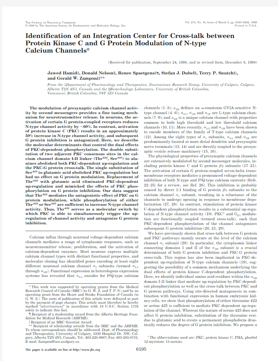
Two putative protein kinase C consensus sites,Thr422and Ser425,are contained with this region of the channel(Fig.1, inset).To test whether one or both of these residues might mediate PKC/G??cross-talk,we replaced both sites with ala-nine residues.Fig.1A depicts current records obtained from the double alanine mutant in the absence and the presence of 100n M somatostatin.Similar to that observed with the wild type channel(Fig.1),activation of somatostatin receptors me-diates a reversible and pronounced inhibition of channel activ-ity,paired with a slowing of activation kinetics and an appar-ent slowing of inactivation.Fig.1B illustrates the effect of PMA on channel activity and on somatostatin modulation of the alanine double mutant.While application of100n M PMA had no detectable effect on channel activity,the subsequent appli-cation of100n M somatostatin produced the same degree of G protein inhibition as that observed in the absence of PMA.Fig. 1,C and D,illustrates the effect of alanine substitution for a number of experiments.There are two effects evident.First, the alanine mutation per se significantly reduces the degree of somatostatin inhibition seen with the wild type channel from 53?5to36?5%(p?0.02)(Fig.1C).Second,while PMA treatment reduced the somatostatin effect for the wild type channel,the somatostatin sensitivity of the double mutant was not altered by PMA(36?5versus37?5%,p?0.86;Fig.1C), indicating that cross-talk between PKC and G protein path-ways is blocked by the double alanine substitution.In addition, the PMA-induced up-regulation observed with the wild type channels was reduced from49?14to6?5%when both PKC consensus sites were simultaneously replaced with alanines (Fig.1D).Thus,the critical structures mediating up-regulation of channel activity and inhibition of direct G protein action appear to reside within overlapping regions of the calcium channel domain I-II linker and contain one or both of Thr422 and Ser425.
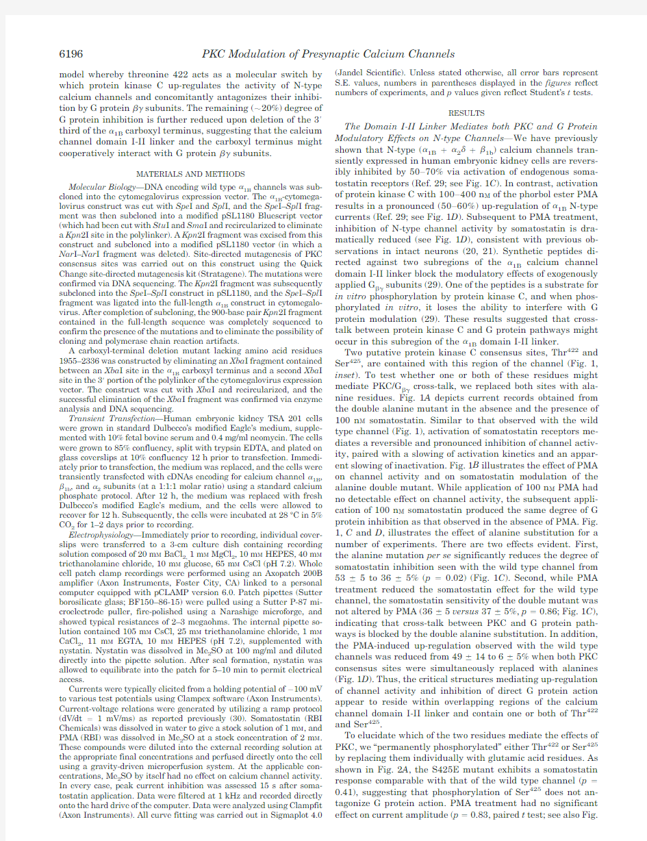
To elucidate which of the two residues mediate the effects of PKC,we“permanently phosphorylated”either Thr422or Ser425 by replacing them individually with glutamic acid residues.As shown in Fig.2A,the S425E mutant exhibits a somatostatin response comparable with that of the wild type channel(p?0.41),suggesting that phosphorylation of Ser425does not an-tagonize G protein action.PMA treatment had no significant effect on current amplitude(p?0.83,paired t test;see also Fig.
PKC Modulation of Presynaptic Calcium Channels 6196
3B ),suggesting that replacing Ser 425with glutamic acid mim-ics a permanently up-regulated state of the channel.Nonethe-less,PMA treatment significantly attenuated the degree of somatostatin inhibition from 46?7to 15?4%(p ?0.004)(Figs.2B and 3A ),suggesting that replacement of Ser 425with glutamic acid does not preclude phosphorylation of the adja-cent Thr 422residue and that cross-talk between PKC and G ??modulation is probably mediated by Thr 422rather than Ser 425.If this is correct,then mimicking phosphorylation of Thr 422(i.e.T422E)should reduce the degree of G protein inhibition
to
F I
G .1.Effect of a double substitution of I-II linker protein kinase C consensus sites for alanines on PKC and G protein action.A ,current records illustrating G protein inhibition of the double alanine mutant.Somatostatin reversibly reduces the peak current amplitude of the mutant channel to 65%of its control value.The currents were leak-subtracted using a p /5protocol.The holding potential was ?100mV,and the test potential was ?20mV.B ,application of PMA does not affect current amplitude and has no adverse effects on G protein inhibition mediated by somatostatin (holding potential ?100mV,test potential ??20mV).Inset ,proposed transmembrane topology of voltage-dependent calcium channels and amino acid sequence of part of the ?1B channel I-II linker.The bar above the amino acid sequence indicates a previously identified putative target region for G protein/PKC cross-talk.The Thr 422and Ser 425residues were substituted to alanine residues.C ,degree of somatostatin (100n M )inhibition of wild type ?1B channel and the alanine (422and 425)mutant channels (each coexpressed with ?1b and ?2)with or without prior application of 100n M PMA.Note that the alanine substitution slightly but significantly reduces the degree of somatostatin inhibition.While PMA reduces the G protein sensitivity of the wild type channel,the G protein inhibition of the double mutant is not affected.D ,up-regulation of channel activity by 100n M PMA.Note that the up-regulation seen with the wild type channel is blocked by the double alanine substitution.Error bars represent S.E.values;the test potential in C and D was ?20mV.
PKC Modulation of Presynaptic Calcium Channels 6197
those levels observed after PKC phosphorylation of the wild-type channel.This is supported by the current records shown in Fig.2,C and D ,and the data presented in Fig.3A .The T422E mutant showed a significantly reduced somatostatin sensitiv-ity,which was comparable in magnitude with that observed with the wild type channel after PMA treatment (WT (with PMA)?20?5%;T422E (without PMA)?20?3%,p ?0.91).These data indicate that replacing Thr 422with a negatively charged side group mimics the antagonistic effect of PKC on G protein inhibition.PMA application did not further affect so-matostatin sensitivity of T422E (T422E (with PMA)?14?3%,p ?0.29).Also,PMA failed to increase the peak current am-plitude of T422E (p ?0.71)(Figs.2D and 3B ),suggesting that similar to S425E,the T422E construct is likely be tonically up-regulated.Overall,the data suggest that whereas only Thr 422is capable of mediating the cross-talk effect,phospho-rylation of either Ser 425or Thr 422is sufficient to fully up-regulate channel activity.
To confirm this hypothesis,we created two additional mu-tants in which Thr 422and Ser 425were substituted individually by alanines in order to further define the relative contributions of the individual PKC consensus sites to the overall action of PKC.As shown in Fig.4A ,T422A exhibits a somatostatin sensitivity that closely parallels that seen with the T422A/S425A double mutant shown in Fig.1.After treatment with 100n M PMA,the degree of somatostatin inhibition of T422A did not decrease significantly (37?5%(without PMA)versus 32?3%(with PMA),p ?0.42)and remained significantly (p ?0.003)larger than that of the PMA-treated wild type channel (20?5%).These data indicate that cross-talk between PKC and G protein pathways is blocked upon selective removal of the Thr 442PKC substrate.Consistent with this notion,removal of the Ser 425PKC substrate (i.e.S425A)did not significantly change G protein sensitivity (p ?0.49),nor did it affect cross-talk between the G protein and PKC pathways (48?7%(without PMA)versus 18?3%(with PMA),p ?0.002,Fig.4A ).Both T422A and S425A exhibited a similar degree of PKC-de-pendent up-regulation (Fig.4B ),which did not differ signifi-cantly from that observed with the wild type channel (p ?0.79).This further supports the notion that N-type channel activity is fully up-regulated upon phosphorylation of either Thr 422or Ser 425,whereas only Thr 422is capable of mediating the cross-talk between PKC and G protein pathways.
Voltage Dependence of G Protein Modulation—To examine the voltage dependence of the T422E mutant,we utilized a ramp protocol to allow the acquisition of complete current-voltage relations without contamination from receptor desen-sitization.Fig.5compares the somatostatin response of wild type channels to that of the T422E mutant at a number of test potentials.In both cases,the effect of somatostatin is depend-ent on membrane potential and is consistent with the direct inhibition of native N-type calcium channels by G proteins (18,23,24,31,32).As evident from Fig.5,somatostatin produced a significantly greater inhibition of the wild type channels at
all
F I
G .2.Effect of individual replacement of I-II linker PKC consensus sites with glutamic acid residues.The holding potential was ?100mV,the test potential was ?20mV,and currents were leak-subtracted using a p /5protocol.A and C ,inhibition of mutant channels by 100n M somatostatin.Note that the threonine substitution dramatically reduces G protein sensitivity.B and D ,effect of PMA on channel activity and on the degree of somatostatin inhibition.Either mutation blocks up-regulation of the channel by PMA.S425E shows a reduced sensitivity to somatostatin inhibition following application of PMA (i.e.cross-talk remains intact),while no additional effect of PMA on G protein inhibition of T422E is evident.
PKC Modulation of Presynaptic Calcium Channels
6198
potentials.Although the T422E mutant is capable of undergo-ing G protein modulation,the voltage dependence of G ??dis-sociation appears be shifted toward more hyperpolarized po-tentials in the mutant channel.
Contribution of the ?1B Carboxyl Region in PKC and G Pro-tein Modulation—Several studies have implicated the carboxyl terminus in the direct G protein modulation of voltage-depend-ent calcium channels (33,34).Furthermore,the carboxyl ter-minus contains several putative protein kinase C consensus sites.To examine the possibility that the carboxyl terminus might contribute to the modulation of N-type calcium channels by PKC and G proteins,we examined a deletion mutant in which the last third of the carboxyl terminus of ?1B (residues 1955–2336)was deleted (?1B ?COOH).Fig.6B depicts a current record obtained with the deletion mutant in the presence and absence of somatostatin.Although somatostatin reduces the
peak current amplitude and mediates the slowing of activation kinetics typical of direct G ??modulation,the degree of inhibi-tion is reduced compared with the wild type channel (from 53?5%to 32?2%,p ?0.02).In contrast,up-regulation by PMA remains intact (Fig.6C and inset ),suggesting that the deleted portion of the carboxyl-terminal does not directly mediate PKC-dependent changes in channel activity.Following pretreatment with PMA,somatostatin application resulted in only a small effect on peak current amplitude (4?2%inhibition),at a test potential of ?20mV (Fig.6D ).Hence,PKC-dependent phos-phorylation in combination with deletion of the carboxyl termi-nus further reduces the degree of G ??modulation.To further examine this observation,we deleted the carboxyl-terminal region of the T422E mutant.As seen from Fig.6D ,this mutant showed only a ?10%inhibition in response to somatostatin at a test potential of ?20mV.The degree of inhibition did not differ significantly from that seen with mutant T422E after PMA treatment (p ?0.1)but was significantly lower than the sensitivity of the ?1B ?COOH construct (p ?0.025).These data indicate that deletion of the carboxyl terminus and replace-ment of Thr 422with glutamic acid produce additive effects on G protein sensitivity.This particular construct did not express well in HEK cells,and we were unable to systematically exam-ine the voltage dependence of somatostatin action.
Overall,the data suggest that the carboxyl region mediates an important role in stabilizing the G ??interaction with the calcium channel ?1subunit,especially when PKC sites in the ?1B domain I-II linker are phosphorylated.
DISCUSSION
Protein Kinase C-dependent Up-regulation Is Mediated by the Calcium Channel I-II Linker—Whole cell currents of exog-enously expressed ?1B N-type channels are up-regulated by activation of protein kinase C by either phorbol esters (such as PMA)or activation of coexpressed metabotropic glutamate re-ceptors (19).Here,we have used application of 100n M PMA to stimulate protein kinase C in human embryonic kidney cells expressing ?1B channels.Consistent with the results of Stea and co-workers (19),PMA application resulted in a pronounced increase in channel activity for wild type ?1B channels that was blocked by pretreatment with staurosporine.In their study,Stea and co-workers (19)were able to confer aspects of PKC sensitivity of ?1B onto the less sensitive ?1A channels by insert-ing the domain I-II linker of ?1B into ?1A .We have previously shown that a fusion protein directed against the ?1B I-II linker region is a substrate for PKC-dependent phosphorylation (29).Two considerations have led us to focus on a pair of PKC consensus sites (Thr 422and Ser 425)located within a 20-amino acid stretch (residues 410–428)of the ?1B I-II linker.First,this stretch of residues is both a substrate for in vitro phosphoryl-ation by PKC and has also been implicated in the PKC-medi-ated antagonism of G protein inhibition of wild type ?1B chan-nels (29).Second,certain amino acid substitutions in the vicinity of the corresponding region in ?1A increases PKC sen-sitivity of ?1A .2
Here,we show that the double substitution of Thr 422and Ser 425for alanines abolishes the PKC-dependent up-regulation of ?1B channels,suggesting that phosphorylation of one or both of these residues is sufficient to mediate up-regulation.Indi-vidual substitutions of these residues for glutamic acid also precluded the effect of PKC stimulation.In contrast,individual substitution of these two residues for alanines had no adverse effect on PKC-dependent up-regulation.These data imply that the effects of phosphorylation of the two PKC consensus sites are nonadditive and that phosphorylation of either Thr 422or
2
G.W.Zamponi and T.P.Snutch,unpublished
observations.
F I
G .3.A,effect of somatostatin on calcium channel N-type channel activity with or without prior application of 100n M PMA.The S425E mutant exhibits a somatostatin sensitivity that is not significantly different from that of the wild type channel.With prior application of PMA,the degree of somatostatin inhibition of both the wild type and the S425E mutant is reduced to similar levels.In contrast,even in the absence of PMA,the T422E mutant exhibits a somatostatin sensitivity that parallels that of a PMA-treated wild type channel,and PMA does not further effect somatostatin sensitivity.The holding potential was ?100mV,and the test potential was typically ?20mV.Error bars represent S.E.values.B ,up-regulation of wild type and mutant chan-nels by 100n M PMA.Note that the glutamic acid substitution of either Thr 422or Ser 425blocks up-regulation by PMA.Experimental conditions were as described for A .
PKC Modulation of Presynaptic Calcium Channels 6199
Ser 425is sufficient to mediate complete up-regulation of chan-nel activity in an all or none manner.At present,the molecular mechanisms by which the phosphorylation event affects chan-nel activity remain to be determined.It is possible that phos-phorylation induces a conformational change in the domain I-II linker that directly affects activation.Alternatively,phospho-rylation of these residues might alter the interaction with the calcium channel ?subunit,which in turn may affect channel activation.Such a mechanism would be consistent with data of Stea and co-workers (19),who reported that the calcium chan-nel ?subunit is required for PKC-dependent up-regulation.Recently,Shistik et al.(35)showed that deletion of N-termi-nal residues 2–46abolished PKC-dependent up-regulation of
rabbit heart ?1C channels expressed in Xenopus oocytes.The N-terminal region of rat brain ?1B N-type calcium channel is 51residues shorter than that of the rabbit heart ?1C channel and thus lacks the motif identified by Shistik et al.(35).Further-more,there is no counterpart to Thr 422present in the rabbit heart ?1C sequence,and the analog to Ser 425(Ser 499in the rabbit heart ?1C sequence)is not part of a PKC consensus motif.This suggests that the molecular mechanism underlying the PKC-dependent modulation of N-type calcium channel ac-tivity is fundamentally different from that for the rabbit car-diac L-type isoform.
Model for the G Protein Inhibition of N-type Calcium Chan-nels—Over the past several years,a number of studies have examined the molecular determinants of G protein modulation of presynaptic calcium channels (18,28,29,31–34,36–40).It is now widely accepted that the G ??subunits are the active G protein species mediating the antagonistic effect on presynap-tic calcium channel activity (28,29,31,32).The G protein ??subunits are able to interact with two separate regions within the calcium channel domain I-II linker (29,36).We have pre-viously suggested that the PKC dependent phosphorylation of one of these two I-II linker G ??binding motifs might mediate the previously identified antagonistic effect of PKC stimulation on G protein sensitivity (29).Here,we present further confir-mation for the involvement of the domain I-II linker region in direct G protein modulation of N-type calcium channels.Each,the double alanine mutant and the T422E and T422A con-structs,exhibited a significantly reduced sensitivity to soma-tostatin-induced G ??modulation.Neither of these substitu-tions resulted in significant changes in current kinetics or half-activation potential,minimizing the possibility of an indi-rect effect due to changes in channel gating.We suggest the possibility that these substitutions more likely reduce the af-finity of the channel for binding G ??.
In addition to the domain I-II linker,the carboxyl
terminus
F I
G .4.Effect of individual alanine substitutions in the I-II linker PKC consensus sites on PKC and G protein inhibition of N-type calcium channels.Error bars represent S.E.values,and numbers in parentheses indicate the numbers of experiments.The dotted lines indicate the levels of G protein inhibition (A )or PKC-dependent up-regulation (B )depicted in Fig.1.A ,inhibition of T422A and S425A by 100n M somatostatin with and without prior application of 100n M PMA.Note that T422A does not permit PKC activation to antagonize somatostatin inhibition,whereas the S425A mutant exhibits the behavior seen with the wild type channel.The level of somatostatin-induced inhibition of T422A closely parallels that observed with the T422A/S425A double mutant examined in Fig.1B .The PKC-dependent up-regulation observed with T422A and S425A is similar to that observed with the wild type channel.The dotted line indicates the level of up-regulation of the wild type
channel.
F I
G .5.Voltage dependence of G protein inhibition for wild type (n ?7)and mutant T422E (n ?12)?1B channels elicited by application of 100n M somatostatin.The data were obtained via ramp protocols as described under “Materials and Methods”15s after somatostatin application.Note that the voltage dependence of soma-tostatin inhibition is shifted toward more hyperpolarizing potentials for the mutant channels,resulting in a significantly greater inhibition of the wild type channels at each potential.Error bars represent S.E.values.
PKC Modulation of Presynaptic Calcium Channels
6200
(33,34)as well as domain I (38)and the amino terminus (39)of the calcium channel ?1subunit have all been implicated in direct G protein modulation.Here,we present corroborating evidence that the carboxyl terminus contributes to a portion of the overall G protein inhibition of N-type channels.A deletion of one-third of the carboxyl-terminal region significantly re-duced but did not eliminate the somatostatin-induced inhibi-tion of ?1B channels.The deleted portion contains a highly conserved motif that was recently implicated in G ??binding to ?1E channels (33).Together with our results,it appears that while the carboxyl terminus probably contributes to G ??bind-ing to N-type calcium channels,the major determinant of G protein action is the domain I-II linker.That the carboxyl terminus contributes to G ??binding could account for the ob-servation that ?1B or ?1E channel constructs containing the ?1C I-II linker remain sensitive to G protein inhibition (33,34)3despite the fact that ?1C I-II linker fusion proteins do not bind G ??.It is possible that the carboxyl region cooperatively en-hances binding to the ?1C I-II linker in these chimeric constructs.
Kinetic modeling of G protein inhibition of single N-type calcium channels has shown that G protein binding results in
a reluctance of channels to undergo transitions from the closed states to channel opening (27).Upon membrane depolarization,G proteins dissociate from the channels prior to opening,and the associated increase in first latency to opening results in a decrease in peak current amplitude.Within the framework of this model,the binding of G ??to the domain I-II linker and perhaps the carboxyl terminus mediates the stabilization of the closed state by inducing a conformational change in the chan-nel protein.The translation of G protein binding into a change in channel function may be mediated by residues located in domain I,consistent with previous studies (34,38,39).
Overall,a model emerges in which G ??interacts with two high affinity regions within the calcium channel domain I-II linker (residues 353–389and 410–428(29,36)and a lower affinity site in the carboxyl terminus (33).PKC-dependent phosphorylation probably destabilizes G ??binding to the sec-ond site within the I-II linker G ??binding domain (i.e.residues 410–428),thereby shifting the voltage dependence of G ??dis-sociation to more hyperpolarized potentials and perhaps reduc-ing the increase in first latency to opening associated with G ??binding (27).Single channel experiments will ultimately be required to confirm any effects of phosphorylation (and of the T422E substitution)on first latency.
Implications of Dual PKC Sites for Calcium Channel Modu-
3
E.Bourinet,personal
communication.
F I
G .6.Effect of deletion of residues 1955–2336of the ?1B carboxyl terminus on G protein and PKC sensitivity.A ,schematic representation of the proposed calcium channel transmembrane topology indicating the deletion of part of the carboxyl terminus region.B ,current record elicited by a step depolarization from ?100to ?20mV and leak-subtracted via a p /5protocol.Deletion of the 3?third of the carboxyl terminus region results in a slowing of inactivation kinetics as well as in a reduced degree of G protein inhibition as evident by application of 100n M somatostatin.C ,time course of up-regulation of current activity by 100n M PMA.The bar graphs depicted in the inset illustrate the degree of PMA up-regulation as defined by the peak current ratio I (with PMA)/I (without PMA)for the wild type channel and the deletion mutant.D ,degree of somatostatin-mediated G protein inhibition of the wild type channel and the deletion mutant in the presence and absence of 100n M PMA.Note that the degree of G protein inhibition is significantly attenuated upon deletion of the 3?portion of the ?1B carboxyl terminus.Pretreatment with 100n M PMA almost completely abolished G protein inhibition at test potentials of ?20mV.A double mutant (?COOH,T422E )also exhibited a reduced G protein sensitivity that did not differ significantly from that seen with T422E after PMA treatment.Error bars represent S.E.values.
PKC Modulation of Presynaptic Calcium Channels 6201
lation—Our data indicate that up-regulation of N-type channel activity occurs via phosphorylation of either Thr422or Ser425, whereas G protein modulation is antagonized by phosphoryla-tion of only Thr422.The selective phosphorylation of Ser425 would result in up-regulation by?50%,and the subsequent stimulation of the G protein pathway would produce a?50% inhibition of the PKC-enhanced current,resulting in an overall inhibition of?25%.Phosphorylation of Thr422(or of both resi-dues simultaneously)would also result in the50%up-regula-tion,but subsequent G protein inhibition would be attenuated to?20%and result in a net up-regulation by?20%.Since activation of PKC and G protein pathways individually would respectively produce a50%increase and a50%decrease in control current levels,Thr422and Ser425may function as an integration center for inputs from PKC and G protein pathways to produce multiple levels of calcium channel activity.Together with recent reports that protein kinase C-dependent phospho-rylation disrupts the interactions between syntaxin and the calcium channel II-III linker(16),this convergence of second messenger pathways directly at the level of the calcium chan-nel?
1
subunit would provide a mechanism to precisely control neurotransmitter release at presynaptic nerve terminals.
REFERENCES
1.Mori,Y.,Friedrich,T.,Kim,M.-S.,Mikami,A.,Nakai,J.,Ruth,P.,Bosse,E.,
Hofmann,F.,Flockerzi,V.,Furuichi,T.,Mikoshiba,K.,Imoto,K.,Tanabe, T.,and Numa,S.(1991)Nature350,398–402
2.Sather,W.A.,Tanabe,T.,Zhang,J.F.,Mori,Y.,Adams,M.E.,and Tsien,
R.W.(1993)Neuron11,291–303
3.Stea,A.,Tomlinson,W.J.,Bourinet,E.,Dubel,S.J.,Vincent,S.R.,and
Snutch,T.P.(1994)Proc.Natl.Acad.Sci.U.S.A.91,10567–10580
4.Williams,M.E.,Brust,P.F.,Feldman,D.H.,Patthi,S.,Simerson,S.,Maroufi,
A.,McCue,A.F.,Velicelebi,G.,Ellis,S.
B.,and Harpold,M.(1992)257,
389–395
5.Dubel,S.J.,Starr,T.V.B.,Hell,J.,Ahlijanian,M.K.,Enyeart,J.J.,Catterall,
W. A.,and Snutch,T.P.(1992)Proc.Natl.Acad.Sci.U.S.A.89, 5058–5062
6.Fujita,Y.,Mynlieff,M.,Dirksen,R.T.,Kim,M.,Niidome,T.,Nakai,J.,
Friedrich,T.,Iwabe,N.,Miyata,T.,Furuichi,T.,Furutama,D.,Mikoshiba, K.,Mori,Y.,and Beam,K.G.(1993)Neuron10,585–598
7.Williams,M.E.,Feldman,D.H.,McCue,A.F.,Brenner,R.,Velicelebi,G.,
Ellis,S.B.,and Harpold,M.M.(1992)Neuron8,71–84
8.Tomlinson,W.J.,Stea,A.,Bourinet,E.,Charnet,P.,Nargeot,J.,and Snutch,
T.P.(1993)Neuropharmacology32,1117–1126
9.Fisher,S.E.,Ciccodicola,A.,Tanaka,K.,Curci,A.,Desicato.,S.,D’Urso,M.,
and Craig,I.W.(1997)Genomics45,340–34710.Soong,T.W.,Stea,A.,Hodson,C.D.,Dubel,S.J.,Vincent,S.R.,and Snutch,
T.P.(1993)Science260,1133–1136
11.Williams,M.E.,Marubio,L.M.,Deal,C.R.,Hans,M.,Brust,P.F.,Philipson,
L.H.,Miller,R.J.,Johnson,E.C.,Harpold,M.M.,and Ellis,S.B.(1994) J.Biol.Chem.269,22347–22357
12.Perez-Reyes,E.,Cribbs,L.L.,Daud,A.,Lacerda,A.E.,Barclay,J.,Williamson,
M.P.,Fox,M.,Rees,M.,and Lee,J.H.(1998)Nature391,896–900
13.Westenbroek,R.E.,Hell,J.W.,Warberm C.,Dubel,S.J.,Snutch,T.P.,and
Catterall,W.A.(1992)Neuron9,1099–1115
14.Westenbroek,R.E.,Sakurai,T.,Elliott,E.M.,Hell,J.W.,Starr,T.V.,Snutch,
T.P.,and Catterall,W.A.(1995)J.Neurosci.15,6403–6418
15.Sheng,Z.,Rettig,T.,Takahashi,M.,and Catterall,W.A.(1994)Neuron13,
1303–1313
16.Yokoyama,C.T.,Sheng,Z.,and Catterall,W.A.(1997)J.Neurocsi.17,
6929–6938
17.Dunlap,K.,and Fischbach,G.D.(1981)J.Physiol.317,519–535
18.Bourinet,E.,Soong,T.W.,Stea,A.and Snutch,T.P.(1996)Proc.Natl.Acad.
Sci.U.S.A.93,1486–1491
19.Stea,A.,Soong,T.W.,and Snutch,T.P.(1995)Neuron5,929–940
20.Swartz,K.J.,Merrit,A.,Bean,B.P.,and Lovinger,D.M.(1993)Nature361,
165–168
21.Swartz,K.J.(1993)Neuron11,305–320
22.Ikeda,S.R.,and Schofield,G.(1989)J.Physiol.409,221–240
23.Bean,B.P.(1989)Nature340,153–156
24.Boland,L.M.,and Bean,B.P.(1993)J.Neurosci.13,515–533
25.Mintz,I.M.,and Bean,B.P.(1993)Neuron10,889–898
26.Hille,B.(1994)Trends Neurosci.17,531–536
27.Patil,P.G.,deLeon,M.,Reed,R.R.,Dubel,S.,Snutch,T.P.,and Yue,D.T.
(1996)Biophys.J.71,2509–2521
28.Zamponi,G.W.,and Snutch,T.P.(1998)Proc.Natl.Acad.Sci.U.S.A.95,
4035–4039
29.Zamponi,G.W.,Bourinet,E.,Nelson,D.,Nargeot,J.,and Snutch,T.P.(1997)
Nature385,442–446
30.Zamponi,G.W.,Bourinet,E.,and Snutch,T.P.(1996)J.Membr.Biol.151,
77–90
31.Herlitze,S.,Garcia,D.E.,Mackie,K.,Hille,B.,Scheuer,T.,and Catterall,
W.A.(1996)Nature280,258–262
32.Ikeda,S.R.(1996)Nature380,255–258
33.Quin,N.,Platano,Olcese,R.,Stefani,E.,and Birnbaumer,L.(1997)Proc.
Natl.Acad.Sci.U.S.A.94,8866–8871
34.Zhang,J.F.,Ellinor,P.T.,Aldrich,R.W.,and Tsien,R.W.(1996)Neuron17,
991–1003
35.Shistik,E.,Ivanina,T.,Blumenstein,Y.,and Dascal,N.(1998)J.Biol Chem.
273,17901–17909
36.DeWaard,M.,Liu,H.,Walker, D.,Scott,V. E.S.,Gurnett, C. A.,and
Campbell,K.P.(1997)Nature385,446–450
37.Page,K.M.,Stephens,G.J.,Berrow,N.S.,and Dolphin,A.C.(1997)J.Neu-
rosci.17,1330–1338
38.Stephens,G.J.,Canti,C.,Page,K.M.,and Dolphin,A.C.(1998)J.Physiol.
509,163–169
39.Page,K.M.,Canti,C.,Stephens,G.J.,Berrow,N.S.,and Dolphin,A.C.(1998)
J.Neurosci.18,4815–4824
40.Herlitze,S.,Hockermann,G.H.,Scheuer,T.,and Catterall,W.A.(1997)Proc.
Natl.Acad.Sci.U.S.A.94,1512–1516
PKC Modulation of Presynaptic Calcium Channels 6202