Activation of Chaperone-mediated Autophagy during Oxidative Stress-2004
Molecular Biology of the Cell
Vol.15,4829–4840,November2004
Activation of Chaperone-mediated Autophagy during Oxidative Stress□D
Roberta Kif?n,*Christopher Christian,*Erwin Knecht,?and Ana Maria Cuervo*?
*Department of Anatomy and Structural Biology,Marion Bessin Liver Research Center,Albert Einstein College of Medicine,Bronx,NY10461;and?Instituto de Investigaciones Citolo′gicas,Fundacio′n Valenciana de Investigaciones Biome′dicas,Valencia4010,Spain
Submitted June11,2004;Revised August3,2004;Accepted August18,2004
Monitoring Editor:Jennifer Lippincott-Schwartz
Oxidatively damaged proteins accumulate with age in almost all cell types and tissues.The activity of chaperone-mediated autophagy(CMA),a selective pathway for the degradation of cytosolic proteins in lysosomes,decreases with age.We have analyzed the possible participation of CMA in the removal of oxidized proteins in rat liver and cultured mouse?broblasts.Added to the fact that CMA substrates,when oxidized,are more ef?ciently internalized into lysosomes, we have found a constitutive activation of CMA during oxidative stress.Oxidation-induced activation of CMA correlates with higher levels of several components of the lysosomal translocation complex,but in particular of the lumenal chaperone,required for substrate uptake,and of the lysosomal membrane protein(lamp)type2a,previously identi?ed as a receptor for this pathway.In contrast with the well characterized mechanism of CMA activation during nutritional stress,which does not require de novo synthesis of the receptor,oxidation-induced activation of CMA is attained through transcriptional up-regulation of lamp2a.We conclude that CMA is activated during oxidative stress and that the higher activity of this pathway under these conditions,along with the higher susceptibility of the oxidized proteins to be taken up by lysosomes,both contribute to the ef?cient removal of oxidized proteins.
INTRODUCTION
Accumulation of oxidized protein is a common feature of aged cells(Dunlop et al.,2002;Grune and Davies,1997; Grune et al.,2001,2002b;Stadtman,2001).Many physiolog-ical and pathological processes lead to the generation of free radicals and consequent damage of intracellular compo-nents,including proteins.In most of these oxidative events, damaged proteins are removed from the cell through deg-radation by proteases(Dunlop et al.,2002).The activity of different intracellular proteolytic systems decreases with age (Terman,1995;Cuervo and Dice,1998a;Friguet et al.,2000; Carrard et al.,2002;Donati et al.,2001;Merker et al.,2001; Friguet,2002;Keller et al.,2002;Ward,2002),and this im-paired activity is considered responsible for the de?cient removal of oxidized proteins in old organisms(Grune,2000; Merker and Grune,2000;Dunlop et al.,2002;Szweda et al., 2002).
The susceptibility of oxidized proteins to proteases changes with the duration and degree of oxidative damage (Dunlop et al.,2002;Mehlhase and Grune,2002).Thus,mild oxidation induces partial protein unfolding and facilitates proteolytic cleavage(Grune et al.,1995,1997).However, persistent or extensive oxidative damage usually promotes protein aggregation,due to the exposure of patches of hy-drophobic amino acids.Once a protein aggregates,it be-comes less susceptible to proteolytic cleavage(Hoff et al., 1993;Grune et al.,1997;Davies,2001;Demasi and Davies, 2003).Kinetics of degradation of oxidized proteins in vitro have been analyzed using different types of proteases (Merker and Grune,2000;Dunlop et al.,2002;Szweda et al., 2002).One of the most extensively analyzed protease in this respect has been the proteasome.Although most of the studies linking the proteasome to the degradation of oxi-dized proteins have been carried out in vitro(Rivett,1985; Davies,2001),recent evidence supports the participation of the proteasome in the removal of oxidized proteins in vivo also(Hosler et al.,2003;Shringarpure et al.,2003).Thus, treatment of culture cells with proteasome inhibitors or an-tisense oligonucleotides against essential proteasome sub-units results in accumulation of oxidized proteins inside cells and diminished rates of cell survival during oxidative stress.The degradation of most oxidized proteins by the proteasome is ATP and ubiquitin independent(Shringar-pure et al.,2003),suggesting that the exposure of hydropho-bic regions in the surface of the protein acts as recognition signal for this protease.
The lysosomal system,the other major proteolytic system in cells,has not been often considered as a possible candi-date for the removal of oxidized proteins because of its lack of selectivity.In mammalian cells three different main mech-anisms contribute to the degradation of intracellular com-ponents inside lysosomes(autophagy)(Dice,2000;Reggiori and Klionsky,2002;Wang and Klionsky,2003;Cuervo, 2004a,b).Two of those mechanisms,macroautophagy and
Article published online ahead of print.Mol.Biol.Cell10.1091/
mbc.E04–06–0477.Article and publication date are available at
https://www.360docs.net/doc/ec1626563.html,/cgi/doi/10.1091/mbc.E04–06–0477.
□D The online version of this article contains supplementary material
accessible through https://www.360docs.net/doc/ec1626563.html,.
?Corresponding author.E-mail address:amcuervo@https://www.360docs.net/doc/ec1626563.html,.
Abbreviations used:CMA,chaperone-mediated autophagy;
GAPDH,glyceraldehide-3-phosphate dehydrogenase;hsc70,heat
shock cognate protein of70kDa;lamp,lysosomal associated mem-
brane protein;MOPS,3-(N-morpholino)propanesulfonic acid;
RNase A,ribonuclease A.
?2004by The American Society for Cell Biology4829
microautophagy,result in the degradation of complete re-gions of the cytosol,including organelles,in the lysosomal lumen.Although degradation of organelles by these auto-phagic pathways can be selective(i.e.,speci?c removal of damaged mitochondria sparing normal functioning ones) (Lemasters et al.,2002),there is no evidence supporting selectivity for the degradation of soluble cytosolic proteins. In contrast,the main characteristic of a third form of auto-phagy,chaperone-mediated autophagy(CMA),is its selec-tivity regarding the substrates(cytosolic proteins)degraded through this pathway(Cuervo and Dice,1998b;Dice,2000; Dice et al.,2003).
CMA is a generalized form of autophagy present in most cell types and tissues(Massey et al.,2004).All the CMA substrate proteins described to date are soluble cytosolic proteins containing a targeting motif biochemically related to the pentapeptide KFERQ(Chiang and Dice,1988;Dice et al.,2003).This motif,present in?30%of the proteins in the cytosol,is recognized by a cytosolic chaperone,the heat shock cognate protein of73kDa(cyt-hsc70)(Chiang and Dice,1988;Chiang et al.,1989).The interaction with this chaperone,modulated by the hsc70cochaperones(Agarra-beres and Dice,2001),targets the substrate to the lysosomal membrane,where it interacts with the lysosomal membrane protein(lamp)type2a(Cuervo and Dice,1996).Substrates need to be unfolded before translocation into the lysosomal lumen,and several cytosolic chaperones associated to the lysosomal membrane have been proposed to assist in the unfolding(Agarraberes and Dice,2001).Translocation of the substrate requires the presence of a variant of hsc70,lys-hsc70,in the lysosomal lumen(Agarraberes et al.,1997; Cuervo et al.,1997)and is followed by the rapid proteolysis of the substrate by resident lysosomal proteases(half-life in the lysosomal lumen of5–10min).
Although some basal level of CMA activity is probably present in most cells,nutritional stress has been shown to maximally activate this pathway(Wing et al.,1991;Cuervo et al.,1995).Activation during nutrient deprivation is associ-ated with higher levels of lys-hsc70in the lysosomal lumen and of lamp2a at the lysosomal membrane(Cuervo et al., 1995;Agarraberes et al.,1997;Cuervo and Dice,2000b). Because the interaction of substrate proteins with lamp2a is a limiting step for this pathway,changes in levels of lamp2a at the lysosomal membrane modulate CMA activity(Cuervo and Dice,2000b,c).Interestingly,all the conditions known to activate CMA,up-regulation of the lysosomal levels of lamp2a does not require de novo synthesis of the protein, but instead it is attained through down-regulation of its degradation and by relocation of a fraction of the protein from the lysosomal lumen into the membrane(Cuervo and Dice,2000b).
In addition to starvation,activation of CMA has also been observed in rat liver and kidney after exposure to gasoline derivatives(Cuervo et al.,1999);in?broblasts from galactosia-lidosis patients,which lack the protective protein/cathepsin A (Cuervo et al.,2003);and in cells overexpressing lamp2a(Cu-ervo and Dice,1996).CMA activity is reduced during renal tubular cell growth(Franch et al.,2001)and in aging(Cuervo and Dice,2000a).The decrease in CMA activity in old cells, known to accumulate oxidized proteins(Cuervo and Dice, 2000a),combined with the fact that activation of CMA during toxic exposure results in the selective degradation of a protein altered by the toxic compound(Cuervo et al.,1999),and with our?nding that in the presence of antioxidants degradation of the inhibitor of the nuclear factor?B by CMA decreases(Cu-ervo et al.,1998),prompted us to analyze a possible role of CMA in the removal of oxidized proteins from cells.We show in this work that oxidation of CMA substrates facilitates their translocation into lysosomes for degradation via CMA and also that CMA itself is activated during oxidative stress.Unexpect-edly,the oxidative stress-mediated activation of CMA is at-tained through a novel mechanism,different from the previous well-characterized activation of CMA in response to nutritional stress.
MATERIALS AND METHODS
Animals and Cells
Male Wistar rats(200–250g)were fed ad libitum or starved for20or48h before sacri?ce.An age-controlled rat strain(Fisher344)was used for the study of age-related changes and3-,12-,and22-mo-old rats were compared. Mild oxidative stress was induced in rats with two single i.p.injections of paraquat(40mg/100g body weight)separated by24h.Lysosomal isolation was carried out24h after the last injection.Mouse?broblasts(NIH3T3)were from the American Type Culture Collection(Manassas,VA).Cells were maintained in DMEM(Sigma-Aldrich,St.Louis,MO)in the presence of10% newborn calf serum.To deprive cells from serum,plates were extensively washed with Hanks’balanced salt solution(Invitrogen,Carlsbad,CA),and fresh medium without serum was https://www.360docs.net/doc/ec1626563.html,d oxidative stress was induced in culture cells by supplementation of the culture medium with100?M H2O2 or40?M paraquat for1h.Assays were carried out12–24h after removing the oxidizing compound.
Chemicals
Sources of chemicals and antibodies were as described previously(Cuervo and Dice,1996;Cuervo et al.,1997;Cuervo and Dice,2000a;Martin et al.,2002). The antibodies against the cytosolic tail of rat and mouse lamp2a and lamp2c were prepared in our laboratory(Cuervo and Dice,1996;Zhang,Bandhyo-padhyay,Kif?n,Massey,and Cuervo,unpublished data).The antibody against mouse lamp1(1D4B)was from the Developmental Studies Hybrid-oma Bank(University of Iowa,Iowa City,IA),and antibodies against hsp90 (SPA-845),hsp72(SPA-810),hsp40(SPA-400),hip(SPA-766),and Bag-1 (AAM-400E)were from Stressgene Biotechnologies(Victoria,BC,Canada). Carbonyl groups in oxidized proteins were detected using the OxyBlot oxi-dized protein detection kit from Chemicon International(Temecula,CA). Glyceraldehyde-3-phosphate dehydrogenase(GAPDH)and ribonuclease A (RNase A)were radiolabeled with[14C]formaldehyde by reductive methyl-ation as described previously(Jentoft and Dearborn,1983).Cytosolic proteins from mouse?broblasts in culture were metabolically radiolabeled by incu-bation with[14C]leucine(2?Ci/ml)at37°C for2d(Cuervo et al.,1997).Cells were disrupted by nitrogen cavitation,and the cytosolic fraction was the supernatant of a centrifugation at100,000?g for1h at4°C.Oxidized radiolabeled cytosolic proteins were prepared by the same procedure from cells treated with different prooxidants for24h before isolation.
Isolation of Lysosomal Fractions
Rat liver lysosomes were isolated from a light mitochondrial-lysosomal frac-tion in a discontinuous metrizamide density gradient(Wattiaux et al.,1978). Lysosomal fractions with different activities for CMA were separated as described previously(Cuervo et al.,1997).Lysosomes from cultured cells were isolated as described previously(Storrie and Madden,1990).Prepara-tions with more than10%broken lysosomes,measured as?-hexosaminidase latency,were discarded.Lysosomal matrices and membranes were isolated after hypotonic shock(Ohsumi et al.,1983).
Uptake and Degradation of Proteins by Isolated Lysosomes
GAPDH or RNase A were incubated in MOPS buffer(10mM3-(N-morpho-lino)propanesulfonic acid[MOPS]pH7.3,0.3M sucrose)with chymostatin-treated lysosomes as described previously(Cuervo et al.,1997).Transport was measured after proteinase K treatment of the samples,SDS-PAGE,and im-munoblot as the amount of GAPDH associated to lysosomes resistant to the protease.In another group of experiments,to overcome any possible changes in the proteolytic susceptibility of the oxidized proteins to proteinase K, substrates were incubated with chymostatin-treated or untreated lysosomes, and uptake was calculated as the difference between the amount of substrate associated to lysosomes(chymostatin-treated lysosomes)and the amount of substrate bound to their membrane(untreated lysosomes)(Salvador et al., 2000).Degradation of[14C]GAPDH(260nM; 1.5?108dpm/nmol), [14C]RNase A(150nM;0.9?108dpm/nmol),or a pool of radiolabeled cytosolic proteins(500dpm/?g)by intact lysosomes or lysosomal matrices was measured as described previously(Terlecky and Dice,1993;Cuervo et al., 1997).After incubation for30min at37°C in MOPS/dithiothreitol(DTT) buffer(10mM MOPS pH7.3,0.3M sucrose,5.4?M cystein,and1mM DTT) (for intact lysosomes)or in0.03M sucrose(for lysosomal matrices),samples
R.Kif?n et al.
Molecular Biology of the Cell 4830
were precipitated in10%trichloroacetic acid and?ltered through a multi-screen assay system(Millipore,Bedford,MA)using a0.45?m pore?lter. Radioactivity in the?ow through and in the?lter was converted to disinte-grations per minute in a WinSpectral1414liquid scintillation analyzer (PerkinElmer Life and Analytical Sciences,Boston,MA)by correcting for quenching using an external standard.Proteolysis was expressed as the percentage of the initial acid-insoluble radioactivity(protein)transformed into acid-soluble radioactivity(amino acids and small peptides)at the end of the incubation.
Rates of degradation of lamp2a in the isolated membranes were followed by immunoblot with a speci?c antibody against the cytosolic tail of lamp2a as described previously(Cuervo and Dice,2000b).Brie?y,isolated lysosomal membranes were incubated in MOPS buffer at37°C,and at different times aliquots were removed and subjected to SDS-PAGE and immunoblot for lamp2a.
GAPDH Oxidation
Metal-catalyzed oxidation of rabbit muscle d-glyceraldehyde-3-phosphate dehydrogenase EC1.2.1.12(Roche Diagnostics,Indianapolis,IN)was carried out as described previously(Rivett and Levine,1990).GAPDH(10mg of protein)was dialyzed against50volumes of HEPES buffer(50mM HEPES, pH7.2,100mM KCl,10mM MgCl2)supplemented or not(Mock)with25mM ascorbic acid and0.1mM FeCl3at37°C.The oxidative reaction was stopped adding1mM EDTA,and the samples were dialyzed against HEPES buffer for 24h with four changes.The two?rst changes were supplemented with1mM EDTA.The ef?ciency of oxidation was monitored by measuring the enzymatic activity of GAPDH after being exposed to the oxidant mixture for different periods of time.As described in Figure2,incubation of GAPDH with the oxidant mixture for30min resulted in70%decrease of its enzymatic activity.
Immunocytochemical Staining
Immuno?uorescence studies of cultured cells were performed following con-ventional procedures(Cuervo and Dice,2000c).Cells grown on coverslips until con?uent and kept in the presence or absence of serum for20h were ?xed with a3%formaldehyde solution,blocked,and then incubated with the primary and corresponding?uorescein isothiocyanate or Cy5-conjugated secondary antibodies as described previously(Cuervo and Dice,2000c). Mounting medium contained4,6-diamidino-2-phenylindole staining to high-light the cellular nucleus.Images were acquired with an Axiovert200?uo-rescence microscope(Carl Zeiss,Thornwood,NY)and subjected to deconvo-lution with the manufacturer’s software.Colocalization was determined using MetaMorph(Universal Imaging,Downingtown,PA).All digital micro-scopic images were prepared using Adobe Photoshop6.0software(Adobe Systems,Mountain View,CA).
mRNA Quanti?cation
Total RNA was extracted from rat livers using the RNeasy protect mini kit (QIAGEN,Valencia,CA)following the manufacturer’s indications and stored at?80°C until use.The?rst strand cDNA was synthesized from1?g of the total RNA with the SuperScript II RNase H Reverse Transcriptase(Invitrogen) and oligo-(dT)12–18primer.A region of the exon5of lamp2a and of actin were ampli?ed with speci?c primers(lamp2a,5?-GTCTCAAGCGCCAT-CATACT-3?[forward]and5?-TCCAAGGAGTCTGTCTTAAGTAGC-3?[re-verse];actin,5?-AAGGACTCCTATAGTGGGTGACGA-3?[forward]and5?-ATCTTCTCCATGTCGTCCCAGTTG-3?[reverse])by using the SYBR green polymerase chain reaction(PCR)kit(PE Biosystems,Warrington,United Kingdom).Ampli?cation of the lamp2a and actin DNA products(120and108 bp,respectively)was measured in real time in a light cycler(SmartCycler; Cepheid,Sunnyvale,CA).For both genes,the presence of a single ampli?ed product was veri?ed by agarose gel electrophoresis,and by analysis of the melting curves of the reverse transcription-PCR reaction.The expression levels of lamp2a in different samples were normalized with respect of those of actin in the same samples.Differences between samples were calculated based in their differences of the cycle numbers to reach a certain?uorescence intensity level.Because the size of the ampli?ed fragments was very similar we did not need to correct for fragment length.Standard PCR was carried out with the same primers but increasing dilutions of the cDNA pool.PCR products were subjected to electrophoresis in1.2%agarose gels and densi-tometry.The densitometric intensity of the lamp2a products was normalized with respect to the intensity of the actin products for the same sample.
General Methods
Protein concentration was determined by the Lowry method(Lowry et al., 1951)by using bovine serum albumin as a standard.Lysosomal enzymatic activities were measured as reported previously(Storrie and Madden,1990). After SDS-PAGE(Laemmli,1970)and immunoblotting(Towbin et al.,1979), the proteins recognized by the speci?c antibodies were visualized by chemi-luminescence methods(Renaissance;PerkinElmer Life and Analytical Sci-ences).Oxidized proteins were visualized after derivatization with DNPH by immunoblot with an antibody against the DNP moiety following the manu-facturer’s recommendation.Densitometric quanti?cation of the immuno-blotted membranes and stained gels was done with an Image Analyzer System(S1800;Inotech,Sunnyvale,CA).Student’s t test was used for statis-tical analyses.
RESULTS
Oxidized Proteins in Lysosomes
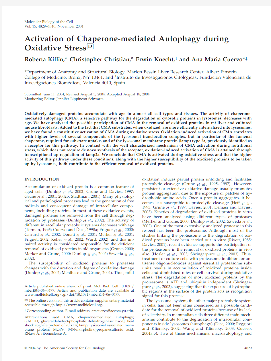
To test the contribution that the lysosomal system,and in particular CMA,might play in the removal of oxidized cytosolic proteins,we?rst analyzed the presence of those proteins inside lysosomes after oxidative stress.We induced mild oxidative stress in rats by treatment with bleomycin, 1,1?-dimethyl-4,4?-bipyridinium dichloride(paraquat),a su-peroxide-generating drug with well-characterized prooxi-dant effects(Haly,1979).To verify the oxidative effect of paraquat in rat liver we compared the content of carbonyl-containing proteins in the cytosol of untreated and para-quat-treated rats(Figure1A,lanes1and2).Although some oxidized proteins can be observed in the cytosol of untreated rats,we found a signi?cant increase in the content of these modi?ed proteins in the cytosol of rats injected with a sub-lethal dose of paraquat for two consecutive days.Lysosomes active for CMA can be isolated from rat liver based on their high content of the lumenal chaperone lys-hsc70required for substrate uptake(Cuervo et al.,1997).When we isolated this particular lysosomal population from treated and un-treated rats and separated their membranes from their lu-menal content,we found carbonyl-containing proteins in both the membrane and lumenal fraction of the lysosomes from paraquat-treated rats(Figure1A,compare lanes lanes 3and5to lanes4and6).Washing the lysosomal membranes with1M NaCl released most of the oxidized proteins(Fig-ure1A,lanes7and8),suggesting that they were likely cytosolic proteins associated to the membrane,rather than the result of oxidation of integral lysosomal membrane pro-teins.Likewise,when we incubated the lysosomal matrices at37°C before electrophoresis to promote the degradation of nonlysosomal proteins in the lysosomal lumen,the level of oxidized proteins in this fraction decreased drastically(Fig-ure1A,lanes9and10).Note that the new lower molecular weight bands detected in the matrices of paraquat-treated animals after the incubation at37°C could be proteolytic products of the proteins detected in the freshly isolated matrices.These results support that the oxidized proteins detected inside the lysosomes were most likely nonlysoso-mal proteins delivered there to be degraded.Interestingly, despite of their cytosolic origin,the electrophoretic pattern of the oxidized proteins associated with the lysosomal frac-tion differs from the ones in the cytosol.Because these dif-ferences are also observed in the membrane associated pro-teins,we do not think that they are due to distinct susceptibility of some of the oxidized proteins to proteases; instead we consider it an indication that a subset of oxidized proteins is selectively taken up by lysosomes.
As reported previously,even in the absence of treatment with a prooxidant agent,oxidized proteins can be detected in normal liver and their content increases with age(Figure 1B).When we analyzed the association of oxidized proteins to lysosomes from livers of3-and9-mo-old rats,we found an increase in the content of oxidized proteins in the lyso-somal lumen with age,similar to the one observed in the cytosol(Figure1B,compare lanes1and2and4and5).In contrast,in the oldest animals(22mo old),for which we have previously described a severe decrease in CMA activity (Cuervo and Dice,2000a),despite the higher content of oxidized proteins in the cytosolic fraction,in proportion,the amount of oxidized proteins detected in lysosomes was
Oxidative Stress and Autophagy
Vol.15,November20044831
lower than in the 9-mo-old animals (Figure 1B,compare lanes 5and 6).Similar assays to the ones described above con?rmed that the oxidized proteins were of extralysosomal origin and could be degraded by the lysosomal proteases (our unpublished data).The last panel of Figure 1B shows the speci?city of the antibody used in these studies against the carbonyl groups,which in nonderivatized samples only weakly cross-reacted with a single protein (compare signal in derivatized (lane 7)and nonderivatized samples (lane 8).
Although macroautophagy,a nonselective form of autoph-agy,also decreases with age (Terman,1995;Donati et al .,2001),the fact that the lysosomal population analyzed has been shown to be very active for CMA and that only a subset of oxidized proteins was detected in lysosomes encouraged us to analyze the role of CMA in the removal of oxidized proteins.
Oxidation of CMA Substrates
We ?rst analyzed whether the oxidation of known CMA substrates facilitated their degradation by CMA.We oxi-dized GAPDH,a well-characterized CMA substrate (Aniento et al .,1993),by incubating this protein with an ascorbic acid/iron oxidizing mixture.After 30min of incu-bation with this mixture,?70%of GAPDH activity was lost (Figure 2A),and carbonyl groups were easily detectable in the puri?ed protein (our unpublished data).We have pre-viously optimized an in vitro system that allows the analysis of binding and uptake of CMA substrates by isolated lyso-somes (Aniento et al .,1993;Cuervo et al .,1997,1999).When we compared the association to lysosomes of untreated GAPDH or of GAPDH exposed for increasing periods of time to the oxidizing mixture,we found an increase in both the amount of protein bound to the lysosomal membrane and translocated into the lysosomal lumen (proteinase K resistant)with longer oxidation times of GAPDH (Figure 2B).Oxidation of ovalbumin,a protein that does not contain a KFERQ-like motif and consequently is not a CMA sub-strate,did not increase its association to lysosomes (our unpublished data).The higher amount of GAPDH detected in the lysosomal lumen could also result from an increased resistance of the oxidized protein to degradation,either by the lysosomal proteases or by proteinase K,which removes the protein associated to the cytosolic side of the lysosomal membrane.However,as shown in Figure 2C,the oxidized protein was even more readily degraded by both the pool of proteases located in the lysosomal lumen and by exog-enously added proteinase K.
An increase in lysosomal translocation of oxidized pro-teins was also evident when instead of using a speci?c CMA substrate,we compared the degradation of a pool of oxi-dized cytosolic proteins,in which ?30%of them are possible CMA substrates (contain KFERQ-related motifs)(Figure 2D).Radiolabeled cytosolic proteins isolated from ?bro-blasts exposed to H 2O 2(Figure 2D,left)or paraquat (Figure 2D,right)were degraded faster than cytosolic proteins from untreated ?broblasts when incubated with intact rat liver lysosomes.Because these differences were greatly reduced when the lysosomal membranes were disrupted before the incubation with the cytosolic proteins,giving free access of the proteases to the substrates,we conclude that,as shown for GAPDH,increased degradation of the oxidized proteins resulted from their higher rates of translocation via CMA into lysosomes.These results support that oxidation of pro-teins that are substrates for CMA facilitates their degrada-tion through this pathway.
Activation of CMA during Oxidative Stress
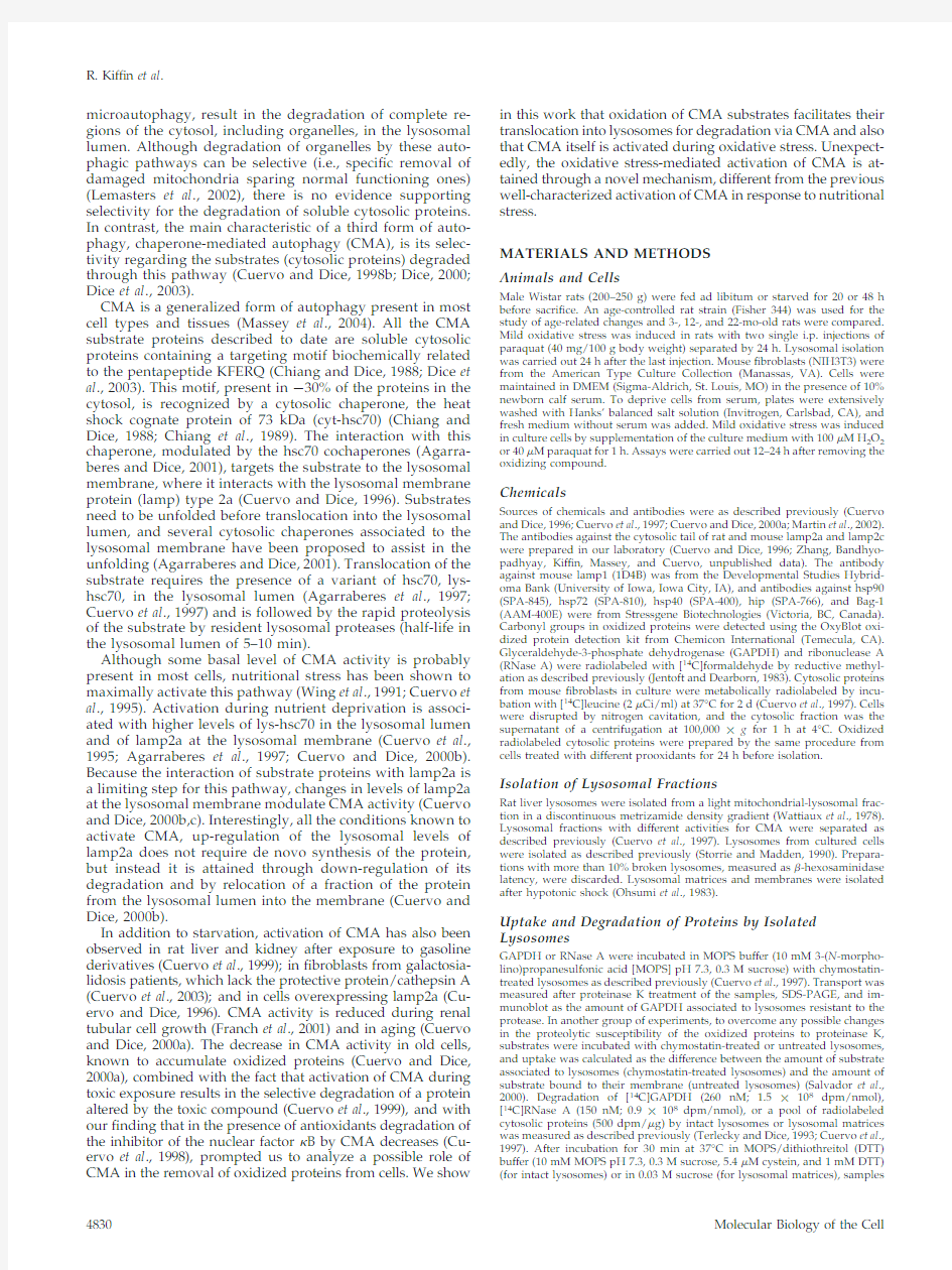
Independently of the effect of oxidation on CMA substrates,which makes them more susceptible to degradation by this pathway,we found a constitutive activation of CMA during oxidative stress.We isolated lysosomes from rats treated with a sublethal dose of paraquat for two consecutive days to generate a mild oxidative stress response.Under these conditions,we did not ?nd signi?cant differences with con-trol in lysosomal membrane stability assessed by measuring ?-hexosaminidase latency (Supplemental Figure 1)
(Storrie
Figure 1.Oxidized proteins in lysosomes active for CMA.(A)Cytosol (20?g of protein)and lysosomal membranes and matrices (10?g of protein)separated after hypotonic shock from lysosomes enriched in hsc70isolated from livers of untreated or paraquat (PQ)-treated rats were derivatized and subjected to SDS-PAGE and immunoblot against 2,4-dinitrophenylhydrazine (DPNH)moieties.To show the protein pattern in all lanes,exposure of lanes 1and 2was 5times shorter than for the other https://www.360docs.net/doc/ec1626563.html,nes 7and 8are duplicate samples of lanes 3and 4,in which membranes were washed with 1M NaCl before https://www.360docs.net/doc/ec1626563.html,nes 9and 10are duplicate samples of lanes 5and 6in which the matrices were incubated for 20min at 37°C before derivatization.No bands were detected in the same samples when derivatization was omitted (our unpublished data).(B)Cytosol (40?g of protein)and lysosomal matrices (20?g of protein)were isolated from livers of 3-,9-and 22-mo-old rats.Content of oxidized proteins in these fractions was analyzed as in A.Exposure of lanes 1–3was 3times shorter than for lanes 4–https://www.360docs.net/doc/ec1626563.html,nes 7and 8show the same sample than lane 5deri-vatized or not with DPNH and immunobloted as the others.The ?lm is overexposed (8times lane 5exposure)to show the low content of protein bands nonspeci?cally recognized by the anti-body.
R.Kif?n et al .
Molecular Biology of the Cell
4832
and Madden,1990)and extralysosomal proteolytic activity (Aniento et al .,1993).Lysosomes isolated from rats exposed to paraquat degraded larger amounts of unmodi?ed
GAPDH than lysosomes isolated from untreated animals (Figure 3A,left).Similar results were observed for RNase A,another well-characterized CMA substrate (Terlecky and Dice,1993;Cuervo et al .,1994)(Figure 3A,right).In both cases,the increase in substrate degradation was no longer observed if the lysosomal membrane was disrupted.In fact,the degradation of both substrates by lysosomal proteases was slightly lower in the paraquat-treated animals.This decrease could result from a decrease in the activity of some lysosomal proteases during oxidative stress,as reported pre-viously (O’Neil et al .,1997;Carr,2001;Crabb et al .,2002).In any case,the higher ability to degrade substrates of the intact lysosomes from paraquat-treated animals was thus a consequence of increased binding/uptake rather than of higher proteolysis in the lysosomal lumen.
We then determined whether oxidative stress could in-crease CMA-mediated degradation of proteins by acting on both the substrates,increasing their ability to be taken up by the lysosomes,and on the lysosomes,increasing their ability to take up substrates.For that purpose,we veri?ed ?rst that,as described above for GAPDH and RNase A,cytosolic proteins from untreated ?broblasts were more readily
de-
Figure 2.Oxidation of CMA substrates facilitates their uptake by lysosomes.(A)GAPDH was exposed to an ascorbic acid/iron oxi-dizing reaction for 30min as described under Materials and Methods .The remaining speci?c enzymatic activity of untreated GAPDH (CTR)and of GAPDH exposed to the oxidant mixture (ASCORB/FeCl 3)or to the dialysis buffer only (MOCK)is shown.Values are mean ?SE of two different preparations with triplicate measure-ments.(B)Unmodi?ed GAPDH (Ctr)or GAPDH exposed to the oxidant mixture for the indicated periods of time was incubated for 30min under standard conditions with intact chymostatin-treated liver lysosomes isolated from 20-h starved rats.Where indicated,samples were treated with proteinase K to remove GAPDH bound to the cytosolic side of the lysosomal membrane.At the end of the incubation,lysosomes were collected by centrifugation and sub-jected to SDS-PAGE and immunoblot for GAPDH.(C)The different forms of GAPDH (50?g)described in A were incubated with lysosomal enzymes (25?g of protein of broken lysosomes;pH 4.5)(left)or proteinase K (0.1?g)(right)at 37or 0°C,respectively.At the indicated times aliquots were taken and subjected to SDS-PAGE and Coomassie Blue staining.Values are the mean ?SE of the densito-metric values for GAPDH from three different experiments.(D)Mouse ?broblasts were metabolically labeled with [3H]leucine for 48h.A group of ?broblasts was exposed to 100?M H 2O 2(left)or 40?M paraquat (Pq)for 1h during the labeling.After cavitation,cytosolic fractions (Cyt)from untreated (Ctr)and H 2O 2or paraquat-treated ?broblasts were prepared and incubated for 30min at 37°C with intact rat liver lysosomes (25?g of protein)or with lysosomes disrupted by a hypotonic shock (broken lysosomes;15?g of pro-tein)under standard conditions.At the end of the incubation pro-teolysis was calculated as the amount of acid precipitable radioac-tivity (protein)transformed in acid soluble (amino acids and small peptides).Values are the mean ?SE of six different experiments (*p ?0.05,**p ?0.01,***p ?
0.001).
Figure 3.Additive effect of mild oxidative stress on CMA sub-strate proteins and on lysosomes.(A)Degradation of [14C]GAPDH (left)and [14C]RNase A (right)by lysosomes isolated from rats treated with paraquat as described under Materials and Methods .Incubations were carried out as described in Fig.2D.Values are the mean ?SE of ?ve to eight different experiments (*p ?0.05,**p ?0.01,***p ?0.001).(B)Degradation of radiolabeled cytosolic pro-teins prepared as in Figure 2D from untreated ?broblasts (control)or from ?broblasts exposed to H 2O 2(left)or paraquat (right),by intact liver lysosomes from untreated rats (Lysosomes control)or from rats treated with paraquat (Lysosomes PQ)as described under Materials and Methods .Proteolysis was calculated as in Figure 2D.Values are mean ?SE of six different experiments.*,differences compared with lysosomes control;?,differences compared with cytosol control;?,differences comparing lysosomes and cytosol control,to lysosomes and cytosol treated (*p ?0.05,**p ?0.01,***p ?0.001).
Oxidative Stress and Autophagy
Vol.15,November 20044833
graded by intact lysosomes from paraquat-treated rats than by lysosomes from untreated rats(Figure3B).Then,we analyzed the effect of combining lysosomes and cytosolic proteins that had been both exposed to the prooxidant. When we incubated the lysosomes from paraquat-treated rats with the cytosolic proteins isolated from?broblasts treated with H2O2(Figure3B,left)or with paraquat(Figure 3B,right),we found that these proteins were degraded even faster.We obtained similar results using lysosomes isolated from H2O2treated?broblasts(our unpublished data).Based on this additive effect,we conclude that not only oxidation-induced changes in the substrate proteins,but also in the lysosomal compartment,are responsible for the higher rates of CMA observed during oxidative stress.
We further analyzed the activation of CMA during oxidative stress by separately analyzing binding and up-take of unmodi?ed substrate proteins by lysosomes iso-lated from paraquat-treated rats.In these studies,to elim-inate any possible differences in the ability of exogenous proteases to remove the protein bound to the cytosolic side of the lysosomal membrane,we compared instead the amount of substrate associated to lysosomes previ-ously treated or not with a strong cocktail of protease inhibitors.In the absence of protease inhibitors the sub-strate that translocates inside the lysosomal lumen is rap-idly degraded by the lysosomal proteases(half-life of proteins in the lysosomal lumen is5–7min;Aniento et al., 1993).Consequently,the protein associated to the lyso-somes at the end of the incubation represents the protein bound to the cytosolic side of the lysosomal membrane (Salvador et al.,2000).In contrast,if lysosomal proteases are inhibited,the protein recovered associated to lyso-somes corresponds to both protein bound and internal-ized into the lysosomal lumen.As described previously, the amount of internalized substrate can then be calcu-lated by subtracting the protein bound from the amount of protein associated(Salvador et al.,2000).Consistent with our previous results,the amount of GAPDH and RNase A bound to the lysosomal membrane was signi?-cantly higher(4-to5-fold increase)in lysosomes from paraquat-treated rats(Figure4A).A two-to threefold increase was also detected in the uptake of both substrates by these lysosomes,when calculated as described above (Figure4A,bottom).We have previously shown that CMA is activated under starvation conditions(Cuervo et al.,1995).When we compared both stimuli,starvation and paraquat,we found that treatment with the prooxidant agent resulted in stronger activation of CMA.Levels of binding and uptake of both substrates were signi?cantly higher in lysosomes isolated from fed rats treated with paraquat than in lysosomes from48-h starved animals.To determine whether the observed increase in CMA during paraquat treatment could be due,at least in part,to some degree of starvation in this group of animals,we mea-sured food-intake in the animals exposed to paraquat.
However,we did not?nd signi?cant differences in food consumption between the paraquat-treated and untreated animals when maintained in ad libitum conditions,sug-gesting that activation of CMA after paraquat treatment was not due to starvation,but to the oxidative stress. Activation of CMA is associated with the relocalization toward the perinuclear region of the lysosomes with higher CMA activity(those with higher levels of lys-hsc70and lamp2a)(Agarraberes et al.,1997;Cuervo and Dice,2000c). As shown in Figure4B,in culture?broblasts maintained in the presence of serum,lamp2a colocalized with other lyso-somal membrane proteins(lamp1is shown here),displaying a typical vesicular punctated pattern.When serum was re-moved from the culture medium,the lamp2a-enriched lyso-somes preferentially localized in the perinuclear region. Treatment with paraquat displayed a similar perinuclear pattern for lamp2a even when the cells were maintained in the presence of serum(Figure4B,right).
These results are consistent with a constitutive activation of CMA during oxidative stress in the two experimental models in which CMA has been better characterized,rat liver and?broblasts in culture(Terlecky and Dice,1993; Cuervo et al.,
1997).
Figure4.Activation of CMA during mild oxidative stress.(A) Association and binding of GAPDH and RNase A to intact lyso-somes from untreated fed rats,48-h starved rats(Strv)or fed rats treated with paraquat(PQ).Lysosomes in lanes1–3were prein-cubated with chymostatin to prevent protein degradation.Incu-bations were carried out in an isotonic buffer,as described under Materials and Methods,and levels of associated proteins were determined by immunoblot of the lysosomes collected by centrif-ugation.Bottom:densitometric quanti?cation(mean?SE)of4–6 immunoblots similar to the ones shown here.Values are ex-pressed as the fold of increase in binding and in uptake(associ-ation-binding)for the lysosomes from starved or paraquat-treated rats,compared with lysosomes from untreated fed animals.*,differences compared with fed animals;?,differences compared with starved animals(*p?0.1,**p?0.05,***p?0.01) (B)Immuno?uorescence for lamp2a(red)and lamp1(green)in mouse?broblasts after removing the serum from the culture medium(serum?)or after treatment with paraquat in cells main-tained in serum supplemented medium.The merged image of both?uorochrome channels is shown.Bar,50?m.
R.Kif?n et al.
Molecular Biology of the Cell 4834
Oxidative Stress-induced Changes in Lysosomes Active for CMA
Several cytosolic chaperones associate with the lysosomal membrane and are required for CMA substrate binding/uptake into lysosomes (Agarraberes and Dice,2001).Al-though the speci?c function of each of these cochaperones in CMA remains unknown,studies with human ?broblasts in culture have revealed that the levels of some of them are up-regulated when CMA is activated by removal of serum,whereas others remain unchanged (Agarraberes and Dice,2001).In lysosomes isolated from livers of rats starved for 48h,in addition to the previously reported increase in lys-hsc70,we found higher levels of hsp90,and,in less extent,of Hip,compared with lysosomes from control-fed animals (Figure 5A).Treatment with paraquat (in fed rats)resulted in similar increase in the lysosomal content of lys-hsc70and of Hip,but only a discrete increase in the content of hsp90,when compared with untreated animals.None of the treatments modi?ed the levels of other associated chap-erones (hsp40or Bag-1),nor did they result in lysosomal association of the inducible form of the hsp70family,hsp72,which was however elevated in the cytosol after paraquat treatment (Figure 5A).Because the function of hsp90in lysosomes is not known,it is not clear why the increase in its lysosomal levels is not as pronounced as the one observed during starvation,but because rates of uptake are still up-regulated under these conditions,it points to a nonlimiting role for hsp90in the translocation complex.
Two components have been shown to be rate-limiting in the uptake of CMA substrates by lysosomes:the chaperone in the lysosomal lumen (lys-hsc70)(Cuervo et al .,1997;Agar-raberes et al .,1997)and the receptor at the lysosomal mem-brane (lamp2a)(Cuervo and Dice,2000b,c).We have previ-ously reported in rat liver the presence of two lysosomal populations with similar morphological and enzymatic characteristics but different CMA activity (Cuervo et al .,1997).The main difference found,so far,between these two groups of lysosomes is the enrichment of lys-hsc70in the lumen of the lysosomes with higher CMA activity (Cuervo et al .,1997).These two groups of lysosomes are normally pu-ri?ed together (they contribute 75and 25%high activity and low activity,respectively,to the fraction that we used in these studies),but can be physically separated by further density and differential centrifugation (Cuervo et al .,1997).The lysosomes with lower CMA activity can become more active under speci?c conditions,such as prolonged (88-h)starvation or aging,which correlates with an increase in their lumenal levels of lys-hsc70(Cuervo et al .,1997;Cuervo and Dice,2000a).Because we found higher levels of lys-hsc70in the lysosomes from paraquat-treated animals,we separated these two lysosomal subpopulations to determine whether there was a net increase in lys-hsc70per lysosome,or it resulted from enrichment of the less active lysosomes in lys-hsc70.As shown in Figure 5B,after treatment with para-quat there were no signi?cant changes in the content of lys-hsc70in the membranes or matrices of the less active group.Accordingly,the ability of this group of lysosomes to selectively take up CMA substrates did not change in para-quat-treated rats (our unpublished data).The percentage of hsc70-enriched lysosomes,determined as recovery of hex-osaminidase activity in this fraction,was similar in un-treated and paraquat-treated rats (our unpublished data).For the more active group of lysosomes,as observed during starvation,levels of lys-hsc70at the lysosomal membrane remained constant,whereas levels of lys-hsc70in the lyso-somal lumen increased approximately fourfold.Separation of the lysosomal membranes and matrices also revealed that hsp90increased in both compartments during
starvation
Figure 5.Changes in lysosome-associated chaperones during mild oxidative stress.(A)Cytosol and lysosomes (75?g of protein)iso-lated from normally fed,48-h starved rats (Stv),or fed rats treated with paraquat (PQ)as labeled were subjected to SDS-PAGE and im-munoblot for the indicated proteins.Right,densitometric quanti?cation (mean ?SE)of the chaperones in the lysosomal fraction in four immunoblots similar to the ones shown here.Levels in lysosomes from fed animals were given an arbitrary value of 1(dotted line).*,differences compared with fed animals (*p ?0.1,**p ?0.05,***p ?0.01).(B)Lyso-somes with high (CMA ?)and low (CMA ?)activity for CMA (Cuervo et al .,1997)were isolated from the same groups of animals as detailed in A.After hypotonic shock and cen-trifugation,membranes and matrices (50?g of protein)were separated and subjected to SDS-PAGE and immunoblot for hsc90(top)or hsc70(bottom).
Oxidative Stress and Autophagy
Vol.15,November 20044835
and after paraquat treatment (Figure 5B,top),but the in-crease in the lumen of lysosomes from paraquat-treated rats was smaller than in starved animals.These differences in the luminal content of hsp90could explain why,when working with total lysosomes (Figure 5A),we found that paraquat induced a lower increase in lysosomal levels of hsp90than starvation.Whether this hsp90in the lysosomal lumen is functional,or whether it is only internalized to be degraded (amino acid sequence analysis of hsc90revealed the pres-ence of two KFERQ-related motifs)remains unknown.
Levels of lamp2a at the lysosomal membrane directly correlate with CMA activity (Cuervo and Dice,2000b).Ly-sosomes isolated from mouse ?broblasts treated with H 2O 2(Figure 6A)or from livers of rats exposed to paraquat (Fig-ure 6B)had higher levels of lamp2a at their membrane than lysosomes isolated from the corresponding untreated con-trols.This increase seems selective for lamp2a,because lev-els of other lysosomal membrane proteins (lamp1shown here)remained unchanged.In agreement with the binding and uptake data (Figure 4A),the increase of lamp2a in the lysosomal membrane induced by paraquat was higher than the one induced by starvation (Figure 6C).For the two other lysosomal membrane proteins analyzed,levels of lamp1remained unchanged in both conditions,and levels of lamp2c were not affected by the treatment with paraquat,but decreased signi?cantly during starvation.Whether this decrease in lamp2c content is related to the increase in lamp2a or it happens independently is currently under in-vestigation.
A Novel Mechanism for Activation of CMA during Oxidative Stress
We have previously shown that CMA activity can be mod-ulated by changes in the levels of lamp2a at the lysosomal membrane (Cuervo and Dice,2000b).In fact,overexpression of lamp2a results in higher rates of CMA proportional to the increase in the receptor protein (Cuervo and Dice,1996,2000c).During starvation,the most extensively studied stim-uli for CMA,the increase of lamp2a at the lysosomal mem-brane does not result from de novo synthesis of the protein.Instead,a decrease in the degradation rate of lamp2a,along with the relocation of part of the lamp2a resident in the lysosomal lumen toward the lysosomal membrane account for most of the lamp2a increase in the membrane (Cuervo and Dice,1996,2000c).To determine whether similar mech-anisms were responsible for the increased levels of lamp2a during oxidative stress,we ?rst compared the rates of deg-radation of lamp2a in membranes of lysosomes from rats fed,starved,or treated with paraquat.In contrast with the signi?cant reduction in lamp2a degradation induced by star-vation,degradation of lamp2a was only slightly slower in rats treated with paraquat than in untreated fed rats (Figure 7A).Likewise,the distribution of lamp2a between mem-brane and matrix was not signi?cantly affected by the treat-ment with paraquat,suggesting that the increased levels of lamp2a in the membrane were not the result of recruitment of the lamp2a normally present in the lumen (Figure 7B).Contrary to starved animals,levels of lamp2a mRNA in the liver of rats treated with paraquat were signi?cantly higher than in untreated rats.We used both a dilution-based quantitative PCR method (Figure 7C)and real-time PCR (Figure 7D)to compare levels of lamp2a mRNA in livers from fed,starved,or paraquat (fed)-treated rats.Treatment with paraquat induced an increase in lamp2a mRNA levels of approximately sixfold in fed animals,whereas starvation resulted in a slight decrease in lamp2a mRNA levels.There-fore,contrary to starvation,where the increase in lamp2a levels did not require new protein to be synthesized,most
of
Figure https://www.360docs.net/doc/ec1626563.html,mp2a levels at the lysosomal membrane increase during mild oxidative stress.(A and B)Immunoblot for lamp2a and lamp1of homogenates (50?g of pro-tein),lysosomal membranes (L.Memb)and lysosomal matrices (L.Matrix)(10?g of protein)isolated from cultured mice ?bro-blasts,exposed or not to 100?M H 2O 2(A)or from liver of rats treated or not with paraquat (PQ).(C)Imunoblot for lamp2a,lamp2c,and lamp1of lysosomal mem-branes isolated from livers of fed rats,48-h starved (Strv)rats,or fed rats treated with paraquat (PQ).Right,densitometric quanti-?cation of six to eight immunoblots as the ones shown here.Values are expressed as fold of the values in fed untreated animals and are the mean ?SE of six different ex-periments.Levels in lysosomes from fed an-imals were given an arbitrary value of 1.*,differences compared with fed animals (*p ?0.05,**p ?0.01,***p ?0.001).
R.Kif?n et al .
Molecular Biology of the Cell
4836
the increase in lamp2a levels detected in paraquat-treated animals was a consequence of de novo synthesis of the protein.
We conclude that activation of CMA is part of the normal oxidative stress response and it contributes to the selective removal of oxidized proteins from the cytosol.The higher rates of CMA observed under these conditions are the com-bined result of an increase in the susceptibility of the pro-teins to be taken up and degraded by lysosomes,and an enhanced ability of the lysosomes for substrate uptake.Un-expectedly,during mild oxidative stress,activation of CMA is mediated by a novel mechanism different from the previ-ously characterized activation of CMA during nutritional stress.DISCUSSION
Our results provide for the ?rst time evidence for the par-ticipation of lysosomes in the removal of oxidized proteins during mild oxidative stress through CMA.This novel role for CMA is supported by the fact that 1)oxidized proteins can be detected in the lumen of lysosomes active for CMA (Figure 1);2)in conditions with declined CMA,such as aging,the amount of oxidized proteins translocated into lysosomes is reduced (Figure 1B);3)oxidized CMA sub-strates bind and are taken up more ef?ciently by isolated lysosomes than their unmodi?ed forms (Figure 2);4)lyso-somes from cells or rats exposed to prooxidants display higher rates of binding and uptake of CMA substrate pro-teins (Figures 3and 4);and 5)blockage of CMA in cultured cells increases their susceptibility to prooxidant compounds and decreases their viability (Massey,Kif?n,and Cuervo,unpublished data).Activation of CMA during oxidative-stress is attained through the up-regulation of speci?c com-ponents of the lysosomal translocation complex (lysosomal chaperones and the lysosomal membrane receptor)(Figures 5and 6).Interestingly,although these changes are similar to the ones described when CMA is activated by nutritional stress,the mechanism involved in the up-regulation is dif-ferent.Therefore,this ?nding suggests that stress-mediated activation of CMA varies depending on the nature of the stress.
Most of the previous studies on the lysosomal role during oxidative stress have focused on the contribution of this subcellular compartment to the oxidative damage,rather than on a possible protective role.In fact,the destabilizing effects of severe oxidative stress (?350?M hydrogen perox-ide)on lysosomes have been well documented (Brunk et al .,1995;Ollinger and Brunk,1995),showing that the release of lysosomal enzymes plays a major role in intracellular dam-age and in cell death under these conditions.Lower concen-trations of hydrogen peroxide (250?M for 30min)still cause leakage of some lysosomal enzymes,although the lysosomal damage is reversible (Brunk et al .,1995;Ollinger and Brunk,1995).In our study,we did not ?nd changes in the stability of the lysosomal membrane,assayed either as release of speci?c enzymes or with a more sensitive proteolytic method.It is possible that the lower doses of hydrogen peroxide and paraquat used in our study (100?M and 40mg/100g body weight,respectively)were enough to orig-inate cytosolic protein damage,but they did not alter the lysosomal compartment.Other possibility is that the initial lysosomal damage still takes place.but due to its reversibil-ity under these conditions (Brunk et al .,1995;Ollinger and Brunk,1995),it is not longer detectable by the time the lyso-somes are isolated (24h after the second injection).In
addition,
Figure 7.A novel mechanism for activation of CMA during mild oxidative stress.(A)Degrada-tion of lamp2a in isolated membranes from nor-mally fed rats,48-h starved rats,or fed rats treated with paraquat (PQ),as indicated under Materials and Methods .Degradation of lamp2a was fol-lowed by immunoblot of the lysosomal mem-branes with an antibody speci?c against its cyto-solic tail,at dif ferent times of the incubation.Values are the mean ?SD of the densitometric quanti?cation of immunoblots from four dif ferent experiments.A representative immunoblot is shown in the top insets.(B)Distribution of lamp2a between the lysosomal membrane and the matrix in lysosomes isolated from normally fed rats,treated or not with PQ.Values are mean ?SE of the densitometric quanti?cation of four immunoblots similar to the one shown in Figure 6B.(C)A 120-nucleotide fragment of lamp2a and a 108-nucleotide fragment of actin were ampli-?ed,through PCR,from increasing concentra-tions of total mRNA isolated from the livers of normally fed,48-h starved,or paraquat-treated fed rats.The electrophoretic pattern of the prod-ucts in a 2%agarose gel is shown.(D)Semiquan-titative real-time PCR was used to compare mRNA expression levels for lamp2a in the same samples as detailed in C.Values were corrected for actin ampli?cation in each samples and are expressed as fold increase compared to mRNA lamp2a values in fed untreated rats (that was given an arbitrary value of 1).Values are mean ?SD for four dif ferent experiments.*,dif ferences compared with fed animals (**p ?0.01,***p ?0.001).
Oxidative Stress and Autophagy
Vol.15,November 20044837
because most of the studies regarding lysosomal stability and oxidative stress have been performed in intact cells,analyzing the leakage of?uorescent probes from the lysosomal compart-ment,we cannot discard that different lysosomal populations might be differently affected by prooxidants.In our assays,the cell fractionation method used has been optimized to isolate a subset of intracellular lysosomes active for CMA(10–15%of total)(Cuervo et al.,1997).Particular characteristics of the CMA-active lysosomes,such as,for example,a lower concen-tration of iron in their lumen,could explain their higher resis-tance to prooxidants.Our studies on CMA-active lysosomes in old animals also support their unique characteristics.Thus, although accumulation of lipofuscin in lysosomes is a com-monly used biomarker of aging,CMA-active lysosomes from old rat livers rarely accumulate this pigment(Cuervo and Dice, 2000a).
The ability of proteasomes to degrade oxidized proteins has been previously well documented in vitro(Rivett,1985;Davies, 2001),and more recently in vivo(Grune et al.,2002a;Hosler et al.,2003;Shringarpure et al.,2003).Most of the studies assessing oxidation-induced changes in lysosomal proteolytic behavior have focused in the analysis of the enzymatic activity of cathe-psins,the lysosomal proteases.Moderate oxidative stress does not signi?cantly change the activity of most cathepsins, whereas more severe oxidizing conditions result in increased or decreased cathepsin activity depending on the cellular con-ditions(Sitte et al.,2000a,b).This lack of correlation between the intracellular accumulation of oxidized proteins and the activity of lysosomal enzymes sets the basis for arguments against the lysosomal participation in removal of oxidized proteins(Sitte et al.,2000a).However,the activity of the lysosomal proteases is a nonlimiting step for any of the forms of autophagy.The intralysosomal concentration of cathepsins is such that,once substrates reach the lysosomal lumen,they are rapidly de-graded.This makes the delivery of substrates the limiting step. Bergamini and colleagues were one of the?rst groups to point out that,if the degradation of proteins by lysosomes,instead of the enzymatic activity of lysosomal proteases,is considered, there is a good inverse correlation between lysosomal proteol-ysis and intracellular content of oxidized proteins(Vittorini et al.,1999).Our current study also supports a good correlation between the intracellular content of oxidized proteins and the delivery of substrate to lysosomes via CMA(binding/uptake). We have previously shown that,in the group of lysosomes active for CMA,the activity of most lysosomal enzymes does not change signi?cantly with age,and yet rates of CMA are lower in aged cells(Cuervo and Dice,2000a).The impaired ability of this group of lysosomes to take up substrates in older animals could explain why there is a lower content of oxidized proteins in their lumen,despite the higher level of oxidized cytosolic proteins(Figure1B).It is unlikely that the lower levels of oxidized proteins detected in the lysosomal lumen of old rats,result from faster degradation of these proteins inside lysosomes,because even when we inhibited lysosomal degra-dation in rats,before lysosomal isolation(by i.p.of leupeptin; our unpublished results),we still found lower content of oxi-dized proteins in the lysosomes from the oldest animals. The contribution of proteasomes to the removal of oxi-dized proteins in vivo has been demonstrated by analyzing the consequences of blocking its catalytic activity in oxidized protein removal(Grune et al.,2002a;Hosler et al.,2003; Shringarpure et al.,2003).This approach,however,cannot be used to evaluate the contribution of autophagy in this pro-cess.Even if the lysosomal proteases are inhibited(either with speci?c protease inhibitors or by raising the intralyso-somal pH),we do not expect to?nd changes in the cytosolic content of oxidized proteins,because only their proteolysis,but not their translocation into the lysosomal lumen,would be blocked.The accumulation of undegraded products in the lysosomal lumen can be tolerated for a long time without affecting their translocation ability.An alternative approach would be to directly inhibit the delivery of the substrates. No chemical inhibitors are available to block CMA.We have recently succeeded in blocking substrate translocation by using RNA interference against the lysosomal receptor in cultured?broblasts(Massey,Kif?n,and Cuervo,unpub-lished data).As mentioned before,these cells have a lower resistance to oxidative stress.However,even in this system, it is dif?cult to asses lysosomal versus proteasome contribu-tion,because blockage of CMA results in changes in the composition of the20S/26S proteasomes and,consequently, in its proteolytic activities(Massey,Kif?n,and Cuervo,un-published data).Likewise,chronic inhibition of protea-somes,similar to the one that occurs in aging,alters the ability of the cells to activate autophagy in response to stress (Ding et al.,2003).Although an experimental challenge,this cross-talking among the different proteolytic systems offers perhaps a more physiological view of what is really hap-pening in the cells during oxidative stress.In the same way that the relative contribution of proteasomes and lysosomes to total protein degradation varies depending on the cell type and on the cellular conditions(Fuertes et al.,2003), participation of these two systems in the removal of oxi-dized proteins is probably also dynamic.In fact,there are now numerous examples of proteins that can be degraded by more than one proteolytic pathway(Cuervo et al.,1998; Lenk et al.,1999).Future studies should be directed at un-derstanding how those?uctuations are regulated and to identify possible ways of stimulating one system to compen-sate for the failure of another.
Whereas during severe oxidative stress,protein aggrega-tion and cross-linking are common events,mild oxidative stress has been shown to generate excellent proteolytic sub-strates(Grune et al.,1995;Gomes-Marcondes and Tisdale, 2002).Our work supports that in the case of CMA,mild oxidation of CMA substrates facilitates not only their deg-radation by lysosomal proteases but also their binding/ translocation across the lysosomal membrane(Figure4A). Although the exact mechanism behind this facilitated up-take is still elusive,we hypothesize that protein unfolding, likely to occur during oxidation(Imai et al.,2003),could accelerate uptake,by diminishing the time required for sub-strate unfolding before translocation.Oxidized substrates were taken up faster by lysosomes even when we did not add cytosolic hsc70in the translocation cocktail(our unpub-lished data).However,because the isolated lysosomes con-tain signi?cant amounts of hsc70in the cytosolic side of the membrane,we cannot discard that partial unfolding might make the KFERQ-motif more accessible for the interaction with hsc70and facilitate in this way unfolding/translocation of the substrate.Our unpublished data showing that manip-ulations,such as partial denaturation of CMA substrates or extensive truncation,increase their rates of uptake into ly-sosomes via CMA(Salvador,Aguado,Cuervo,and Knecht, unpublished data),also support the proposed facilitating role of partial protein unfolding under mild oxidative con-ditions.Whether the activation of CMA under these condi-tions is a response to the presence of these partially unfolded proteins in the cytosol,or it is mediated by other cytosolic components generated during the oxidative stress remains to be elucidated.
An interesting remaining question is how the cells decide whether an oxidized protein should be degraded by the proteasome or in the lysosomal compartment.About30%of
R.Kif?n et al.
Molecular Biology of the Cell 4838
cytosolic proteins contain a KFERQ-motif,suggesting that, in theory,this would be the subset of proteins degraded through CMA during mild oxidative stress(Figure1A). However,a very intriguing idea proposed by Gracy et al., 1998is that some amino acid modi?cations,such as deami-dation and oxidation,could result in the generation of KFERQ-like containing motifs in proteins previously lacking this sequence.As other targeting motifs,the KFERQ-like motif is degenerate,resulting from the combination of a basic,a hydrophobic and an acid residue with a fourth basic or hydrophobic residue,?anked on either side by a glu-tamine.Oxidation for example of a histidine to aspartic acid could provide the acid residue necessary to complete a KFERQ-like motif.On the other hand,the same process could eliminate KFERQ-like motifs in proteins that normally are substrates for this pathway,resulting in their impaired degradation in conditions such as aging.This modi?cation of the targeting motif,added to the fact that CMA activity decreases with age,could explain the lower content of oxi-dized proteins in the lumen of CMA active lysosomes in old rats,despite the higher levels of oxidized proteins in their cytosol.Our laboratory is currently trying to identify changes in the targeting motif of CMA substrates with age using a shot-gun proteomic approach.
Particularly exciting is the?nding that although activation of CMA during oxidative and nutritional stress have the same consequences on the levels of the lysosomal transloca-tion components,up-regulation,at least for the lysosomal receptor,obeys different mechanisms.During starvation CMA is activated to supply the cells with amino acids re-quired for the synthesis of essential proteins.Under these conditions,de novo synthesis of lamp2a to increase CMA rates is probably not a reasonable option due to the shortage of amino acids.Instead,more conservative mechanisms (down-regulation of lamp2a degradation and intralysoso-mal relocation)are adopted(Cuervo and Dice,2000b).If oxidative damage takes place under normal nutritional con-ditions,as the supply of amino acids would not be compro-mised,de novo synthesis of lamp2a is up-regulated to acti-vate CMA(Figure7C).De novo synthesis might be advantageous under these conditions,because it provides a faster mechanism for CMA activation.
In conclusion,we have identi?ed for the?rst time a role of CMA as part of the oxidative stress response.Because CMA activity is severely impaired during aging,we hypothesize that part of the accumulation of oxidized proteins observed in old organisms may result from the malfunctioning of CMA.Future efforts aimed to restore normal CMA activity in old cells would help to understand the relevance of the cross-talk among different proteolytic pathways,and the compensatory mechanisms activated when the activity of one or more of these pathways is compromised.
ACKNOWLEDGMENTS
We gratefully acknowledge Dr.Fernando Macian and the other members of our laboratory for valuable suggestions and comments.This work was sup-ported by the National Institutes of Health/National Institute on Aging grant AG-021904,by an Ellison Medical Foundation Research Award(to A.M.C.), and by the Ministerio de Ciencia y Tecnolog?′a of Spain grants BMC2001-0816 and SAF2002-00206and Fondo de Investigacio′n Sanitaria grant RGDM-G031212(to E.K.).R.K.is a National Institutes of Health/National Institute on Aging postbachelor fellow.
REFERENCES
Agarraberes,F.A.,and Dice,J.F.(2001).A molecular chaperone complex at the lysosomal membrane is required for protein translocation.J.Cell Sci.114, 2491–2499.Agarraberes,F.,Terlecky,S.R.,and Dice,J.F.(1997).An intralysosomal hsp70 is required for a selective pathway of lysosomal protein degradation.J.Cell Biol.137,825–834.
Aniento,F.,Roche,E.,Cuervo,A.M.,and Knecht,E.(1993).Uptake and degradation of glyceraldehyde-3-phosphate dehydrogenase by rat liver lyso-somes.J.Biol.Chem.268,10463–10470.
Brunk,U.T.,Zhang,H.,Dalen,H.,and Ollinger,K.(1995).Exposure of cells to nonlethal concentrations of hydrogen peroxide induces degeneration-re-pair mechanisms involving lysosomal destabilization.Free Radic.Biol.Med. 19,813–822.
Carr,A.C.(2001).Hypochlorous acid-modi?ed low-density lipoprotein inac-tivates the lysosomal protease cathepsin B:protection by ascorbic and lipoic acids.Redox Rep.6,343–349.
Carrard,G.,Bulteau,A.L.,Petropoulos,I.,and Friguet,B.(2002).Impairment of proteasome structure and function in aging.Int.J.Biochem.Cell Biol.34, 1461–1474.
Chiang,H.L.,and Dice,J.F.(1988).Peptide sequences that target proteins for enhanced degradation during serum withdrawal.J.Biol.Chem.262,6797–6805.
Chiang,H.L.,Terlecky,S.R.,Plant,C.P.,and Dice,J.F.(1989).A role for a 70-kilodalton heat shock protein in lysosomal degradation of intracellular proteins.Science246,382–385.
Crabb,J.W.,O’Neil,J.,Miyagi,M.,West,K.,and Hoff,H.F.(2002).Hy-droxynonenal inactivates cathepsin B by forming Michael adducts with active site residues.Protein Sci.11,831–840.
Cuervo,A.M.(2004a).Autophagy:in sickness and in health.Trends Cell Biol. 14,70–77.
Cuervo,A.M.(2004b).Autophagy:many paths to the same end.Mol.Cell. Biochem.263,55–72
Cuervo,A.M.,and Dice,J.F.(1996).A receptor for the selective uptake and degradation of proteins by lysosomes.Science273,501–503.
Cuervo,A.M.,and Dice,J.F.(1998a).How do intracellular proteolytic systems change with age?Front.Biosci.3,25–43.
Cuervo,A.M.,and Dice,J.F.(1998b).Lysosomes,a meeting point of proteins, chaperones,and proteases.J.Mol.Med.76,6–12.
Cuervo,A.M.,and Dice,J.F.(2000a).Age-related decline in chaperone-medi-ated autophagy.J.Biol.Chem.275,31505–31513.
Cuervo,A.M.,and Dice,J.F.(2000b).Regulation of lamp2a levels in the lysosomal membrane.Traf?c1,570–583.
Cuervo,A.M.,and Dice,J.F.(2000c).Unique properties of lamp2a compared to other lamp2isoforms.J.Cell Sci.113,4441–4450.
Cuervo,A.M.,Dice,J.F.,and Knecht,E.(1997).A population of rat liver lysosomes responsible for the selective uptake and degradation of cytosolic proteins.J.Biol.Chem.272,5606–5615.
Cuervo,A.M.,Hildebrand,H.,Bomhard,E.M.,and Dice,J.F.(1999).Direct lysosomal uptake of alpha2-microglobulin contributes to chemically induced nephropathy.Kidney Int.55,529–545.
Cuervo,A.M.,Hu,W.,Lim,B.,and Dice,J.F.(1998).IkB is a substrate for a selective pathway of lysosomal proteolysis.Mol.Biol.Cell9,1995–2010. Cuervo,A.M.,Knecht,E.,Terlecky,S.R.,and Dice,J.F.(1995).Activation of a selective pathway of lysosomal proteolysis in rat liver by prolonged starva-tion.Am.J.Physiol.269,C1200–C1208.
Cuervo,A.M.,Mann,L.,Bonten,E.,d’Azzo,A.,and Dice,J.(2003).Cathepsin A regulates chaperone-mediated autophagy through cleavage of the lysoso-mal receptor.EMBO J.22,12–19.
Cuervo,A.M.,Terlecky,S.R.,Dice,J.F.,and Knecht,E.(1994).Selective binding and uptake of ribonuclease A and glyceraldehyde-3-phosphate de-hydrogenase by rat liver lysosomes.J.Biol.Chem.269,26374–26380. Davies,K.J.(2001).Degradation of oxidized proteins by the20S proteasome. Biochimie83,301–310.
Demasi,M.,and Davies,K.J.(2003).Proteasome inhibitors induce intracellu-lar protein aggregation and cell death by an oxygen-dependent mechanism. FEBS Lett.542,89–94.
Dice,J.,Finn,P.,Majeski,A.,Mesieres,N.,and Cuervo,A.(2003).Chaperone-mediated autophagy.In:Autophagy,ed.D.J.Klionsky,Georgetown,TX: Landes Bioscience,158–177.
Dice,J.F.(2000).Lysosomal Pathways of Protein Degradation,Austin,TX: Landes Bioscience.
Oxidative Stress and Autophagy
Vol.15,November20044839
Ding,Q.,Dimayuga,E.,Martin,S.,Bruce-Keller,A.J.,Nukala,V.,Cuervo, A.M.,and Keller,J.N.(2003).Characterization of chronic low-level protea-some inhibition on neural homeostasis.J.Neurochem.86,489–497. Donati,A.,Cavallini,G.,Paradiso,C.,Vittorini,S.,Pollera,M.,Gori,Z.,and Bergamini,E.(2001).Age-related changes in the autophagic proteolysis of rat isolated liver cells:effects of antiaging dietary restrictions.J.Gerontol.56, B375–B383.
Dunlop,R.A.,Rodgers,K.J.,and Dean,R.T.(2002).Recent developments in the intracellular degradation of oxidized proteins.Free Radic.Biol.Med.33, 894–906.
Franch,H.A.,Sooparb,S.,Du,J.,and Brown,N.S.(2001).A mechanism regulating proteolysis of speci?c proteins during renal tubular cell growth. J.Biol.Chem.276,19126–19131.
Friguet,B.(2002).Protein repair and degradation during aging.Sci.World J. 2,248–254.
Friguet,B.,Bulteau,A.L.,Chondrogianni,N.,Conconi,M.,and Petropoulos, I.(2000).Protein degradation by the proteasome and its implications in aging. Ann.N.Y.Acad.Sci.908,143–154.
Fuertes,G.,Martin de Llano,J.,Villarroya,A.,Rivett,A.J.,and Knecht,E. (2003).Changes in the proteolytic activities of proteasomes and lysosomes in human?broblasts produced by serum withdrawal,amino-acid deprivation and con?uent conditions.Biochem.J.375,75–86.
Gomes-Marcondes,M.,and Tisdale,M.(2002).Induction of protein catabo-lism and the ubiquitin-proteasome pathway by mild oxidative stress.Cancer Lett.180,69–74.
Gracy,R.,Talent,J.,and Zvaigzne,A.(1998).Molecular wear and tear leads to terminal marking and the unstable isoforms of aging.J.Exp.Zool.282, 18–27.
Grune,T.(2000).Oxidative stress,aging and the proteasomal system.Bioger-ontology1,31–40.
Grune,T.,and Davies,K.J.(1997).Breakdown of oxidized proteins as a part of secondary antioxidant defenses in mammalian cells.Biofactors6,165–172. Grune,T.,Klotz,L.O.,Gieche,J.,Rudeck,M.,and Sies,H.(2001).Protein oxidation and proteolysis by the nonradical oxidants singlet oxygen or per-oxynitrite.Free Radic.Biol.Med.30,1243–1253.
Grune,T.,Reinheckel,T.,and Davies,K.J.(1997).Degradation of oxidized proteins in mammalian cells.FASEB J.11,526–534.
Grune,T.,Reinheckel,T.,Joshi,M.,and Davies,K.J.(1995).Proteolysis in cultured liver epithelial cells during oxidative stress.Role of the multicatalytic proteinase complex,proteasome.J.Biol.Chem.270,2344–2351.
Grune,T.,Reinheckel,T.,Li,R.,North,J.A.,and Davies,K.J.(2002a).Protea-some-dependent turnover of protein disul?de isomerase in oxidatively stressed cells.Arch.Biochem.Biophys.397,407–413.
Grune,T.,Stolzing,A.,Jakstadt,M.,McLaren,J.S.,Speakman,J.R.,and Szweda,P.A.(2002b).Proteolysis,free radicals,and aging.Free Radic.Biol. Med.33,259–265.
Haly,T.(1979).Review of the toxicity of paraquat.Clin.Toxicol.14,1–46. Hoff,H.F.,Zyromski,N.,Armstrong,D.,and O’Neil,J.(1993).Aggregation as well as chemical modi?cation of LDL during oxidation is responsible for poor processing in macrophages.J Lipid Res.34,1919–1929.
Hosler,M.R.,Wang-Su,S.T.,and Wagner,B.J.(2003).Targeted disruption of speci?c steps of the ubiquitin-proteasome pathway by oxidation in lens epithelial cells.Int.J Biochem.Cell Biol.35,685–697.
Imai,J.,Yashiroda,H.,Maruya,M.,Yahara,I.,and Tanaka,K.(2003).Protea-somes and molecular chaperones:cellular machinery responsible for folding and destruction of unfolded proteins.Cell Cycle2,585–590.
Jentoft,N.,and Dearborn,D.(1983).Protein labeling by reductive alkylation. Methods Enzymol.91,570–579.
Keller,J.N.,Gee,J.,and Ding,Q.(2002).The proteasome in brain aging.Age Res Rev.1,279–293.
Laemmli,U.(1970).Cleavage of structural proteins during the assembly of the head of the bacteriophage T4.Nature227,680–685.
Lemasters,J.J.,Qian,T.,He,L.,Kim,J.S.,Elmore,S.P.,Cascio,W.E.,and Brenner,D.A.(2002).Role of mitochondrial inner membrane permeabilization in necrotic cell death,apoptosis,and autophagy.Antioxid.Redox Signal.4, 769–781.
Lenk,S.E.,Susan,P.P.,Hickson,I.,Jasionowski,T.,and Dunn,W.A.,Jr.(1999). Ubiquitinated aldolase B accumulates during starvation-induced lysosomal proteolysis.J.Cell Physiol.178,17–27.Lowry,O.H.,Rosebrough,N.J.,Farr,A.L.,and Randall,R.J.(1951).Protein measurement with the Folin phenol reagent.J.Biol.Chem.193,265–275. Martin,A.,Joseph,J.A.,and Cuervo,A.M.(2002).Stimulatory effect of vita-min C on autophagy in glial cells.J.Neurochem.82,538–549.
Massey,A.,Kif?n,R.,and Cuervo,A.M.(2004).Pathophysiology of chaper-one-mediated autophagy.Int.J.Biochem.Cell Biol.36,2420–2434. Mehlhase,J.,and Grune,T.(2002).Proteolytic response to oxidative stress in mammalian cells.Biol.Chem.383,559–567.
Merker,K.,and Grune,T.(2000).Proteolysis of oxidised proteins and cellular senescence.Exp.Gerontol.35,779–786.
Merker,K.,Stolzing,A.,and Grune,T.(2001).Proteolysis,caloric restriction and aging.Mech.Ageing Dev.122,595–615.
Ohsumi,Y.,Ishikawa,T.,and Kato,K.(1983).A rapid and simpli?ed method for the preparation of lysosomal membranes from rat liver.J.Biochem.93, 547–556.
Ollinger,K.,and Brunk,U.T.(1995).Cellular injury induced by oxidative stress is mediated through lysosomal damage.Free Radic.Biol.Med.19, 565–574.
O’Neil,J.,Hoppe,G.,Sayre,L.M.,and Hoff,H.F.(1997).Inactivation of cathepsin B by oxidized LDL involves complex formation induced by binding of putative reactive sites exposed at low pH to thiols on the enzyme.Free Radic.Biol.Med.23,215–225.
Reggiori,F.,and Klionsky,D.J.(2002).Autophagy in the eukaryotic cell. Eukaryot.Cell1,11–21.
Rivett,A.,and Levine,R.(1990).Metal-catalyzed oxidation of Escherichia coli glutamine synthetase:correlation of structural and functional changes.Arch. Biochem.Biophys.278,26–34.
Rivett,A.J.(1985).Preferential degradation of the oxidatively modi?ed form of glutamine synthetase by intracellular mammalian proteases.J.Biol.Chem. 260,300–305.
Salvador,N.,Aguado,C.,Horst,M.,and Knecht,E.(2000).Import of a cytosolic protein into lysosomes by chaperone-mediated autophagy depends on its folding state.J.Biol.Chem.275,27447–27456.
Shringarpure,R.,Grune,T.,Mehlhase,J.,and Davies,K.J.(2003).Ubiquitin conjugation is not required for the degradation of oxidized proteins by proteasome.J.Biol.Chem.278,311–318.
Sitte,N.,Merker,K.,Von Zglinicki,T.,Davies,K.J.,and Grune,T.(2000a). Protein oxidation and degradation during cellular senescence of human BJ ?broblasts:part II–aging of nondividing cells.FASEB J.14,2503–2510. Sitte,N.,Merker,K.,Von Zglinicki,T.,Grune,T.,and Davies,K.J.(2000b). Protein oxidation and degradation during cellular senescence of human BJ ?broblasts:part I–effects of proliferative senescence.FASEB J.14,2495–2502. Stadtman,E.R.(2001).Protein oxidation in aging and age-related diseases. Ann.N.Y.Acad.Sci.928,22–38.
Storrie,B.,and Madden,E.(1990).Isolation of subcellular organelles.Methods Enzymol.182,203–225.
Szweda,P.A.,Friguet,B.,and Szweda,L.I.(2002).Proteolysis,free radicals, and aging.Free Radic.Biol.Med.33,29–36.
Terlecky,S.,and Dice,J.F.(1993).Polypeptide import and degradation by isolated lysosomes.J.Biol.Chem.268,23490–23495.
Terman,A.(1995).The effect of age on formation and elimination of autoph-agic vacuoles in mouse hepatocytes.Gerontology41(suppl2),319–326. Towbin,H.,Staehelin,T.,and Gordon,J.(1979).Electrophoretic transfer of proteins from polyacrylamide gels to nitrocellulose sheets:procedures and some https://www.360docs.net/doc/ec1626563.html,A76,4350–4353.
Vittorini,S.,Paradiso,C.,Donati,A.,Cavallini,G.,Masini,M.,Gori,Z., Pollera,M.,and Bergamini,E.(1999).The age-related accumulation of protein carbonyl in rat liver correlates with the age-related decline in liver proteolytic activities.J.Gerontol.A Biol.Sci.Med.Sci.54,B318–B323.
Wang,C.W.,and Klionsky,D.J.(2003).The molecular mechanism of autoph-agy.Mol.Med.9,65–76.
Ward,W.F.(2002).Protein degradation in the aging organism.Prog.Mol. Subcell.Biol.29,35–42.
Wattiaux,R.,Wattiaux-De Coninck,S.,Ronveaux-Dupal,M.F.,and Dubois,F. (1978).Isolation of rat liver lysosomes by isopycnic centrifugation in a metri-zamide gradient.J.Cell Biol.78,349–368.
Wing,S.S.,Chiang,H.L.,Goldberg,A.L.,and Dice,J.F.(1991).Proteins con-taining peptide sequences related to KFERQ are selectively depleted in liver and heart,but not skeletal muscle,of fasted rats.Biochem.J.275,165–169.
R.Kif?n et al.
Molecular Biology of the Cell 4840
Effect of paraquat on lysosomal stability. Lysosomes isolated from livers of untreated rats or rats treated with 40mg/kg body weight or 80mg/kg body weight paraquat, as labeled, were incubated in an isotonic buffer at 37 °C. At different times the release of β-hexosaminidase into the incubation medium was measured by standard fluorometric procedures. Values are expressed as percentage of total activity in the lysosomal fraction and are mean + S.E. for 3 different experiments. Note that the higher doses of paraquat, but not the lower one (used through this work), results in increased release of the enzyme both right after isolation (time 0) and during the incubation at 37 °C.
51015202530
05101520
Time (min)
H e x o s a m i n i d a s e A c t i v i t y (% o f t o t a l i n )l y s o s o m e s )
《驱动电机及控制技术》课程标准-电气自动化专业
《电机驱动技术》课程标准 一、课程基本信息 二、课程定位与作用 (一)课程定位 《电机驱动技术》课程的开设是通过深入企业调研,与专业指导委员会专家共同论证,根据工作任务与职业能力分析,以必须、够用为度,以掌握知识、强化应用、培养技能为重点,以机电一体化相关工作任务为依据设置本课程。 (二)课程的作用 《电机驱动技术》课程是机电一体化专业必修的一门专业核心课程。是在电工电子、电力拖动等课程基础上,开设的一门综合性较强的核心课程,其任务是使学生掌握常用电动机的结构及其控制方法,培养学生对常用电动机的结构原理分析及控制策略的设计能力;对学生进行职业意识培养和职业道德教育,提高学生的综合素质与职业能力,增强学生适应职业变化的能力,为学生职业生涯的发展奠定基础。 三、课程设计理念 《电机驱动技术》课程的设计以生产实际中的具体案例为主,其服务目标是以就业为导向,以能力为本位,以素质为基础。注重实用性,坚持以实为本,避开高深理论推导和内部电路的过细研究,适当降低理论教学的重心,删除与实际工作关系不大的繁冗计算,注重外部特性及连线技能,同时兼顾对学生素质、能力的培养,做到既为后续课程服务,又能直接服务于工程技术应用能力的培养。 四、课程目标 学生通过学习《电机驱动技术》课程,使学生能掌握机电设备常使用的几种电动机--直流电动机、交流感应电动机、交流永磁电动机和开关磁阻电动机的结构、原理及应用以及驱动电动机的结构及其控制方法。熟悉电机调速、分析及控
制。结合生产生活实际,培养学生对所学专业知识的兴趣和爱好,养成自主学习与探究学习的良好习惯,从而能够解决专业技术实际问题,养成良好的工作方法、工作作风和职业道德。 【知识目标】 掌握驱动电机的结构原理及应用,掌握功率变换器电路及其应用技术,驱动电机控制技术及新型电机的结构特点与选用。 【能力目标】 能对对驱动电机各种控制电路进行选择、应用和设计,能够准确描述各种电机控制技术的控制原理及特点,并针对不同电机选用不同的控制方式。 【素质目标】 能整体把握驱动电机及控制技术的应用及在日后的工作中解决实际问题。培养学生实事求是的作风和创新精神,培养学生综合运用所学知识分析问题和解决问题的能力,培养学生一丝不苟的工作作风和良好的团队协作精神。 五、课程内容设计 根据学院对机电一体化专业人才培养方案的要求,结合就业岗位的技能需求,按照职业教育理念,本课程设计了三个教学项目,具体内容如下:
驱动电机与控制技术技术试卷(A)
院 年 学期新能源汽车驱动电机技术课程试卷 共 3 页第 1 页题次 一 二 三 四 总分 得分 第一部分.概念辨析模块 请判断下列说法是否正确,正确在括号内画“√”,错误则在括号内画“×” (共25分,每空1分) ( )1、新能源汽车要求驱动电机体积小、质量轻,具有高可靠性和寿命长。 ( )2、新能源汽车无需要求驱动电机全速段高效运行。 ( )3、电机驱动系统一般由电动机、功率变换器、传感器和控制器组成。 ( )4、直流电机一般具有电刷装置和换向器。 ( )5、电刷装置的作用是把直流电压、直流电流引入或引出。 ( )6、磁导率是表示物质导磁性能的参数。 ( )7、直流电机的工作原理是通电直导线在磁场中受力。 ( )8、交流异步电机的工作原理是由三相交流电在定子绕组中产生旋转磁场,从而在鼠笼中产生感应电流,从而在磁场中受力。 ( )9、永磁同步电机的工作原理是通过电子开关电路产生旋转磁场,转子根据磁阻最小的原理进行旋转。 ( )10、无刷直流电机的工作原理是通过电子开关产生旋转磁场,转子跟随磁场旋转。 ( )11、开关磁阻电机的工作原理是三相交流电在定子绕组中产生旋转磁场,由永磁铁构成的转子跟随旋转磁场旋转。 ( )12、直流电机调速性能好,启动转矩大。 ( )13、直流电机控制复杂,易磨损。 ( )14、交流异步电机具有高可靠性,制造成本高。 ( )15、无刷直流电机无换向器和电刷,结构简单牢固,尺寸和质量小,基本免维护。 ( )16、开关磁阻电机一般定子凸极比转子凸极少两个。 ( )18、开关磁阻电机的成本相对而言最低。 ( )19、功率二极管基本结构和工作原理与电子电路中的二极管都是相同的。 ( )20、占空比指的是电力电子开关的导通时间与开关周期之比。 ( )21、直流斩波电路只有降压斩波电路。 ( )22、PWM 整流电路采用脉冲宽度调制控制,能够实现电能双向变换。 ( )23、轮毂电机结构简单、布置灵活,车辆的空间利用率高,传动系统效率高。 ( )24、开关磁阻电机的噪音较大。 ( )25、永磁同步电机和无刷直流电机的转子结构相似,都是由永磁铁组成。 第二部分.基本知识模块 下列题目只有一个正确答案,请选择正确答案并将代码填写在括号里。 (共15分,每题1分) 1.交流异步电机的转速为( )r/min 。 A 4000-6000 B 12000-15000 C 4000-10000 D >15000 2.永磁同步电机的转速为( )r/min 。 A 4000-6000 B 12000-15000 C 4000-10000 D >15000 3.磁通所通过的路径称为( ) A 磁感线 B 磁场强度 C 磁路 D 磁阻 4.用于制造永久磁铁和扬声器的磁钢的是( )。 A 硬磁材料 B 软磁材料 C 矩磁材料 D 普通材料 5.用于制造计算机中磁存储元件的磁芯、磁棒和磁膜等的是( )。 A 硬磁材料 B 软磁材料 C 矩磁材料 D 普通材料 6.用于制造电动机、变压器和继电器的铁芯的是( )。 A 硬磁材料 B 软磁材料 C 矩磁材料 D 普通材料 7.右图的电路符号所示为( )。 A 功率二极管 B 功率MOSFET C IGBT D GTR 8.功率MOSFET 指的是( )。 系 班 级 姓 名 学 号 命题教师 教研室负责人 系 负责人 试卷类型 A ………………………………………密封线………………………………………密封
《驱动电机及控制技术》教学大纲
《驱动电机及控制技术》教学大纲 一、授课对象 本课程适用于汽车服务系新能源汽车制造与装配专业(中、高级)班三年制 二、课程学时 总学时108课时,6课时/周,1学期授完。 三、课程的任务和目的 本课程是中等职业学校电子技术应用与维修专业教材,是一门机电类专业课程。其任务是:使学生掌握常用电动机的结构及其控制方法,培养学生对常用电动机的维护、保养与检修的技能和解决实际问题的能力;对学生进行职业意识培养和职业道德教育,提高学生的综合素质与职业能力,增强学生适应职业变化的能力,为学生职业生涯的发展奠定基础。 本课程目的是:使学生能掌握电动类、制冷类日用电器中主要使用的三种电动机——单相异步电动机、直流电动机和单相串励电动机的结构、原理及应用,以及电动类、制冷空调类电器专用电动机的结构及其控制方法。熟悉对上述电动机进行维护、保养与检修。结合生产生活实际,培养学生对所学专业知识的兴趣和爱好,养成自主学习与探究学习的良好习惯,从而能够解决专业技术实际问题,养成良好的工作方法、工作作风和职业道德。 四、课程内容和要求 第一章:直流电动机8课时 1.教学内容: 第一节:直流电动机的结构和分类 第二节:直流电动机的工作原理与运行特性 第三节:直流电动机的起动、反转和调速。 2.教学要求与建议:了解直流电动机的基本结构和分类,掌握直流电动机的基本工作原理,理解直流电动机的起动、反转、调速的原理和方法,初步了解直流电动机常见故障的检修方法。 第二章:单相异步电动机10课时 1.教学内容: 第一节:异步电动机的结构和工作原理 第二节:单相异步电动机的分类 第三节:单相异步电动机的反转和调速 2.教学要求与建议:了解单相异步电动机的基本结构,掌握单相异步电动机的基本工作原理,理解异步电动机的分类和起动方式,了解单相异步电动机的反转、调速的原理和方法,初步了解单相异步电动机常见故障及其检修方法。 第三章:单相串励电动机12课时 1、教学内容 第一节:单相串励电动机的结构和运转原理 第二节:单相串励电动机的运行特性 第三节:单相串励电动机的反转和调速 2、教学要求与建议:理解单相串励电动机的基本结构和工作原理,了解单相串励电动机的主要特点和应用。 第四章:三相异步电动机16课时 1.教学内容: 第一节:三相异步电动机的结构和工作原理
生化常见异常结果及原因分析
生化常见异常结果及原因分析 反应曲线波动、跳动 一、几乎所有反应曲线跳动 1、电源接地不良:主电源接地不良、光电盒接地不良; 2、反应盘进水:真空泵压力密封圈磨损、真空泵电源接触不良、管接头处漏气、管路漏气、吸废液钢管堵塞、单向阀坏、比色杯破裂; 3、搅拌杆不搅拌:搅拌电机坏、搅拌电机线接触不良、搅拌杆顶住比色杯底; 4、光路歪:灯泡装歪、比色杯装歪、温控锅装歪、透镜装歪; 5、试剂加入异常:试剂针堵塞、试剂注射器脱落、试剂针接头处脱落、快速接头脱落; 6、一次性比色杯不干净;
7、阳光直射反应光电系统; 8、外界干扰因素:如发动机,电钻等。 二、只有个别波长反应曲线跳动 电源接地不良:主电源接地不良、光电盒接地不良。 三、只有个别项目反应曲线跳动 试剂异常:试剂变混浊、变色或试剂放错位置; 试剂经搅拌后起较多气泡,挡住光路。 反应曲线异常 一、几乎所有反应曲线形状正确,反应曲线平稳,但基本无反应 样本未加入:样本针堵塞、样本注射器脱落、样本针接头处脱落、快速接头脱落。
二、个别项目反应曲线形状正确,反应曲线平稳,但基本无反应 第二试剂未加入:第二试剂放错位置 三、个别项目反应曲线形状改变,但反应曲线平稳 试剂加入异常:试剂盘固定销钉脱落、试剂注射器漏气。 四、个别项目反应曲线形状改变,剧烈上升或下降,反应曲线或平稳,或小幅波动 试剂异常:试剂性能差或失效,最易发生在ALP、GGT、AMY等项目上。 五、个别测试反应曲线形状改变,剧烈上升或下降,但反应曲线平稳 1、试剂间交叉污染:最易发生在TG、TC、Glu和Bun等项目上;
2、样本异常:样本中含有某些药物或干扰成分,最易发生在TB、DB等项目上。 反应曲线正常,结果重复性差 一、几乎所有项目重复性差 1、样本加入异常:样本针半堵塞、样本注射器中有气泡、样本针接头处密封不严、样本针内壁阀关闭不严; 2、试剂加入异常:试剂针半堵塞、试剂注射器中有气泡、试剂针接头处密封不严、试剂针内壁阀关闭不严; 3、搅拌异常:搅拌电机坏、搅拌电机线接触不良、搅拌杆顶住比色杯底; 4、试剂/样本内外壁清洗不够,交叉污染大:液泵压力下降、第二级过滤器堵塞; 5、反应盘温度不稳定:反应盘温度波动较大。 二、个别项目重复性差
临床生化检验中反应曲线的应用分析
临床生化检验中反应曲线的应用分析 发表时间:2018-07-03T17:25:04.580Z 来源:《中国研究型医院》2018年5卷2期作者:栾春红 [导读] 临床生化检验中应用反应曲线,不仅可以提高生化检验结果的准确性,而且可以节省检验时间。 黑龙江省伊春市桃山林业局职工医院黑龙江伊春市 152514 【摘要】目的探讨反应曲线在临床生化检验中的应用效果。方法选择2016年7月至2017年7月来我院进行生化检验的患者122例,随机分为观察组和对照组两组,每组61例,对照组采用常规生化检验和检验报告,观察组在与对照组相同的基础上采用反应曲线,比较两组检验结果准确率及检验时间。结果观察组检验准确率为93.44%,显著高于对照组的54.10%,差异有统计学意义(P<0.05);观察组检验时间显著少于对照组,差异有统计学意义(P<0.05)。结论临床生化检验中应用反应曲线,不仅可以提高生化检验结果的准确性,而且可以节省检验时间。 【关键词】反应曲线;临床生化检验;应用效果 Application Analysis of Response Curve in Clinical Biochemical Examination Objective To investigate the effect of reaction curve in clinical biochemical examination. Methods 122 patients who came to our hospital from July 2016 to July 2017 for biochemical examination were randomly divided into two groups, the observation group and the control group, each group with 61 cases. The control group used the conventional biochemical test and test report. The observation group used the reaction curve on the same basis as the control group. The accuracy and time of the test results were compared between the two groups. Results The accuracy of the test in the observation group was 93.44 %, which was significantly higher than the control group's 54.10 %. The difference was statistically significant(P<0.05) In the observation group, the test time was significantly less than that of the control group, and the difference was statistically significant(P<0.05) Conclusion The application of response curve in clinical biochemical test can not only improve the accuracy of biochemical test results, but also save the test time. [Keywords] reaction curve; Clinical biochemical examination; Application Effect 全自动生化分析仪是临床上应用十分广泛的生化检验仪器,该仪器在临床检验中的应用不仅有效提高了临床检验工作效率,同时也促使临床检验工作更加趋于标准化和规范化,在很大程度上促进了临床生化检验的发展[1]。但在临床实际生化检验工作中,存在多数检验人员只对其进行常规检测和维护保养,而忽略了对其实时曲线检测系统的应用。笔者通过对该院生化检验者采用反应曲线,并与同期采用常规生化检验和检验报告者进行了比较,现做如下报告: 1资料与方法 1.1一般资料选择2016年7月至2017年7月来我院进行生化检验的患者122例,其中男性66例,女性56例,年龄14-54岁,平均年龄(43.9±6.5)岁;随机分为观察组和对照组两组,每组61例,两组患者在年龄、性别等一般资料方面比较,差异无统计学意义,P>0.05,具有可比性。 1.2方法运用贝克曼AU5800型全自动生化分析仪和北京利德曼公司生产的检测试剂和校准品,定期开展质控品检测,以提高检测结果的控制力。对照组采用常规生化检验和检验报告,由临床医务人员来分析生化检验报告,对疾病进行判断和确定。观察组在与对照组相同的基础上采用反应曲线,临床医务人员应认真观察生化检验中空白试剂、质控品和标准品的反应曲线,分析生化检验反应曲线出现异常的原因,让检验结果得以有效提升。 1.3观察指标比较两组检验准确率及检验时间。 1.4统计学方法采用SPSS17.0软件对数据进行统计分析,对计数资料率的比较采用x2检验,计量资料组间比较采用t检验,当P<0.05时,为差异有统计学意义。 2结果 2.1两组检验准确率比较如表1所示,观察组检验准确率为9 3.44%,显著高于对照组的5 4.10%,差异有统计学意义(P<0.05)。 表1 两组检验准确率比较(例,%) 2.2两组检验时间比较如表2所示,观察组检验时间显著少于对照组,差异有统计学意义(P<0.05)。 表2 两组检验时间比较 3讨论 在临床生化检验中应用全自动生化分析仪能让检验工作效率显著提高,但是由于机械设备机械化工作的影响,生化检验的准确率较差,通过反应曲线能对生化检验中的异常情况进行及时发现,进而来制定科学、合理的改善对策。在检验碱性磷酸酶时,检验结果为 224U/L,分析反应曲线发现,在加入试剂后反应曲线表现为一定程度的下降, 因此需重新开展碱性磷酸酶检验,第二次的碱性磷酸酶检验结果为77U/L,反应曲线无异常,结果显示在首次生化检验中仪器设备的状态不稳定;在胆红素检验中,反应曲线表现为波浪状,和该时段其他检测项目的检验结果类似,出现这种情况可能是因为电源灯在该时段内的使用时间较长,出现电源灯不稳定或者老化现象,所以应对电源灯进行及时更换。在进行碱性磷酸酶检验时,试剂空白处的吸光度高达21241,但是试剂说明书规定生化检验中所用试剂的空白处吸光度不能大于8000,结果显示试剂发生了变质。因此在临床生化检验中,应连续监测试剂空白处的吸光度,同时结合该季度反应曲线,进而来对曲线稳定性进行了解,让生化检验的结果准确性得以保证[2]。部分标
电动车驱动电机和控制技术综述
电动车驱动电机及其控制技术综述 摘要:简述了电动车驱动系统及特点,在此基础上详细分析并比较了电动车主要电气驱动系统,着重介绍了一种深埋式永磁同步电动机及其控制系统,最后简要概述了电动车电气驱动系统的发展方向。 1 概述 电动车是一种安全、经济、清洁的绿色交通工具,不仅在能源、环境方面有其独特的优越性和竞争力,而且能够更方便地采用现代控制技术实现其机电一体化的目标,因而具有广阔的发展前景。 现有电动车大致可以分为以下几个主要部分:蓄电池、电池管理、充电系统、驱动系统、整车管理系统及车体等。驱动系统为电动车提供所需的动力,负责将电能转换成机械能。无论何种电动车的驱动系统,均具有基本相同的结构,都可以分成能源供给子系统、电气驱动子系统、机械传动子系统三部分,其中电气驱动子系统是电动车的心脏,主要包括电动机、功率电子元器件及控制部分。如图1所示。 其中,电动车驱动系统均具有相同或相似的功能模块,如图2所示。 2 电动车电气驱动系统比较 电动机的类型对电气驱动系统以及电动车整体性能影响非常大,评价电动车的电气驱动系统实质上主要就是对不同电动机及其控制方式进行比较和分析。目前正在应用或开发的电动车电动机主要有直流电动机、感应电动机、永磁无刷电动机、开关磁阻电动机四类。由这四类电动机所组成的驱动系统,其总体比较如下表所示。 电动车电气驱动系统用电动机比较表 下面分别对这几种电气驱动系统进行较为详细地分析和阐述。 2.1 直流驱动系统
直流电动机结构简单,具有优良的电磁转矩控制特性,所以直到20世纪80年代中期,它仍是国内外的主要研发对象。而且,目前国内用于电动车的绝大多数是直流驱动系统。 但普通直流电动机的机械换向结构易产生电火花,不宜在多尘、潮湿、易燃易爆环境中使用,其换向器维护困难,很难向大容量、高速度发展。此外,电火花产生的电磁干扰,对高度电子化的电动汽车来说将是致命的。此外,直流电动机价格高、体积和重量大。随着控制理论和电力电子技术的发展,直流驱动系统与其它驱动系统相比,已大大处于劣势。因此,目前国外各大公司研制的电动车电气驱动系统已逐渐淘汰了直流驱动系统。 2.2 感应电动机驱动系统 2.2.1 感应电动机 电动车感应电动机与一般感应电动机相比较具有以下特征: (1)稳定运行时,与一般感应电动机工况相似。 (2)驱动电动机没有一般感应电动机的起动过程,转差率小,转子上的集肤效应不明显。 (3)运行频率不是50hz,而是远远在此之上。 (4)采用变频调速方式时,转速与极数之间没有严格对应关系。 为此,电动车感应电动机设计方面如下特点: (1)尽力扩大恒转矩区,使电动机在高速运转时也能有较高转矩。而要提高转矩,则需尽量减小定转子之间的气隙,同时减小漏抗。 (2)更注重电动机的电磁优化设计,使转矩、功率和效率等因素达到综合最优。 (3)减少重量、体积,以增加与车体的适配性。 2.2.2 控制技术 应用于感应电动机的变频控制技术主要有三种:v/f控制、转差频率控制、矢量控制。20世纪90年代以前主要以pwm方式实现v/f控制和转差频率控制,但这两种控制技术因转速控制范围小,转矩特性不理想,而对于需频繁起动、加减速的电动车不太适宜。近几年
《驱动电机及控制技术》教学大纲
驱动电机及控制技术》教学大纲 一、授课对象本课程适用于汽车服务系新能源汽车制造与装配专业(中、高级)班三年制 二、课程学时 总学时108 课时,6课时/ 周,1 学期授完。 三、课程的任务和目的本课程是中等职业学校电子技术应用与维修专业教材,是一门机电类专业课程。其任务是:使学生掌握常用电动机的结构及其控制方法,培养学生对常用电动机的维护、保养与检修的技能和解决实际问题的能力;对学生进行职业意识培养和职业道德教育,提高学生的综合素质与职业能力,增强学生适应职业变化的能力,为学生职业生涯的发展奠定基础。 本课程目的是:使学生能掌握电动类、制冷类日用电器中主要使用的三种电动机——单相异步电动机、直流电动机和单相串励电动机的结构、原理及应用,以及电动类、制冷空调类电器专用电动机的结构及其控制方法。熟悉对上述电动机进行维护、保养与检修。结合生产生活实际,培养学生对所学专业知识的兴趣和爱好,养成自主学习与探究学习的良好习惯,从而能够解决专业技术实际问题,养成良好的工作方法、工作作风和职业道德。 四、课程内容和要求 第一章:直流电动机8 课时 1.教学内容:第一节:直流电动机的结构和分类第二节:直流电动机的工作原理与运行特性第三节:直流电动机的起动、反转和调速。 2.教学要求与建议:了解直流电动机的基本结构和分类,掌握直流电动机的基本工作原理,理解直流电动机的起动、反转、调速的原理和方法,初步了解直流电动机常见故障的检修方法。 第二章:单相异步电动机10 课时 1. 教学内容:第一节:异步电动机的结构和工作原理第二节:单相异步电动机的分类第三节:单相异步电动机的反转和调速2.教学要求与建议:了解单相异步电动机的基本结构,掌握单相异步电动机的基本工作原理,理解异步电动机的分类和起动方式,了解单相异步电动机的反转、调速的原理和方法,初步了解单相异步电动机常见故障及其检修方法。 第三章:单相串励电动机12 课时 1、教学内容第一节:单相串励电动机的结构和运转原理第二节:单相串励电动机的运行特性第三节:单相串励电动机的反转和调速 2、教学要求
《驱动电机及控制技术》课程标准
江苏安全技术职业学院汽车运用安全管理专业 《驱动电机及控制技术》课程标准 一、课程性质 本课程是三年制高等职业学校新能源汽车运用与维修专业必修的一门专业核心课程。是在汽车电工电子、汽车机械基础等课程基础上,开设的一门综合性较强的核心课程,其任务是使学生掌握常用电动机的结构及其控制方法,培养学生对新能源汽车常用电动机的结构原理分析及控制策略的设计能力;对学生进行职业意识培养和职业道德教育,提高学生的综合素质与职业能力,增强学生适应职业变化的能力,为学生职业生涯的发展奠定基础。 二、学时与学分 本课程建议课时为80课时,本课程的总学分为5学分。 三、课程设计思路 本课程学习方式的多样化。推行项目教学、案例教学、工作过程导向教学等教学模式,分知识模块来实施。 1、课程定位 本课程的开设是通过深入企业调研,与专业指导委员会专家共同论证,根据工作任务与职业能力分析,以必须、够用为度,以掌握知识、强化应用、培养技能为重点,以新能源汽车相关工作任务为依据设置本课程。 2、目标确立 依据新能源汽车运用与维修专业人才培养方案中确定的培养目标、综合素质、职业能力,按照知识与技能、过程与方法、情感态度与价值观三个维度,突出核心素养和关键能力,结合本课程的性质和职业教育课程教学的最新理念,确定课程目标。 3、教学内容确定 依据《驱动电机及控制技术》课程所对应工作的基本内容,将本课程划分为驱动电机基础知识、常用驱动电机、功率变换器、功率变换器应用技术、驱动电机控制技术和新型驱动电机等几大部分,在设计上强调学生学习自主性。内容上以任务为导向,强化知识与信息的应用,弱化知识的了解与背诵;教学指导上合乎以学生为中心,重视学习成果的展示分享,让学习者在享受成就感的前提下,兴趣盎然地完成学习任务,达到单元学习目标。 四、课程目标 学生通过学习本课程,使学生能掌握新能源汽车中主要使用的几种电动机--直流电动机、交流感应电动机、交流永磁电动机和开关磁阻电动机的结构、原理及应用,以及新能源汽车驱动电动机的结构及其控制方法。熟悉对上述调速、分析及控制。结合生产生活实际,培养学生对所学专业知识的兴趣和爱好,养成自主学习与探究学习的良好习惯,从而能够解决专业技术实际问题,养成良好的工作方法、工作作风和职业道德。 核心素养和关键能力目标:
驱动电机与控制技术技术试卷(A)
院 年 学期新能源汽车驱动电机技术课程试卷 共 3 页第 1 页 题次 一 二 三 四 总分 得 第一部分.概念辨析模块 请判断下列说法是否正确,正确在括号内画“√”,错误则在括号内画“×” (共25分,每空1分) ( )1、新能源汽车要求驱动电机体积小、质量轻,具有高可靠性和寿命长。 ( )2、新能源汽车无需要求驱动电机全速段高效运行。 ( )3、电机驱动系统一般由电动机、功率变换器、传感器和控制器组成。 ( )4、直流电机一般具有电刷装置和换向器。 ( )5、电刷装置的作用是把直流电压、直流电流引入或引出。 ( )6、磁导率是表示物质导磁性能的参数。 ( )7、直流电机的工作原理是通电直导线在磁场中受力。 ( )8、交流异步电机的工作原理是由三相交流电在定子绕组中产生旋转磁场,从而在鼠笼中产生感应电流,从而在磁场中受力。 ( )9、永磁同步电机的工作原理是通过电子开关电路产生旋转磁场,转子根据磁阻最小的原理进行旋转。 ( )10、无刷直流电机的工作原理是通过电子开关产生旋转磁场,转子跟随磁场旋转。 ( )11、开关磁阻电机的工作原理是三相交流电在定子绕组中产生旋转磁场,由永磁铁构成的转子跟随旋转磁场旋转。 ( )12、直流电机调速性能好,启动转矩大。 ( )13、直流电机控制复杂,易磨损。 ( )14、交流异步电机具有高可靠性,制造成本高。 ( )15、无刷直流电机无换向器和电刷,结构简单牢固,尺寸和质量小,基本免维护。 ( )16、开关磁阻电机一般定子凸极比转子凸极少两个。 ( )18、开关磁阻电机的成本相对而言最低。 ( )19、功率二极管基本结构和工作原理与电子电路中的二极管都是相同的。 ( )20、占空比指的是电力电子开关的导通时间与开关周期之比。 ( )21、直流斩波电路只有降压斩波电路。 ( )22、PWM 整流电路采用脉冲宽度调制控制,能够实现电能双向变换。 ( )23、轮毂电机结构简单、布置灵活,车辆的空间利用率高,传动系统效率高。 ( )24、开关磁阻电机的噪音较大。 ( )25、永磁同步电机和无刷直流电机的转子结构相似,都是由永磁铁组成。 第二部分.基本知识模块 下列题目只有一个正确答案,请选择正确答案并将代码填写在括号里。 (共15分,每题1分) 1.交流异步电机的转速为( )r/min 。 A 4000-6000 B 12000-15000 C 4000-10000 D >15000 2.永磁同步电机的转速为( )r/min 。 A 4000-6000 B 12000-15000 C 4000-10000 D >15000 3.磁通所通过的路径称为( ) A 磁感线 B 磁场强度 C 磁路 D 磁阻 4.用于制造永久磁铁和扬声器的磁钢的是( )。 A 硬磁材料 B 软磁材料 C 矩磁材料 D 普通材料 5.用于制造计算机中磁存储元件的磁芯、磁棒和磁膜等的是( )。 A 硬磁材料 B 软磁材料 C 矩磁材料 D 普通材料 6.用于制造电动机、变压器和继电器的铁芯的是( )。 A 硬磁材料 B 软磁材料 C 矩磁材料 D 普通材料 7.右图的电路符号所示为( )。 A 功率二极管 B 功率MOSFET C IGBT D GTR 8.功率MOSFET 指的是( )。 系 班 级 姓 名 学 号 命题教师 教研室负责人 系负责人 试卷类型 A ………………………………………密封线………………………………………密封
驱动电机及控制技术教学大纲
驱动电机及控制技术教 学大纲 集团标准化小组:[VVOPPT-JOPP28-JPPTL98-LOPPNN]
《驱动电机及控制技术》教学大纲 一、授课对象 本课程适用于汽车服务系新能源汽车制造与装配专业(中、高级)班三年制 二、课程学时 总学时108课时,6课时/周,1学期授完。 三、课程的任务和目的 本课程是中等职业学校电子技术应用与维修专业教材,是一门机电类专业课程。其任务是:使学生掌握常用电动机的结构及其控制方法,培养学生对常用电动机的维护、保养与检修的技能和解决实际问题的能力;对学生进行职业意识培养和职业道德教育,提高学生的综合素质与职业能力,增强学生适应职业变化的能力,为学生职业生涯的发展奠定基础。 本课程目的是:使学生能掌握电动类、制冷类日用电器中主要使用的三种电动机——单相异步电动机、直流电动机和单相串励电动机的结构、原理及应用,以及电动类、制冷空调类电器专用电动机的结构及其控制方法。熟悉对上述电动机进行维护、保养与检修。结合生产生活实际,培养学生对所学专业知识的兴趣和爱好,养成自主学习与探究学习的良好习惯,从而能够解决专业技术实际问题,养成良好的工作方法、工作作风和职业道德。 四、课程内容和要求 第一章:直流电动机8课时 1.教学内容: 第一节:直流电动机的结构和分类 第二节:直流电动机的工作原理与运行特性 第三节:直流电动机的起动、反转和调速。 2.教学要求与建议:了解直流电动机的基本结构和分类,掌握直流电动机的基本工作原理,理解直流电动机的起动、反转、调速的原理和方法,初步了解直流电动机常见故障的检修方法。 第二章:单相异步电动机10课时 1.教学内容: 第一节:异步电动机的结构和工作原理 第二节:单相异步电动机的分类 第三节:单相异步电动机的反转和调速 2.教学要求与建议:了解单相异步电动机的基本结构,掌握单相异步电动机的基本工作原理,理解异步电动机的分类和起动方式,了解单相异步电动机的反转、调速的原理和方法,初步了解单相异步电动机常见故障及其检修方法。 第三章:单相串励电动机12课时 1、教学内容 第一节:单相串励电动机的结构和运转原理 第二节:单相串励电动机的运行特性
电动车驱动电机及其控制技术综述
电动车驱动电机及其控制技术综述电动车驱动电机及其控制技术综述 2009-5-29 15:51:35 关注率:234 简述了电动车驱动系统及特点,在此基础上详细分析并比较了电动车主要电气驱动系统,着重介绍了一种深埋式永磁同步电动机及其控制系统,最后简要概述了电动车电气驱动系统的发展方向。 1 概述 电动车是一种安全、经济、清洁的绿色交通工具,不仅在能源、环境方面有其独特的优越性和竞争力,而且能够更方便地采用现代控制技术实现其机电一体化的目标,因而具有广阔的发展前景。 现有电动车大致可以分为以下几个主要部分:蓄电池、电池管理、充电系统、驱动系统、整车管理系统及车体等。驱动系统为电动车提供所需的动力,负责将电能转换成机械能。无论何种电动车的驱动系统,均具有基本相同的结构,都可以分成能源供给子系统、电气驱动子系统、机械传动子系统三部分,其中电气驱动子系统是电动车的心脏,主要包括电动机、功率电子元器件及控制部分。如图1所示。 其中,电动车驱动系统均具有相同或相似的功能模块,如图2所示。 too busy to come over, this is very responsible. Next to the sanitation department signed removal contracts, in strict accordance
with the provisions of the contract. The back street alleys and suburban , to do a good job in environmental health at the same time, do a good job in a timely public facilities maintenance, repair, install, completely solve the piles of garbage, sewage crosscurrent, heidengxiahuo, potholes and other phenomena, do the streets clean, orderly, the convenience of the public life. To some five on the improvement of the urban river construction site. Now our urban river environment really let people see not bottom go to, than the works of Lao She's "Longxugou ditch" is not strong to where to go. The next step, we should insist on treating the symptoms, the strict implementation of the "long river" responsibility system, strengthen the river daily cleaning, planting a number of decomposition and green vegetation, so that Hanoi no garbage, riverside, no debris, the river no smell. At the same time, it is necessary to focus on the effect a permanent cure, accelerate the rain sewage diversion, to carry out the system of comprehensive remediation, accomplish treat a river, into a piece of landscape. The building housing the departments should strengthen the construction site construction supervision, supervise the implementation of construction dust dust measures, the public security traffic police department To