Science-2014-Hom-94-8
湖南省爬行动物新纪录———华西腹链蛇及其系统发育分析

1909年,Boulenger [1]发现棕黑腹链蛇(Am-phiesma sauteri )。
1962年,Malnate [2]厘定了棕黑腹DOI:10.16605/ki.1007-7847.2023.07.0177湖南省爬行动物新纪录———华西腹链蛇及其系统发育分析收稿日期:2023-07-21;修回日期:2023-10-01;网络首发日期:2024-01-22基金项目:湖南高望界国家级自然保护区陆生脊椎动物调查与小灵猫种群监测与研究(GWJ202301);湖南省古丈县域生物多样性调查与研究(GZ202201)作者简介:黄杰(2001—),男,湖南邵阳人,学生,E-mail:*****************;*通信作者:吴涛(1992—),男,苗族,湖南湘西土家族苗族自治州人,讲师,主要从事动物分类及行为生态研究,E-mail:****************;张佑祥(1966—),男,苗族,湖南湘西土家族苗族自治州人,副教授,主要从事动物学研究,E-mail:*****************。
黄杰1,张自亮2,李辉3,杨鑫宇1,胡小龙1,唐依萍1,刘昕1,刘慧1,张佑祥1*,吴涛1*(1.吉首大学生物资源与环境科学学院,中国湖南吉首416000;2.湖南高望界国家级自然保护区管理局,中国湖南湘西自治州416308;3.湖南师范大学生命科学学院脊椎动物学实验室,中国湖南长沙410081)摘要:2023年5月24日于湖南省高望界国家级自然保护区(28°41′36″N,110°09′30″E;212m)采集到东亚腹链蛇属(Hebius )一雌性物种标本,经形态特征比较,符合华西腹链蛇(Hebius maximus )形态描述;基于线粒体cytb 基因构建的东亚腹链蛇属部分物种的贝叶斯系统发育树显示,该标本与华西腹链蛇(H.maximus )聚为一支,其遗传距离为1.7%~2.2%。
等离子体发射光谱法测定蕨麻中人体必需金属元素含量的研究
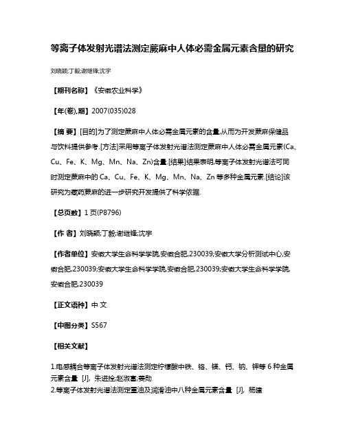
等离子体发射光谱法测定蕨麻中人体必需金属元素含量的研究刘晓颖;丁毅;谢继锋;沈宇
【期刊名称】《安徽农业科学》
【年(卷),期】2007(035)028
【摘要】[目的]为了测定蕨麻中人体必需金属元素的含量,从而为开发蕨麻保健品
与饮料提供参考.[方法]采用等离子体发射光谱法测定蕨麻中人体必需金属元素(Ca、Cu、Fe、K、Mg、Mn、Na、Zn)含量.[结果]结果表明,等离子体发射光谱法可同
时测定蕨麻中的Ca、Cu、Fe、K、Mg、Mn、Na、Zn等多种金属元素.[结论]该
研究为藏药蕨麻的进一步研究开发提供了科学依据.
【总页数】1页(P8796)
【作者】刘晓颖;丁毅;谢继锋;沈宇
【作者单位】安徽大学生命科学学院,安徽合肥,230039;安徽大学分析测试中心,安
徽合肥,230039;安徽大学生命科学学院,安徽合肥,230039;安徽大学生命科学学院,
安徽合肥,230039
【正文语种】中文
【中图分类】S567
【相关文献】
1.电感耦合等离子体发射光谱法测定柠檬酸中铁、铬、镁、钙、钠、钾等6种金属元素含量 [J], 朱进拴;赵淑富;姜勋
2.等离子体发射光谱法测定重油及润滑油中八种金属元素含量 [J], 杨建
3.ICP-MS同时测定当归中12种人体必需微量元素及5种重金属元素的含量 [J], 王涵;董庆海;吴福林;谭静;李平亚;刘金平;林红强
4.高效液相色谱法测定不同产地蕨麻中蕨麻苷含量 [J], 董培;崔颖;顾军;龚海英;李灵芝
5.电感耦合等离子体发射光谱法测定甘肃五大宗药材中人体必需微量元素 [J], 欧阳晓玫;何英梅;朱俊儒;贺军权;马潇;丁永辉
因版权原因,仅展示原文概要,查看原文内容请购买。
2014年度诺贝尔化学奖获得者的科学技术方法研究

渊上接第 35 页冤
揖参考文献铱
咱员暂闫鹏展,刘仲鹏,梁永慈,等.在线教师培训中微型学习资源设计研究[J].中国电
化教育,2014(2):84-87.
咱圆暂李小刚,王运武,马德俊,等.微型学习视野下的微课程设计及教学应用研究[J].
现向成人学习者的微型学习资源分类研究[J].中国远程教育, 2011(1).
Science & Technology Vision
科技视界
2014 年度诺贝尔化学奖获得者的 科学技术方法研究
余冰倩 渊南京中医药大学袁江苏 南京 210046冤
揖摘 要铱科学领域的成功都是通过不断地实验和多次的失败孕育而生的遥 因此研究方法就格外重要袁直接或间接地决定着实验的效率和 最终结果遥 默尔纳采用观察法成功成为世界首位测量单个荧光分子吸收的科学家袁本茨格的实验法和史蒂芬窑赫尔运用假设法都突破了 Abbe 衍射极限袁这三位 2014 年度诺贝尔化学奖的获得者的成功都与各自正确的实验方法有着密切的联系遥
们发出荧光遥 同样的袁只有一部分分子会发光袁我们记录下每一次发光 分子的图像遥 这一过程被一再重复遥 当本茨格最终将所有这些图像叠 加在一起时袁他得到了溶酶体外膜结构的超高分辨率图像遥 这张图像 的分辨率远远超出了 Abbe 衍射极限所限定的值遥由此可见袁实验和科 研的成功联系密切遥
要想在前人研究的基础上进行突破袁 光进行观察和实验是不够 的袁还要根据观察和实验的结果进行大胆假设袁这就是假设法遥 1990 年在海德堡大学获得博士学位之后袁 史蒂芬窑赫尔一直在设想超越一 个多世纪前提出的 Abbe 极限的方法遥 当他在一本量子光学书中读到 有关受激发射的内容时袁一种全新的想法在他的脑海中逐渐成型遥 他 设想中的技术方案袁也就是所谓 野受激发射减损技术冶渊STED冤中计划 采用闪光来激发所有的荧光分子袁随后利用另外一次闪光让所有分子 荧光熄灭要 要要那些位于中部位置上纳米尺度空间内的除外遥 当进行记 录时则只记录下这一部分遥 让这一光束扫过整个样品表面袁并连续记 录光强信息袁就有可能得到一张整体图像遥 每次允许发出荧光的空间 区域越小袁最后得到的图像分辨率便越高遥 从原理上说袁对于光学显微 成像的极限再也不复存在了遥 2000 年袁他证明了自己的技术方法在实 际工作中是可行的遥 当时他对大肠杆菌进行了摄像袁其分辨率是此前 任何光学显微镜都从来未能达到过的遥 史蒂芬窑赫尔根据读到的受激 发射内容袁假设出了一种技术方案要要 要野受激发射减损技术冶袁其在原 理上突破了光学显微成像的极限说法遥
介孔二氧化硅纳米颗粒结合神经干细胞构成靶向光敏药物运输载体用于肿瘤治疗

介孔二氧化硅纳米颗粒结合神经干细胞构成靶向光敏药物运输载体用于肿瘤治疗张卫佳;陈家树【期刊名称】《中国组织工程研究》【年(卷),期】2018(022)006【摘要】BACKGROUND:Neural stem cells (NSCs),which exert no promoting effect on tumor growth and break through the blood brain barrier to deliver drugs into tumor tissues,are considered as a promising tumor targeted drug delivery vehicle.OBJECTIVE:To develop a hybrid delivery system composed of NSCs and moseporous silica nanoparticles for photosensitizer delivery,and to test if the system can be used for tumor therapy.METHODS:The photosensitizer,zinc phthalocyanine,was encapsulated in mesoporous silica nanoparticles.(1) Cytophagy experiment:NSCs were incubated with mesoporous silica nanoparticles (0,10,50,100,200 mg/L) loaded with zinc phthalocyanine for 6 hours,and fluorescence microscope was employed to observe the nanoparticles inside the cells.(2) Cytotoxicity test:NSCs incubated with mesoporous silica nanoparticles at various concentrations (0,10,50,100,200 mg/L) which loaded with or without zinc phthalocyanine for 6 hours,followed by 3 days of normal culture.Then,the cells were harvested for MTT assay.(3) Retention of nanoparticles within the NSCs:100 mg/L mesoporous silica nanoparticles loaded with zinc phthalocyanine were co-cultured with NSCsfor 6 hours.Then,the cells were normally cultured for 12,24,and 72 hours,and observed with fluorescence microscope.(4) Zinc phthalocyanine excitation in vitro:100 mg/L mesoporous silica nanoparticles loaded with or without zinc phthalocyanine were co-cultured with NSCs for 6 hours.The cells were then normally cultured for 12 hours and irradiated withlaser.Microscope was employed to observe cell morphology.(5)Tumor cell killing experiment:NSCs cells were cultured with 100 mg/L mesoporous silica nanoparticles loaded with zinc phthalocyanine,then mixed with MCF7 cells for 12 hours,and irradiated with laser.After that,the cells were cultured for another 12 hours and cell death was observed under fluorescence microscopy.RESULTS AND CONCLUSION:(1) After co-cultured with the calls for 6 hours,nanoparticies could be found in the cytoplasm and the number was increased with the concentration of nanoparticles.(2) The nanoparticles with or without zinc phthalocyanine loaded at the concentration of < 100 mg/L showed no toxicity to NSCs.(3) After 72 hours of co-culture,the nanoparticles in the cytoplasm was decreased in number,but still could be found.(4) Laser irradiation could damage the cell membrane of NSCs co-cultured with mesoporous silica nanoparticles loaded with zinc phthalocyanine.(5) A large number of MCF7 cells died after tumor cells were co-cultured with NSCs that were cultured with mesoporous silica.To conclude,the hybrid system composed of NSCs and mesoporous silica nanoparticles loaded with zinc phthalocyanine can serve as a great potential tumor-targeted delivery vehicle for photodynamic therapy.%背景:神经干细胞对肿瘤细胞没有显著促生长作用,并且其可突破血脑屏障将药物运送到颅内肿瘤组织,目前神经干细胞被大量用于肿瘤药物靶向运输载体研究.目的:利用介孔二氧化硅纳米颗粒结合神经干细胞形成一个杂合的药物运输载体,探讨该载体是否能够用于光能药物靶向运输.方法:将光敏药物-酞菁锌包裹于介孔二氧化硅纳米颗粒中.①细胞吞噬实验:采用含不同质量浓度(0,10,50,100,200 mg/L)载酞菁锌介孔二氧化硅纳米颗粒培养液培养神经干细胞6h,采用荧光显微镜观察细胞内颗粒;②细胞毒性实验:采用含不同质量浓度(0,10,50,100,200 mg/L)载酞菁锌介孔二氧化硅纳米颗粒(或单纯介孔二氧化硅纳米颗粒)培养液分别培养神经干细胞6h,再常规培养3d,MTT法检测细胞增殖;③纳米粒子细胞内滞留时间实验:采用含100 mg/L介孔二氧化硅纳米颗粒的培养液培养神经干细胞6h,再常规培养12,24,72 h,利用荧光显微镜观察细胞内颗粒;④体外激发细胞内药物实验:采用含100 mg/L载酞菁锌介孔二氧化硅纳米颗粒(或单纯介孔二氧化硅纳米颗粒)培养液培养神经干细胞6h,再常规培养12h,以激光照射细胞,利用显微镜观察照射前后的细胞形态;⑤肿瘤细胞杀伤实验:采用含100 mg/L载酞菁锌介孔二氧化硅纳米颗粒培养液培养神经干细胞,再加入乳腺癌细胞MCF7共培养12h,以激光照射细胞后继续培养12h,通过荧光显微镜观察细胞死亡情况.结果与结论:①神经干细胞胞质中有纳米粒子存在,并且随着纳米粒子质量浓度的增加,细胞吞噬的纳米粒子量也增加;②对于有或没有包裹酞菁锌的纳米颗粒,在质量浓度小于100 mg/L时,对神经干细胞活性无明显影响;③培养72 h后,仍然有相当数量的纳米粒子聚集在细胞内;④载酞菁锌介孔二氧化硅纳米颗粒培养的细胞,激光照射后细胞膜发生明显破损;⑤载酞菁锌介孔二氧化硅纳米颗粒培养的神经干细胞与乳腺癌细胞MCF7大量死亡;⑥结果表明,介孔二氧化硅纳米颗粒结合神经干细胞形成的药物运输载体,可用于定点杀死肿瘤细胞.【总页数】6页(P883-888)【作者】张卫佳;陈家树【作者单位】中山大学新华学院,广东省广州市523145;中山大学药学院,广东省广州市510006【正文语种】中文【中图分类】R318【相关文献】1.介孔二氧化硅纳米颗粒结合神经干细胞构成靶向光敏药物运输载体用于肿瘤治疗[J], 张卫佳;陈家树;;2.负载光敏剂的生物可降解纳米药物载体用于肿瘤的光动力治疗 [J], 何艳梅;施文强;蔡胜胜;梅蘅;曹俊3.刺激响应和靶向型介孔二氧化硅纳米颗粒递送抗肿瘤药物的研究进展 [J], 郭燕;刘浩;李姗姗;贺冠迪;陈强;梅其炳;卢婷利;武祥龙4.加载促生长激素神经肽的介孔二氧化硅纳米颗粒药物载体对高糖环境下神经元细胞增殖的影响 [J], 段淏;赵玉武5.介孔二氧化硅纳米粒作为中药控释和靶向载体在肿瘤治疗中的应用进展 [J], 李楠;刘岩;郭盼;黄瑞;刘志东因版权原因,仅展示原文概要,查看原文内容请购买。
《2024年高效液相色谱联用四极杆轨道阱质谱分析柏树花粉过敏原蛋白》范文

《高效液相色谱联用四极杆轨道阱质谱分析柏树花粉过敏原蛋白》篇一一、引言随着环境问题日益突出,花粉过敏现象逐渐成为影响人们生活质量的重要问题。
柏树花粉作为常见的过敏原之一,其过敏原蛋白的准确检测与鉴定对于预防和治疗花粉过敏具有重要意义。
高效液相色谱(HPLC)和四极杆轨道阱质谱(Q-TRAP)联用技术的运用,为柏树花粉过敏原蛋白的检测与鉴定提供了强有力的技术支持。
本文旨在介绍并分析这种联用技术的高效应用。
二、高效液相色谱(HPLC)高效液相色谱是一种常用于分离、纯化和定量分析复杂混合物的方法。
其基本原理是利用混合物中各组分在固定相和流动相之间的分配系数不同,进行连续洗脱分离。
对于柏树花粉过敏原蛋白的检测,HPLC可初步实现蛋白的分离与纯化,为后续的质谱分析提供条件。
三、四极杆轨道阱质谱(Q-TRAP)四极杆轨道阱质谱是一种高灵敏度、高分辨率的质谱技术,广泛应用于生物大分子的检测与鉴定。
其工作原理是利用电场和磁场的作用,使带电粒子在飞行过程中发生能量变化,从而实现质量分析和元素鉴定。
对于柏树花粉过敏原蛋白的质谱分析,Q-TRAP能够准确检测蛋白的分子量、元素组成及肽谱等信息,为过敏原蛋白的鉴定提供有力依据。
四、HPLC联用Q-TRAP质谱分析柏树花粉过敏原蛋白HPLC与Q-TRAP质谱联用技术,通过将两者的优势相结合,可实现柏树花粉过敏原蛋白的高效、准确检测与鉴定。
首先,HPLC对柏树花粉样品进行初步分离与纯化,得到目标蛋白组分;然后,通过Q-TRAP质谱对目标蛋白进行详细分析,包括分子量、元素组成、肽谱等信息。
通过对比标准数据库或已知过敏原蛋白信息,可实现柏树花粉过敏原蛋白的准确鉴定。
五、实验结果与讨论通过HPLC联用Q-TRAP质谱技术对柏树花粉样品进行分析,我们成功分离并鉴定了多种柏树花粉过敏原蛋白。
实验结果表明,该联用技术具有高灵敏度、高分辨率和高通量的特点,可实现柏树花粉过敏原蛋白的快速、准确检测与鉴定。
银离子与壳聚糖形成水凝胶的作用机制研究

0 引言
经过 几 十 年 的 研 究 发 现, 壳 聚 糖 ( chitosan,
CTS) 对于碱金属几乎没有配位能力,而对于一些过
些研究并没有深入到壳聚糖水凝胶的微观结构,也
没有从分子结构方面对有金属离子参与的水凝胶
进行论证。 因而笔者通过本次具体实验数据和科
渡金属和重金属离子却具有很强的配位能力,比如
stretching vibration of C O in the amide I band and the bending vibration of amino group in chitosan,
respectively. As the content of Ag in hydrogel gradually increased,The peak intensity ratio between 1 640 cm
398 1 eV,corresponding to N in NH⁃acetyl group and N on free amino group,respectively. The diffraction peaks
of CTS at 2θ = 13° and 20° in the wide⁃angle X⁃ray diffraction pattern corresponded to intermolecular and
通信作者:李鹏,工程师。 E⁃mail:lipeng@ bimt org cn
spectroscopy and wide⁃angle X⁃ray diffraction. Results A
wide absorption band at 3 500 cm to 3 200 cm
-1
-1
美国化学家利用质谱分析法阅读遗传密码

美国化学家利用质谱分析法阅读遗传密码
佚名
【期刊名称】《世界经济科技》
【年(卷),期】1992(000)015
【总页数】2页(P47-48)
【正文语种】中文
【中图分类】Q349.55
【相关文献】
1.利用质谱分析法从酶谱鉴定嗜热真菌木聚糖酶 [J], 吴亚宁;刘刚;余少文;李水明;王娟
2.普利茅斯-詹姆斯镇-威廉斯堡-约克镇找寻美国强大的遗传密码 [J], 王凡;刘东平;
3.遗传密码轮——遗传密码表的另一表达方式 [J], 方振伟;王方
4.线粒体遗传密码及基因组遗传密码的对称分析 [J], 陈惟昌;陈志华;王自强;刘伟;左琳
5.最新研制的药物:生物化学家已破译出一半遗传密码 [J], Garp.,B;朱嘉鸣
因版权原因,仅展示原文概要,查看原文内容请购买。
《2024年《科学》杂志研究》范文

《《科学》杂志研究》篇一一、引言随着科技的飞速发展,纳米技术逐渐成为了研究领域的重要分支。
作为一种新兴的技术手段,纳米材料在生物医学领域的应用愈发广泛。
《科学》杂志近期的几篇研究报告中,着重探讨了新型纳米材料在生物医学中的重要作用。
本文将通过一篇范文的形式,详细介绍这些研究成果。
二、新型纳米材料的制备与性质近年来,研究者们开发出了一种新型的纳米材料,具有优异的生物相容性和独特的物理化学性质。
这种纳米材料主要由金属氧化物、碳基材料等构成,具有高比表面积、良好的生物可降解性等特点。
其制备方法也相对简单,可以通过溶胶-凝胶法、化学气相沉积法等方法实现规模化生产。
三、新型纳米材料在药物传递中的应用新型纳米材料在药物传递方面表现出显著的优势。
研究表明,通过将药物负载于纳米材料中,可以有效地提高药物的稳定性和生物利用度。
同时,纳米材料还能够穿透细胞膜,将药物直接送达靶点,减少药物在体内的分布和代谢过程,从而提高治疗效果。
此外,纳米材料还可以通过调节药物释放速率,实现药物的持续释放和精准控制。
四、新型纳米材料在肿瘤诊断与治疗中的应用肿瘤是当前医学领域的重点研究领域之一。
新型纳米材料在肿瘤诊断与治疗中也发挥了重要作用。
通过将具有诊断功能的分子或药物与纳米材料结合,可以实现对肿瘤的早期诊断和精准治疗。
例如,利用纳米材料的光学性质和磁学性质,可以实现肿瘤的荧光成像和磁共振成像。
同时,通过将光敏剂、放射线等治疗手段与纳米材料结合,可以实现对肿瘤的局部治疗和微创手术。
五、结论新型纳米材料在生物医学领域的应用前景广阔。
通过对其制备方法、性质以及在药物传递、肿瘤诊断与治疗等方面的应用进行深入研究,我们可以发现其巨大的潜力和价值。
然而,纳米材料在生物医学应用中也存在诸多挑战和问题,如安全性评估、体内代谢过程等仍需进一步研究和探讨。
相信在未来的研究中,随着科学技术的不断发展和进步,新型纳米材料在生物医学领域的应用将会取得更大的突破和进展。
- 1、下载文档前请自行甄别文档内容的完整性,平台不提供额外的编辑、内容补充、找答案等附加服务。
- 2、"仅部分预览"的文档,不可在线预览部分如存在完整性等问题,可反馈申请退款(可完整预览的文档不适用该条件!)。
- 3、如文档侵犯您的权益,请联系客服反馈,我们会尽快为您处理(人工客服工作时间:9:00-18:30)。
DOI: 10.1126/science.1253320, 94 (2014);345 Science Erik F. Y. Hom and Andrew W. Murray mutualismNiche engineering demonstrates a latent capacity for fungal-algalThis copy is for your personal, non-commercial use only.clicking here.colleagues, clients, or customers by , you can order high-quality copies for your If you wish to distribute this article to othershere.following the guidelines can be obtained by Permission to republish or repurpose articles or portions of articles): July 6, 2014 (this information is current as of The following resources related to this article are available online at/content/345/6192/94.full.html version of this article at:including high-resolution figures, can be found in the online Updated information and services, /content/suppl/2014/07/02/345.6192.94.DC1.htmlcan be found at:Supporting Online Material /content/345/6192/94.full.html#related found at:can be related to this article A list of selected additional articles on the Science Web sites /content/345/6192/94.full.html#ref-list-1, 16 of which can be accessed free:cites 60 articles This article /content/345/6192/94.full.html#related-urls 1 articles hosted by HighWire Press; see:cited by This article has been/cgi/collection/microbio Microbiologysubject collections:This article appears in the following registered trademark of AAAS.is a Science 2014 by the American Association for the Advancement of Science; all rights reserved. The title Copyright American Association for the Advancement of Science, 1200 New York Avenue NW, Washington, DC 20005. (print ISSN 0036-8075; online ISSN 1095-9203) is published weekly, except the last week in December, by the Science o n J u l y 7, 2014w w w .s c i e n c e m a g .o r g D o w n l o a d e d f r o mbasement membrane at P0(fig.S11,B and C).However,at P7,endocardial cells and their deriv-atives (labeled by Tie2-Cre or VE Cad-CreER)were no longer aligned with COL3A1strands but rather took up intramyocardial positions (P7in fig.S11,B and C).To better visualize the pro-cess by which P0endocardium transforms into intramyocardial VECs at P7,we studied an inter-mediary stage (P3)when hearts contain both unremodeled trabeculae (Fig.4B)and actively compacting regions (Fig.4C).We labeled devel-opmental intermediates by treating Apln-CreER;Rosa26mTmG/+mice with tamoxifen at P1.5(Fig.4A).Trapped endothelial cells remote from the basement membrane adopted the morphology of individual capillary-like cells and were marked by the VEC genetic lineage tracer (green fluores-cent protein,see GFP in Fig.4C),whereas endo-thelial cells facing the ventricular lumen and residing on the basement membrane retained sheetlike morphology and did not express VEC lineage tracer,consistent with endocardial iden-tity (Fig.4B).These observations suggest that myocardial compaction traps sheets of endocar-dial cells,which convert to the VEC lineage and translocate to an intramyocardial location.We investigated conditions that might promote endocardium to VEC transition in the neonatal heart.Hypoxyprobe,a hypoxia-sensitive chem-ical probe,indicated that rapid expansion of the compact myocardium by trabecular coales-cence in the first several postnatal days of life creates a hypoxic environment within the inner myocardial wall (fig.S12).Expression of hypoxia inducible factor 1a (Hif1a )and vascular endo-thelial growth factor A Vegfa ,genes known to be up-regulated by hypoxia,increased in the inner myocardial wall of the P1and P3neonatal hearts (fig.S12,B and C).This corresponds to the region in which we observed endocardial to VEC lineage conversion,suggesting that hypoxia and its resulting up-regulation of the key angio-genic factor Vegfa contribute to this process.Our work reveals a mechanism by which trabec-ular coalescence and endocardial-to-VEC lineage conversion drive vascular expansion in the post-natal heart.Why does coronary vascular growth in this setting rely on this alternative mechanism,rather than occurring through more typical angio-genic sprouting from preexisting vessels?The transition from fetal to postnatal circulation acute-ly increases the hemodynamic burden on the left ventricle.To accommodate this increased workload,we reason that mammals developed trabecular myocardium as a reservoir of new cardiomyo-cytes that is quickly recruited after birth through myocardial compaction to increase neonatal left ventricular mass.In addition to cardiomyocytes,this myocardial reservoir also contains coronary vessel precursors in the form of endocardial cells.Trabeculae coalesce during neonatal myocardial compaction causes regional hypoxia that stim-ulates the trapped neonatal endocardial cells to form the vascular supply for the newly compacted myocardium.This mechanism likely allows more rapid vascular and myocardial growth than angio-genic sprouting of the first CVP from the periphery.Understanding this endogenous mechanism for rapidly developing a functional vascular supply has important implications for cardiac diseases and cardiac regenerative medicine (4).REFERENCES AND NOTES1.P.R.Riley,N.Smart,Cardiovasc.Res.91,260–268(2011).2. B.A.Yi,O.Wernet,K.R.Chien,J.Clin.Invest.120,20–28(2010).3.N.Smart et al .,Nature 445,177–182(2007).4. E.R.Porrello et al .,Science 331,1078–1080(2011).5.H.S.Bennett,Am.J.Anat.60,27–53(1936).6.K.Red-Horse,H.Ueno,I.L.Weissman,M.A.Krasnow,Nature464,549–553(2010).7.T.C.Katz et al .,Dev.Cell 22,639–650(2012).8. B.Wu et al .,Cell 151,1083–1096(2012).9.P.Riley,Nature 464,498–499(2010).10.G.del Monte,P.Richard,Cell 151,932(2012).11.X.Tian et al .,Cell Res.23,1075–1090(2013).12.D.Sedmera,T.Pexieder,M.Vuillemin,R.P.Thompson,R.H.Anderson,Anat.Rec.258,319–337(2000).13.H.Elmasri et al .,FASEB J.23,3865–3873(2009).ACKNOWLEDGMENTSWe thank K.Red-Horse,Y.Chen,and N.Jin for insightful discussions and R.Adams,H.Zeng,Z.Yang,T.Quertermous,J.Rossant,and A.Nagy for mouse strains.This work was supported by National Basic Research Program of China(2012CB945102and 2013CB945302),National Natural Science Foundation of China (91339104,31271552,31222038,31301188),Chinese Academy of Sciences (Hundred Talents Program and KSCX2-EW-R-09),Shanghai Pujiang Program (11PJ1411400)and Basic Research Key Project (14JC1407400),Organization Department of the CPC Central Committee Bajian Talents Program,AstraZeneca,Sanofi-Aventis Shanghai Institutes for Biological Sciences (SA-SIBS)Fellowship,Postdoc Fund (SIBS-2013KIP311,China-2013M541561),NIH (2R01HL094683),and American Heart Association Established Investigator Award to W.T.P.SUPPLEMENTARY MATERIALS/content/345/6192/90/suppl/DC1Materials and Methods Figs.S1to S12References (15–25)29January 2014;accepted 22May 2014tions between different species involving persistent physical contact and physio-logical coupling —are central to many evo-lutionary and ecological innovations(1–3).These include the origin of eukaryotic cells,the colonization of land by plants,coral reefs,and the gut microbiota of insects and animals (4,5).Despite their ubiquity and importance,we understand little about how mutualistic sym-bioses form between previously free-living orga-nisms (5,6).Like speciation,the birth of novel symbioses has rarely been witnessed,making itfore symbiosis begins or if chance ecological encounters initiate new symbioses (5,7).Such “ecological fitting ”(8,9)occurs when both a particular environment and previously evolved traits allow a set of species to complement each other,giving rise to novel interactions without the need for prior coevolutionary adaptation.We tested two genetically tractable organisms,the budding yeast Saccharomyces cerevisiae and the green alga Chlamydomonas reinhardtii ,to determine if a reciprocal exchange of carbon and nitrogen would lead to obligate mutualism between algae and fungi such as those that oc-cur naturally (10–13).In our scheme (Fig.1A),S.cerevisiae metabolizes glucose to carbon di-oxide (CO 2),a carbon source that C.reinhardtii fixes via photosynthesis,and C.reinhardtii reduces nitrite (NO 2–)into ammonia (NH 3)(14),which yeast can use as a nitrogen source.Coculturing944JULY 2014•VOL 345ISSUE 6192 SCIENCE1Department of Molecular and Cellular Biology,HarvardUniversity,Cambridge,MA 02138,USA.2Faculty of Arts and Sciences Center for Systems Biology,Harvard University,Cambridge,MA 02138,USA.*Corresponding author.E-mail:erik@;amurray@ †Present address:Department of Biology,University of Mississippi,University,MS 38677,USA.RESEARCH |REPORTSSCIENCE 4JULY 2014•VOL 345ISSUE 619295reinhardtii .exchange.carbon C.reinhardtii under different coculture conditions demonstrates that obligate mutualism can arise without any genetic engineering of metabolic pathways.T op:cartoons of the different conditions tested;middle:cell density of yeast RESEARCH |REPORTSexperiments (15)indicate that by preventing access to atmospheric CO 2,S.cerevisiae and C.reinhardtii become obligate mutualists (Fig.1B).This mutualism depends on the metabolic capa-bilities of the two organisms:S.cerevisiae cannot use nitrite as a nitrogen source and C.reinhardtii cannot use glucose as a carbon source.Cell pro-liferation did not require genetic engineering or fine-tuning of nutrient concentrations or starting ratios of the two species (Fig.1B and figs.S1and S2)and failed when either species (Fig.1B,con-ditions 2and 3),glucose,or nitrite was omitted from the experiment (Fig.1B,conditions 4and 5).Agitation attenuates this mutualism (Fig.1B,con-dition 6),suggesting the importance of cell-cell proximity and spatial structure in establishing successful cooperation (16).Thus,a simple envi-ronmental change can induce free-living orga-nisms to be mutualistic without requiring adaptive coevolution.In our scheme,mutualism can be obligate or facultative depending on the environment.Ac-cess to atmospheric CO 2makes C.reinhardtii a facultative mutualist by removing its dependence on S.cerevisiae for carbon (Fig.1B,condition 7),but thenitrogen.In this environment,algal proliferation is improved by the presence of glucose-metabolizing,CO 2-generating budding yeast whereas yeast pro-liferation is reduced,although not extinguished (Fig.1B,compare conditions 7and 8).Conversely,adding ammonia (as ammonium chloride)to airtight cocultures allows budding yeast to prolif-erate independently of the alga whereas the alga remains dependent on the yeast for carbon.Under these conditions,S.cerevisiae (~4hours doubling time in our conditions)outproliferates C.reinhardtii (≥12hours doubling time)and drives the alga to near extinction (fig.S1,con-dition 15).These results suggest that stable meta-bolic mutualisms require that the faster-growing species be obligately dependent on nutrients pro-duced by its slower-growing partner.The engineered obligate mutualism between S.cerevisiae and C.reinhardtii is not limited to our initial choice of input nutrient concentra-tions.Successful mutualisms were established over nearly two orders of magnitude in glucose and nitrite concentrations (Fig.2).However,this resulted in complex population dynamics.We observed undulations and variations in stability lation cycles predicted for mutualistic systems (17).Other carbon (e.g.,galactose)or nitrogen (e.g.,nitrate)sources,although less effective,also sustain mutualism between S.cerevisiae and C.reinhardtii (fig.S3).We also demonstrate that many different as-comycetous yeast and four Chlamydomonas species,spanning over 300million years of evolutionary divergence in each clade,can form mutualisms (Fig.3).Nearly all yeast species we examined form synthetic obligate mutualisms with C.reinhardtii ,although with different degrees of productivity (Fig.3A).Mutualistic productivity,as assessed by total cell counts,did not correlate with a yeast ’s preference for a fermentative or respiratory lifestyle (Fig.3A),whether a yeast strain was isolated from soil (a potential habitat shared with C.reinhardtii ),intrinsic growth rate,or nitrite-mediated inhibition of growth (fig.S4and table S1).Thus,we observe that mutualisms can be phylogenetically broad,but that the degree of success depends on species-specific traits.Two yeast species and the alga Chlorella vulgaris did not form obligate mutualisms (Fig.3).C.vulgaris ,which can use glucose as a carbon S.cerevisiae ,whereas964JULY 2014•VOL 345ISSUE SCIENCEcapacity for mutual-broad.phylogenetic tree of fungal spe-and modified from work (15)]paired with (CC-1690,21gr),in 9-day-old co-by dashed linesunder 110m mol m –2s –1of confidence interval;are shown to the of Ascomycota are in-yeasts (exhibiting a even under aero-”,and weakly Crabtree-is a nonfermenting (NF)are indicated by a light connected dots (seefig.S5).cultivars (green bars at right)T 95%confidence interval;hexagon).Descriptions of strains RESEARCH |REPORTSHansenula polymorpha ,a yeast that can use nitrite as a sole nitrogen source,outproliferated C.reinhardtii .The yeast Kluveromyces polysporus failed to form an obligate mutualism with C.reinhardtii .This yeast can grow in an ammonium-supplemented coculture medium,suggesting that it fails to cooperate with C.reinhardtii likely be-cause it either cannot grow at the low ammonia sitive to nitrite inhibition at such low ammonia levels (fig.S4).Neurospora crassa and Aspergillus nidulans are genetically tractable filamentous fungi that can use nitrite as a nitrogen source (18).The ability of these fungi to reduce nitrite keeps wild-type strains from forming obligate mutualisms with C.reinhardtii .However,mu-tants that cannot reduce nitrite did form obligate gene function in one species could be comple-mented through mutualism (7,19).We observed that the filamentous fungi formed macroscopic structures (fig.S5)such that the fungal hyphae were decorated with C.reinhardtii cells (Fig.4,A and B,and movies S1to S6).How-ever,physical associations between fungus and alga form even in the absence of any metabolic dependency (figs.S6and S7and movies S7to S16).Electron microscopy of interactions be-tween C.reinhardtii and A.nidulans ,which shares a most recent common ancestor with lichenous fungi within the class Eurotiomycetes (10),revealed a tight fungal-algal contact inter-face (Fig.4,C and D)reminiscent of wall-to-wall interfaces between fungal and algal cells in ex-tant lichens (20).The walls of C.reinhardtii cells in contact with A.nidulans hyphae are less heavily stained and appear thinner than C.reinhardtii cells cultured separately (Fig.4E),possibly be-cause of locally secreted A.nidulans cell wall –remodeling enzymes.We saw no evidence of any morphologically complex tissue structures,such as those seen in many lichens,nor of fungal hy-phae penetrating algal cells (11,20).Thus,these synthetic mutualisms may result in physical com-plexes but they do not appear to form elaborate morphological structures at the cellular or or-ganismal level.The ease with which fungal-algal mutualisms were created suggests that ecological interac-tions may be relatively easy to establish (21).Fur-thermore,they do not require a prior facultative,commensal,or parasitic stage,or coevolutionary adaptation (5–7,22,23).Our understanding of how “ecologically framed ”pairs of species can be created in response to environments that force them to depend on each other will be useful in the emerging field of synthetic ecology (24,25),as well as for understanding the assembly of microbial communities in cases of disturbed or invaded habitats.REFERENCES AND NOTES1.J.N.Thompson,Science 284,2116–2118(1999).2.J.L.Bronstein,in The Princeton Guide to Ecology ,S.A.Levin et al.,Eds.(Princeton Univ.Press,Princeton,NJ,2009),pp.233–238.3.R.M.Brucker,S.R.Bordenstein,Trends Ecol.Evol.27,443–451(2012).4.S.Paracer,V.Ahmadjian,Symbiosis:An Introduction toBiological Associations (Oxford Univ.Press,New York,2000).5. A.E.Douglas,The Symbiotic Habit (Princeton Univ.Press,Princeton,NJ,2010).6.J.L.Sachs,R.G.Skophammer,J.U.Regus,Proc.Natl.Acad.Sci.U.S.A.108(suppl.2),10800–10807(2011).7.J.J.Morris,R.E.Lenski,E.R.Zinser,MBio 3,e00036-e12(2012).8. D.H.Janzen,Oikos 45,308–310(1985).9.S.J.Agosta,J.A.Klemens,Ecol.Lett.11,1123–1134(2008).10.F.Lutzoni,M.Pagel,V.Reeb,Nature 411,937–940(2001).11.R.Honegger,in Fungal Associations ,B.Hock,Eds.(Springer,Berlin,2012),pp.287–339.12.D.L.Hawksworth,Bot.J.Linn.Soc.96,3–20(1988).13.J.Kohlmeyer,E.J.A.Kohlmeyer,in Marine Mycology:TheHigher Fungi (Academic Press,New York,1979),pp.70–78.14.M.P.Azuara,P.J.Aparicio,Plant Physiol.71,286–290(1983).15.Materials and methods are available as supplementarymaterials on Science Online.16.M.J.I.Müller,B.I.Neugeboren,D.R.Nelson,A.W.Murray,Proc.Natl.Acad.Sci.U.S.A.111,1037–1042(2014).17.J.N.Holland,D.L.DeAngelis,Ecology 91,1286–1295(2010).18.J.C.Slot,D.S.Hibbett,PLOS ONE 2,e1097(2007).SCIENCE 4JULY 2014•VOL 345ISSUE 619297Fig.4.C.reinhardtii physically associates with N.crassa and A.nidulans .Representative light micrographs of the periphery of fungal-algal associations formed in obligate mutualistic coculture.C.reinhardtii cells (green)stick to hyphae (white filaments)of (A )N.crassa (FGSC 11007D nit-4)or (B )A.nidulans (TS003crnA-crnB-).(C to F )Representative transmission electron micrographs reveal a simple wall-to-wall interface between C.reinhardtii (Cre )cells and A.nidulans (Ani )hyphae.Opposed arrows indicate the thickness of fungal cell walls,and opposed colored T-bars indicate those of algal cells [(C):51T 10nm;(D):60T 7nm;mean T SD].(E)C.reinhardtii grown in monoculture (160T 20nm;blue T-bars)or (F)unattached C.reinhardtii isolated from the supernatant of the same coculture {T-demarcations:reference monoculture cell wall thickness [blue dashed;see (E)];(2)core (heavy)cell wall staining (red):50T 4nm;(3)diffuse cell wall staining (purple):260T 30nm)}(15).Labeled intracellular components:c,chloroplast;e,eyespot;g,Golgi;m,mitochondria;n,nucleus;p,pyrenoid;and v,vacuole.RESEARCH |REPORTS19.M.J.Wade,Nat.Rev.Genet.8,185–195(2007).20.R.Honegger,New Phytol.103,785–795(1986).21.J.M.Gómez,M.Verdú,F.Perfectti,Nature 465,918–921(2010).22.W.Harcombe,Evolution 64,2166–2172(2010).23.K.L.Hillesland,D.A.Stahl,Proc.Natl.Acad.Sci.U.S.A.107,2124–2129(2010).24.N.Klitgord,D.Segrè,PLOS Comput.Biol.6,e1001002(2010).25.B.Momeni,C.C.Chen,K.L.Hillesland,A.Waite,W.Shou,Cell.Mol.Life Sci.68,1353–1368(2011).ACKNOWLEDGMENTSWe thank Q.Justman,B.Stern,A.Pringle,S.Sasso,M.Dayel,N.Collins,J.Hess,M.Mueller,G.Frenkel,S.Kryazhimskiy,P.Boynton,J.Calarco,D.Chiang,Y.Eun,K.Foster,R.Losick,W.Tong,Y.Katz,and members of the Murray and Nelson labs for helpful feedback.We thank D.Thompson,M.Dunham,F.Winston,and N.Rhind for yeast strains;T.Schinko and J.Strauss forA.nidulans strains;and the Fungal Genetics Stock Center (Kansas City,MO)for fungal strains.We thank P.Rogers,M.Tam,andB.Tilton (Faculty of Arts and Sciences Center for Systems Biology FACS Core);B.Goetze,C.Kraft,andD.Richardson (Harvard Center for Biological Imaging);M.Yankova and S.King (Central Electron Microscopy Facility,University of Connecticut Health Center)for their resources and assistance;and U.Goodenough for her help in interpreting electron micrographs.Supported in part by a Jane Coffin Childs postdoctoral fellowship toE.F.Y.H.and by the National Institute of General Medical Sciences Center for Modular Biology (NIH grant P50-GM068763).Additional data materials.E.F.Y.H.conceived the project,performed theexperiments,and analyzed the data.E.F.Y.H.and A.W.M.devised the research and wrote the manuscript.A.W.M.supported and provided input throughout all stages of this work.SUPPLEMENTARY MATERIALS/content/345/6192/94/suppl/DC1Materials and Methods Figs.S1to S7Tables S1to S8Movies S1to S16References (26–71)13March 2014;accepted 22May 2014folding and maturation of transmembrane and secreted proteins (1,2).Elevated phys-iological demand for protein folding can cause misfolded proteins to accumulate in the ER lumen —a condition called ER stress.The unfolded protein response (UPR)senses such stress and mediates cellular adaptation by ex-panding the ER ’s protein-folding capacity while decreasing its synthetic load.Protein kinase R (PKR)–like kinase (PERK)and inositol-requiring enzyme 1a (IRE1a )are two key metazoan UPR sensors (1,2);residing in the ER membrane,each has a lumenal domain that detects misfolded polypeptides.PERK harbors a cytoplasmic kinase moiety that phosphorylates eukaryotic translation-translation but promotes synthesis of preferred factors —including ATF4,which activates the UPR transcription factor CCAAT/enhancer-binding pro-tein homologous protein (CHOP),among other genes.IRE1a has both kinase and endoribonuclease (RNase)cytoplasmic moieties (3).The kinase con-trols RNase activity,which mediates regulated IRE1a -dependent decay (RIDD)of ER-associated mRNAs (4)and generates the UPR transcription factor X-box binding protein 1spliced (XBP1s).Certain pathological conditions can cause irre-solvable ER stress (5),often leading to apoptotic cell death (1,2,6).Two interconnected signaling cascades control apoptosis:the intrinsic,mitochon-drial pathway,and the extrinsic,death-receptor pathway (7).Each engages distinct proteases,called initiator caspases,to activate a common set of executioner caspases (8).Unmitigated ER stress regulates the intrinsic pathway via several Bcl-2family proteins (1,2,6,9,10).Furthermore,IRE1a cleaves specific micro-RNAs to derepress caspase-2expression (11);however,caspase-2may be dis-pensable for ER stress-induced apoptosis (12),which leaves the underlying initiation mecha-nisms obscure.Experiments with biological and pharmacolog-ical ER stressors revealed consistent activation of the pivotal initiator in the extrinsic (8)(Fig.1).The bacterial AB5subtilase SubAB induces pathophysiological ER by cleaving the chaperone BiP (13).SubAB dose-dependent BiP depletion and ER evident by CHOP and XBP1s up-regulation,KMS11multiple myeloma cells (Fig.1A).In with data that PERK activity persists,IRE1a activation is transient (14),CHOP elevated,whereas XBP1s declined by 24SubAB also induced activation of caspase-8caspase-3by 24hours,evident by cleaved and poly(ADP ribose)polymerase (PARP)SubAB substantially increased caspase-8caspase-3/7enzymatic activity,and DNA —an apoptotic hallmark (fig.S1,A C).Brefeldin-A (BfA)—an inhibitor of ER-to-trafficking —similarly induced ER stress,activation,and apoptosis in SK-MES-1lung cells (Fig.1B and fig.S1,D to F).The ER calcium –adenosine triphosphatase thapsigargin (Tg)induced persistent and transient XBP1s expression in wild-and in Bax −/−HCT116colon carcinoma cells;whereas apoptosis required Bax ,caspase-8acti-vation did not (Fig.1,C and D,and fig.S1,G to I).Moreover,small interfering RNA (siRNA)deple-tion of caspase-8,but not caspase-2,blocked activation of caspase-3/7and apoptosis by di-verse ER stressors (Fig.1,E and F,and fig.S1,J to O).Caspase-8activates the Bcl-2family protein Bid to engage the intrinsic pathway via Bax (15,16).Full-length Bid declined in association with Tg-induced caspase-8activation (fig.S1I),which indicated Bid processing.Bid siRNA knock-down commensurately attenuated Tg-induced apoptosis,whereas caspase-8siRNA inhibited both Bid processing and apoptosis (fig.S1,P to S).Tg also up-regulated Bim (fig.S1I)as reported (10);however,caspase-8and Bid processing oc-curred much earlier,which suggests that Bim might support later apoptotic signals.Thus,caspase-8plays a pivotal role,whereas caspase-2appears dispensable,during apoptosis induction by un-mitigated ER stress.Upon binding of cognate extracellular ligands,the death receptors Fas,DR4,or DR5nucleate a death-inducing signaling complex (DISC)at the plasma membrane,which activates caspase-8via984JULY 2014•VOL 345ISSUE 6192 SCIENCE1Cancer Immunology,Genentech,Inc.,1DNA Way,South San Francisco,CA 94080,USA.2Howard Hughes MedicalInstitute,University of California,San Francisco,CA 94158,USA.3Department of Biochemistry and Biophysics,University of California,San Francisco,CA 94158,USA.4Research Centre for Infectious Diseases,School ofMolecular and Biomedical Science,University of Adelaide,South Australia,5005,Australia.*These authors contributed equally to this work.†Corresponding author.E-mail:peter@ (P.W.);aa@ (A.A.)RESEARCH |REPORTS。