清华大学Elisa实验报告
ELISA生化实验报告

ELISAObjective:1.To know about the basic operation and thoery of themeasurement of ELISA.2.To learn how to use microplate reader, how to dilute thesolution in certain ratio quickly.3.Understand the advantage of ELISA to measure theconcentration of protein.Theory:In the general procedure, the antigen was absorbed by the solid support, then the antibody we want to test was added in. Then the conjugated enzyme was added in to label the antibody. Then the substrate of the enzyme was added in and caused the formation of colored chemicals. Then the amount of antibody cloud be calculated by light absorption of spectrophotometer.In this experiment, we use Rabbit-anti-human IgG antiserum as first antibody. It can combine with Goat-antirabbit IgG-HRP, which can facilitate the oxidation of OPD. The colored product absorbs light at 492 nm. So the concentration of products can be measured by spectrophotometry. We can get a linear relation and cauculate the concentration.Reagens:1.Coating buffer (Carbonate-Bicarbonate buffer, pH 9.6) :2.Phosphate buffered saline (PBS)3.Washing buffer (PBS-T): PBS + 0.05% Tween 20.4.Blocking solution: 0.5% BSA in PBS.5.First antibody: Rabbit-anti-human IgG antiserum (1/400~1/12800).6.Peroxidase conjugated secondary antibody: Goat-anti-rabbit IgG-HRP (1:40000).7.Substrate: substrate buffer(fresh):8.Stopping reagent: 2mol/L H2SO4.Equioment:1.microplate reader2.microtiter plate3.PipetteProcedure:Antigen Coating1. Prepare human IgG solution at 0.025mg/ml in carbonate-bicarbonate buffer (coating buffer).2. Pipette 0.2 ml of the above solution to each well of the 96-well microtiter plate from 2B-D to 10 B-D. Incubate at 37 ℃for 30-45 min.3. Remove the coating solution. Wash three times with PBS-T (0.2 ml/well).4. Blocking step: pipette 0.02% (w/v) non-fat dry milk in-PBST (0.2ml/well) to each well of the 96-well microtiter plate from 2B-D to 11 B-D except 10 B-D. Keep the plate at Room Temperature for 5-10 min. Wash as mentioned above.B. Primary Antibody Reaction5. Dilute the primary (first) antibodies, Rabbit-anti-human IgG, in PBS-T. (different dilutions of 1st Ab., 1:400~1:12800).6. Add 0.2 ml of the diluted first antibody to each well according to Figure 1.Note: Add sample A (1st Ab. of unknown concentration) to well 8B-D. Negative control should be included (9B-D, No first Ab.).7. Incubate at 37 ℃for 1 hour.8. Wash three times with PBS-T (0.2 ml/well).C. Application of Secondary Antibody9. Dilute (1:40,000) the peroxidase conjugated Goat-anti-rabbit IgG-HRP (secondary antibody) in PBS-T. Add 0.2 ml of this solution to each well.10. Incubate at 37 ℃for 30 min.11. Wash three times with PBS-T (0.2 ml/well).D. Substrate Preparation12. During the last incubation and immediately before use, dissolve o-Phenylenediamine in Citric acid-sodium hydrogen phosphate buffer; add 30% H 2 O 2 before use.E. Development13. Add 0.2 ml of the freshly prepared substrate to each well.14. Orange-yellow color should develop in positive wells after 3-5 minutes.15. Stop reaction with 50 μl per well of preferred stopping reagent and read at 490 nm in a microplate reader.The element of each well was shown in the table 1.Table 1Data:Table 2. A490 of the sampleNo. dilution of the sample 1 sample 2 sample 3 average first antibody1 1/400 1.252 1.307 1.433 1.3312 1/800 1.079 1.294 1.295 1.2233 1/1600 1.186 0.910 1.070 1.0554 1/3200 0.939 0.954 0.818 0.9045 1/6400 0.698 0.722 0.783 0.7346 1/12800 0.646 0.577 0.526 0.5837. 1/4000 0.801 0.730 0.788 0.7738 - 0.023 0.021 0.020 0.0219 1/400 1.123 1.350 1.177 1.21710 1/400 0.133 0.140 0.242 0.172 Then Drawing the curves of absorbency and the concentration of the series of diluted first antibody.The dilution of sample7 as calculate is 5560.DiscussionAs we can see in the result, when the concentration of the first antibody decrease, A will decrease, too. And the –lg(ratio) and A follow a linear relation approximately. The result can be explainedthat with equal amount of antigen and 2nd antibody in every well, (in exception of Group 9and10) the concentration of 1st antibody dominates the product yield in restricted time. The higher first antibody added into the well, the more second-antibody-peroxidase complex attached to the well. So we can get the higher concentration of colored product. And the aborsance changes as lambert-beer law.The result of No..9 is similar No.1 but lower than it, blocking will decrease the influence of the other irrelevant protein’s adsorption.The result of No.8 is so low,,nearly zero,because there is no first antibody in No.8, and No.10 is low because without the antigen, the adsoption of first antibody will be much lower.Easy to see, our curve is not a perfect linear relation. And the calculation of the No.7 is different from the theoric so much. There may be several reasons.1.When we pipette the stopping reagent, I spilled alittle of the first sample, the height of the liquid isnot absolutely equal with the others.2.The stopping method mayt be too late so thecolored product was generated too much, whichinfluences the linearity.3.The move of the microtiter plate leads to thebubble in the sulotion, which may influence theresult.Literature references:《ELISA》 《The practical approach of biochemistry》Bingbin Yu。
ELISA实验报告
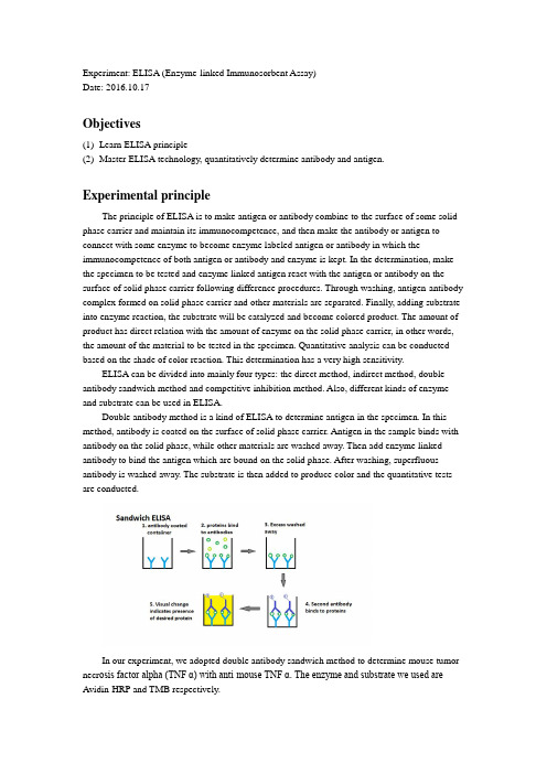
Experiment: ELISA (Enzyme-linked Immunosorbent Assay)Date: 2016.10.17Objectives(1)Learn ELISA principle(2)Master ELISA technology, quantitatively determine antibody and antigen.Experimental principleThe principle of ELISA is to make antigen or antibody combine to the surface of some solid phase carrier and maintain its immunocompetence, and then make the antibody or antigen to connect with some enzyme to become enzyme labeled antigen or antibody in which the immunocompetence of both antigen or antibody and enzyme is kept. In the determination, make the specimen to be tested and enzyme linked antigen react with the antigen or antibody on the surface of solid phase carrier following difference procedures. Through washing, antigen-antibody complex formed on solid phase carrier and other materials are separated. Finally, adding substrate into enzyme reaction, the substrate will be catalyzed and become colored product. The amount of product has direct relation with the amount of enzyme on the solid phase carrier, in other words, the amount of the material to be tested in the specimen. Quantitative analysis can be conducted based on the shade of color reaction. This determination has a very high sensitivity.ELISA can be divided into mainly four types: the direct method, indirect method, double antibody sandwich method and competitive inhibition method. Also, different kinds of enzyme and substrate can be used in ELISA.Double antibody method is a kind of ELISA to determine antigen in the specimen. In this method, antibody is coated on the surface of solid phase carrier. Antigen in the sample binds with antibody on the solid phase, while other materials are washed away. Then add enzyme linked antibody to bind the antigen which are bound on the solid phase. After washing, superfluous antibody is washed away. The substrate is then added to produce color and the quantitative tests are conducted.In our experiment, we adopted double antibody sandwich method to determine mouse tumor necr osis factor alpha (TNF α) with anti-mouse TNF α. The enzyme and substrate we used are Avidin-HRP and TMB respectively.Reagents and equipment(1)Equipments40-well reaction plate coated with anti-mouse TNF α; liquid transfer gun (1ml, 200μl); thermotank (37℃).(2) ReagentsWashing liquid (PBST); sealing fluid; antigen (1000pg/ml); anti-mouse TNF α; TMB;Avidin-HRP; 1M H2SO4.Method of operation(1) Empty out the solution in the plate and clean the plate with washing liquid for three times. Top up each hole and flap on absorbent paper to clean away the remaining liquid in each hole. Add 200μl sealing fluid in each hole and put the plate in 37℃ for 30mins.(2) Wash the plate for 3 times. Add reagents in 8 holes according to the table: (number refer to the co ncentration of antigen added, each hole is added 100μl liquid)1 2 3 4 5 6 7 831.25(this hole is antibody-free) Washingliquid31.25 62.5 125 250 500 1000Put the plate in 37℃ for 45mins.(3) Wash the plate for 3 times, add 100μl antibody in each hole, put the plate in 37℃ for 30mins.(4) Wash the plate for 3 times, add 100μl enzyme in each hole, put the plate in 37℃ for 20mins.(5) Wash the plate for 5 times, add 100μl substrate in each hole, put the plate in 37℃ for 5mins. Observe the coloration and take a picture.(6) Add 50μl 1M H2SO4 in each hole, observe the color change and take a picture.Notes:(1)All reagents shall be kept in dark place at 2-8℃.(2)Liquid feeding shall be limited at the bottom of holes instead of wall, without spillage.(3)When cleaning the plate manually, do not spill out the liquid in the holes in case that theadjacent holes pollute each other to cause false positive.(4)The results are only valid within 30mins after the termination of test.(5)Experiment wastes shall be treated as infectious samples.Discussion(1)From the result, we can see the gradient shade of coloration, which means the shade of coloration is in proportion to the concentrate of antigen. The exact shade of coloration can be detected by spectrophotometer, and be compared with the standard curve to get the concentrate value.(2)Every time when cleaning the plate, the washing liquid must be completely removed.Questions(1)Analyze the effect of impure enzyme labeled antibody to the testing result of antigen. (Double Antibody Sandwich Method)If the impurity can not be bond with antigen, it has no affect to the result; if the impurity can be bond with antigen, it will cause the result lower than the real concentrate.(2)Compare the most suitable testing objects of IFA and ELISA.IFA is used in vivo to locate a certain antigen; while ELISA is used in vitro condition to determine the presence and concentrate of a certain antigen or antibody.。
生化实验-ELISA-清华

现代生物学导论-生化实验报告-ELISA 生医9 田雪霏2009013189 (同组人:于淼)一、实验名称:酶联免疫吸附测定二、实验原理:ELISA是一种免疫测定(immunoassay,IA)。
基础:抗原或抗体的固相化及抗原或抗体的酶标记。
加入酶反应的底物后,底物被酶催化成为有色产物,产物的量与标本中受检物质的量直接相关,由此进行定性或定量分析。
此次实验采用间接法,是检测抗体最常用的方法。
这一方法的基本原理是:①使抗原或抗体结合到某种固相载体表面,并保持其免疫活性。
②使抗原或抗体与某种酶连接成酶标抗原或抗体,这种酶标抗原或抗体既保留其免疫活性,又保留酶的活性。
在测定时,把受检标本(测定其中的抗体或抗原)和酶标抗原或抗体按不同的步骤与固相载体表面的抗原或抗体起反应。
用洗涤的方法使固相载体上形成的抗原抗体复合物与其他物质分开,最后结合在固相载体上的酶量与标本中受检物质的量成一定的比例。
加入酶反应的底物后,底物被酶催化变为有色产物,产物的量与标本中受检物质的量直接相关,故可根据颜色反应的深浅刊物定性或定量分析。
由于酶的催化频率很高,故可极大地放大反应效果,从而使测定方法达到很高的敏感度。
三、实验仪器和材料:1. 聚苯乙烯微量细胞培养板(平板, 96孔)。
2. 酶联免疫检测仪3. 辣根过氧化物酶羊抗兔IgG。
4. 包被液:0.05 mol/L 碳酸缓冲液(pH 9.6):Na2CO30.15克,NaHCO30.293克,蒸馏水稀释至100 毫升。
5. 稀释液(PBS-Tween):NaCl 8克,KCl 0.2克,KH2PO4 0.2克,Na2HPO4·12H2O 2.9g,Tween-20,0.5毫升,蒸馏水加至1000毫升。
6. 洗涤液:同稀释液。
7. 封闭液:0.5 % (质量分数)BSA(用PBS配制)。
8. 邻苯二胺溶液(底物):0.1mol/L 柠檬酸(2.1克/100毫升), 取6.1毫升,0.2 mol/L Na2HPO4·12H2O (7.163克/100毫升),取6.4毫升,加蒸馏水12.5毫升,取邻苯二胺8毫克(溶解);临用前加30 % (体积分数)H2O240 微升。
elisa实验报告结果分析

Elisa实验报告结果分析1. 引言Elisa(酶联免疫吸附实验)是一种常用于检测和定量分析生物样本中特定分子的方法。
本文旨在对实验结果进行分析,并根据分析结果得出结论。
2. 实验设计在本次实验中,我们选择了特定的生物分子作为目标,通过Elisa方法来检测样本中的该分子含量。
实验过程中,我们遵循了以下步骤: 1. 准备样本和试剂:收集样本并处理,准备所需的试剂。
2. 涂覆酶标板:将待测样本加入酶标板孔中,使样本中的目标分子与酶标板表面发生特异性结合。
3. 洗涤:去除未结合的物质。
4. 加入检测抗体:加入特异性抗体,与已结合的目标分子发生反应。
5. 加入标记物:加入标记物,用于检测目标分子的存在。
6. 洗涤:去除未结合的物质。
7. 反应底物:加入底物,观察颜色变化。
8. 停止反应:加入停止液停止反应。
9. 测量:使用酶标仪或光谱仪测量吸光度。
3. 结果分析根据实验结果,我们得到了一系列吸光度值。
下面我们将对吸光度值进行分析,并根据实验设计和已有知识得出结论。
3.1. 标准曲线分析实验中通常会使用一系列已知浓度的标准样品来构建标准曲线。
通过测量标准样品的吸光度值,我们可以绘制出标准曲线。
在实验中,我们可以使用标准曲线来确定未知样本中目标分子的浓度。
3.2. 样本浓度分析根据实验设计,我们在酶标板中加入了待测样本,并测量了其吸光度值。
通过标准曲线,我们可以将吸光度值转化为目标分子的浓度。
根据样本中目标分子的浓度,我们可以进行进一步的分析。
3.3. 结果验证为了验证实验结果的准确性和可靠性,我们可以进行重复实验或与其他方法进行比较。
此外,还可以进行质控实验来评估实验的可重复性和准确性。
4. 结论通过对Elisa实验结果的分析,我们得出以下结论: 1. 根据标准曲线分析,我们可以确定未知样本中目标分子的浓度。
2. 样本浓度分析结果显示,样本中目标分子的含量为X单位。
3. 实验结果经过验证,具有较高的准确性和可靠性。
ELISA实验报告(20200623015910)
Experime nt: ELISA (En zyme-li nked Immuno sorbe nt Assay) Date: 2016.10.17Objectives(1) Learn ELISA principle(2) Master ELISA tech no logy, qua ntitatively determ ine an tibody and an tige n.Experimental principleThe principle of ELISA is to make antigen or antibody combine to the surface of some solid phase carrier and maintain its immuno compete nee, and the n make the an tibody or an tige n to connect with some en zyme to become en zyme labeled an tige n or an tibody in which the immun ocompete nee of both an tige n or an tibody and en zyme is kept. In the determ in ati on, make the specimen to be tested and enzyme linked antigen react with the antigen or antibody on the surface of solid phase carrier follow ing differe nee procedures. Through wash ing, an tige n-an tibody complex formed on solid phase carrier and other materials are separated. Finally, adding substrate into en zyme react ion, the substrate will be catalyzed and become colored product. The amount of product has direct relation with the amount of enzyme on the solid phase carrier, in other words, the amount of the material to be tested in the specime n. Quan titative an alysis can be con ducted based on the shade of color react ion. This determ in ati on has a very high sen sitivity.ELISA can be divided into mai nly four types: the direct method, in direct method, double an tibody san dwich method and competitive in hibiti on method. Also, differe nt kinds of en zyme and substrate can be used in ELISA.Double antibody method is a kind of ELISA to determine antigen in the specimen. In this method, antibody is coated on the surface of solid phase carrier. Antigen in the sample binds with antibody on the solid phase, while other materials are washed away. Then add enzyme linked antibody to bind the antigen which are bound on the solid phase. After washing, superfluous an tibody is washed away. The substrate is the n added to produce color and the qua ntitative tests are con ducted.I YY HIV Y I I YY IVisu-al chang* in^licafEai pngsanci ofprotsinIn our experiment, we adopted double antibody sandwich method to determine mouse tumor necrosis factor alpha (TNF a ) wrtbuaraiiTNF a . The enzyme and substrate we used areAvidi n-HRP and TMB respectively.Sandwich ELISALeditedcofiiaiineif2L prataini bind to去 E HCB » wash«d4.轴讯曲 antllwdhrbindite proleinaReagents and equipment(1) Equipments40-well react ion plate coated with an ti- mouse TNF a ; liquid tran sfer gun (1ml, 200 卩 I); thermotank (37 C ).⑵ Reage ntsWashing liquid (PBST); sealing fluid; antigen (1000pg/ml); anti- mouse TNF a ; TMB; Avidin-HRP; 1M H 2SO 4.Method of operation(1) Empty out the solution in the plate and clean the plate with washing liquid for three times. Top up each hole and flap on absorbe nt paper to clea n away the rema ining liquid in each hole. Add 200l sealing fluid in each hole and put the plate in 37C for 30mins.(2) Wash the plate for 3 times. Add reage nts in 8 holes accord ing to the table: (nu mber refer to the12 3 4 5 6 7 8 31.25(this hole is antibody-free)Wash ing liquid31.2562.51252505001000Put the plate in 37 for 45mins.Observe the colorati on and take a picture.(6) Add 50 l 1M HSO 4 in each hole, observe the color change and take a picture.⑶ Wash the plate for 3 times, add 100(4) Wash the plate for 3 times, add 100(5) Wash the plate for 5 times, add 100 l antibody in eaictelpdaepioit37 C for 30mins.l enzyme in each hole, putth £pfate2i0in3i7s.Notes:(1) All reagents shall be kept in dark place at 2-8 °C .(2) Liquid feeding shall be limited at the bottom of holes instead of wall, without spillage.(3) When cleaning the plate manually, do not spill out the liquid in the holes in case that theadjacent holes pollute each other to cause false positive.(4) The results are only valid within 30mins after the termination of test.(5) Experiment wastes shall be treated as infectious samples.Discussion(1) From the result, we can see the gradient shade of coloration, which means the shade of coloration is in proportion to the concentrate of antigen. The exact shade of coloration can be detected by spectrophotometer, and be compared with the standard curve to get the concentrate value.(2) Every time when cleaning the plate, the washing liquid must be completely removed.Questions(1) Analyze the effect of impure enzyme labeled antibody to the testing result of antigen. (Double Antibody Sandwich Method)If the impurity can not be bond with antigen, it has no affect to the result; if the impurity can be bond with antigen, it will cause the result lower than the real concentrate.(2) Compare the most suitable testing objects of IFA and ELISA.IFA is used in vivo to locate a certain antigen; while ELISA is used in vitro condition to determine the presence and concentrate of a certain antigen or antibody.。
ELISA实验报告

ELISA实验报告实验目的:本实验旨在通过ELISA(酶联免疫吸附试验)技术来检测细胞培养上清液中的蛋白质含量,从而了解该细胞株的蛋白质表达水平,进一步研究相关的细胞分子机制。
实验原理:酶联免疫吸附试验是一种常用的免疫学实验方法,通过特异性抗体与待测物结合来实现对特定蛋白质的检测。
该实验主要分为三个步骤:包被抗原、特异性抗体结合和酶底物显色。
实验材料:1.待测细胞上清液2.包被抗原3.特异性抗体4.酶标记的二抗5.酶底物6.缓冲液7.清洗缓冲液8.吸光度测定仪实验步骤:1.包被抗原的处理:将抗原溶液加入微孔板孔中,使其均匀附着在孔壁上。
然后将孔中的液体弃去,用清洗缓冲液进行孔的清洗。
2.探针的结合:将待测样品加入已经包被抗原的孔中,使其与抗原结合。
然后将孔中的液体弃去,用清洗缓冲液进行孔的清洗。
3.酶标记的二抗结合:将酶标记的二抗加入已经含有特异性抗体的孔中,使其与特异性抗体结合。
然后将孔中的液体弃去,用清洗缓冲液进行孔的清洗。
4.酶底物加入:将酶底物加入到孔中,使其在酶的作用下发生显色反应。
5.吸光度测定:使用吸光度测定仪读取吸光度值,根据吸光度值可以推断待测样品中蛋白质的含量。
实验结果:经过ELISA实验,我们得到了待测细胞上清液中蛋白质的含量。
根据吸光度值和标准曲线的对照,我们可以计算出待测样品中蛋白质的浓度。
实验结论:实验分析:本次实验利用ELISA技术成功检测了待测细胞上清液中的蛋白质含量。
该方法具有高灵敏度、高特异性、操作简单等优点,可以在生物医学、生物工程等领域广泛应用。
但需要注意的是,ELISA实验在操作过程中需要严格控制实验条件,避免交叉污染和误差的产生。
实验改进:为了进一步提高实验的准确性和可靠性,可以进行以下改进:1.增加重复次数,提高数据的可靠性和稳定性;2.使用更加准确的仪器和试剂;3.对实验流程进行优化,减少操作的差异性。
总结:本次实验通过ELISA技术成功检测了待测细胞上清液中的蛋白质含量,得出了蛋白质表达水平较高的结论。
elisa酶联免疫吸附试验报告
.elisa酶联免疫吸附实验报告一.实验目的酶联免疫吸附测定(enzyme-linked immunosorbent assay 简称ELISA)是在免疫酶技术(immunoenzymatic techniques)的基础上发展起来的一种新型的免疫测定技术,ELISA过程包括抗原(抗体)吸附在固相载体上称为包被,加待测抗体(抗原), 再加相应酶标记抗体(抗原),生成抗原(抗体)--待测抗体(抗原)--酶标记抗体的复合物,再与该酶的底物反应生成有色产物。
借助分光光度计的光吸收计算抗体(抗原)的量。
待测抗体(抗原)的定量与有色产生成正比。
二.实验原理用于免疫酶技术的酶有很多,如过氧化物酶,碱性磷酸酯酶,β-D-半乳糖苷酶,葡萄糖氧化酶,碳酸酐酶,乙酰胆碱酯酶,6-磷酸葡萄糖脱氧酶等。
常用于ELISA法的酶有辣根过氧化物酶,碱性磷酸酯酶等,其中尤以辣根过氧化物酶为多。
由于酶摧化的是氧化还原反应,在呈色后须立刻测定,否则空气中的氧化1 / 11.作用使颜色加深,无法准确地定量。
辣根过氧化物酶(HRP)是一种糖蛋白,每个分子含有一个氯化血红素(protonhemin)区作辅基。
酶的浓度和纯度常以辅基的含量表示。
氯化血红素辅基的最大吸收峰是403nm,HRP酶蛋白的最大吸收峰是275nm,所以酶的浓度和纯度计算式是(已知HRP的A(1cm 403nm 1%)=25,式中1%指HRP百分浓度为100ml含酶蛋白1g,即10mg/ml,所以,酶浓度以mg/ml 计算是HRP的A(1cm 403nm mg/ml=2.5)HRP纯度(RZ)=A403nm/A275nm纯度RZ(Reinheit Zahl)值越大说明酶内所含杂质越少。
高纯度HRP的RZ值在3.0左右,最高可达3.4。
用于ELISA检测的HRP的RZ值要求在3.0以上。
ELISA的基本原理有三条:(1)抗原或抗体能以物理性地吸附于固相载体表面,可能是蛋白和聚苯乙烯表面间的疏水性部分相互吸附,并保持其免疫学活性;(2)抗原或抗体可通过共价键与酶连接形成酶结合物,而此种酶结合物仍能保持其免疫学和酶学活性;(3)酶结合物与相应抗原或抗体结合后,可根据加入底物的颜色反应来判定是否有免疫反应的存在,而且颜色反应的深浅是与标本中相应抗原或抗体的量成正比例的,因此,可以按底物显色的程度显示试验结果。
elisa实验报告实验目的
elisa实验报告实验目的Title: The Purpose and Procedure of ELISA ExperimentIntroductionEnzyme-Linked Immunosorbent Assay (ELISA) is a widely used technique in the field of immunology and biochemistry. It is used to detect the presence of specific proteins, such as antibodies, antigens, and hormones, in a sample. The purpose of this experiment is to understand the principles and procedure of ELISA and to demonstrate its application in detecting a specific antigen. Materials and MethodsIn this experiment, we used a commercial ELISA kit to detect the presence of a specific antigen in a sample. The kit included microplates coated with a capture antibody specific to the antigen of interest, a detection antibody conjugated to an enzyme, a substrate solution, and a stop solution. The procedure involved several steps, including coating the microplate with the capture antibody, blocking non-specific binding sites, adding the sample and the detection antibody, washing the plate to remove unbound substances, adding the substrate solution to produce a color change, and finally stopping the reaction and measuring the absorbance at a specific wavelength.ResultsThe results of the ELISA experiment showed a significant increase in absorbance at the specific wavelength, indicating the presence of the antigen in the sample. This demonstrated the specificity and sensitivity of the ELISA technique indetecting the target protein.DiscussionELISA is a highly sensitive and specific technique that has a wide range of applications in research, clinical diagnostics, and pharmaceutical development. It is a valuable tool for detecting and quantifying proteins in biological samples. The principles of ELISA can be adapted to detect various targets, making it a versatile and widely used method in the field of biomedical sciences. ConclusionIn conclusion, the ELISA experiment successfully demonstrated the principles and procedure of this technique and its application in detecting a specific antigen. The results obtained validated the sensitivity and specificity of ELISA in detecting the target protein. This experiment provided valuable insights into the practical application of ELISA and its importance in biomedical research and diagnostics.。
- 1、下载文档前请自行甄别文档内容的完整性,平台不提供额外的编辑、内容补充、找答案等附加服务。
- 2、"仅部分预览"的文档,不可在线预览部分如存在完整性等问题,可反馈申请退款(可完整预览的文档不适用该条件!)。
- 3、如文档侵犯您的权益,请联系客服反馈,我们会尽快为您处理(人工客服工作时间:9:00-18:30)。
Elisa实验报告
实验名称:
酶联免疫吸附剂测定
实验原理:
酶联免疫吸附测定是一种免疫测定技术。
测定中,先使抗原吸附在固相载体上,然后加待测的抗体,再用某种酶标记抗体,形成抗原-抗体-酶标记抗体的“双抗体夹心”,此时酶仍保有活性,同时标记抗体亦有免疫活性。
之后再加入酶的底物,在酶的催化下产生反应并产生有色物,颜色深浅与待测物质的量直接相关。
至此,酶的催化放大作用与免疫反应的特异性相完美结合,提高了测定的准确性与灵敏度。
实验材料与试剂:
1.聚苯乙烯微量细胞培养板(平板, 96孔)。
2.酶联免疫检测仪。
3.辣根过氧化物酶羊抗兔IgG。
4.包被液:0.05 mol/L 碳酸缓冲液(pH 9.6):
Na2CO3 0.15g,
NaHCO3 0.293g,
蒸馏水稀释至100 ml。
5.稀释液(PBS-Tween):
NaCl 8g,
KCl 0.2g,
KH2PO4 0.2g,
Na2HPO4·12H2O 2.9g,
Tween-20,0.5ml,
蒸馏水加至1000ml。
6.洗涤液:同稀释液。
7.封闭液:0.5 % (质量分数)BSA(用PBS配制)。
8.邻苯二胺溶液(底物):
配制:
0.1 mol/L 柠檬酸(2.1g/100ml), 取6.1ml,
0.2 mol/L Na2HPO4·12H2O (7.163g/100ml),取6.4ml,
加蒸馏水12.5ml,
取邻苯二胺8mg(溶解);
临用前加30 % (体积分数)H2O2 40μl。
9.终止液:2 mol/L H2SO4。
实验步骤:
1.包被抗原:用包被液将抗原作适当稀释, 一般为1~10μg/孔,每孔加200μl, 37 ℃温育30min。
2.洗涤:倒尽板孔中液体,加200μl洗涤液,反复三次,最后将反应板倒置在吸水纸上,使孔中洗涤液流尽。
3.加封闭液200μl,37 ℃温育30min。
4.洗涤同2。
5.加被检血清:用稀释液将被检血清作几种稀释,每孔200μl。
同时作稀释液对照。
37 ℃温育30min。
6.洗涤同2。
7.加辣根过氧化物酶羊抗兔IgG, 每孔200μl, 37 ℃温育30min。
8.洗涤同2。
9.加底物:邻苯二胺溶液加200μl,室温暗处5min。
10.加终止液:每孔50μl。
11.观察结果:用酶联免疫检测仪记录OD490nm读数。
实验结果
数据如下:
P.S:紫色数据表示异常
实验讨论:
1无一抗(10)数据值存在异常。
理论上应趋近于零。
可能是由于受污染所致
实验现象比较清晰,随浓度由浓到稀,颜色由深黄渐变为浅黄。
2线性关系比较明显。
但斜率随浓度减小而有所增大。
这可能是在使用取液器时有气泡混入,导致实际浓度低于理论上的浓度。
3组号(2)(4)(5)中3个样本数据差别较大,可能是因为配血清时混合不够均匀所致。
这个实验采用了间接法测定,利用酶的催化放大作用提高了检测的灵敏度。
作为一名精仪系的学生,这给予我的启示就是,在直接测量精度如果遇到瓶颈时,没有合适的方法提高精度,那么可以考虑寻找一个“中介”,而这个“中介”应该具有“在变量产生微小变化时能放大这种变化,或者用另一种形式体现这种变化”的特性,从而测量变得可行。
而该实验中,最终效果就是我们可以直接通过颜色深浅大致看出浓度的不同。
这一点很值得思考。
另外由于操作不熟练、不仔细,也带来了一些粗大误差,如无一抗(10)。
这是我们实验中应该避免的。