脑静脉畸形的MRI诊断
放射学征象 “海蛇头征或水母头征”的影像诊断意义

放射学征象“海蛇头征或水母头征”的影像诊断意义
海蛇头征或水母头征
【英文】Caput Medusae Sign海蛇头征或水母头征海蛇头征为脑静脉血管瘤或静脉畸形在增强CT和磁共振上较特异的征象。
主要表现为脑实质内放射状排列的管状结构,朝一个共同点聚拢。
见图。
【病理基础】脑静脉血管瘤是颅内静脉系统最常见的变异,是由放射状排列的异常髓静脉汇入中央扩张的静脉干所组成,周围是正常的神经组织。
CT增强或MRI上特征性表现是海蛇头样的深部髓静脉汇集到单根粗大的引流静脉,然后汇入表浅皮层静脉或硬膜窦。
【鉴别诊断】海蛇头征是脑静脉血管瘤或静脉畸形较特征的表现,鉴别诊断主要与小的动静脉畸形鉴别。
动静脉畸形平扫CT常可见钙化,可合并出血,CTV可显示供血动脉、畸形血管团及引流静脉,不出现典型的海蛇头状形态。
图1海蛇头征MR图像
T1WI对比增强轴位图像显示脑实质内会聚的管状结构,代表了室管膜下静脉引流到右颞叶静脉血管瘤(箭)。
静脉畸形——精选推荐

静脉畸形静脉畸形静脉畸形的⽔母征或者叫海蛇头征,其DSA诊断要点:放射状扩张的髓静脉和粗的的引流静脉在晚期出现,显影时间延长。
⾎管造影是CVM的最佳诊断⽅法,其典型表现是在静脉期中出现许多细⼩扩张的髓静脉呈放射状汇⼊⼀条或多条粗⼤的引流静脉。
后者通常经表浅的⽪层静脉进⼊静脉窦,或向深部进⼊深静脉系统,表现为“⽔母头”征,或称为“伞状”,“车辐状”,“星簇状”改变。
在静脉早期出现,持续到静脉晚期,髓静脉在静脉中期显⽰最清。
Yasargi总结了CVM的诊断标准:1、缺乏供⾎动脉;2、病灶出现在静脉期;3、许多细⼩扩张的髓静脉;4、经扩张的脑贯穿静脉(表浅型)或室管膜下静脉(深部型)引流。
根据髓静脉的部位CVM分为:1、⽪层表浅型,引流⼊浅静脉系统或浅深静脉同时引流;2、⽪层下型,引流⼊深静脉系统或深浅静脉同时引流;3、脑室旁型,引流⼊室管膜下深静脉系统。
根据引流静脉的类型分为表浅型和深部型。
幕上浅型经⽪层静脉进⼊静脉窦,幕上深型注⼊侧脑室上外侧⾓的室管膜下静脉。
幕下浅型向⼩脑蚓静脉或⼩脑表⾯静脉引流。
幕下深型向第四脑室侧隐窝静脉,前中央静脉或桥横静脉引流。
⼥性,53岁,主因“突发额颞部胀痛3天”急诊以“脑室出⾎”于2008-02-04收⼊院。
⾎管畸形的分类:AVM静脉畸形(静脉瘤)海绵状⾎管瘤⽑细⾎管扩张症另⼀例:⼜⼀例FIG 1. A,Noncontrast axial CT scan shows an isoattenuated extraaxial lesion anterior tothe right temporal pole. B, Contrast-enhanced axial CT scan shows intense enhancementof the lesion. C, Contrast-enhanced axial CT scan shows multiple subependymalveins joining to form collector veins unifying into a tortuous channel in theregion of the right sylvian fissure and continuing to the region of theanterior temporal enhancing extraaxial lesion. Abnormal enhancing vessels arealso seen in the left frontal region.FIG 2. A, -B,Right and left internal carotid artery angiograms show a normal arterial phase. FIG 3. A, -B,Anteroposterior view of early and late venous phases of the right internalcarotid artery angiograms shows the presence of 2 varices and the venousangioma.FIG 4. Lateralview of the venous phase of the right internal carotid artery angiogram shows aclassic “Medusa head” appearance with a draining vein varix in the region ofthe right superficial middle cerebral veinFIG 5. Lateralview of the venous phase of the left internal carotid artery angiogram shows aclassic “Medusa head” appearance draining into the superior sagittal sinus.其实MR+C对发育性静脉畸形诊断价值极⾼FIG. 1. Left andCenter: Unenhanced CT scans obtained at the level of the pons, demonstrating atubular, high-density structure corresponding to the clot in the draining veinof the venous angioma. This malformation was located deep in the left middlecerebellar peduncle in the vicinity of the fourth ventricle. Right: UnenhancedCT scan obtained after the patient’s condition had worsened, revealing asecondary obstructive hydrocephalus.FIG. 2. AxialT2-weighted MR images at the level of the pons exhibiting a high signalintensity corresponding to the nonhemorrhagic subcortical infarction of thevermis and left cerebellum. The tubular hypointensity and round shapecorrespond to the clot in the drainingvein (arrows).FIG. 3. Axialthree-dimensional spoiled gradient–recalled acquisition MR sequence revealingthe venous angioma with its typical caput medusae appearance resulting from theconvergence of small veins (arrowheads) on a large, thrombosed draining vein.FIG. 4. Axial T2-weightedMR images obtained at 1 month postadmission,demonstrating partial resolution ofthe cerebellar infarct nd an area of high signal intensity related to an oldclot in the draining vein.(arrows)FIG. 5. Drawing ofparasagittal section showing the location of the patient’s venous angioma withits drainage vein (dv) and collector trunk (ct). Other main veins of theposterior fossa that could drain possible venous angiomas are also marked. Amv= anterior medullary vein; apmv = anterior pontomesencephalic vein; irtv = inferiorretrotonsillar vein; ivv = inferior vermian vein; lbv = lateral brachial vein;pcev = precentral vein; pcuv = preculminate vein;psv = posterior spinal vein;pv = petrosal vein; srtv = superior retrotonsillar vein; svv = superior vermianvein; tpv = transverse pontine vein; vlr = vein of lateral recess of fourthventricle.CT诊断静脉畸形Fig. 1. a., b.Brain CT without IV contrast administration showing hyperdense hematoma (largearrow) in the left high frontal lobe, interspersed with multiple smallpunctate-like calcifications (small arrows). Moderate-degree perifocal edemacan also be noted (arrowheads). c., d. Post-contrast CT at the same anatomiclevels showing abnormal contrast-opacified vasculature (large arrowheads).Fig. 2. Leftcommon carotid digital subtraction cerebral angiograms: A-C (frontalprojection, in early arterial, late arterial, and venous phases, respectively),D-F (lateral projection, in early arterial, late arterial, and venous phases,respectively) showing the paradoxical dilated left ACA and MCA (small blackarrows), the nidus of AVM (large black arrows), the draining vein of AVM (largewhite arrows, the descending limb; long thin white arrows, the ascending limb),the umbrella-like venous malformation and its stem vein (large whitearrowheads), the engorged cortical draining vein (small white arrows), and thedeep draining vein of the venous malformation (small white arrowheads) whichdrains to the internal cerebral vein (black arrowheads). It is worth noting thatin the frontal projection, the ascending limb of the draining vein of the AVMwas superimposed over the stem vein of the venous malformation (hollow arrow).Picture 1. Brain,venous vascular malformation. Coronal T1-weighted contrast-enhanced image obtainedin a patient who had undergone surgery in the past for an arteriovenousmalformation (AVM) shows bilateral developmental venous anomalies (DVAs) andthe classic caput medusa appearance. Note the signal intensity abnormality inthe inferior right cerebellar hemisphere due to the prior surgery.Picture 2. Brain,venous vascular malformation. Coronal T1-weighted contrast-enhanced imageclearly shows the draining vein and associated venous network of adevelopmental venous anomaly (DVA).Picture 3. Brain,venous vascular malformation. Axial proton density–weighted image in the samepatient as image 2 demonstrates the high signal intensity of the draining vein,which is typical on images obtained with this sequence. Note the yin-yangappearance of the vessel with an area of decreased signal intensity adjacent tothe area with increased signal intensityPicture 4. Brain,venous vascular malformation. Axial proton density–weighted image in the samepatient as Image 2 and Image 3 shows an area of marked signal intensity loss inthe right cerebellum adjacent to the developmental venous anomaly (DVA). Thisfinding is consistent with a coexistent cavernous angioma.Picture 5. Brain,venous vascular malformation. Coronal T1 postcontrast demonstrates a typicallocation for a DVA, here within the periventricular white matter. Thismalformation drained into a cortical vein along the parietal convexity.Picture 6. Brain,venous vascular malformation. Axial postcontrast image from the same patient asin Image 5 demonstrates the fine network of feeder veins that converge into thesingle draining vein.Picture 7. Brain,venous vascular malformation. Axial T2 image from same patient as Images 5 and6 shows that the DVA can be subtle. In this patient, the draining vein is largeenough to have a flow void on the image. The parenchymal abnormality is typicallynot visible.Picture 8. Brain,venous vascular malformation. Axial fluid-attenuated inversion recovery showssome artifactual increased signal within the vessel, which can aid in detectionof DVAs on noncontrasted studies.Picture 9. Brain,venous vascular malformation. On fast low-angle shot images, both the venouscluster and the draining vein may have mild susceptibility artifact (althoughnot as much as hemosiderin) secondary to the deoxyhemoglobin within theslow-flowing veins (arrows). Picture 10. Brain,venous vascular malformation. Axial T1 postcontrast demonstrates a large DVAoriginating from the frontal lobe white matter. Note the cluster of smallvessels that form the large draining vein..Picture 11. Brain,venous vascular malformation. Slightly higher image in the same patient asImage 10. The large draining vein is noted to drain into the superior sagittalsinus。
颅脑各病变MRI表现与诊断描述
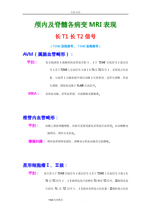
颅内及脊髓各病变MRI表现长T1长T2信号(T1WI呈低信号,T2WI呈高信号)AVM(属脑血管畸形):平扫:见毛线团状(或蜂窝状血管流空影),(于T1WI呈低信号(或无信号)且于T2WI上呈高信号)或(长T1长T2信号),无明显占位征象,与血管(大脑前或中或后动脉)关系密切,边界欠清晰,形态欠规则,团块状边缘于FLAIR呈高信号。
MRA:见供血动脉、异常血管团、引流静脉及静脉窦。
椎管内血管畸形:平扫:双侧上颈段脊髓增粗,内部可见排列紊乱异常流空血管团。
由双侧椎动脉颅内、颅外分支供血。
增强扫描:畸形血管团明显强化,清晰显示供血动脉及引流静脉。
星形细胞瘤Ⅰ、Ⅱ级:平扫:见片状(于T1WI呈低信号(或无信号)且于T2WI上呈高信号)或(长T1长T2信号),(Ⅰ级周边见片状稍长T1稍长T2信号、Ⅱ级周边见片状长T1长T2信号),(Ⅰ级未见明显占位征象;Ⅱ级轻度占位征象,周围组织见稍受压)。
诊断要点:1)肿瘤直接造成的信号强度及占位征象。
2)Ⅰ、Ⅱ级坏死囊变少,占位效应轻,强化程度低。
3)Ⅲ、Ⅳ级信号多不均匀,占位效应重,强化明显。
4)小脑星形细胞瘤,多位于小脑半球,囊肿有瘤,瘤中有囊,坏死囊变多见,占位效应重,强化明显。
(分级主要依据累及范围,形态,钙化、坏死程度,占位效应,主要根据DWI扩散程度)。
少突胶质细胞瘤:平扫:见片状异常信号灶,于T1WI呈低信号,T2WI上呈高信号,边界清晰,形态规则,周边见小片状长T1长T2信号,轻度占位效应(Ⅰ级)、占位征象重(Ⅱ级)。
增强扫描:明显强化。
诊断要点:1)多发于幕上半球。
2)钙化少,水肿重,囊变,出血,强化明显。
3)多见于成年人。
鉴别:星形、钙化性脑膜瘤、室管膜瘤、钙化性AVM、海绵状血管瘤、结核球。
室管膜肿瘤:第四脑室多见。
平扫:于第几…..脑室(侧脑室、或脑池、或脑实质)见斑片状异常信号灶,T1WI呈低信号(或等信)T2WI呈高信号(为主),内见多个片状长T1长T2信号区(囊变)。
MRI在脑动静脉畸形合并自发性出血诊断中的应用

i f e y s r e y a e r o f e y DS a d 1 p d a rc p te twh a y i a m r d b u g r ,7 e s we e c n i d b A, s m r n e i t ai n o h d t p c lMR1fn i g ie ty r c ie k ie r d o i d n s d r c l e e v d i n f a i— t e a y Re u t n e to a h r p . s ls Co v n i n MRIs o d t a h mal b o d s p l re e d c a s r i en a o d t e h ma o s we e f u d l h we h tt e s l l o - u p y a t r s a o re d a n v i r un h e tma r o n i n
r a e u d r n o v n i n h g n e we t c n e to a MR ma i g a in s u d r e t a d to a n a c d M R c n .Ofa lt e p te t,5 e e r o — l i g n .8 p te t n e w n d iin e h n e l sa s h a i n s a s we e c n l s
【 bt c】 bet e o i us h ai ty f R i a n e i n s f e ba a e oe os a o ao s C V s a— A s atQ j i s s te e b i g g n h a oio c er r r vnu l r t n (A M )s r c v T d c f i oM m i it d g s r l t s t i m fm i
大脑动静脉畸形诊断金标准

大脑动静脉畸形诊断金标准
大脑动静脉畸形(AVM)是一种罕见但严重的血管疾病,它会导致动脉和静脉之间的异常连接。
这种异常连接可能导致血管破裂和出血,进而引发中风或其他严重并发症。
因此,对于患有AVM的患者来说,早期的诊断和治疗至关重要。
为了确诊大脑动静脉畸形,医生通常会依靠一系列的临床表现和影像学检查。
在这里,我们将介绍一些诊断AVM的金标准。
1. 磁共振成像(MRI),MRI是诊断AVM的首选影像学检查。
通过MRI可以清晰地显示出大脑血管的结构,帮助医生确定血管异常的位置和大小。
2. 磁共振血管成像(MRA),MRA是一种特殊的MRI技术,可以更清晰地显示血管的情况,有助于确定AVM的血流情况和血管供应区域。
3. CT血管造影(CTA),CTA是一种通过计算机断层扫描来观察血管的影像学检查,可以提供关于AVM的血流情况和血管结构的详细信息。
4. 数字减影血管造影(DSA),DSA是一种介入性的影像学检查,通过在血管内注入造影剂来观察血管的情况,是诊断AVM的金标准之一。
除了影像学检查,医生还会根据患者的临床症状和体征来进行诊断。
一旦诊断出AVM,医生会根据患者的具体情况制定个性化的治疗方案,可能包括手术、介入治疗或放射治疗等。
总之,对于怀疑患有大脑动静脉畸形的患者来说,及早进行全面的临床评估和影像学检查是至关重要的。
只有通过精准的诊断,患者才能及时接受有效的治疗,最大限度地降低患者的风险。
脑血管畸形的MRI诊断_0
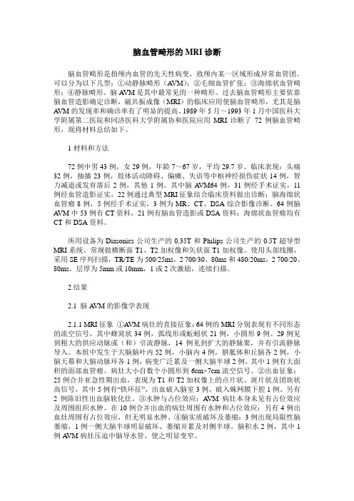
脑血管畸形的MRI诊断脑血管畸形是指颅内血管的先天性病变,致颅内某一区域形成异常血管团。
可以分为以下几型:①动静脉畸形(A VM);②毛细血管扩张;③海绵状血管畸形;④静脉畸形。
脑A VM是其中最常见的一种畸形。
过去脑血管畸形主要依靠脑血管造影确定诊断,磁共振成像(MRI)的临床应用使脑血管畸形,尤其是脑A VM的发现率和确诊率有了明显的提高。
1989年5月~1993年1月中国医科大学附属第二医院和同济医科大学附属协和医院应用MRI诊断了72例脑血管畸形,现将材料总结如下。
1材料和方法72例中男43例,女29例,年龄7~67岁,平均29.7岁。
临床表现:头痛32例,抽搐23例,肢体活动障碍、偏瘫、失语等中枢神经损伤症状14例,智力减退或发育落后2例,其他1例。
其中脑A VM64例,31例经手术证实,11例经血管造影证实,22例通过典型MRI征象结合临床资料做出诊断;脑海绵状血管瘤8例,5例经手术证实,3例为MR、CT、DSA综合影像诊断。
64例脑A VM中53例有CT资料,21例有脑血管造影或DSA资料;海绵状血管瘤均有CT和DSA资料。
所用设备为Diasonics公司生产的0.35T和Philips公司生产的0.5T超导型MRI系统。
常规做横断面T1、T2加权像和矢状面T1加权像。
使用头部线圈,采用SE序列扫描,TR/TE为500/25ms,2 700/30、80ms和480/20ms,2 700/20、80ms。
层厚为5mm或10mm,1或2次激励,连续扫描。
2结果2.1 脑A VM的影像学表现2.1.1 MRI征象①A VM病灶的直接征象:64例的MRI分别表现有不同形态的流空信号,其中蜂窝状34例,弧线形或蚯蚓状21例,小圆形9例。
29例见到粗大的供应动脉或(和)引流静脉,14例见到扩大的静脉窦,并有引流静脉导入。
本组中发生于大脑脑叶内52例,小脑内4例,胼胝体和丘脑各2例,小脑天幕和大脑动脉环各1例,病变广泛累及一侧大脑半球2例,其中1例有大面积的面部血管瘤。
MRI对脑血管疾病的诊断大有作用
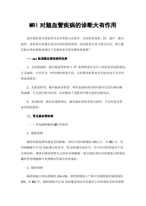
MRI对脑血管疾病的诊断大有作用现在能检查头颅血管有无异常的方法很多,比如血管造影、CT、超声、磁共振等。
各检查手段都有其存在的价值和优势,这些检查手段又相互补充。
那么磁共振头颅血管检查相比于其他检查手段有哪些优势呢?一、mri检查脑血管疾病的优势1、无电离辐射。
磁共振血管检查与CT血管检查以及介入科的血管造影相比,它无辐射,可以作为一种常规的筛查手段,且短期内的重复多次检查也不会对身体造成损害。
2、无需造影剂。
磁共振血管检查,利用血液的流动效应就可以进行脑动脉的成像,它无需注射对比剂,从而避免了造影剂可能引起的过敏反应。
3、无创检查。
跟血管造影相比,磁共振血管检查是无创的,不会给患者带来有创的损害。
二、常见脑血管疾病(一)常见脑肿瘤的MRI的检查1、脑胶质瘤脑胶质瘤是颅内最常见的肿瘤,其约占颅内肿瘤的46%左右。
在MRI中,星形细胞瘤平扫T1加权像呈低信号,T2加权像呈高信号,信号均匀程度取决于其内部结构。
增强扫描侵润性生长的星形细胞瘤一般无强化或仅有轻微斑点样强化,囊性星形细胞瘤可见肿瘤实性部分明显强化。
2、脑转移瘤脑转移瘤占颅内肿瘤的10%-15%,恶性肿瘤病人尸检中发现肿瘤有脑转移约30%。
在MRI中,脑转移瘤平扫T1加权像见颅内多发散在小环形或结节样等或稍低信号影,瘤周水肿可十分明显,病灶多位于皮质或皮质下;T2加权像病灶表现为不规则形高信号。
增强扫描可见轻到中度环形或结节样强化。
3、脑膜瘤脑膜瘤是最常见的非胶质性原发性颅内肿瘤,其发病率仅次于脑胶质瘤,占颅内肿瘤的15%—20%。
MRI上典型的脑膜瘤多呈质地均匀、边缘清楚的等T1和等T2信号,少数表现为稍长T1及稍短T2信号;T2加权像常见肿瘤边缘有一低信号边缘带,多为肿瘤纤维包膜或肿瘤血管所致。
增强扫描见中度或明显强化;邻近脑膜也有强化,称“脑膜尾征”。
脑胶质瘤、脑转移瘤、脑膜瘤是脑肿瘤中三种很常见的肿瘤,MRI检查显像对于早期发现。
脑静脉畸形有哪些症状?

脑静脉畸形有哪些症状?*导读:本文向您详细介绍脑静脉畸形症状,尤其是脑静脉畸形的早期症状,脑静脉畸形有什么表现?得了脑静脉畸形会怎样?以及脑静脉畸形有哪些并发病症,脑静脉畸形还会引起哪些疾病等方面内容。
……*脑静脉畸形常见症状:慢性头痛、昏迷、颅内出血、步态不稳*一、症状大多数病人临床上很少有症状或出血表现,经常为偶然发现脑内病灶,但后颅窝的脑静脉畸形常引起临床表现。
症状的发生依其部位而定,幕上病灶多有慢性头痛、癫痫、运动障碍或感觉障碍。
幕下病灶多表现为步态不稳或其他后颅窝占位症状,小脑病灶更容易出血。
脑静脉畸形发生的出血主要为脑内和脑室内出血。
主要临床表现有:1.癫痫是最常见的临床表现,主要为癫痫大发作。
2.局限性神经功能障碍表现为单侧肢体轻瘫,可伴有感觉障碍。
3.慢性头痛。
4.颅内出血一般认为脑静脉畸形出血率在15%~20%,幕下病灶比幕上病灶更易于出血。
病人突然剧烈头痛,昏迷或偏瘫。
*二、诊断根据临床表现及典型的静脉性血管畸形在血管造影的表现、CT扫描、MRI扫描的表现,一般可做出诊断。
*以上是对于脑静脉畸形的症状方面内容的相关叙述,下面再看下脑静脉畸形并发症,脑静脉畸形还会引起哪些疾病呢?*脑静脉畸形常见并发症:脑静脉畸形、动脉瘤*一、并发病症最常见伴发海绵状血管瘤。
文献报道海绵状血管瘤中20%~30%伴有静脉畸形。
组织学上区分二者的标准是病变血管间是否存在正常脑组织以及血管管腔的大小。
脑静脉畸形也可伴发其他血管性或非血管性病变,如肿瘤、脱髓鞘疾病、动脉瘤、AVM、硬膜动静脉瘘、烟雾病及头面眼的血管病变等。
静脉畸形常引流远离这些病灶的正常脑组织的回流血液,少数情况下也引流这些病灶本身。
*温馨提示:以上就是对于脑静脉畸形症状,脑静脉畸形并发症方面内容的介绍,更多疾病相关资料请关注疾病库,或者在站内搜索“脑静脉畸形”可以了解更多,希望可以帮助到您!。