MIQE-2009
沸石分子筛的性能特点

Fig. Stereoscan of zeolite X crystal
Fig. Stereoscan of zeolite X crystal about 50 m in size showing spinel-type contact twin and spheroids of zeolite P
•Байду номын сангаас溶液中旳反应:
25
① 骨架Si、Al可用Ga、P等取代→杂原子取代分子筛② 可调变表面酸性及其他活性中心旳强度和浓度,或者调变分子筛表面旳吸附性质,从亲水性到疏水性。 如:阳离子互换→酸性分子筛、碱性分子筛 a、取得酸性:Na型 → H型 例如:NaY → HY 互换剂:NH4NO3、也可直接用酸溶液进行互换。 b、取得较强碱性: Na型 → K、Rb、Cs型 互换剂:碱金属旳硝酸盐等可分解型盐类。 碱性强弱:NaY< KY< RbY< CsY、NaX< KX< RbX< CsX
• 水热转化
Table. Steam stability of zeolite XaCation Form % Exchange Structureb Adsorptionc K+ 77 - 60 % - 89 % Na+ 100 - 80 % - 84 % Ca2+ 84 - 60 % - 71 % Ce3+ 77 no change - 21 % a Loose powder (300 C, 8 hr in 100% steam) b Determined from loss in intensity of selected X-ray powder reflections c As determined from argon adsorption at -183 C and 700 torr
MIQE指南
Bulletin CN 5859
ԛິࠅݴǖ ںǖԛ࡛ۏ൶ኪؾୟ 113 ࡽ
ᆀྪዐ႐ 702 ࣆۉǖ010 - 8267 5748 دኈǖ010 - 6252 9800 ᆰՊǖ100086
Fleige S and Pfaffl MW (2006). RNA integrity and the effect on the real-time qRTPCR performance. Mol Aspects Med 27, 126-139.
Garson JA et al. (2009). Unreliable real-time PCR analysis of human endogenous retrovirus-w (HERV-W) RNA expression and DNA copy number in multiple sclerosis. AIDS Res Hum Retroviruses 25, 377-378; author reply 379-381.
Bio-Rad’s real-time thermal cyclers are licensed real-time thermal cyclers under Applera’s United States Patent 6,814,934 B1 for use in research, human in vitro diagnostics, and all other fields except veterinary diagnostics. This product is covered by one or more of the following U.S. patents or their foreign counterparts owned by Eppendorf AG: U.S. Patent Nos. 6,767,512 and 7,074,367.
pcr引物设计的基本理念 -回复

pcr引物设计的基本理念-回复PCR引物设计的基本理念PCR(聚合酶链反应)是一种常用的分子生物学技术,用于扩增DNA序列。
在PCR过程中,引物起着至关重要的作用,引物的设计质量直接影响PCR反应的效果。
因此,正确的PCR引物设计是成功进行PCR实验的基本前提。
本文将介绍PCR引物设计的基本理念,并分步回答如何设计合适的PCR 引物。
第一步:确认目标序列PCR实验通常是为了扩增某个特定的DNA序列,因此首先需要确定所需扩增的目标序列。
这个目标序列可以是基因片段、特定的DNA结构、启动子区域等。
在确认目标序列时,需要注意目标序列的长度和特异性,确保其能准确而特异地被引物扩增。
第二步:确定引物的长度引物通常由20到30个核苷酸组成,过短的引物可能不稳定,无法正确与模板DNA特异性结合,过长的引物则可能导致PCR产物过多的副产物。
因此,一般情况下引物长度约为18到24个核苷酸,适当选择引物长度有利于PCR的优化。
第三步:评估引物的理化性质引物的理化特性包括引物的GC含量、熔解温度(Tm)和自身引物结合能力。
高GC含量的引物更稳定,但也更难设计;Tm值表征了引物与模板DNA杂交的稳定性,通常理想的Tm值为50-65;自身引物结合能力的评估可以避免引物之间的非特异性结合。
多种软件可用于计算引物的理化性质,并为引物设计提供参考。
第四步:检查引物的特异性引物的特异性是PCR引物设计的关键点之一。
为了确保引物仅扩增目标序列,没有互补的序列存在于其他地方,可以使用引物设计软件进行特异性检查。
软件可以检查引物在基因组中的互补性和互补的副产物,以确保引物只扩增目标序列。
第五步:考虑引物间的配对引物在PCR反应中需要与目标序列的两端结合,因此两个引物(前向引物和反向引物)需要在目标序列上互补配对。
在设计引物时,需要确保两个引物的Tm值相近,以避免一个引物过早或过晚离开DNA链。
此外,还需要考虑引物之间的距离和可能的PCR产物长度,以确定引物的相对位置。
PCR程序两步法和三步法的区别是

PCR程序两步法和三步法的区别是引言聚合酶链反应(PCR)是一种在分子生物学领域广泛应用的技术,用于扩增DNA片段。
PCR可以使用两步法或三步法进行,这两种方法在程序设置和反应条件上有所区别。
本文将探讨PCR程序两步法和三步法之间的区别。
PCR的原理在介绍两步法和三步法之前,我们先来了解PCR的基本原理。
PCR的反应体系中包含待扩增的DNA模板,引物(primer),DNA聚合酶,反应缓冲液和dNTPs(脱氧核苷酸)。
通过一系列温度循环,PCR使DNA模板的目标区域反复复制,产生大量的目标DNA片段。
PCR程序的两步法两步法是最早采用的PCR程序方法之一,它包括三个主要步骤:变性、退火和延伸。
其中,变性步骤将高温(通常为94°C)用于使DNA模板两条链分离,即变性为两条单链。
接下来是退火步骤,将温度降低至引物能与目标DNA序列互补匹配的温度。
在这个温度下,引物与单链DNA发生互补结合。
最后是延伸步骤,将温度升高至DNA聚合酶的最适工作温度,使得DNA聚合酶能够从引物的3’端开始合成新的DNA链。
这样,PCR程序的一个循环就完成了。
两步法的优点是操作简便、反应时间相对较短,适用于一些简单的PCR反应。
然而,由于变性和延伸是在同一温度下进行,引物可能在变性温度下结合到非特异性的DNA序列上。
这会导致非特异性产物的形成,并可能影响PCR的结果。
PCR程序的三步法为了提高PCR的特异性,研究人员开发了三步法。
三步法在两步法的基础上增加了一个退火步骤,具体分为变性、退火和延伸三个步骤。
与两步法不同的是,退火步骤的温度通常要高于引物与目标DNA互补的温度。
这样可以使引物只与特异性的DNA序列结合,进一步提高PCR的特异性。
三步法相对于两步法来说,能够减少非特异性产物的产生,提高PCR的准确性和特异性。
它在一些复杂的PCR反应中表现良好,特别是在需要区分高度相似的DNA序列时。
结论PCR程序的两步法和三步法是在PCR技术发展过程中出现的两种主要方法。
9家国内外数字PCR技术领军企业
9 家国内外数字PCR技术领军企业自从1985年美国科学家Kary Mullis发明PCR方法以来,该方法已经成为生命科学研究领域中最常规的实验方法之一。
PCR是一种用于放大扩增特定的DNA 片段的分子生物学技术,它可看作是生物体外的特殊DNA复制,PCR的最大特点,是能将微量的DNA 大幅增加。
利用这一特性,可以将样本中微量DNA进行大量扩增(如血液,唾液,病原体等) ,用于下游多种应用,如克隆表达、芯片杂交、突变检测、测序等等。
最传统的PCR采用琼脂糖电泳的方式对PCR产物进行分析,但存在着操作繁琐、只适用于定性研究、交叉污染风险大等不足。
为了避免上述传统PCR的缺陷,并对基因的表达水平进行定量分析,1992 年诞生了荧光定量实时PCR。
定量PCR (Quantification PCR)在PCR扩增的同时对反应体系中的荧光信号进行实时收集,通过三个参数间(荧光信号-Cq 值-靶基因的起始浓度)的关系,最终确定靶基因的拷贝数或基因的表达水平。
由于定量PCR的分析最终结果依赖于Cq值,2009年The MIQE Guides(Clinical Chemistry 55 :4 611-622)的发表明确了定量实验整个流程标准化的重要性,否则就会出现实验结果无法重复,甚至结果互相矛盾的状况。
更重要的该文献还给出了如何规范定量PCR流程的checklist,其核心目的就是在规范的实验流程、统一的设计思路下,得到的结果才具有可重复性,也具有可比性。
但依赖于Cq 值仍然是目前定量PCR最大的技术瓶颈,在这个意义上所谓的“定量”也只是相对的。
而且在低拷贝靶分子、模板浓度差异细微的条件下,其检测的灵敏度、精确度都受到了限制。
1999年霍普金斯大学的Bert Vogelstein教授以Digital PCR为题发表的文章被视为最早的采用数字PCR概念进行研究的文献。
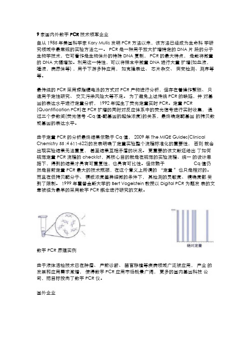
数字PCR原理实例由于液体活检技术已在肿瘤、产前诊断、器官移植等疾病领域广泛被应用,产业的发展和应用需求激增,使得数字PCR应用市场钱景广阔,更多的国内基因科技公司,把目标投向了数字PCR仪。
反应蛋白测定范围

反应蛋白测定范围1. 引言反应蛋白是一类广泛存在于生物体内的蛋白质,其在细胞内外的生理和病理过程中起着重要的调节作用。
反应蛋白测定范围是指通过实验手段对反应蛋白进行测量的范围,可以帮助研究人员了解反应蛋白在不同生理状态下的变化情况,从而揭示其在疾病发生和发展中的作用。
2. 反应蛋白测定方法目前常用的反应蛋白测定方法主要包括免疫学方法和分子生物学方法。
2.1 免疫学方法免疫学方法是通过特异性抗体与目标蛋白结合来进行测定的。
常见的免疫学方法有酶联免疫吸附试验(ELISA)、免疫印迹(Western blotting)等。
2.1.1 酶联免疫吸附试验(ELISA)ELISA是一种基于酶标记技术原理的免疫分析方法,可用于定量或半定量测定反应蛋白的含量。
ELISA具有灵敏度高、特异性好、操作简便等优点,广泛应用于临床诊断和科学研究中。
2.1.2 免疫印迹(Western blotting)免疫印迹是一种通过将目标蛋白从复杂的混合物中分离出来,并通过特异性抗体进行检测的方法。
免疫印迹可以用于检测反应蛋白的表达水平和分子量,并可进一步定量分析。
2.2 分子生物学方法分子生物学方法是通过对反应蛋白基因进行检测和分析来间接推断其表达水平和功能。
2.2.1 聚合酶链式反应(PCR)PCR是一种可以扩增目标基因片段的技术,通过检测PCR产物的数量来推断目标基因在样本中的表达水平。
PCR方法具有高灵敏度、高特异性等优点,常用于研究反应蛋白基因在不同组织或细胞中的表达差异。
2.2.2 实时荧光定量PCR(qPCR)qPCR是一种结合了PCR技术和荧光探针技术的方法,可以实时监测PCR反应过程中的荧光信号强度,并根据荧光信号强度推断目标基因的表达水平。
qPCR具有高灵敏度、高准确性等优点,广泛应用于反应蛋白的定量分析。
3. 反应蛋白测定范围的意义反应蛋白作为一类重要的调节蛋白,在生理和病理过程中起着关键作用。
测定其范围可以帮助我们了解以下几个方面:3.1 生理状态下的变化通过测定不同生理状态下反应蛋白的表达水平,可以揭示其在正常生理过程中的变化规律。
MIQE检查表说明书

Table 3. The Minimum Information for Publication of QuantitativeReal-Time PCR Experiments (MIQE) checklist for reviewers, and editors (Bustin et al. 2009)MIQE checklist (Bustin et al. 2009)aI t e m t o c h e c k I m p o r t a n c e I t e m t o c h e c k I m p o r t a n c e Experimental design qPCR oligonucleotidesDefinition of experimental and control groups EPrimer sequencesENumber within each groupE RTPrimerDB identificationnumberD NAAssay carried out by the core or investigator’s laboratory? D NAProbe sequencesD d NAAcknowledgment of author’s contributions DLocation and identity of anymodificationsE NASample Manufacturer of oligonucleotidesDDescription E Purification method D ∅Volume/mass of sampleprocessedD qPCR protocolMicrodissection or macrodissection E NAComplete reactionconditionsE SMProcessing procedureE Reaction volume andamount of cDNA/DNAE SMIf frozen, how and how quickly? EPrimer, (probe), Mg2+, anddNTP concentrationsE SMIf fixed, with what and how quickly? E NAPolymerase identity andconcentrationE SMSample storage conditions andduration (especially for FFPE b samples) E SMBuffer/kit identity andmanufacturer E SMNucleic acid extraction Exact chemical composition D ∅of the bufferProcedure and/or instrumentation EAdditives (SYBR Green I,DMSO, and so forth)E SMName of kit and details of any modifications E Manufacturer ofplates/tubes and catalognumberD SMSource of additional reagents used D NAComplete thermocyclingparametersE SMDetails of DNase or RNase treatment EReaction setup(manual/robotic)D SMContamination assessment (DNA or RNA) EManufacturer of qPCRinstrumentE SMNucleic acid quantification E qPCR validationInstrument and methodE Evidence of optimisation(from gradients)D OKPurity (A260/A280)D SM Specificity (gel, sequence,melt, or digest)E SMYieldD SM For SYBR Green I, Cq ofthe NTCERNA integrity: method/instrument ECalibration curves withslope and y interceptE SM*RIN/RQI or Cq of 3’ and 5’ transcripts E SMPCR efficiency calculatedfrom slopeE SMElectrophoresis tracesD ∅CIs for PCR efficiency orSED ∅Inhibition testing (Cq dilutions, spike, or other) E ∅r2 of calibration curveE SM*Reverse transcription Linear dynamic range E ∅Complete reaction conditions E SM Cq variation at LOD E NAAmount of RNA and reaction volume E SMCIs throughout rangeD NAPriming oligonucleotide (if using GSP) and concentration E SMEvidence for LODE NAReverse transcriptase and concentration E SMIf multiplex, efficiency andLOD of each assayE NATemperature and time E SM Data analysisManufacturer of reagents and catalog numbers D SMqPCR analysis program(source, version)E SMCqs with and without reverse transcription D c OKMethod of Cq determinationE SMStorage conditions of cDNAD SM Outlier identification and dispositionE SM*qPCR target information Results for NTCs EGene symbolE Justification of number andchoice of reference genesESequence accession numberE Description of normalization methodELocation of ampliconD ∅Number and concordance of biological replicatesDAmplicon lengthE Number and stage (reverse transcription or qPCR) of technical replicatesEIn silico specificity screen (BLAST, and so on) ERepeatability (intraassayvariation)E ∅Pseudogenes,retropseudogenes, or other homologs? D NAReproducibility (interassayvariation, CV) D ∅Sequence alignment D ∅Power analysis D ∅Secondary structure analysis of amplicon D ∅Statistical methods forresults significanceELocation of each primer by exon or intron (if applicable) E NASoftware (source, version)EWhat splice variants are targeted? E NACq or raw data submissionwith RDMLD SM*a All essential information (E) must be submitted with the manuscript. Desirable information (D) should be submitted if available. If primers are from RTPrimerDB, information on qPCR target, oligonucleotides, protocols, and validation is available from that source.b FFPE, formalin-fixed, paraffin-embedded; RIN, RNA integrity number; RQI, RNA quality indicator; GSP, gene-specific priming; dNTP, deoxynucleoside triphosphate.c Assessing the absence of DNA with a no-reverse transcription assay is essential when first extracting RNA. Once the sample has been validated as DNA free, inclusion of a no-reverse transcription control is desirable but no longer essential.d Disclosure of the probe sequence is highly desirable and strongly encouraged; however, because not all vendors of commercial predesigned assays provide this information, it cannot be an essential requirement. Use of such assays is discouraged.Information is included in the text.OK Test was performed but evidence is missing.∅ Information is not available.NA Not applicable.SM Information is provided in Supplementary Materials (* if requested).SampleSample storage conditions and duration: 5 yrs at -80 °CNucleic acid extractionPurity (A260/A280)Colony # 1 17 24 4 11 15 23 16 251.96 1.97 1.772.03 2.07 1.6 1.99 1.91na24/01/0124/01/02 2.01 1.98 1.95 2.02 2.01 1.79 1.73 1.97naYield (ng/µL) in 20 µLColony # 1 17 24 4 11 15 23 16 2523.8 83.970.6 102.6150.573.5261.9150.7na24/01/0124/01/02 123.992 130.8144.1118.819.7152.9140.1naRIN/RQI or Cq of 3’ and 5’ transcriptsColony # 1 17 24 4 11 15 23 16 258.9 8.7 8.3 7.8 8.9 6.1 7.2 na na 24/01/0124/01/02 9.1 9.5 8.3 8.2 6.9 6.2 9.3 9.3 na Reverse transcriptionComplete reaction conditions: Reactions were prepared on iceAmount of RNA and reaction volume: Amount of RNA ranged from 150-560 ng in 20 µL reactionPriming oligonucleotide (if using GSP) and concentration: Oligo(dT)20 (2.5 µM), random hexamers (2.5 ng/µL)Reverse transcriptase and concentration: SuperScript TM III RT (400 U/µL) Temperature and time: 10 min @ 25 °C + 30 min @ 50 °C + 5 min @ 85 °C + ice Manufacturer of reagents and catalog numbers: Invitrogen (Cat. No. 11765) Storage conditions of cDNA: -20 °CqPCR protocolComplete reaction conditions: Reactions were prepared at room temperature Reaction volume and amount of cDNA/DNA: 10 µL SYBR GreenER TM mix, 2 µL primers, 2 µL 5x diluted cDNA, 6 µL DEPC waterPrimer, (probe), Mg2+, and dNTP concentrations: Primers at 4 µM, ∅Polymerase identity and concentration: Platinum®Taq DNA Polymerase, ∅Buffer/kit identity and manufacturer: SYBR®GreenER™ Two-Step qRT-PCR Kit Universal (Invitrogen)Additives (SYBR Green I, DMSO, and so forth): SYBR® GreenER™Manufacturer of plates/tubes and catalog number: Corbett Research 0.2 mL PCR Tubes, Cat. no. 3001-001Complete thermocycling parameters: 2 min @ 50 °C + 10 min @ 95 °C + (15 sec @ 95 °C + 60 sec @ 60 °C) x40 + melt 60-95 °CReaction setup (manual/robotic): Using the Corbett CAS-1200 robotManufacturer of qPCR instrument: Corbett Research, Corbett Rotor-Gene 6000qPCR validationSpecificity (gel, sequence, melt, or digest): Melt curves from 60-95 °C on CorbettRotor-Gene 6000PCR efficiency calculated from slopeGenes Ctg1913 Am Cat Am Ferri Am CD151 Am Trib Am SW Am CTL Am Ch Ctg3235 GAPDH rpL13 rpL9 rpS7 Efficiency 1.98 1.91 1.98 1.95 1.98 1.95 1.95 1.93 1.74 1.87 1.64 1.88 1.93 Data analysisqPCR analysis program (source, version): Corbett Rotor-Gene 6000 softwareMethod of Cq determination: Manual positioning above background signal。
反转录pcr程序

反转录pcr程序反转录聚合酶链反应(Reverse Transcription Polymerase Chain Reaction,简称RT-PCR)是一种用于合成cDNA的分子生物学技术。
它结合了反转录和聚合酶链反应(PCR),可从RNA模板合成对应的cDNA。
该技术在分子生物学中被广泛应用,特别是在基因表达分析、病毒检测和疾病诊断等方面。
以下是关于反转录PCR的相关内容。
一、反转录PCR原理反转录PCR的主要原理是利用病毒逆转录酶(Reverse Transcriptase)将RNA模板反转录合成对应的cDNA。
逆转录酶可合成一条cDNA链,作为PCR反应的起始物。
接下来,使用DNA聚合酶进行PCR扩增,即将cDNA扩增为足够数量的DNA分子。
最终通过PCR产物的检测和分析,可以获得RNA模板的相关信息。
二、反转录PCR技术步骤1. 提取RNA:从样品中提取所需的RNA,如细胞、组织、血液等。
2. 反转录反应:将RNA通过逆转录酶转录为cDNA,需要使用引物(primers)和反转录酶。
3. 酶反应停止:用高温杀死逆转录酶。
4. PCR扩增:使用DNA聚合酶进行PCR反应,通过引物特异性扩增cDNA。
5. 检测分析:通过电泳、荧光定量、实时荧光PCR或基因测序等技术对PCR产物进行分析,获得所需信息。
三、应用领域反转录PCR被广泛应用于各个领域,以下是一些常见的应用:1. 基因表达分析:通过检测RNA的表达,可以了解特定基因的转录水平,揭示基因在生理或病理过程中的功能。
2. 病毒检测:可通过反转录PCR检测病毒RNA,如HIV、乙肝病毒等,并进行病毒分类和定量分析。
3. 诊断疾病:反转录PCR在疾病的早期诊断中有重要应用,例如检测癌症标志物、感染病原体等。
4. 揭示基因调控机制:通过分析RNA转录的变化,可以了解特定基因的转录起始位点、剪接异构体和可变剪接等。
四、RT-PCR与PCR的区别RT-PCR是从RNA模板合成cDNA,然后进行PCR扩增。
- 1、下载文档前请自行甄别文档内容的完整性,平台不提供额外的编辑、内容补充、找答案等附加服务。
- 2、"仅部分预览"的文档,不可在线预览部分如存在完整性等问题,可反馈申请退款(可完整预览的文档不适用该条件!)。
- 3、如文档侵犯您的权益,请联系客服反馈,我们会尽快为您处理(人工客服工作时间:9:00-18:30)。
The MIQE Guidelines:M inimum I nformation for Publication of Q uantitative Real-Time PCR E xperimentsStephen A.Bustin,1*Vladimir Benes,2Jeremy A.Garson,3,4Jan Hellemans,5Jim Huggett,6 Mikael Kubista,7,8Reinhold Mueller,9Tania Nolan,10Michael W.Pfaffl,11Gregory L.Shipley,12Jo Vandesompele,5and Carl T.Wittwer13,14BACKGROUND:Currently,a lack of consensus exists on how best to perform and interpret quantitative real-time PCR(qPCR)experiments.The problem is exac-erbated by a lack of sufficient experimental detail in many publications,which impedes a reader’s ability to evaluate critically the quality of the results presented or to repeat the experiments.CONTENT:The Minimum Information for Publication of Quantitative Real-Time PCR Experiments(MIQE) guidelines target the reliability of results to help ensure the integrity of the scientific literature,promote con-sistency between laboratories,and increase experimen-tal transparency.MIQE is a set of guidelines that de-scribe the minimum information necessary for evaluating qPCR experiments.Included is a checklist to accompany the initial submission of a manuscript to the publisher.By providing all relevant experimental conditions and assay characteristics,reviewers can as-sess the validity of the protocols used.Full disclosure of all reagents,sequences,and analysis methods is neces-sary to enable other investigators to reproduce results. MIQE details should be published either in abbreviated form or as an online supplement.SUMMARY:Following these guidelines will encourage better experimental practice,allowing more reliable and unequivocal interpretation of qPCR results.©2009American Association for Clinical ChemistryThe fluorescence-based quantitative real-time PCR (qPCR)15(1–3),with its capacity to detect and mea-sure minute amounts of nucleic acids in a wide range of samples from numerous sources,is the enabling tech-nology par excellence of molecular diagnostics,life sci-ences,agriculture,and medicine(4,5).Its conceptual and practical simplicity,together with its combination of speed,sensitivity,and specificity in a homogeneous assay,have made it the touchstone for nucleic acid quantification.In addition to its use as a research tool, many diagnostic applications have been developed,in-cluding microbial quantification,gene dosage determi-nation,identification of transgenes in genetically mod-ified foods,risk assessment of cancer recurrence,and applications for forensic use(6–11).This popularity is reflected in the prodigious number of publications reporting qPCR data,which invariably use diverse reagents,protocols,analysis methods,and reporting formats.This remarkable lack of consensus on how best to perform qPCR ex-periments has the adverse consequence of perpetu-ating a string of serious shortcomings that encumber its status as an independent yardstick(12).Techni-cal deficiencies that affect assay performance include the following:(a)inadequate sample storage,prep-aration,and nucleic acid quality,yielding highly variable results;(b)poor choice of reverse-transcription primers and primers and probes for the PCR,leading to inefficient and less-than-robust assay performance;1Centre for Academic Surgery,Institute of Cell and Molecular Science,Barts and the London School of Medicine and Dentistry,London,UK;2Genomics Core Facility,EMBL Heidelberg,Heidelberg,Germany;3Centre for Virology,Depart-ment of Infection,University College London,London,UK;4Department of Virology,UCL Hospitals NHS Foundation Trust,London,UK;5Center for Medical Genetics,Ghent University Hospital,Ghent,Belgium;6Centre for Infectious Diseases,University College London,London,UK;7TATAA Biocenter,Go¨teborg, Sweden;8Institute of Biotechnology AS CR,Prague,Czech Republic;9Seque-nom,San Diego,California,USA;10Sigma–Aldrich,Haverhill,UK;11Physiology Weihenstephan,Technical University Munich,Freising,Germany;12Quantita-tive Genomics Core Laboratory,Department of Integrative Biology and Phar-macology,University of Texas Health Science Center,Houston,Texas,USA; 13Department of Pathology,University of Utah,Salt Lake City,Utah,USA; 14ARUP Institute for Clinical and Experimental Pathology,Salt Lake City,Utah,USA.*Address correspondence to this author at:3rd Floor Alexandra Wing,The Royal London Hospital,London E11BB,UK.Faxϩ44-(0)20-73777283;e-mail s.a.bustin@.Received October20,2008;accepted January27,2009.Previously published online at DOI:10.1373/clinchem.2008.11279715Nonstandard abbreviations:qPCR,quantitative real-time PCR;MIQE,Minimum Information for Publication of Quantitative Real-Time PCR Experiments;RT-qPCR,reverse transcription–qPCR;FRET,fluorescence resonance energy trans-fer;C q,quantification cycle,previously known as the threshold cycle(C t), crossing point(C p),or take-off point(TOP);RDML,Real-Time PCR Data Markup Language;LOD,limit of detection;NTC,no-template control.Clinical Chemistry55:4611–622(2009)Special Report611and(c)inappropriate data and statistical analyses,gen-erating results that can be highly misleading.Conse-quently,there is the real danger of the scientific litera-ture being corrupted with a multitude of publications reporting inadequate and conflicting results(13).The publication(14)and retraction(15)of a Science “Breakthrough of the Year2005”report provides a disquieting warning.The problem is exacerbated by the lack of information that characterizes most reports of studies that have used this technology,with many publications not providing sufficient experimental detail to permit the reader to critically evaluate the quality of the results presented or to repeat the experi-ments.Specifically,information about sample acquisi-tion and handling,RNA quality and integrity,reverse-transcription details,PCR efficiencies,and analysis parameters are frequently omitted,whereas sample normalization is habitually carried out against single reference genes without adequate justification.The aim of this document is to provide authors, reviewers,and editors specifications for the minimum information,set out in Table1,that must be reported for a qPCR experiment to ensure its relevance,accu-racy,correct interpretation,and repeatability.MIQE (Minimum Information for Publication of Quantita-tive Real-Time PCR Experiments,pronounced mykee) is modeled on similar guidelines drawn up for DNA microarray analysis(16),proteomics experiments (17),genome sequence specification(18),and those under discussion for RNA interference work(19,20) and metabolomics(21),all of which are initiatives co-ordinated under the umbrella of MIBBI(Minimum Information for Biological and Biomedical Investiga-tions,)(22).Compulsory inclu-sion of a common reporting language to allow data sharing is not proposed,although it is envisaged that a future update of these guidelines could include such a recommendation.Rather,these guidelines target the reliability of results to help ensure the integrity of the scientific literature,promote consistency between lab-oratories,and increase experimental transparency. They should be read in conjunction with recent publi-cations that deal in depth with the issue of qPCR stan-dardization(23–26).1.NomenclatureA few terms require standardization to ensure clarification:1.1We propose that the abbreviation qPCR be usedfor quantitative real-time PCR and that RT-qPCR be used for reverse transcription–qPCR.Applying the abbreviation RT-PCR to qPCR causes confu-sion and is inconsistent with its use for conven-tional(legacy)reverse transcription–PCR.1.2Genes used for normalization should be referred toas reference genes,not as housekeeping genes.1.3TaqMan probes should be referred to as hydrolysisprobes.1.4The term FRET probe(fluorescence resonance en-ergy transfer probe)refers to a generic mechanism in which emission/quenching relies on the interac-tion between the electron-excitation states of2flu-orescent dye molecules.LightCycler-type probes should be referred to as dual hybridization probes.1.5The Oxford English Dictionary lists only quantifica-tion,not quantitation;therefore,the former is the proper word.1.6The nomenclature describing the fractional PCRcycle used for quantification is inconsistent,with threshold cycle(C t),crossing point(C p),and take-off point(TOP)currently used in the literature.These terms all refer to the same value from the real-time instrument and were coined by competing manu-facturers of real-time instruments for reasons of product differentiation,not scientific accuracy or clarity.We propose the use of quantification cycle(C q),according to the RDML(Real-Time PCRData Markup Language)data standard(http:// )(27).2.Conceptual ConsiderationsTo explain and justify the guidelines,we find it useful to review a number of key issues surrounding qPCR experiments:2.1Analytical sensitivity refers to the minimum num-ber of copies in a sample that can be measured accurately with an assay,whereas clinical sensitivity is the percentage of individuals with a given disor-der whom the assay identifies as positive for that condition.Typically,sensitivity is expressed as the limit of detection(LOD),which is the concentra-tion that can be detected with reasonable certainty (95%probability is commonly used)with a given analytical procedure.The most sensitive LOD the-oretically possible is3copies per PCR(28),assum-ing a Poisson distribution,a95%chance of includ-ing at least1copy in the PCR,and single-copy detection.Experimental procedures typically in-clude sample-processing steps(i.e.,extraction) and,when required,reverse transcription.If the volume changes and the efficiencies of these steps are accounted for,the most sensitive LOD theoret-ically possible can be expressed in units relevant to the experiment,such as copies per nanogram of tissue.Experimental results less than the theoreti-Special Report612Clinical Chemistry55:4(2009)MIQE Guidelines for qPCR Special ReportClinical Chemistry55:4(2009)613cally possible LOD should never be reported.It also follows that results of“0”are meaningless and misleading.LOD estimates in qPCR analyses are complicated by the logarithmic nature of C q,be-cause C q is undefined when the template concen-tration is zero.Appropriate determination and modeling of the LOD in the qPCR is the focus of continued research(26).2.2Analytical specificity refers to the qPCR assay de-tecting the appropriate target sequence rather than other,nonspecific targets also present in a sample.Diagnostic specificity is the percentage of individu-als without a given condition whom the assay iden-tifies as negative for that condition.2.3Accuracy refers to the difference between experi-mentally measured and actual concentrations, presented as fold changes or copy number estimates.2.4Repeatability(short-term precision or intraassayvariance)refers to the precision and robustness of the assay with the same samples repeatedly ana-lyzed in the same assay.It may be expressed as the SD for the C q variance.Alternatively,the SD or the CV for copy number or concentration variance may be used.CVs should not be used with C q s, however(29).2.5Reproducibility(long-term precision or interas-say variance)refers to the variation in results between runs or between different laboratories and is typically expressed as the SD or CV of copy numbers or concentrations.C q values gen-erated from different runs are subject to inher-ent interrun variation(30);hence,reporting in-terrun C q variation is not appropriate.Publications describing mRNA concentrations for target genes should make it clear precisely what the targets are.The transcripts of most human genes and many genes in other multicellular organisms are alter-natively spliced(31,32),and these splicing variants specify alternative protein isoforms,with variation in splicing patterns reported in different tissues or at dif-ferent developmental stages.Consequently,single exon–based RT-qPCR assays may detect a number of splice variants,whereas intron-spanning primers may be more selective but may miss some splice variants altogether.Most recently,autosomal nonimprinted genes that display allelic imbalance in their expression have been described(33).Taken together,these find-ings imply that use of an RT-qPCR assay that simply targets one or at most2exons of an mRNA is no longer sufficient to describe the expression level of a particular gene.Consequently,sequence information for primers must be provided together with an assessment of their specificity with respect to known splice variants and single-nucleotide polymorphism positions docu-mented in transcript and single-nucleotide polymor-phism databases.For primer sets selected from the RTprimerDB database(34,35),this is easily done by consulting the RTprimerDB Web site(http://www. ),which contains all the relevant infor-mation.For commercial assays,lot information and the providers’experimental validation criteria are re-quired.The reporting of results for nonvalidated com-mercial assays and assays that have been validated only in silico are strongly discouraged.It must be remembered that detection of the pres-ence of an mRNA provides no information on whether that mRNA will be translated into a protein or,indeed, whether a functional protein is translated at all.Immunohistochemistry,western blotting,or other protein-quantification methods are not always able to corroborate quantitative cellular mRNA data. It is now well established that there is frequently a lack of concordance between mRNA-and protein-concentration data(36),which is particularly true for mRNAs that specify proteins that are part of multi-function protein complexes(37).Finally,it has be-come clear that knowledge of the presence and func-tion of specific microRNAs is as important to understanding gene expression as being able to quan-tify the mRNA species(38).It is also necessary to be aware that most quantita-tive RNA data are not absolute,but relative.Thus,the reference genes or materials used for standardization are critical,and any assessment of the validity of an RT-qPCR experiment must also consider the appropri-ateness of the relative-quantification reference.There-fore,the development of universal reference DNA and RNA calibration materials,although very helpful (39,40),will not be a universal panacea(41,42).Much of the variance in reported expression val-ues produced in RT-qPCR experiments is not simply due to variation in experimental protocols but is caused by corrections applied by various data-processing algorithms,each of which makes its own assumptions about the data.Consequently,although qPCR has frequently been proclaimed a touchstone or a gold standard,in practice this“standard”is a variable one,and the reporting of results requires considerable sophistication of analysis and interpretation(43). 3.Research vs Diagnostic Applications Applications of qPCR technology can be broadly di-vided into research and diagnostic applications.Re-search applications usually analyze a wide range of targets with a fairly low throughput and many dif-ferent sample types.The main parameters that need to be addressed relate to assay analytical sensitivitySpecial Report614Clinical Chemistry55:4(2009)and specificity,which in this context refer to how many target copies the assay can detect and whether the no-template controls(NTCs)are reliably nega-tive,respectively.In contrast,diagnostic applications usually ana-lyze a limited number of targets,but require high-throughput protocols that are targeted at only a few sample types.Although all of the considerations that apply to research applications also apply to diagnostic assays,clinical-diagnostic assays have a number of ad-ditional requirements that need to be considered. These requirements include information on analytical sensitivity and specificity that in this context refers to how often the assay returns a positive result when a target is present and how often it is negative in the absence of the target.Furthermore,the accuracy and precision within and between laboratories is often monitored by external QC programs.Additional clin-ical laboratory requirements include criteria for gener-ating reportable results,whether repeated measure-ments are made on samples,data on the resolution of false-positive/false-negative data,and the similarity of results from multiple laboratories that use the same and different technologies.Thus far,only a couple of interlaboratory comparisons have been performed, and both of these studies emphasized the need for stan-dardization of qPCR diagnostic assays(44,45).An-other interlaboratory exercise is planned within the Eu-ropean Framework7project:SPIDIA(Standardisation and Improvement of Generic Pre-analytical Tools and Procedures for In-Vitro Diagnostics;http://www. spidia.eu).4.Sample Acquisition,Handling,and Preparation Sample acquisition constitutes the first potential source of experimental variability,especially for exper-iments targeting RNA,because mRNA profiles are eas-ily perturbed by sample-collection and-processing methods.There is some suggestion that fresh tissue can be stored on ice without major effects on RNA quality and concentration(46),but although this supposition may be true for some mRNAs and tissues,it may not be universally applicable.Hence,it is better to be cautious. Consequently,it is important to report in detail where the tissue sample was obtained and whether it was pro-cessed immediately.If the sample was not processed immediately,it is necessary to report how it was pre-served and how long and under what conditions it was stored.A brief description of the sample is also essential. For example,microscopical examination of a tumor biopsy will reveal what percentage of the biopsy is made up of tumor cells,and this information should be reported.Nucleic acid extraction is a second critical step. Extraction efficiency depends on adequate homogeni-zation,the type of sample(e.g.,in situ tissue vs log-phase cultured cells),target density,physiological sta-tus(e.g.,healthy,cancerous,or necrotic),genetic complexity,and the amount of biomass processed. Therefore,it necessary that details of the nucleic acid–extraction method be provided and that the methods used for measuring nucleic acid concentration and as-sessing its quality be described.Such details are partic-ularly crucial for RNA extracted from fresh frozen laser-microdissected biopsy samples,because varia-tions in tissue-preparation procedures have a substan-tial effect on both RNA yield and quality(47).5.QC of Nucleic Acids5.1.RNA SAMPLESQuantification of RNA in the extracted samples is im-portant,because it is advisable that approximately the same amounts of RNA be used when comparing differ-ent samples.There are several quantification proce-dures in common use,however,including spectropho-tometry(NanoDrop;Thermo Scientific),microfluidic analysis(Agilent Technologies’Bioanalyzer,Bio-Rad Laboratories’Experion),capillary gel electrophoresis (Qiagen’s QIAxcel),or fluorescent dye detection(Am-bion/Applied Biosystems’RiboGreen).The methods produce different results,making it unwise to try to compare data obtained with the different methods (48).The preferred method for quantifying RNA uses fluorescent RNA-binding dyes(e.g.,RiboGreen), which are best for detecting low target concentrations. In any case,it is advisable to measure all samples with a single method only and to report this information.It is also important to test for and report the extent of genomic-DNA contamination and to record the threshold cutoff criteria for the amounts of such con-tamination that are tolerable.It is essential to report whether the RNA sample has been treated with DNase (including the type of DNase used and the reaction conditions)and to report the results from a compari-son of C q s obtained with positive and no–reverse tran-scription controls for each nucleic acid target.It is also essential to document the quality assess-ment of RNA templates.The only situation in which this requirement does not apply is when the quantity of total RNA extracted is too low to permit quality assess-ment.This situation arises when RNA is extracted from single cells,plasma,other cell-free body fluids,some laser-captured samples,or clarified tissue culture me-dium.It also applies in cases in which extraction and RT-qPCR steps are performed as a continuous,single-tube experiment.Key information to report includes data on RNA quantity,integrity,and the absence ofMIQE Guidelines for qPCR Special ReportClinical Chemistry55:4(2009)615reverse transcription or PCR inhibitors.It is worth re-membering that RNA degrades markedly in vivo,ow-ing to the natural regulation of mRNAs in response to environmental stimuli(49).This source of RNA deg-radation is beyond the control of the researcher;one of its manifestations is that even high-quality RNA sam-ples can show differential degradation of individual mRNAs.The A260/A280ratio must be measured in a buffer at neutral pH,but such measurement is not sufficient if the nucleic acid is to be used for quantitative analysis, especially when the aim is to measure minor differ-ences(Ͻ10-fold)in cellular mRNA concentrations. The absorbance ratio does provide an indication of RNA purity,because the presence of DNA or residual phenol alters the ratio.Instead,one should provide gel electrophoresis evidence at the least or,better yet,re-sults from a microfluidics-based rRNA analysis(50)or a reference gene/target gene3Ј:5Јintegrity assay(51). The advantage of the use of a Bioanalyzer/Experion system to calculate an RNA integrity number or an RNA quality indicator number is that these measures provide quantitative information about the general state of the RNA sample.It is important to bear in mind,however,that these numbers relate to rRNA quality and cannot be expected to be an absolute mea-sure of e of a3Ј:5Јassay requires that the PCR efficiencies of both assays be virtually identical (51)and not be subject to differential inhibition.This assay also necessitates the establishment of a threshold criterion that delineates the RNA quality sufficient to yield reliable results.Ideally,the assay should target a panel of“integrity reference genes,”probably without introns,with a3Ј:5Јthreshold ratio of approximately 0.2–5.Clearly,further work is required to generate a universally applicable,cost-effective,and simple pro-tocol for assessing RNA integrity.Inhibition of reverse-transcription activity or PCR should be checked by dilution of the sample(prefera-bly)or use of a universal inhibition assay such as SPUD (52,53).If the RNA sample is shown to be partially degraded,it is essential that this information be re-ported,because the assay’s sensitivity for detecting a low-level transcript may be reduced and relative differ-ences in the degradation of transcripts may produce incorrect target ratios.5.2.DNA SAMPLESIn general,degradation is much less of an issue with DNA;however,it is important to be able to assess the extent of DNA degradation for forensic applications, i.e.,in cases in which harsh environmental conditions at scenes of crimes or mass disasters or at sites involving missing-person cases may have degraded the chemical structure of DNA.The small amplicon size of qPCR assays helps to minimize assay-related problems,but methods have been developed that provide a quantita-tive measurement of DNA quality(54)and should be considered for such specialized purposes.The potential for inhibition is a more generally applicable variable that must be addressed in a publi-cation,and it is important to ensure that no inhibitors copurified with the DNA will distort results,e.g., pathogen detection and their quantification(55).Al-though such approaches such as spiking samples with positive controls(52)can be used to detect inhibition, different PCR reactions may not be equally susceptible to inhibition by substances copurified in nucleic acid extracts(56,57).Consequently,it is better to routinely use dilutions of nucleic acids to demonstrate that ob-served decreases in C q s or copy numbers are consistent with the anticipated result and to report these data. 6.Reverse TranscriptionThe reverse-transcription step introduces substantial variation into an RT-qPCR assay(58,59).Hence,it is essential that a detailed description of the protocol and reagents used to convert RNA into cDNA be provided. This documentation must include the amount of RNA reverse-transcribed,priming strategy,enzyme type, volume,temperature,and duration of the reverse-transcription step.It is recommended that the reverse-transcription step be carried out in duplicate or tripli-cate and that the total RNA concentration be the same in every sample(58).7.qPCRThe following information must be provided for qPCR assays:database accession numbers of each target and reference gene,the exon locations of each primer and any probe,the sequences and concentra-tions of each oligonucleotide,including the identi-ties,positions,and linkages of any dyes and/or mod-ified bases.Also required are the concentration and identity of the polymerase,the amount of template (DNA or cDNA)in each reaction,the Mg2ϩconcen-tration,the exact chemical compositions of the buffer(salts,pH,additives),and the reaction vol-ume.The investigators must also identify the instru-ment they used and document all of the PCR cycling conditions.Because the consumables used affect thermal cycling,it is necessary to identify the use of single tubes,strips,or plates,and their manufactur-ers.The degree of transparency of the plasticware used,e.g.,white or clear,is also important,because different plastics exhibit substantial differences in fluorescence reflection and sensitivity(60).When plates are used,the method of sealing(heat bondingSpecial Report616Clinical Chemistry55:4(2009)vs adhesives)can affect the evaporation of samples at the plate perimeter and should therefore be documented.Because PCR efficiency is highly dependent on the primers used,their sequences must be published. This requirement is perfectly feasible even with com-mercial primers,because there is a precedent for companies to make their primer and probe se-quences available(/ research_with_integrity.asp).In addition,submission to public databases such as RTprimerDB is strongly encouraged;over time,these databases could become universal clearinghouses.7.1.SECONDARY STRUCTUREThe structure of the nucleic acid target(e.g.,stem and loop secondary RNA structure)has a substantial im-pact on the efficiency of reverse transcription and the PCR.Therefore,the positions of primers,probes,and PCR amplicons must take the folding of RNA tem-plates into consideration.Sequences should be checked with nucleic acid–folding software,e.g.,mfold for DNA (/cgi-bin/dna-form1.cgi)or RNA(/applications/ mfold/cgi-bin/rna-form1-2.3.cgi).Ideally,the folding structures should be made available to reviewers.7.2.SPECIFICITYIn silico tools such as BLAST or equivalent specificity searches are useful for assay design.Any appreciable homology to pseudogenes or other unexpected targets should be documented and provided as aligned se-quences for review;however,specificity must be vali-dated empirically with direct experimental evidence (electrophoresis gel,melting profile,DNA sequencing, amplicon size,and/or restriction enzyme digestion).Algorithms for predicting an oligonucleotide’s melting temperature(T m)are useful for initial design, but the practical optimum temperature for annealing must be determined experimentally.Although primer optimization has become unfashionable,it is clear that poor annealing optimization has a large effect on assay quality(51).A marked presence of primer dimers pro-duces a lower PCR efficiency in probe-based assays and may generate false positives in assays based on SYBR Green I.Some evidence for primer optimization should be provided to reviewers,ideally in the form of annealing temperature or Mg2ϩgradients,and be pre-sented as C q values,plots of fluorescence vs cycle num-ber,and/or melting curves(61).7.3.CONTROLS AND QUANTIFICATION CALIBRATORSIn addition to the no–reverse transcription control in RT-qPCR assays mentioned above,additional controls and/or quantification calibrators are required for all qPCR reactions.NTCs detect PCR contamination when probes are used and can also distinguish unin-tended amplification products(e.g.,primer dimers) from the intended PCR products in SYBR Green I re-actions.NTCs should be included on each plate or batch of samples,and conditions for data rejection be established.For example,NTCs with C q sՆ40could be ignored if the C q for the lowest concentration un-known is35.Positive controls in the form of nucleic acids ex-tracted from experimental samples are useful for mon-itoring assay variation over time and are essential when calibration curves are not performed in each run.Quantification calibrators may be purified target molecules,such as synthetic RNA or DNA oligonucle-otides spanning the complete PCR amplicon,plasmid DNA constructs,cDNA cloned into plasmids,RNA transcribed in vitro,reference RNA pools,RNA or DNA from specific biological samples,or internation-ally recognized biological standards(as they become available).Dilutions should be carried out into defined concentrations of carrier tRNA(yeast or Escherichia coli at10–100ng/L).For detection of human patho-gens,calibrators can be diluted into negative control sample RNA or DNA,or they can be diluted into healthy human plasma,after which lysis may be carried out in the presence of carrier tRNA.Serial dilutions of a particular template can be prepared as stock solutions that resist several freeze–thaw cycles.A fresh batch should be prepared when a C q shift of0.5–1.0is de-tected.Alternatively,solutions for calibration curves can be stored for a week at4°C and then discarded.For diagnostic assays,the qPCR should include an independently verified calibrator,if available,that lies within the linear interval of the assay.Positive and neg-ative extraction controls are also recommended.7.4.ASSAY PERFORMANCEThe following assay performance characteristics must be determined:PCR efficiency,linear dynamic range, LOD,and precision.7.4.1.PCR efficiency.Robust and precise qPCR assays are usually correlated with high PCR efficiency.PCR efficiency is particularly important when reporting mRNA concentrations for target genes relative to those of reference genes.The⌬⌬C q method is one of the most popular means of determining differences in con-centrations between samples and is based on normal-ization with a single reference gene.The difference in C q values(⌬C q)between the target gene and the refer-ence gene is calculated,and the⌬C q s of the different samples are compared directly.The2genes must be amplified with comparable efficiencies for this com-MIQE Guidelines for qPCR Special ReportClinical Chemistry55:4(2009)617。