Experimental research on blast power of fiber reinforced anti-hard target warhead
马头山羊耳组织成纤维细胞系的建立

遗传育种与繁殖832024.4·0 引言马头山羊是恩施州内肉羊养殖的主要品种,以个体较大,肉和板皮质量优良而著称,其肉用性能好,在全年放牧的情况下,12月龄阉羊体重可达40 kg 左右,18月龄可达50~60 kg ,如适当补料,可达80~90 kg 。
12月龄屠宰率为57.87%,肉质细嫩,味道鲜美。
马头山羊板皮质地优良,结构致密,拉力强、弹性好、油性足,为重要的出口物资,也是重要的试验材料。
目前,优良畜禽的选育主要采用“优胜劣汰”的模式,周期较长、需要饲养的种羊多而提高选育成本,制约了遗传改良的步伐。
1996年,利用体细胞克隆技术培育的克隆羊“多莉”在英国问世,“多莉”的性状与提供体细胞的母体相同,这为培育性状优良的畜禽提供依据。
成纤维细胞是结缔组织中的主要细胞之一,拥有动物完整的遗传信息,易获得且对动物伤害不大,体外培养增殖较快,是进行体细胞克隆的理想细胞系。
建立和保存性状优良马头山羊的成纤维细胞系对快速扩繁出大量优秀的马头山羊意义重大。
本研究选取性别不同的1岁马头山羊作为试验对象,利用纤维蛋白酶消化法建立马头山羊耳部组织成纤收稿日期:2024-01-15基金项目:湖北省恩施州科技计划项目(XYJ2020000027)作者简介:申红春(1982-),女,汉族,湖北恩施人,硕士,副教授,从事基础兽医研究。
*通信作者简介:陈潇飞(1990-),男,汉族,山西襄汾人,硕士,畜牧师,研究方向:动物遗传育种与繁殖。
申红春,曾璐瑶,李杨,等.马头山羊耳组织成纤维细胞系的建立[J].现代畜牧科技,2024,107(4):83-85. doi :10.19369/ki.2095-9737.2024.04.023. SHEN Hongchun ,ZENG Luyao ,LI Yang ,et al .Establishment of Ear Marginal Tissue Fibroblast Cell Line of Matou Goat[J].Modern Animal Husbandry Science & Technology ,2024,107(4):83-85.马头山羊耳组织成纤维细胞系的建立申红春1,曾璐瑶1,李杨1,李美发1,陈潇飞2*(1. 恩施职业技术学院,湖北 恩施 445000;2. 恩施土家族苗族自治州农业科学院,湖北 恩施 445000)摘要:马头山羊是武陵山脉的地方良种,以个体较大,肉和板皮质量优良而著称,是湖北省重点保护的品种。
实验性牙齿移动中骨细胞的死亡

Cell Death of Osteocytes Occurs in Rat Alveolar Bone during Experimental Tooth MovementM.Hamaya,1I.Mizoguchi,1Y.Sakakura,2T.Yajima,2Y.Abiko 31Department of Orthodontics,School of Dentistry,Health Sciences University of Hokkaido,Kanazawa 1757,Ishikari-Tobetsu,Hokkaido 061-0293,Japan 2First Department of Oral Anatomy,School of Dentistry,Health Sciences University of Hokkaido,Kanazawa 1757,Ishikari-Tobetsu,Hokkaido 061-0293,Japan 3Department of Oral Pathology,School of Dentistry,Health Sciences University of Hokkaido,Kanazawa 1757,Ishikari-Tobetsu,Hokkaido 061-0293,JapanReceived:2October 2000/Accepted:24July 2001/Online Publication:28January 2002Abstract.The purpose of this study was to examine the morphological changes in alveolar bone osteocytes on the pressure side during experimental tooth movement,using quantitative evaluation on hematoxylin and eosin-stained sections,the TUNEL method,confocal laser scanning microscopy (CLSM),and transmission elec-tron microscopy.In 8-week-old Wistar rats,the left ®rst molar was forced to move mesially with an average load of 10g by a nickel-titanium superelastic wire.After 6hours,nuclear condensation and fragmentation appeared in osteocytes adjacent to the hyalinized peri-odontal ligament (PDL).These cells showed TUNEL-positive reaction.The number of osteocytes with apoptosis progressively increased up to 1day.At 1and 2days,cytoplasmic and nuclear destruction and distri-bution within the lacunae occurred and increased up to 4days.The proportion of necrotic osteocytes and near empty lacunae peaked at 2and 4days,respectively.At 7days,necrotic osteocyte and empty lacunae numbers returned to the level of control bone,probably due to resorption of the alveolar bone containing apoptotic and necrotic osteocytes.Ultrastructually,the osteocytes showed apoptotic morphology at 6and 12hours and 1day;at 2and 4days,several osteocytes exhibited char-acteristics of necrosis and destructive images of the surrounding bone matrix,which resulted in enlarge-ment of the lacunae.The present results demonstrate that osteocytes in alveolar bone adjacent to the hyali-nized PDL underwent cell death via apoptosis and ``secondary necrosis''during orthodontic tooth move-ment,which may be associated with the subsequent bone resorption.Key words:Tooth movement ÐCell death ÐApoptosis ÐNecrosis ÐOsteocyte.Orthodontic tooth movement is a multistep biological process characterized by sequential reactions of peri-odontal tissue against biomechanical forces [1±3].Orth-odontic force produces two biomechanically di erentregions in periodontal tissues:pressure and tension sides.On the pressure side,the biological events are as follows:disturbance of blood ¯ow in the compressed periodontal ligament (PDL),cell death in the compressed area of the PDL (hyalinization of the PDL),resorption of the hy-alinized tissue by macrophages,and undermining bone resorption by osteoclasts beside the hyalinized tissue,which ultimately results in tooth movement [1±6].On the tension side,blood ¯ow is activated where the PDL is stretched,which promotes osteoblastic activity and bone formation [1±6].At present,although the exact mecha-nism of tooth movement is not clear,two possible con-trol elements are thought to act as stimuli,i.e.,biological electrical and chemical signals [3].Osteocytes are buried within the bone matrix and comprise approximately 90%of all bone cells [7].in vivo and in vitro studies have demonstrated that os-teocytes have a variety of functions including the transport of ions or nutrients via their cytoplasmic processes,calcium uptake,regulation of osteoclastic activity,and probably also mechanotransduction [8±13].Morphologically,osteocytes form complicated cellular networks by gap junctions between neighboring osteo-cytes and/or osteoblasts on bone surfaces [14,With respect to the biology of tooth movement,much attention has been focused on osteoblasts,osteo-clasts,and ®broblasts in the PDL,especially their re-lated biological factors [3].However,little research has been carried out on osteocytes in alveolar bone,except regarding the expression of connexin 43and osteopontin [5,6].It is assumed that the environmental changes in periodontal tissue during tooth movement in¯uence cellular activity,metabolism,and communication of alveolar bone osteocytes.The purpose of this study was to examine the mor-phological changes in alveolar bone osteocytes on the pressure side during experimental tooth movement usingCalcif Tissue Int (2002)70:117±126DOI:10.1007/s002230010021Calci®ed Tissue InternationalÓ2002Springer-Verlag New York Inc.quantitative evaluation on hematoxylin and eosin-stained sections,the TUNEL method [16,17],a ¯uorescence technique with phalloidin [14,15]and 4',6-diamidine-2-phenylindole hydrochloride (DAPI)[17]and transmission electron microscopy (TEM).We demonstrated that osteocytes in alveolar bone adja-cent to the hyalinized PDL undergo cell death via apoptosis and necrosis during orthodontic tooth movement.Materials and Methods Experimental Tooth MovementEight-week-old male Wistar rats were used in this study.All animal experiments were conducted in accordance with ``The guidelines for the care and use of laboratory animals''of Health Sciences University of Hokkaido.Experimental tooth movement was performed using a straight nickel-titanium wire,which has a superelastic property and can deliver a continuous light force [4].At ®rst,bucco-lingual oriented grooves were made on the crown of the ®rst and second molars of the maxilla at an interval of 3.0mm.Then,both ends of the straight nickel-titanium wire (0.01inch in diameter and 12mm in length)were attached into the grooves using an adhasive resin (Fig.1).In this force system,the left ®rst molar was forced to move mesially.The wire initially generated 10.6 2.6g (mean SD),when measured by a dial tension gauge (Mitutoyo,Tokyo,Japan).The experimental period was 3,6,12hrs,1,2,4and 7days after the application of force.Twelve animals were used for each experimental period.Twelve 8-week-old rats were used for controls (0day).Tissue PreparationAfter tooth movement,the animals were perfused from the ascending aorta with 4%paraformaldehyde in 0.1M phosphate bu er,pH 7.4,(PFA;nine animals for each group)or 2%paraformaldehyde and 2%glutaraldehyde in 0.1M cacodylate bu er,pH 7.4,(PGA;three animals for each group)for 30min under pentobarbital anesthesia.After removing the maxillae,the specimens were further ®xed in the same ®xative at 4°C overnight,and decalci®ed with 10%ethylenediamine tetraacetic acid (EDTA)solution for approximately 1month at 4°C.For morphological analysis and TUNEL reaction of os-teocytes,six PFA-®xed specimens for each group were rinsed with PBS,dehydrated with a series of ethanol solutions,im-mersed in xylene,and then embedded in para n.The serial horizontal sections (6l m thick)were cut parallel to the occlusal plane of the maxilla and mounted on silanized glass slides.For confocal laser scanning microscopy,three PFA-®xed specimens for each group were thoroughly rinsed with PBS,immersed in 10%sucrose solution,embedded in Tissue-Tek pound (Miles Scienti®c,Elkhart,Ind.,USA),then frozen in a mixture of dry ice and acetone.The six serial sec-tions were cut horizontally at a thickness of 30l m with a cryotome (Leica,Heidelberg,Germany),mounted on silanized glass slides and stored at )30°C until use.For TEM,three PGA-®xed specimens for each group were thoroughly rinsed with 0.1M cacodylate bu er.The specimens were sliced at a thickness of approximately 1mm with a saw microtome (SP 1600,Leica).The sections were post®xed with 1%OsO 4for 1hour at room temperature and rinsed with distilled water.They were then dehydrated in a graded series of ethanol,immersed in propylene oxide,and embedded in Epon 812resin.Quantitative Analysis of Osteocytes and Osteocyte Lacunae After depara nization,every ®fth section,at the level of the alveolar bone crest to a depth of 180l m,was stained with hematoxylin and eosin and used for quantitative analysis of osteocytes and osteocyte ing six rats for each group,six sections per animal were scored for the proportion of osteocytes displaying apoptotic and necrotic morphology and for the proportion of empty lacunae.Apoptotic osteocytes were identi®ed using a criteria based on nuclear morphology,such as presence of chromatin condensation (pyknosis)and/or nuclear fragmentation.Necrotic osteocytes were de®ned as the cells with nuclear and cytoplasmic swelling and distribution within osteocyte lacunae.The alveolar bone corresponding to the mesial side of the disto-buccal root (area enclosed by box),in which hyaliniza-tion of the PDL was always observed at the early experimental period,was analyzed in this study (Fig.2).The area was a100-Fig.1.Schematic drawing of the experimental appliance used for tooth movement in this ing a straight Ni-Ti orthodontic wire,the ®rst molar was forced to move mesially at an average load of 10g.The large arrow indicates the di-rection of tooth movement.I:upper incisor,Ml:upper ®rst molar,M2:upper second molar,M3:upper thirdmolar.Fig.2.Photomicrograph of horizontal section of alveolar bone 6hrs after the application of force,stained with haematoxylin and eosin.Hyalinization of the PDL appeared in the com-pressed PDL corresponding to the mesial side of the disto-buccal root of the left ®rst molar.The alveolar bone corre-sponding to the disto-buccal root (area enclosed by box in the ®gure)as analyzed for morphology of osteocytes and osteocyte lacunae.The area was the square of 100l m ´200l m,the ¯atwise side of which was located pararell to the bucco-lingual long axis of the disto-buccal rge arrow indicates the direction of tooth movement.B:alveolar bone.H:hyalinized PDL on the pressure side,R:root of the ®rst molar,T:tension side of PDL.Bar =200l m.l m ´200-l m square,with the 200-l m side parallel to the bucco-lingual surface of the disto-buccal root.The right side of the maxilla without the appliance served as the contralateral control for quantitative analysis.For quantitative analysis of osteocyte lacunae area,mi-crophotographs of the observation area described above were obtained at ´2000magni®cation,including printing enlarge-ment.On each of the pictures,the outline of osteocyte lacunae containing intact (contralateral control side at 6and 12hours,1,2,and 4days),apoptotic (experimental side at 6and 12hours),necrotic (experimental side at 1and 2days)and nearly disappeared osteocytes (experimental side at 4days)was traced.The area of the lacunae was measured with an image scanner (GT-9600,Epson Co.,Nagano,Japan)connected to a personal computer and an image-analyzing program (NIH Image 1.54,public dominant software).Statistical AnalysisResults are shown as the mean SEM.Data were compared by a two-way analysis of variance (ANOVA).If the analysis showed a signi®cant di erence,the di erences between con-tralateral and experimental sides at each time point were as-sessed by a paired Student's t -test.P values <0.05were considered signi®cant.Detection of DNA Fragmentation by the TUNEL Method DNA fragmentation in para n-embedded sections (twosec-tionsper animal)was detected by the TUNELmethod,as de-scribed previously [16,17].Brie¯y,depara nized sections were treated with 20l g/mL proteinase K (Boehringer Mannheim)in 10mM Tris-HCl bu er,pH 7.4,at RT for 20min,and then incubated with 2%H 2O 2for 7min at room temperature (RT)to block endogenous peroxidase activity.After rinsing with dis-tilled water,the sections were immersed in TdT bu er,pH 7.2,(0.5M sodium cacodylate,10mM CoCl 2,1mM DTT)(GIB-CO BRL,Grand Island,NY)for several minutes.The sections were then incubated with TdT (0.3equivalent U/l l)(GIBCO)containing biotin-16-UTP (biotin-16-20-deoxyuridine-50-tri-phosphate,0.04nM/ml)(Boehringer Mannheim)and TdT bu er diluted 5times in distilled water at 37°C for 90min.After extensive rinsing with PBS and blocking with 10%normal goat serum,the sections were incubated with avidin-biotin-peroxi-dase (ABC)complex for 30min at RT.After a thorough rinsing in PBS,sections were reacted with 3,3-diaminobenzidine and hydrogen peroxide for 4min at RT,rinsed in distilled water,and counterstained with methyl green.Positive controls were sections that had been pretreated with 1U/l l of DNase I (Promega,Madison,WIS)for 10min at RT.Negative controls were stained with solutions from either transferase or the ABC peroxidase was omitted from the reaction mixtures.Confocal Laser Scanning MicroscopyTo examine the morphological details of the osteocytes,the frozen sections (two sections per animal)were stained for F-actin with Alexa Fluor ä488-labeled phalloidin (Molecular Probes,Eugene,OR)diluted to 0.7l g/ml with PBS con-taining 0.05%Triton X 100for 2days at 4°C [14,15].After several rinses with PBS,nuclei were stained with DAPI (Boehringer Mannheim,Mannheim,Germany)diluted to 1l g/ml with PBS [18].Sections that were not stained with phalloidin or DAPI were used for controls.The sections were rinsed in PBS,mounted with Perma Fluor (Lipshaw,Pitts-burgh,PA,USA),and observed with a confocal laser scan-ning microscope (CLSM;Leica,TCS-SP).The wavelengths of the excitation light used for visualization of the ¯uorescence of Alexa Fluor ä488and DAPI were 488nm and 347nm (UV),respectively.The epi¯uorescence mode was used for the observation.Transmission Electron Microscopic ObservationThe resin sections (two or three sections per animal)were cutat a thickness of approximately 800A,stained with uranyl acetate and lead citrate,and examined with a transmission electron microscope (Hitachi,Tokyo,Japan).ResultsLight Microscopic ObservationSix hours after the application of force,the PDL cor-responding to the distobuccal root of the ®rst molar was compressed in the mesial direction,and hyalinized PDL,which was stained uniformly and lightly with eosin,mentation ®rst appeared in osteocytes adjacent to the hyalinized tissues at 6hours cyto-plasmic and nuclear destruction was observed in several osteocytes at the super®cial layer of the alveolar bone (Fig.3b)and increased at days 2and 4(Fig.3c,d).At days 4and 7,the alveolar bone os-teocyte was resorbed by mining the hyalinized tissues,and PDLwas removed by Quantitative Analysis of OsteocytesIn control animals (day 0),the proportion of apoptotic,necrotic,and disappeared osteocytes was 3.5%,2.0%,and 0.9%,respectively (Table 1).There were no signi®cant di erences among the contralateral controls at each time point.The number of osteocytes with apoptosis signi®-cantly increased at 6hours and progressively increased up to 1day.At day 1,the proportion of osteocytes with cytoplasmic and nuclear distribution in the lacunae was 10.7%and peaked at day 2(Fig.3b).At day 4,the pro-portion of necrotic osteocytes decreased,but near-empty lacunae increased up to 25.8%.At day 7,the number of apoptotic and necrotic osteocytes nearly reached the level of the contralateral control,suggesting resorption of the alveolar bone containing necrotic osteocyte.The average area of osteocyte lacunae ranged from to 43.0l m 2to 43.6l m 2in the alveolar bone corresponding to the disto-buccal root of the contralateral control sides (Table 2).The size of lacunae containing apoptotic os-teocytes of the experimental side after 6and 12hours was almost the same as that of the contralateral side.The size of lacunae containing necrotic osteocytes and near empty lacunae signi®cantly increased at 2and 4days.TUNEL ReactionIn the controls,very little TUNEL-positive cells were present in the alveolar bone,the PDL,or bone marrow,and these were stained only lightly for methyl green (Fig.4a).At 6hours after the application of orthodontic force,TUNEL-positive cells ®rst appeared in hyali-nized tissue on the pressure side.Several TUNEL-positive osteocytes were also observed in the alveolar bone adjacent to the hyalinized tissue (Fig.4b,e).TUNEL staining was seen in shrunken or fragmented nuclei and the osteocytic cytoplasm was stained weakly.At days 1and 2,the number of TUNEL-positive cells increased and the TUNEL-positive reaction spread into the osteocyte lacunae (Fig.4c,f).The alveolar bone containing TUNEL-positive osteocytes was resorbed by adjacent osteoclasts at day 4and almost disappeared at day 7(Fig.4d).Confocal Laser Scanning MicroscopyThe ¯uorescence of Alexa Fluor ädin and DAPI appeared as green and red,respectively.Although nuclear staining with DAPI was originally blue,it was processed to red to enhance the contrast.In the controls,osteocytes were connected to neighboring osteocytes and/or osteoblasts on the alveolar bone sur-face with complicated cytoplasmic processes (Fig.5a).Especially,young osteocytes,which were located in the In the experimental animals,several osteocytes ad-jacent to hyalinized tissues showed chromatin conden-sation,nuclear fragmentation,cell shrinkage,andTable 1.Proportion of intact,apoptotic,necrotic,and disappeared osteocytes in the alveolar bone corresponding to the mesial side of the disto-buccal root during orthodontic tooth movement a%Intact osteocytes%Apoptotic osteocytes %Necrotic osteocytes %Disappeared osteocytes Time Control side Experimental side Control side Experimental side Control side Experimental side Control side Experimental side 0day 93.9 0.993.6 1.0 2.6 0.6 3.5 0.7 2.2 0.3 2.0 0.3 1.3 0.40.9 0.43hrs 94.0 1.089.7 1.0 2.4 1.2 5.6 0.9b 2.2 0.3 2.5 0.7 1.4 0.4 2.2 0.96hrs 92.9 1.184.9 2.1b 2.5 0.810.7 0.9b 2.6 0.4 2.4 0.4 2.0 0.7 2.0 0.412hrs 92.6 0.573.9 3.1b 2.6 0.618.0 1.3b 2.2 0.8 5.2 l.0b 2.6 0.6 2.9 0.31day 92.7 1.464.7 3.1b 2.2 1.419.7 1.6b 2.9 0.810.7 1.1b 2.2 1.4 4.9 0.7b 2days 92.2 1.157.1 4.5b 2.2 0.37.2 1.0b 3.5 0.930.7 0.5b 2.1 0.3 5.0 0.6b 4days 92.2 1.660.5 2.3b 2.9 1.2 3.6 0.3 2.7 0.510.1 1.1b 2.2 1.125.8 1.2b 7days93.51.084.21.4b2.00.93.30.42.50.55.30.9b2.00.97.20.7baResults from evaluation on hematoxylin and eosin-stained sections.Data show percentage of intact,apoptotic,necrotic and disappeared osteocytes.Values represent mean SEM (n =6)bSigni®cantly di erent from contralateral control,P <0.05Fig.3.Photomicrographs of horizontal sections of alveolar bone stained with haematoxylin and eosin.(a)six hours after the application of force.Chromatin condensation and nuclear fragmentation of osteocytes (arrows)adjacent to the hyalinized PDL appeared.(b)One day.Several osteocyte lacunae in the super®cial alveolar bone contained disrupted nuclei and cytoplas-mic ?tul>contents (arrowheads).(c)At two days,the number of nearly empty lacunae (arrowhead)increased.(d)The alveolar bone containing empty lacunae (arrowhead)was resorbed by osteoclasts.B:alveolar bone,H:hyalinized PDL,O;osteoclast.Bar =30l m.disruption of cytoplasm and cell processes,which are characteristicof apoptosis,at 6hours (Fig.5c).At 12hours,the ¯uorescent intensity of phalloidin decreased in the cytoplasm and cytoplasmic processes of osteo-cytes (Fig.5d,e),and the staining for phalloidin and DAPI almost disappeared at day 1(Fig.5f).Further-more,both the number and distribution of empty la-cunae increased at 2days (Fig.5g).Ultrastructural ObservationThe detailed morphological characteristics of osteocytes were visualized at the electron microscopic level.In the controls,osteocytes had oval and relatively large nuclei,and showed a high karyocytoplasmic ratio (Fig.6a).Chromatin was aggregated in the inner nuclear mem-brane and was dispersed into the center.The cytoplasm contained several mitochondria and ill-developed rough endoplasmic mina limitans were observed continuously along the lacunar walls,with no interrup-tion.In the experimental rats,chromatin condensation,nuclear fragmentation,and disappearance of cytoplas-mic processes were observed in several osteocytes adja-cent to the hyalinized PDL at 6hours.(Fig.6b).The number of osteocytes with these ultrastructural features and cell shrinkage remarkably increased at day 1(Fig.6c).At 2days,several osteocytes showed an intermedi-ate ultrastructure between apoptosis and necrosis;they not only showed chromatin condensation,but also swelling of mitochondria and rough endoplasmic retic-ulum,vesiculation of Golgi apparatus,loss of plasma membrane,and freely distributed cytoplasmic contents (Fig.6d,e).At days 2and 4,several empty lacunae were observed in the alveolar bone and they contained ir-regularly shaped cell remnants and collagen ®brils (Fig.6f).In addition,the lacunae had expanded and discon-tinuous lamina limitans were observed along the lacunar walls.DiscussionTwo distinct types of cell death are recognized on the basis of morphological criteria:necrosis and apoptosis.Necrosis is accidental cell death,which is induced by le-thal,chemical,biological,or physical events.It is char-acterized by cytoplasmic swelling,membrane rupture and organelle dissolution with relative integrity of the nuclear structure [19,20].Apoptosis is a gene-directed biological process which shows extensive nuclear and cellular fragmentation with a relatively intact cell mem-brane and exposes novel surface molecules for phagocy-tosis [19±21].Recently,apoptosis has been shown to account for a broad spectrum of cell death during mor-phogenesis and in response to various physiological,pathological,and pharmacological agents [19±21].In the present study,several characteristics of apop-tosis,including chromatin condensation,nuclear frag-mentation,disappearance of cytoplasmic processes,and cell shrinkage,were observed at 6hours after the ap-plication of force.It is well known that these morpho-logical changes re¯ect the ®nal phase in a complex biochemical cascade of apoptosis and are associated with the multi-gene caspase (cysteinyl aspartate-speci®c proteinase)family [22].In this study,TUNEL-positive osteocytes appeared within 6hours and the staining intensity of phalloidin disappeared at day 1,probably due to the cleavage of DNA and actin ®laments by caspases.In addition to morphological changes,lad-dering of DNA fragments in agarose gel is a diagnostic criterion for apoptosis [14].In this study,however,laddering of DNA fragments was not available because of technical di culties;the alveolar bone containing apoptotic osteocytes was adjacent to the PDL,which also underwent apoptosis and was very limited in size.With respect to the cell death of osteogenic cells,necrosis was observed in osteoblasts of fetal rat calvaria and mouse long bone [23,24],and in osteocytes under ischemia [25],bone fracture [26]and fatigue loading [27].Table 2.Area of osteocyte lacunae in the alveolar bone corresponding to the mesial side of the disto-buccal root during ortho-dontic tooth movement a 6and 12hrs 1and 2days4days Lacunae with intactosteocytes at control side (l m 2)Lacunae with apoptotic osteocytes atexperimental side (l m 2)Lacunae with intact osteocytes at control side (l m 2)Lacunae with necroticosteocytes atexperimental side (l m 2)Lacunae with intact osteocytes at control side (l m 2)Empty lacunae at experimental side (l m 2)43.6 1.743.2 1.643.0 1.151.3 1.6b43.0 1.671.6 1.7baResults from evaluation on hematoxylin and eosin-stained sections.Data show area of lacunae containing intact (contralateral control side at 6and 12hrs,1and 2days and 4days),apoptotic (experimental side at 6and 12hrs),necrotic (experimental side at 1and 2days),and near disappeared osteocytes (experimental side at 4days).Values represent mean SEM bSigni®cantly di erent from the contralateral control side,P <0.05Clearly de®ned osteocyte apoptosis was ®rst reported in the alveolar bone of neonatal rodents [28].TUNEL-positive osteocytes accounted for 1.1%of these cells in normal alveolar bone,23.4%of which were in contact with osteoclasts.Osteocyte apoptosis was also observed under pathological conditions,such as in adult human hypertrophic bone,osteophyte [27],estrogen withdraw-al-induced [30,31],and glucocorticoid-induced oste-oporosis [32].To our knowledge,this is the ®rst evidence that osteocytes in alveolar bone adjacent to the hyali-nized PDL undergo cell death via apoptosis during orthodontic tooth movement.Two days after loading,while characteristic apop-totic cells decreased,the osteocytes exhibited charac-teristics of necrosis,such as swelling of mitochondria and endoplasmic reticulum,vesiculation of Golgiap-Fig.4.Photomicrographs of the TUNEL reaction in alveolar bone.(a)Control animal (0day).Very few TUNEL-positive cells (arrow)were present in the bone,PDL,or bone marrow.(b)Six hours after the application of force.TUNEL-positive osteocytes (arrows)®rst appeared in the alveolar bone adjacent to hyalinized PDL on the pressure side.(c)At one day,the number of TUNEL-positive cells increased.(d)Four days.The alveolar bone was resorbed by osteoclasts and the number of TUNEL-positive cells decreased.(e)Six hours.Higher magni®cation of osteocytes with TUNEL-positive nuclei.(f)Higher magni®cation of osteocytes at 2days.The TUNEL-positive reaction distributed within the lacunae (arrowhead).B:alveolar bone,H:hyalinized PDL,P:PDL,R:root of the ®rst molar.Bar (a±d)=50l m.Bar (e,f)=15l m.paratus,loss of plasma membrane and freely distributed vesicular structures [23,24],as well as nuclear changes.Recently,a switch from apoptosis to necrosis was elic-ited by a caspase inhibitor or mitochondrial perme-ability transition inhibitor,suggesting that apoptosis and necrosis share a common pathway,where the ®nal result depends on caspases [33,34].In addition,these ®ndings are similar to recently reported ``secondary necrosis,''in which apoptotic cells undergo necrosis if they are not phagocytosed quickly [33].Roach and Clarke [36]reported a similar phenomenon in growth plate cartilage of the long bone.Like chondrocytes,osteocytes in vivo are surrounded by calci®ed bone ma-trix,which would make it di cult for neighboring cells to access and eliminate the apoptotic bodies.Unlike necrotic cell death,apoptosis results in the formation of sealed cell fragments (apoptotic bodies),thus preventing the uncontrolled release of lysosomal enzymes and an in¯ammatory reaction of the surrounding tissues [35].In this study,disruption of lamina limitans,appearance of collagen ®brils in lacunae,and enlargement of lacunae were observed at the late stage,suggesting that there-Fig.5.Fluorescence photomicrographs of alveolar bone stained with Alexa-488-labeled phalloidin (green)and DAPI (red).(a,b)Control animal.Osteocytes were connected to neighboring osteocytes and/or osteoblasts on the alveolar bone surface with complicated cytoplasmic processes.(c)Six hours after the application of force.Osteocytes (arrows)lost contact with PDL cells with cytoplasmic processes.(d,e)Twelve hours.Chromatin condensation,nuclear fragmentation,and cell shrinkage were observed in osteocytes adjacent to the hyalinized tissue.(f)One day.The intensity of staining for phalloidin and DAPI further decreased.(g)Two days.The staining for phalloidin and DAPI almost disappeared in alveolar bone in the observation area,except for bone marrow cells.B:alveolar bone,M:bone marrow,H:hyalinized PDL,P:PDL,R:root of the ®rst molar.Bar (a,c,d,f,g)=10l m.Bar (b,e)=5l m.lease of lysosomal enzymes after necrosis may destroy the surrounding bone matrix (osteolysis).Several studies have indicated that osteocytes express various proteins,including b -actin,osteocalcin,conn-exin 43,c-fos ,c-jun,insulin-like growth factors I (IGF-I),nitric oxide,and prostaglandin E2[9,10].They are mechano-sensitive and exhibit an immediate response to mechanical stress with an increase in the expression of speci®c molecules [10±12].Orthodontic force spreads into the alveolar bone as mechanical stress [1±3]and elicits an increase in connexin 43[5]and osteopontin mRNA in osteocytes [6].However,mechanicalstressFig.6.Photomicrographs by transmission electron microscopy of osteocytes.(a)Control animal.Normal osteocyte with oval and relatively large nucleus but small amounts of cytoplasm,(b)Six hours.Nuclear chromatin condensation,nuclear fragmentation,and disappearance of cytoplasmic processes were observed in osteocytes adjacent to the hyalinized PDL.(c)One day.Frag-mentation of condensed nuclei and cytoplasm were observed.(d,e)Two days.Condensed and fragmented nuclei,loss of plasma membrane,and freely distributed cytoplasmic contents were observed.(f)Four cunae contained irregularly shaped cell remnants.The lacunae increased in size due to absorption of the lacunar wall (arrows).(a±f)x 5000.。
二道沟金矿采场岩爆控制试验

( 1. 东北大学 资源与土木工程学院 , 辽宁 沈阳 110819; 2. 辽宁二道沟黄金矿业有限责任公司 , 辽宁 朝阳 122126)
摘
要 : 二道沟金矿三条急倾斜极薄矿脉平行产出 , 应用浅孔留矿法与削壁充填法开采 , 随着采深增大 ,
岩爆现象逐渐增加 , 严重影响采矿生产的正常进 行 从调查分析岩爆采场入手 , 总结出二道沟金矿岩爆发生的 三大必要条件 , 即岩体受高应力作用、 应变能的高度积蓄与其突然释放 , 揭示出上盘楔形体的附加应力是引起 高应 力的主要原因 , 提出了卸压控制 采场岩 爆的技术 方案 , 并在 现场进行 了工业 试验 , 取得了 显著的 卸压 效 果 , 使岩爆采场得到了正常开采 关 键 词 : 高应力 ; 坚硬岩体 ; 岩爆控制 ; 崩落卸压 ; 锚杆支护 文献标志码 : A 文章编号 : 1005 - 3026( 2012) 06 - 0891 - 04 中图分类号 : T D 324. 2
Fi g. 1 图 1 矿 体剖面图 The profi le map of the ore - body
表 1 采场岩爆情况统计表 Table 1 The statistics of rockbursts happened in stopes 采场 编号 1301 1311 1410 1411 1510 1511 1530 1540 1600 1630 采场位置 1 1 1 1 1 1 1 3 1 1 号脉 号脉 号脉 号脉 号脉 号脉 号脉 号脉 号脉 号脉 采矿方法 削壁充填法 削壁充填法 削壁充填法 削壁充填法 削壁充填法 削壁充填法 削壁充填法 削壁充填法 削壁充填法 削壁充填法 2005200520082006200820082011200820092010回采时间 10~ 200703~ 200601~ 200804~ 200711~ 200904~ 200805 至今 04~ 200903~ 201002~ 201104 06 08 04 03 10 04 01 02 上中段采场及采矿方法 ( 1201) 浅孔留矿法 ( 1211) 浅孔留矿法 ( 1301) 浅孔留矿法 ( 1311) 削壁充填法 ( 1411) 削壁充填法 ( 1432) 浅孔留矿法 ( 1431) 浅孔留矿法 未采 ( 1510) 削壁充填法 ( 1530) 削壁充填法 岩爆情况 拉底 后岩爆 拉底 后岩爆 拉底 后岩爆 拉底 后岩爆 采高 15~ 16 m 停采 采高 15~ 16 m 停采 回采过程 中多次岩爆 回采过程 中多次岩爆 回采过程 中多次岩爆 浅采 1. 6 m 高时岩爆
现场混装乳化炸药爆破破岩机理分析及其工程应用

第35卷第4期2020年12月矿业工程研究Mineral Engineering ResearchVol.35No.4Dec.20200oi:1043522/j.c56i.l674-5276.2020.04401现场混装乳化炸药爆破破岩机理分析及其工程应用卢军!,马元军(葛洲坝易普力四川爆破工程有限公司,四川成都610000)摘要:为提高现场混装乳化炸药爆破效果,以某石灰石矿为背景,采用理论分析方法研究其爆轰波、爆破冲击波及爆破压缩波的作用机理,计算得到其对爆破大块率的影响,并提出合适的布孔方式及孔网参数.研究表明:某石灰石矿山采用现场混装乳化炸药爆破时,炮孔中的爆轰压力为10.04GPa,炸药对周边岩体的爆破初始冲击压力为1349GPa,爆破冲击压力及拉伸应力对岩体的影响区域分别为14,14m;采用梅花形布孔,孔网参数设置为5mx4m时,爆破块度分布更集中,块度破碎更充分,大块率较参数优化前降低1347%.关键词:现场混装乳化炸药;爆破冲击;孔网参数;布孔方式;大块率中图分类号:TD2354文献标志码:A文章编号:1672-9102(2020)04-0001-05Mechanism Analysis and Engineering Application of Blasting Fragmentation for On-sitt Mixed Emulsion ExplosivesLu Jun,MaYuanjun(Gezhouba Explosive Sichuan Blasting Engineering Co.,Ltd.,Chengdu610000,China)Abstract:In order to improve the blasting effect of on-site mixed emulsion explosive,taking a limestone mine as the reseerch background,the action mechanism of detonation wave,blasting shock wave and blasting compression wave are studied by theoreticcl analysit method.The influence of blasting bouldeo ratio is obtained by celculation,the appropaaie I io I c arrangemeni mode and I o I c network parametera are proposed.R cu O s show that the detonation passua in the blast hok is10.04GPa and the initim impact pressure on surrounding rock mass is1349GPa.The aree of impact pressure and hnsile stress on rock masses is14and1.1m especthely. When plum blossom shaped holes are used and the hcOe network parametere are set at5mX4m,the blasting fraamentation distriVuhon is more concentrated and the fragmentation is more sufficient,the block ratio is decreesed by13.47%compared with that before optimization.Keywords:on-site mixed emulsion explosives%blasting impact%hcOe network parametere%hcOe arangement%block ratio自1627年,奥地利人葛期帕尔•温德首次将炸药应用于煤矿开采以来,经过几百年的发展,爆破法已成为矿山开采最主要的方法[1].伴随着爆破法的推广应用,工业炸药也陆续更新换代,最初的黑火药,逐步由代那买特、硝铵炸药所替代•硝铵炸药由于安全、可靠、威力大,特别是现场混装乳化炸药生产工艺简单,其制造、运输、使用等环节均为炸药半成品,无雷管、机械等感度,安全可靠,且生产工艺高效、环保,因此广泛应用于露天大型矿山爆破开采.收稿日期:2020-08-16通信作者$E-maiV****************2矿业工程研究2020年第35卷现场混装乳化炸药流动性大,主要呈耦合装药结构,其配方可以根据矿岩的性质调整,因此研究其与矿岩匹配性对于爆破效果提升至关重要.国内外大量学者分别从现场混装乳化炸药原材料性质'$,3(、配方'#旳、装药结构'7,8]等方面研究了其对爆破效果的影响,并提出了针对性的措施•但是针对现场混装乳化炸药爆区爆破参数的设计仍采用传统的经验公式⑼,对于现场混装乳化炸药破岩机理及影响范围研究较少,相关爆破参数的优选理论支撑不足•基于此,本文以某石灰石矿山为背景,研究现场混装乳化炸药爆破应力波传播规律,分析其破岩机理,为爆破参数的优化提供理论依据・1现场混装乳化炸药爆轰冲击性能分析某石灰石矿山采用现场混装乳化炸药进行爆破作业,工艺简单.首先在地面集中制备站制备水相(硝酸铵水溶液)、油相(柴油及乳化剂)、敏化剂(亚硝酸钠),然后将水相、油相、敏化剂分别装入BCRH-15型现场混装乳化炸药车的不同罐体内,现场混装乳化炸药车进入爆破区域后,通过螺杆泵将水相、油相搅拌均匀,形成W/O型抗水乳胶基质,输入炮孔时添加敏化剂,10~15min后现场混装乳化炸药在炮孔中敏化发泡,成为具备爆炸性能的乳化炸药•具体配比:水相溶液中!(硝酸铵):!(水)=82%:18%,油相溶液中!柴油):!(SP-80)=80%:20%,敏化剂中!(亚硝酸钠):!(水)=25%:75%,炸药密度为1.15g/cm3,水相吸晶点温度为63°C.现场混装乳化炸药装药完成后,在起爆具爆炸能作用下,炸药爆炸并以较快的速度达到爆轰,其爆轰波传播过程符合ZND模型,如图1所示.爆轰波在炮孔传播过程中,以D表示爆轰波速度,以p H,P h,“H,$H,e H及P o,P o,"0,$0,%)分别表示爆轰产物及炸药的密度、压力、运动速度、温度和比热力学能(如图1所示)•在爆轰波传播过程中,爆轰波阵面前后单位质量炸药遵循质量、动量及能量守恒定律'10(:&H'&0=(e H-e0)+(*H-*0);(1)p(")==P((2)P h_P0=p(D~"0)("h-"0)-(3)式中:&0,&h分别为炸药、爆轰产物单位质量热力学能,E*0,*h分别为炸药、爆轰产物单位质量的化学能,J.采用Microtrap孔内爆速仪对现场混装乳化炸药爆速进行测试,得到"=6051.6m/s.将相关参数代入式(1)~式(3),可得现场混装乳化炸药爆轰压力ph=10.04GPa.1—爆轰产物;2—反应区;3—现场混装乳化炸药;4—压力曲线;5—(C-J)面;6—冲击波面图1柱状耦合装药爆轰ZND模型2现场混装乳化炸药爆破应力波传播特征2.1爆轰波对岩体初始冲击荷载现场混装乳化炸药装入炮孔后呈流体状,根据应力波传播特征,爆轰波在炮孔壁发生透射及反射,透射波向岩体内部继续传播,反射波则在爆轰产物中传播,如图2所示.透射波向岩体深处传播,对周边岩体产生动力扰动,因此,研究爆轰波对岩体的冲击荷载实际上就是研究爆轰波作用于孔壁的透射波的冲击荷载.第#期卢军,等:现场混装乳化炸药爆破破岩机理分析及其工程应用31—爆轰产物;2—现场混装乳化炸药;3—炮孔壁;4—爆轰波头;5—入射波;6—反射波;7—透射波图2柱状装药爆轰波冲击荷载透射波均遵循质量、动量和能量守恒,参照式(1)~式(3),得到透射波压力(岩体初始冲击荷载)为1+N-—式中:P2为爆轰波对岩体初始冲击荷载,MPa;N为比例系数,该石灰石属中风化灰岩,取1.2;P s为岩体密度,取2670kg/m3;为岩体中弹性波波速,取4644m/s.将相关参数代入式!4),计算得到p=13.79GPa.2.2现场混装乳化炸药爆破应力波衰减规律炸药爆炸后,产生大量高温高压气体作用于炮孔周边的岩体,在距炮孔中心较近的范围内(—7.0),岩体变形过程复杂,呈类似流体变形状态,在该区域内,高温高压气体的能量快速释放,影响范围较小•在r#7R a附近,爆轰波产生的冲击波在岩体中很快形成陡峭的波阵面[11],具有较高的冲击压力,冲击波继续传播的过程中,冲击压力开始衰减,当冲击荷载衰减至小于岩体抗压强度时,冲击压力转换为压缩应力,压缩应力对岩体压缩产生拉应力,压缩应力小于岩体抗压强度,不会使岩体产生破坏,但是因压缩产生的拉应力大于岩体抗拉强度,促使岩体出现拉伸破坏.根据文献[10,11]爆破应力波衰减理论公式,分别得到爆破压缩应力P及切向拉应力#的特征方程:P2二);(5式中:P为压缩应力,MPa;为径向压应力,MPa;#为初始冲击压力,MPa;-为比距离;$为压力衰减指数,爆破冲击波的衰减指数$#3;A r为爆破应力计算点与爆轰波波阵面的相对距离,!r=r-7R%,其中r 为爆破应力计算点距炮孔中心的距离,m;R%为炮孔半径,.%=0.069m.式中:#为切向拉应力,MPa;"为岩石泊松比,取0.28.将相关参数代入式!5)~式(7),得到爆破压应力、拉应力与距炮孔中心距离的反比关系如图3所示.现场混装乳化炸药爆破后,首先产生爆破冲击压力,爆破冲击波压力P由13.79GPa迅速衰减至40.20MPa (图3a所示),衰减的距离为1.0m,此后爆破冲击波继续衰减形成爆破压缩波,爆破压缩波压应力小于岩体抗压强度,不会对岩体产生破坏,但是压缩产生横向拉应力,导致岩体破坏,拉应力由6.9MPa逐步衰减至2.0MPa时(图3b所示),拉应力对岩体不再产生破坏,拉应力破岩范围为1.1m,爆破应力破岩范围为2.1m.4矿业工程研究2020年第35卷3工程应用3.1方案优化根据经验,某石灰石矿爆破孔排距设计范围为(4~6) mx ( 3~5) m ,为提高爆破效果,一般采用大孔距、 小排距•选取几种典型的爆破参数及炮孔布置形式进行混装乳化炸药破岩机理分析.不同的布孔方式下爆破应力破岩范围如图4所示.当孔排距为5 mX4 m 时,梅花形布孔方式对比长方 形布孔,相邻炮孔起爆后,中间区域未受冲击,且拉裂的区域较小并呈狭长分布,该区域产生爆破大块率的 概率较小,更利于控制爆破块度.(a )梅花形布孔(b )长方形布孔图4不同布孔方式爆破应力破岩范围当炮孔采用梅花形布孔时,不同孔排距导致相邻炮孔间未受扰动区域面积各不相同,如图5所示.当孔排 距6 mX4 m 时(如图5a ),相邻炮孔间未受扰动的区域最大,大块率发生概率最大;当孔排距4 mX4 m 时(如 图5c ),相邻炮孔破裂区域重叠,可能导致炮孔爆炸能更多应用于岩石过度破碎,产生大量粉矿,不利于铲装; 当孔排距5mX4 m 时(如图5b ),能量利用率最高,且炮孔间岩石破碎较充分,发生大块率概率较小.(a) 6 m X 4 m (b) 5 m x 4 m图5不同爆破参数爆破应力破岩范围4m(c) 4 m x 4 m因此,基于现场混装乳化炸药爆破应力破岩机理,采用孔排距为5 mX4 m 的梅花形布孔方式,更利于 充分破岩, 提高爆破效果.第4期卢军,等:现场混装乳化炸药爆破破岩机理分析及其工程应用53.2应用效果分析为进一步直观对比分析不同孔网参数条件下混装乳化炸药爆破时,该石灰石矿大块率的分布特征,选 取常用的6 mX4 m 和优化推荐的5 mX4 m 孔网参数进行爆破效果对比分析,爆破单耗均取04 kg/m 3.进 行混装乳化炸药装药并起爆后,利用爆破块度软件对爆堆表面大块率进行分析,如图6所示.(a)原参数爆破块度 (b)优化后爆破块度图6爆破参数优化前后岩石爆破块度对2种爆破参数起爆后大块率进行分析后,其爆破块度累计质量百分比如图7所示.参数优化前后,爆破块度 在矿山要求的10 - 100 mm 内所占比例分别为72.13%, 8244%,超过100 mm 的所占比例分别为27.47% ,1440%. 由此可见,基于现场混装乳化炸药破岩机理,优化爆破孔 网参数后,爆破块度分布更集中,大块率降低13.57%.4结论1)分析并计算得到现场混装乳化炸药耦合柱状装药结构爆轰压力及其对周边岩体爆破冲击压力,为现场混装 乳化炸药爆轰能定量计算及配方优化提供了思路.00O O O OO OOO O987654321 %、£0皿*径44除图7参数优化前后爆破块度对比2)现场混装乳化炸药爆破冲击压力随着应力波向外传播,冲击压力逐步衰减为压缩应力,冲击压力对周边岩体产生冲击破碎,压缩产生的拉应力对周边岩体产生拉裂破碎.3)研究表明梅花形布孔较长方形布孔爆破效果更佳,针对某石灰石矿提出了梅花形布孔适合的孔网 参数,有效降低了爆破大块率.参考文献:[1] 李有良,郝志坚,姜庆洪.工业炸药生产技术'M ].北京:北京理工大学出版社,2015.[2] 卢文川,孟昭禹,马军,等.乳化剂和油相材料对现场混装乳化炸药基质稳定性的影响'J ].爆破器材,2019,48(6) $7-12.[3] 张家田,高锡敏,黄胜松.混装乳化炸药敏华助剂对爆破效果的影响研究'J ].采矿技术,2020,20(5):161-163.[4] 李杰,刘露,赵明生,等.基于混装乳化炸药配方调整改善爆破效果的研究[J].矿业研究与开发,2020,40(5) $27-31.[5] Huang S S , Zhao M S , Zhang Y P , st aO Experimental Study on the Performance oO on-site Mixed Emulsion Explosives andRock Impedancc Matching [ J ]. American Journal oO Scientific Research and Essays , 2020,5( 26) : 1-7.[6] 黄麟,田丰,田惺哲,等.抗低温地下混装乳化炸药工艺配方研究[J ].工程爆破,2018,24(5):35-39.[7] 余红兵,赵明生,周桂松,等.混装乳化炸药不同孔径水孔装药结构研究'J ].爆破,2018,35(4):104-123.[8] 李斌,马元军,胡劲松,等.某铁矿大孔径中深孔爆破装药结构对比试验[J ].现代矿业,2019,35( 12):117-119.[9] 汪旭光.爆破手册'M ].北京:冶金工业出版社,2010.[10] 戴俊.岩石动力学特征与爆破理论'M ] 4匕京:冶金工业出版社,2014.[11] 杨仁树,丁晨曦,王雁冰,等.爆炸应力波与爆生气体对被爆介质作用效应研究[J ].岩石力学与工程学报,2016,35(s2) :3501-3505.。
人乳牙牙髓干细胞膜片在组织工程牙髓再生中的实验研究

人乳牙牙髓干细胞膜片在组织工程牙髓再生中的实验研究作者:谢娜陆磊姬小婷王丹杨唐成芳李子夏胡妍李旋来源:《中国美容医学》2022年第12期[摘要]目的:選用人乳牙牙髓干细胞(Stem cells derived from exfoliated deciduous teeth,SHED)作为种子细胞构建细胞膜片,探索适宜SHED进行牙髓再生所需的微环境,并且比较人乳牙牙髓干细胞膜片和细胞-聚乙丙交酯(PLGA)复合物(SHED-PLGA)两种不同的构建方法在组织工程牙髓再生中的效果。
方法:采用酶解组织块法培养SHED,有限稀释法进行纯化,条件诱导液诱导SHED细胞膜片,并将其置于处理过的牙本质基质片段的根管内,在裸鼠体内异位构建年轻恒牙牙髓再生的实验模型。
结果:制备的牙本质基质片段分别负载SHED-PLGA复合物和SHED细胞膜片后在裸鼠体内异位移植3个月,经免疫组化染色后可观察到牙本质基质片段的根管内壁有一层新形成的牙本质样基质和单层不连续排列的成牙本质细胞样细胞,通过灰度扫描分析可知:SHED细胞膜片组的组织生成量明显高于SHED-PLGA组。
结论:利用条件诱导液可以形成SHED细胞膜片,SHED细胞膜片和SHED-PLGA均可在体内形成牙髓-牙本质复合体样结构,SHED细胞膜片组的组织生成量明显高于SHED-PLGA组,细胞膜片可以为牙髓再生提供新的实验方法。
[关键词]组织工程;人乳牙牙髓干细胞;支架材料;细胞膜片;牙髓再生[中图分类号]R329.2 [文献标志码]A [文章编号]1008-6455(2022)12-0116-05Experimental Research on Regeneration of Dental Pulp with Stem Cells Derived from Exfoliated Deciduous TeethXIE Na1,LU Lei1,JI Xiaoting2,WANG Danyang1,TANG Chengfang1,LI Zixia1,HU Yan1,LI Xuan1(1.School of Stomatology,Xi'an Medical University,Xi'an 710021,Shaanxi,China;2.Department of Stomatology,Affiliated Hospital of Shaanxi University of Chinese Medicine,Xianyang 712000,Shaanxi,China)Abstract: Objective SHED were used as seed cells to construct cell membrane, and the microenvironment for pulp regeneration was explored. The effects of two different construction methods, cell membrane and cell scaffold composite, on pulp regeneration of tissue engineering were compared. Methods SHED were cultured by enzymolysis and purified by limited dilution method. The membrane of SHED cells was induced by conditioned medium and placed in the root canal of the treated dentin matrix fragment. The experimental model of pulp regeneration of youngpermanent teeth was established in nude mice. Results After loading SHED-PLGA complex and SHED cell membrane into nude mice for 3 months. Immunohistochemical staining showed that there was a new layer of dentin-like matrix and a single layer of non continuous odontoblast-like cells on the inner wall of the root canal of the dentin matrix fragment. The amount of tissue formation in SHED membrane group was significantly higher than that in SHED-PLGA group. Conclusion SHED membrane and SHED-PLGA can form pulp dentin complex structure in vivo, and the tissue production of SHED membrane group is significantly higher than that of SHED-PLGA group,which can provide a new experimental method for pulp regeneration.Key words: tissue engineering; stem cells derived from exfoliated deciduous teeth (SHED); support materials; cell membrane; dental pulp regeneration牙髓再生是目前牙齿组织再生中最具挑战性的课题之一,这是由于牙髓组织稳定的内部结构和自身有限的修复能力所致。
丙戊酸钠对大鼠海马神经元癫痫样放电后细胞外信号调节激酶磷酸化水平的影响

丙戊酸钠对大鼠海马神经元癫痫样放电后细胞外信号调节激酶磷酸化水平的影响徐祖才;王学峰;雷显泽;徐忠祥;徐平【摘要】目的细胞外信号调节激酶(extracellular signal-regulated kinase1/2,ERK1/2)参与癫痫的发生,但其与抗癫痫药物之间的关系不明确,文中旨在观察丙戊酸钠对大鼠海马神经元癫痫样放电后磷酸化ERK1/2(p-ERK1/2)的影响.方法取24h内新生Wistar大鼠,雌雄不拘,迅速断头取脑.建立神经元癫痫样放电模型,将神经元分为空白对照组和丙戊酸钠组,量效实验中,于神经元癫痫样放电前30min时加入不同浓度的丙戊酸钠(50mg/L、75mg/L、100mg/L),运用免疫荧光技术测定p-ERK1/2在不同浓度时的表达;时效实验中,分别于癫痫样放电前30min,放电后0min、30min、2h和6h加入50mg/L丙戊酸钠,采用 Wester blot观察p-ERK1/2的变化.结果量效实验中,不同浓度的丙戊酸钠均能降低ERK1/2的磷酸化水平,且无显著性差异.时效实验中,于放电前30min时加入丙戊酸钠对ERK1/2的磷酸化水平抑制最明显,与以后各时间点间都有显著性差异.结论海马神经元癫痫样放电后ERK1/2被过度持久的激活,在早期小剂量有效浓度的丙戊酸钠能显著抑制此反应中ERK1/2的磷酸化水平.%Objective Extracellular signal-regulated kinase l/2(ERKl/2) plays a role in the occurrence of epilepsy , but the mechanism of the involvement of ERK1/2 and its association with antiepileptic drugs remain unclear . The aim of this study is to investi -gate the effects of valproate sodium on ERK 1/2 phosphorylation (p-ERKl/2) after hippocampal neuronal epileptiform discharge in rats. Methods The epileptiform discharge model of the neuron was established in female and male neonate Wistar rats by rapid de -capitation. The neurons weredivided into a blank control and a valproate sodium group , the latter incubated with valproate sodium at 50, 75 and 100 mg/L 30 min before epileptiform discharge in the concentration response experiment, and the expression of p-ERKl/2 at different concentrations detected using immunofluorescence technique . In the time-response experiment, the neurons were incubated with valproate sodium at 50 mg/L 30 min before and 0 min, 30 min, 2 h and 6 h after epileptiform discharge. Changes in p-ERKl/2 were observed by Western blot. Results In the concentration response experiment, valproate sodium decreased the level ofrnp-ERKl/2, with no significant difference among different concentrations. In the time-response experiment, the inhibitory effect of valproate sodium on p-ERKl/2 was the most significant at 30 min before epileptiform discharge , as compared with the other time points . Conclusion ERK1/2 could be excessively and persistently activated after epileptiform discharge of neurons , and low-dose effective concentration of valproate sodium could suppress p-ERKl/2 at an earlier time of epileptiform discharge of neurons .【期刊名称】《医学研究生学报》【年(卷),期】2012(025)011【总页数】4页(P1124-1127)【关键词】丙戊酸钠;细胞外信号调节激酶;神经元;癫痫样放电【作者】徐祖才;王学峰;雷显泽;徐忠祥;徐平【作者单位】563003,遵义,遵义医学院附属医院神经内科;400016重庆,重庆医科大学附属第一医院神经内科;563003,遵义,遵义医学院附属医院神经内科;563003,遵义,遵义医学院附属医院神经内科;563003,遵义,遵义医学院附属医院神经内科【正文语种】中文【中图分类】R742.1ERK1/2是丝裂原活化蛋白激酶家族中重要成员之一,是一类丝/苏氨酸蛋白激酶,可将细胞外信号通过级联方式传递至细胞内,在多种疾病的发病机制和病理生理过程中起关键作用。
FAE战斗部毁伤威力评价的试验研究
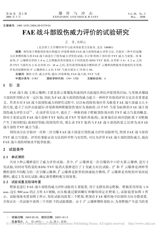
图 3 1 # 云爆弹试验时靶板的 变形情况 Fig . 3 T ypical defor mations of the plates in 1# FA E exper iment
1 mm 钢 板在 1 云 爆 弹 和 等药 量 的 RDX/ T NT 装药火箭弹作用下的变形挠度值 w f 如图 4 所 示。对于 RDX/ T NT 装药, 靶板在炸距 R 较近的情 况下 产生 了较 大的 塑性 变形 , 如在 炸距 3 m 处 , RDX/ T N T 装药靶板的变形挠度约为 1# 云爆弹的 1. 18 倍 , 而随着炸距的增大 , 靶板的变形量 迅速减 小。对于云爆弹 , 靶板在炸距较近的情况下产生的 塑性变形比 RDX/ T NT 装 药小 , 但随 着炸 距的 增 大, 靶板变形量的下 降较缓慢, 如在 炸距约 4. 1 m 处, 1# 云爆弹靶板的变形挠度已为 RDX/ T NT 装药 的 1. 24 倍。由图 4 可知 , 炸距 3. 8 m 之前 , RDX/ T NT 装药的靶板变形挠度大于云爆弹; 炸距 3. 8 m 之后 , 云爆弹的靶板变形挠度大于 RDX/ T NT 装药。 RDX/ T N T 装药和云爆弹的靶板变形情况充分体现了点爆炸和分布式爆炸的不同。与云爆弹相
#
4
FAE 战斗部毁伤威力评价
图 5 2 # 及 3 # 云爆弹毁伤威力评 价试验数据
根据超压 冲量准则
[ 3]
, 对应于某一毁伤等级有
Fig . 5 Ex per imental data of 2 # and 3 # F AE w arheads
一系列超压 p 和比冲量 i + 值的组合 , 这些达到同 一毁伤等级的( p , i+ ) 点的轨迹形成等毁伤曲线。当某一给定战斗部在某一距离处的冲击波峰值超压 和比冲量均大于或等于某一等毁伤曲线的对应值时, 则对该目标产生相应等级的毁伤。不同毁伤等级 下靶板等毁伤曲线的表达形式可用下式表示 , 即[ 3] ( p - p j ) ( i+ - ij ) = D j
科技英语翻译考试题目
Part I: Translate the Chinese in the brackets into English. (15%)1. ____________________(爱因斯坦相对论)is the only one which can explain such phenomena.2. Rate of penetration was found to ____________________ (与……成正比) the net pressure applied by the tool.3. Being a ______________(非良导体/绝缘体), rubber is often used in cables.4. Properly speaking, ___________(并非所有的物质) exist in three states.5. An electric current produces ______________(磁场) around it.6. With the result of automation, productivity has ______________ (增加了5倍) in that factory.7. Copper and aluminum are the best conductors of electric current ______________(仅次于银).8. The neutron has __________________(既不带正电荷,也不带负电荷).9. When the water temperature is increased, it vaporized more quickly until it reaches ________(沸点).10. A __________(变压器) is a very useful device, even though it can be sued only with alternating current.Part Ⅱ: Choose the better rendition for each of the following sentences.(20%)1.由于我在会上发表论文,如蒙介绍有关会议的详细情况,不胜感激。
- 1、下载文档前请自行甄别文档内容的完整性,平台不提供额外的编辑、内容补充、找答案等附加服务。
- 2、"仅部分预览"的文档,不可在线预览部分如存在完整性等问题,可反馈申请退款(可完整预览的文档不适用该条件!)。
- 3、如文档侵犯您的权益,请联系客服反馈,我们会尽快为您处理(人工客服工作时间:9:00-18:30)。
Experimental research on blast power of fiber reinforced anti-hard target warheadBin Liang,Jie-qun Zhou *,Gao-peng Feng,Yong-gang LuInstitute of Systems Engineering,China Academy of Engineering Physics,Chinaa r t i c l e i n f oArticle history:Received 12January 2017Received in revised form 27March 2017Accepted 17April 2017Available online 19April 2017Keywords:Carbon fiber composite Effect of blastingExperimental investigation Low collateral damagea b s t r a c tFiber reinforced anti-hard-target warhead is a new-type sample munition,which is only designed based on theoretical analysis and numerical simulation in laboratory.This warhead consists of carbon com-posite casings and high explosive,which can greatly reduce the damage to objects outside the damage range.In order to evaluate its blasting damage effect on concrete target,the three types of charges were researched by means of experiment,which are bare charge,charge with carbon composite material shell and charge with steel shell.Experimental results show that the peak overpressure of charge with carbon fiber composite shell is higher than that of charge with steel shell,but is lower than that of bare charge in the case of the same TNT equivalence.No fragments and fragment effect exist for distant target under the condition of charge with carbon fiber composite shell.However,the experimental result of the charge with steel shell is completely contrary.According to the blast effect in the concrete target,the charge with carbon composite material shell is optimal in matched impedance and detonation propagation.Also,the effective energy produced by the detonation of explosive with carbon composite material shell is the largest.©2017Published by Elsevier Ltd.This is an open access article under the CC BY-NC-ND license (http:///licenses/by-nc-nd/4.0/).1.IntroductionIn recent years,the low-collateral-damage munitions have been badly needed,which has good performance in reducing destruction outside the damage range,while enhancing destructive force on the target.To achieve this goal,it is necessary to design a weapon that could penetrate hard targets as deeply as a steel-shelled pro-jectile does,while could restrain the damage of the blast within a small radius.Fiber reinforced anti-hard-target warhead is a new-type sample warhead,which is only designed based on theoret-ical analysis and numerical simulation in laboratory.This munition consists of carbon composite casings and high explosive,which can greatly reduce the damage to objects outside the damage range.The blasting damage of fiber-reinforced composite warhead has already become a hot topic in development of conventional weapon and design of defense engineering,yet little information is available about the blasting effect on targets [1].Therefore,it is signi ficant to the development of new-concept warhead and the design of defense engineering,including the study of blasting-damage evaluation for fiber reinforced composite warhead.The phenomena that may occur during the new-concept warhead blast in air and concrete were preliminary analyzed,including the damage of target and the ef ficiency of loaded ex-plosives [2].The damage of target is caused by blast wave and fragments [3,4].However,the quasi-static failure of concrete target and the damage ef ficiency of fiber-reinforced composite warhead in the concrete structure are highly non-linear transient phenomena,which are dif ficult to be studied by using theoretical and numerical methods.So the physical experiments play a vital role in the characterization of such problems.The results obtained from the physical experiments represent the ef ficiency of blasting damage and the failure rules of concrete target for the different charge shells.The present work is intended to evaluate the damage charac-teristics of fiber reinforced anti-hard target warhead,i.e.,explosion ef ficiency in air,damage abilities of targets and damage effect of concrete targets.Explosion tests were conducted to examine the failure region of concrete target subjected to inner blast and get the overpressure curves of air blast.Then,the radius of damage and the craters of concrete targets due to bare charge,charge with com-posite material shell and charge with steel shell explosive are compared.*Corresponding author.E-mail address:zhoujiequn1414@ (J.-q.Zhou).Peer review under responsibility of China OrdnanceSocietyContents lists available at ScienceDirectDefence Technologyjou rna l homepage:www.elsevier.com/locate/dt/10.1016/j.dt.2017.04.0022214-9147/©2017Published by Elsevier Ltd.This is an open access article under the CC BY-NC-ND license (/licenses/by-nc-nd/4.0/).Defence Technology 13(2017)212e 2182.Blast experiment2.1.Experimental samplesFor enhancing the energy delivery to a target while controlling the radius of damage area as well,a sample wasfilled with the Composition B charge.The Composition B has been used in ammunition because it is a kind of powerful and very insensitive explosive.It is highly unlikely to explode accidentally.The experimental samples were designed for the comparative analysis of three types of charge,i.e.,bare charge,charge with carbonfiber composite material shell and charge with steel shell. The analysis focuses on the explosion efficiency in air,the damage ability of targets and the damage effect on concrete targets.The structures of charges are shown in Fig.1,and their parameters are listed in Table1.The properties of composite material are listed in Table2.2.2.Test layout2.2.1.Air explosion test layoutThe air explosion test layout for overpressures of three charges is shown in Fig.2.The loaded sample is hanged over a support,and its axis is vertical to a horizontal plane.Distance between the bottom of sample and the ground is1.5m.Horizontal distances among overpressure sensors and loaded sample axis are0.4m, 0.6m and1.0m,respectively.Gauges designed to measure these pressures must be robust enough to survive for the total recording time,typically from100m s upwards.In order to avoid unnecessary fragment impact damage,the silicon piezoelectric sensors and charges are placed in different angles according to the estimated dispersion angles of fragments.The sensors are placed at1.3m, 1.1m and0.9m above the ground,and the guide lines of silicon piezoelectric sensors are guarded by steel tube and sandbags.2.2.2.Test of blasting in concrete targetThe schematic diagram of an experimental setup for explosion in concrete target is given in Fig.3.The concrete specimens put on the ground are500mm thick and1600mmÂ1600mm square plates,and their compressive strength is35MPa.A predrilled hole at target center for charges is30mm in diameter and200mm in length.A high speed camera is used to record the forming process of blasting crater.The diameter and volume of blasting crater could be used to evaluate the damage effect of different charges.3.Experimental resultsIn the experiments,the silicon piezoelectric sensors were used to measure the shock wave over pressure caused by the explosive charge in the air.The processes of blasting damage and fracture of concrete target was recorded using high speed camera.3.1.Air blast experimental resultsA detonator is detonated at the top of charge.For the bare charge and the charge with carbonfiber shell,there is no damage to the sensor,sensor bracket,concrete cylindrical target and concrete protection board after blasting.The carbonfiber shell debris can not be seen around the damage range.It can be inferred from the combustible carbonfiber shell material and the previous round of tests[2],that the carbonfiber shell is burning after blasting.When the air is driven by the explosiveflows at high speed,the blast wave is followed by an exponential decay of pressure as a steep pressure occurs.Besides,explosions fragments could cause structural damage.A metal case in contact with explosive is usually broken into chunky fragments with the different dimensions in different directions.The measured initial velocity of fragments is 2km/s e3km/s.The combination with the blast wave and the fragments causes the damage to target[2,3].The target damage due to blasting of the charge with steel shell is depicted in Fig.4.From the distribution of test pieces,it can be seen that the explosion fragments produce penetration damage to steel hoop on the outer surface of the steel cylindrical concrete target and inner side of Longmen,which is2m far from the charge center.A3D numerical simulation model,which includes high explo-sive material,shell,air and ground,was established using AUTO-DYN3D.High explosives were modeled using the Jones e Wilkins e Lee(JWL)equation of state as follows:p¼C11ÀuR1veÀR1vþC21ÀuR2veÀR2vþu ev(1)where p is hydrostatic pressure;v is specific volume;e is specific internal energy;and C1,R1,C2,R2and u are material constants.The values of the material constants for many common explosives were determined from dynamic experiments and are available in AUTODYN.In the present simulation,C1,R1,C2,R2and u are assumed as3.74Â105MPa,4.15,3.75Â105MPa,0.9,and0.35, respectively[5].Air is modeled by the ideal gas equation of state,in which the pressure is related to the energy byp¼ðgÀ1Þr e(2) where g is constant;r is air density;and e is the specific internal energy.In the simulation,the standard properties of air from AUTODYN material library are utilized,i.e.,air density r¼1.225kg/ m3and g¼1.4.The initial internal energy of air is assumed to be 2.068Â105kJ/kg[5].In the experiment,the waveform of overpressure is recorded by a test instrument,and the measured electrical signal is converted to the overpressure signal.The measured results of overpressure and thefitted curves of peak overpressure are shown in Table3,Fig.5 and Fig.6,respectively.In Table3,R is the distance from the charge center,P t,P c and t are the experimental value ofpeak Fig.1.Schematic diagram of samples.B.Liang et al./Defence Technology13(2017)212e218213overpressure,the numerically simulated value,and the time cor-responding to the peak overpressure,respectively.Because the test sensor layout is reasonable,the sensors and cable protection pipes were avoided to be destroyed by explosive fragments.Three air blast testing overpressure curves were suc-cessfully obtained.The sensors are numbered 1e 6according to the distance from charge axis.Since the location of the Sensor 1is about 50mm higher than Sensor 2(seeing Table 3),it is closer to the explosive center,which is the reason for the values measured by Sensor 1are greater than those measured by Sensor 2.The overpressure of explosion shock caused by carbon fiber shell wave is about two times of that caused by steel shell charge shown in Table 3,and the peak overpressure of blast shock wave caused by the bare charge is signi ficantly higher than that caused by charge with case,the former is about two times of the latter.The peak overpressure is signi ficantly different although the height differ-ence of two channels is only about 50mm.The main reason is that the overpressure decays rapidly in the near field of explosion shock wave.Fig.5shows the test of pressure-time curves at 0.4m from the charge center.Fig.6represents the fitting curves of air blast over-pressures (P t )of three charges with the distance (R ).On the basis of Table 3and Fig.5,the propagation velocity of explosion shock wave in the bare charge is larger than that of shelled charges,while the propagation velocity of explosion shock wave in the carbon fiber shell is faster than that of steel shell charge.The numerical results agree well with the results in Table 3.The experimental results demonstrate that the distribution and propagation of overpressure are closely related to the charge structure.The overpressure and propagation velocity of shock wave in the bare charge are higher than those in the charge with shell.Meanwhile,the overpressure and propagation velocity of shock wave in the carbon fiber shell charge are higher than those in the steel shell.In Ref.[1],the explosion model about shell density andTable 1Parameters of three charges.SampleShell Comp.BLoading ratio Total mass/gMaterialThickness/mm Mass/g Sample 1ee 54154Sample 2carbon fiber composite material 4540.47115Sample 3Steel4540.15360Table 2Properties of composite material.MaterialYoung's modulus Poisson's ratio Normal wave impedance/(kg $(m 2s)À1)Longitudinal/GPaLateral/GPa Longitudinal Lateral Composite material13310.40.330.293.73Â106Fig.2.Schematic diagram of air blast experimentallayout.yout of explosive charge in the slab.B.Liang et al./Defence Technology 13(2017)212e 218214thickness was proposed based on the physical process of blast effect,which can be used to estimate the effect of charge structure on the strength of shock wave,and the in fluences of the density and thickness of charge shell on the intensity of air explosion shock wave (or energy utilization)were analyzed.Theoretical analysis proves that the shell thickness and density are the main factors affecting the energy utilization of air blast under the same loading.The ratio of energy using for the shell damage and driven fragments to the total energy of explosive is not 0.01e 0.03in the engineering,but increases with the increase in the shell thickness ratio (ratio of shell thickness to explosive thickness)and density ratio (ratio of shell material density to explosive density),as shown in Fig.7.The reasons for the differences are mainly two aspects:firstly,a part of energy generated by explosion of charge with shell has been used for driving shell,so that the explosion shock wave pressure of the charge with case is lower than that of thebareFig.4.Damage of steel fragments from the air blast.Table 3Experimental and numerically simulated results of air blast about different charges.Charge structure R /m 0.40.61.0bare chargesensor 1#2#3#4#5#6#P t /MPa 0.7930.4550.2450.2180.1820.176P c /MPa 0.7240.6110.3000.182t /ms 0.284/0.3200.702 1.577Charge with composite material shellP t /MPa 0.4390.2520.1980.1940.1700.150P c /MPa 0.4040.3600.2170.154t /ms 0.420/0.4680.936 1.916Charge with steel shellP t /MPa 0.1590.1490.1180.0920.0660.054P c /MPa 0.2570.2240.1820.144t /ms0.578/0.5291.0892.133Fig.5.The testing overpressure-time curves of the threecharges.Fig.6.Overpressure fitting curves of the three charges [5].B.Liang et al./Defence Technology 13(2017)212e 218215charge.The quality and density of the charge with carbon fiber composite material shell are relatively lower,and a part of the shell is ablated during explosion,so the driving energy of frag-ments is relatively smaller.As for the charge with steel shell,a large amount of explosive energy is consumed in the fragment driving,so the energy diffusing in the air decreases after blasting compared with that of the carbon fiber shell.Secondly,the normal wave impedance of carbon fiber shell is in consonance with that of explosive or explosion product,meanwhile,the normal wave impedance of steel shell material is dissimilar to those of explo-sives and air.3.2.The blasting damage to concrete target with chargeThree tests were performed to explore the driving abilities of three kinds of charge structures.High speed photographs were taken at the same frequency (50,000frames/sec)for recording the explosion in concrete target,as shown in Fig.8.It can be seen that there exists a certain leakage and pressure relief of explosive products from the predrilled holes on a concrete target.The initial time is detonator ignition time.For the bare charge explosion,there is no explosion flame in concrete gun mouth.However,there exist explosion flames for both the carbon fiber charge and steel shelled charge.The explosion flame of steel shelled charge is more obvious and lasting longer,which is caused by the circular constraint of the charge.For the bare charge,the diameter of explosion hole is larger than the diameter of cylinder explosive,and the explosive products are not bound by the circum restraint,that is why the explosion gas leakage along the axis of charge is later.In the condition of shelled charges,there is a circular restriction,therefore,the explosion products leak from the initiation point along the axis to the charge explosion hole,which leads to different level explosion flames.The strength of carbon fiber shell is weaker than that of the steel shell,and the explosive products leaked along the axial direction are less than that of the steel-shelled charge,therefore the explosion flame is weaker and shorter.In this paper,the de finition of charge energy distribution ratio is that the ratio of axial and radial energy distributions after explo-sion.Through the analysis of high speed photographs (seeing Fig.8),it can be found that the charge energy distribution ratioisFig.7.Ratio of energyutilization.Fig.8.High speed photographs of blasting in concrete for the three charges.B.Liang et al./Defence Technology 13(2017)212e 218216different,and the distributed energy of carbon fiber shelled charge is larger than that of the steel shelled charge and less than that of the bare charge since the axial constraint are different.Three blasting craters in the concrete target induced by explo-sion are shown in Fig.9.The speci fic sizes of blasting craters are listed in Table 4.There are some differences in blasting craters among the three kinds of charge.For the bare charge explosion,the ratio of bottom radius to mouth radius (R a )is the smallest,i.e.,the funnel taper is the smallest.The blasting funnel taper is the largest for steel shelled charge,and is in the middle for the carbon fiber shell counterpart.Moreover,the blasting crater depth (H ),the funnel area (S ),pit crater volume (V )all have the same rules.It can be seen from Table 4that the blasting crater sizes and shapes caused by the three kinds of charges are different.From the principle of shock wave propagation in multilayer media [6],it can be known that the surface wave impedance of carbon fiber com-posite shelled charge is more closer to those of the explosives (or explosive products)and concrete than the steel shelled material,so that the wave impedance of carbon fiber composite shelled charge is more matching with explosive,and the initial overpressure caused by the explosion of carbon fiber shelled charge is higher than that of the steel shelled one.It is a matter of controlling the energy and putting it to better use.Carbon fiber composite is lightweight,and the weight of car-bon fiber composite case will account for only 10to 20percent of total weight of munitions.From the explosion energy distribution,the strength of the carbon fiber shell material is lower than that of the steel shell material,the consumption of shell fracture energy in explosion is relatively low,at the same time,the explosion product leakage of carbon fiber shell is relative less than the counterpart of the steel shell,so that the effective explosion energy to target damage of carbon fiber shell is higher than that of the steel shell.The axial constraint of three charges are different,so that the utilization rate of carbon fiber shelled charge is higher than that of steel shelled charge and lower than that of bare charge.4.Conclusions and discussionIn the present work,the effect of carbon fiber sheet reinforce-ment on the damage performance of concrete target was investi-gated.The local damage degree of concrete plates subjected to inner explosion was estimated by using experimental method.The following conclusions can be concluded from the aboveexperimental results.Under the condition of the same quantity of explosives,the overpressure and the propagation velocity of shock wave in the carbon fiber composite shelled charge are higher than those in the steel shell,and are lower than those in the bare charge.The ex-plosion fragments of carbon fiber composite shelled charge could not produce damage to target.The steel shelled charge has killing effect on distant target.In the case of the explosion damage effect of charge blasting in concrete target,the carbon fiber shelled charge is better than the steel shelled charge in the impedance matching,and is more conducive to the propagation of blast shock wave.In the case of the same quantity of explosives,the effective energy (impulse),the radial and circular energy ratio of the carbon fiber shell charge to the concrete target is higher than that of the steel charge.It can be concluded that the damage ef ficiency of the carbon fiber shelled charge is higher than that of the steel shelled charge and less than that of the bare charge.The carbon fiber shell charge in the air explosion will not pro-duce a lot of lethal damage elements (such as fragments),which can be used for the urban environment.In view of the carbon fiber reinforced armor-piercing warhead having the characteristics above,the anti-hard-target projectile with head made of high strength steel or body using the composite shell has excellent damage effect on concrete target,which is superior to the tradi-tional steel shell.Furthermore,the explosive damage effect will be further improved when the loading ratio of warhead is increased greatly.Therefore,the application of fiber reinforced warhead technology with high filling ratio is defective to facing the chal-lenges from forti fications fortress concrete targets,which signi fi-cantly enhances the damage effect of blasting on target.The designed new monition with carbon fiber composite case and enhanced-blast explosive increases the impulse delivered to the intended target and that eliminates collateral damagecausedFig.9.Photographs of concrete craters.Table 4Blasting craters sizes of concrete.Parameters of crater bare case carbon fiber composite case steel case depth/mm 160150120area/cm 2246212951225volume/cm 3864039783105B.Liang et al./Defence Technology 13(2017)212e 218217by shell fragments.The experimental results well demonstrate that, even though the new munition produces a more powerful blast,the range of its damage footprint is smaller than that of conventional warheads.AcknowledgementsThis research is partially sponsored by the Chinese-NSF Foun-dation(11672278)and Chinese-NDTF(B1520132012).The authors would like to thank Prof.X.W.Chen at China Academy of Engi-neering Physics for valuable joint discussion on explosive engi-neering.The authors are also grateful to reviewers for discerning comments on this paper.References[1]Goldsmith Werner.Review Non-ideal projectile impact on targets.Int J ImpactEng1999;22:95.395.[2]Liang B,Chen ZF,Lu YG.Numerical simulation and experimental investigationof blast effect of explosive charge covered with different materials shell.J PL Univ Sci Technol Sci Ed2007;8(5):429e34.[3]Liang B,Chen ZF,Lu YG.Investigation of blast effect of explosive charge withdifferent shell material.Chin J Explos&Propellants2008;31(1):6e11.[4]Ohkubo K,Beppu M,Ohno T,Satoh K.Experimental study on the effectivenessoffiber sheet reinforcement on the explosive-resistant performance of concrete plates.Int J Impact Eng2008;35:1702e8.[5]Shi Yanchao,Hao Hong,Li Zhong-Xian.Numerical simulation of blast waveinteraction with structure columns.Shock Waves2007;17:113e33.[6]Lili Wang.Foundation of stress waves.Beijing:National defense Industry press;2005.p.35e60.B.Liang et al./Defence Technology13(2017)212e218 218。