early stage of pilsed-laser growth of silicon microcolumns and microcones in air and SF6
秦皇岛柳江地区长龙山组石英砂岩物_省略_屑锆石U_Pb_Hf同位素的证据_第五春
第30卷第1期2011年1月岩石矿物学杂志ACTA PET ROLOGICA ET M INERALOGICAVol.30,No.1:1~12Jan.,2011#专题研究#秦皇岛柳江地区长龙山组石英砂岩物质源区组成)))来自碎屑锆石U-Pb-Hf同位素的证据第五春荣,孙勇,刘养杰,韩伟,戴梦宁,李永项(大陆动力学国家重点实验室,西北大学地质学系,陕西西安710069)摘要:在秦皇岛市柳江地区出露最古老的沉积岩为青白口系长龙山组石英砂岩,该石英砂岩与下伏的新太古代钾质花岗岩呈不整合接触。
LA-I CPM S锆石U-Pb测年显示该区长龙山组石英砂岩中碎屑锆石年龄分布在2635~ 2487M a之间,其物质源区较单一。
与北京)蓟县标准剖面相比,本研究区在较长的一段地质时期为古陆壳的剥蚀区,直至新元古代早期,又沉积了长龙山组滨海相碎屑岩。
碎屑锆石Hf同位素组成显示,它们的源区物质虽然有不同程度的壳幔混合,但主要来自于古老的地壳物质再循环,暗示在~215Ga其碎屑物质源区的地壳已经达到一定的规模和厚度。
关键词:华北克拉通;青白口系;长龙山组;石英砂岩;碎屑锆石中图分类号:P597;P588.21文献标识码:A文章编号:1000-6524(2011)01-0001-12The protolith nature of quartz sandstone from Changlongshan Formationin Liujiang area,Q inhuangdao City:evidence of U-Pb and Hf-isotopefrom detrital zirconsDIWU Chun-rong,SU N Yong,LIU Yang-jie,HAN Wei,DAI Meng-ning and LI Yong-x iang (State K ey Labor ator y of Continental Dynamics,Department of Geology,No rthw est U niv ersity,Xi'an710069,China)Abstract:With the development of the in situ zircon SHRIM P U-Pb dating method and laser ablation ICP-M S, the study of the age distribution of detrital zircons from sedimentary rocks has become an effective w ay to find the source area of clastic material and also become an important topic all over the w orld.How ever,during zircon laser ablation(LA)-ICPM S dating,the U-Pb isotopic fractionation remains one of the principal obstacles for precise zircon dating.The ex periments performed by the authors demonstrate that a low laser frequency(5Hz or 6Hz)would reduce element fractionation during laser ablation.The U-Pb ages of international zircon standards GJ-1and the laboratory zircon standard SK10-2w ere measured by LA-ICP-M S in20L m spot size at5Hz laser frequency.The reduce mean206Pb/238U ages obtained in our analysis are in good agreement w ith the published v alues.Quartz sandstone from Changlongshan Formation of Qingbaikou System,the oldest metasedimentary rock in Liujiang area of Q inhuangdao City,w as deposited unconform ably on the Neoarchean potassic granite. Detrital zircons from the quartz sandstone have207Pb/206Pb ages of2635Ma to2487Ma.A comparison w ith the Beiijng-Jix ian stratatigrphic section,from Early Proterozoic,the study area experienced a long period of w eathering and erosion,and then began to deposit on the surface and become the source area of clastic sediments收稿日期:2010-03-26;修订日期:2010-05-24基金项目:国家自然科学基金项目(40902056);西北大学大陆动力学国家重点实验室科技部专项作者简介:第五春荣(1977-),男,博士,从事前寒武纪地质和同位素年代学研究,E-mail:diw uchunrong@。
pre-gastrulation developmental

pre-gastrulation developmentalWhat is Pre-gastrulation Developmental Phase?Pre-gastrulation developmental phase refers to the early stage in embryonic development before the formation of the gastrula. During this critical phase, various crucial events occur that lay the foundation for the subsequent formation of the three germ layers that give rise to the different tissues and organs in the developing embryo. In this article, we will explore the pre-gastrulation developmental phase in detail, discussing its key stages and the processes that take place during this time.1. Fertilization and Cleavage:The pre-gastrulation phase begins with fertilization, where a sperm fuses with an egg to form a zygote. Following fertilization, the zygote undergoes cleavage, a process of rapid cell divisions. These divisions result in the formation of blastomeres, smaller cells that make up the blastula.2. Blastula Formation:As cleavage continues, the blastomeres divide and rearrange, leading to the formation of a hollow ball-like structure called ablastula. The blastula consists of an outer layer of cells, known as the trophoblast, and an inner cell mass.3. Compaction and Morula Formation:During this stage, the blastomeres undergo a process called compaction, where they tightly adhere to each other, forming a compacted ball of cells called a morula. Compaction is crucial for the subsequent differentiation of embryonic cells.4. Blastocyst Formation:At this point, the morula undergoes further cell divisions and differentiation, resulting in the formation of a blastocyst. The blastocyst consists of two distinct cell populations: the inner cell mass (ICM) and the outer trophoblast layer. The ICM gives rise to the embryo, while the trophoblast layer contributes to the formation of extraembryonic structures such as the placenta.5. Implantation:The blastocyst moves towards the uterine lining and undergoes implantation, a process where it buries itself into the endometrium. This establishes a connection between the embryo and the maternal blood supply, allowing for nutrient and gas exchange.6. Formation of the Three Germ Layers:Following implantation, the pre-gastrulation phase progresses further as the blastocyst differentiates into the three germ layers: ectoderm, mesoderm, and endoderm. This process is known as gastrulation. The ectoderm gives rise to the nervous system, skin, and other ectodermal tissues. The mesoderm gives rise to the skeletal system, muscles, heart, and blood vessels. The endoderm gives rise to the gastrointestinal tract, respiratory system, and other endodermal tissues.7. Germ Layer Migration and Differentiation:During gastrulation, cells from each of the three germ layers undergo migration and differentiation to form specific tissues and organs. For example, ectodermal cells migrate to form the neural tube, which develops into the brain and spinal cord. Mesodermal cells differentiate to form muscles, bones, and internal organs. Endodermal cells give rise to the lining of the digestive and respiratory tracts.8. Organogenesis:As gastrulation progresses, the three germ layers continue todifferentiate and form the rudiments of various organs. This process, known as organogenesis, involves intricate cell interactions, proliferation, and remodeling to shape and develop organs such as the heart, lungs, liver, and kidneys.In conclusion, the pre-gastrulation developmental phase is a crucial period in embryonic development. It involves key events such as fertilization, cleavage, blastula, and blastocyst formation, implantation, gastrulation, and organogenesis. These processes play a fundamental role in establishing the basic body plan of the developing embryo, paving the way for its subsequent growth and differentiation into a complex multicellular organism.。
椎间盘退变潜在治疗因子成骨蛋白-1研究进展
椎问盘组织再生研究的关键是要在椎 间盘退变的早期 作出诊断并给与治疗 , 椎问盘完全失去再生基础就只能通过 外科手段处理或应用体外培养的组织工程化椎间盘进行替 代, 后者在目前实现的困难较大。现阶段对椎间盘退变的机
后其含量大幅下降。O - 主要增加蛋 白聚糖 的生成 , P1 从 而恢复髓核的含水量及其粘弹性_ 1 。有研究报道分别对针 刺家兔椎问盘退变模型口 青春期家 “、 、 ]体外藻酸盐珠口 j 应用 O -, P1发现蛋白聚糖均有增加, 且椎问盘高度也有增加
或重建。蛋 白聚糖含量的增加可 以改变椎间隙环境 的 p H
制仍不十分 明确 , 整个椎间隙是一个缺少血供、 呈低氧酸性 的环境 , 椎问盘 自身修复能力差。目前研究认为椎问盘退变
不同时间段的髓核和纤维环细胞的蛋白聚糖合成口 这对于 , 需作椎间盘修复的老年椎问盘退变患者无疑是个好消息, 他 们处于椎问盘退变 的主要年龄段。还有两项研究口 结果 显示 ,P1 O - 能修复经 白介素一 和软骨素酶 A C作用后的髓 1 B 核细胞和纤维环细胞 的基质, 使其 中的生物大分子恢复, 甚
值, 从而降低疼痛 的敏感 ] P1的其他作用还包括抑 。O - 制炎症因子( I ) 如 L1等的炎性作用, 从而减轻疼痛口]促进 ; 胶原生成的作用相对蛋白聚糖较弱l 1 。O - 是否能促进细 P1 胞增殖, 各种文献报道不一。早期文献报道认为 O - 不能 P1 促纤维细胞分裂 但现在的观点有所改变 , 1 , 人为地持续给
泛生子胶质瘤基础 项(glioma basic)
泛生子胶质瘤基础项(glioma basic)全文共四篇示例,供读者参考第一篇示例:泛生子胶质瘤基础项是一种高度恶性的颅内肿瘤,起源于神经胶质细胞。
这种类型的肿瘤在成人和儿童中均有发生,约占所有脑瘤的20-30%。
泛生子胶质瘤基础项通常表现为快速生长和浸润性生长,对治疗的反应也较为有限,给患者的生存率带来极大挑战。
泛生子胶质瘤基础项的发生机制至今尚不完全清楚,但已有研究表明,多种因素可能参与包括基因突变、环境因素和神经胶质细胞的异常增殖等。
在研究这种疾病的过程中,科学家们发现了一些与泛生子胶质瘤基础项相关的重要基因,例如EGFR、IDH1和PTEN等。
这些基因的突变和异常表达与泛生子胶质瘤基础项的发生和发展密切相关。
临床上,泛生子胶质瘤基础项通常会出现头痛、恶心、呕吐、视觉障碍、语言障碍等症状。
MRI检查是最常用的检测方法,可以清晰地显示肿瘤的位置、大小和形态。
治疗方面,手术切除通常是首选的治疗方法,但由于泛生子胶质瘤基础项浸润性生长的特点,手术切除并不能完全治愈患者。
放疗和化疗也常常与手术切除相结合,以提高治疗效果。
泛生子胶质瘤基础项的治疗结果仍然不容乐观,临床研究表明,患者的生存率往往不高,术后复发的可能性也较大。
科学家们正在不断探索新的治疗方法,如靶向药物治疗、免疫治疗和基因治疗等。
这些新的治疗方法有望为泛生子胶质瘤基础项患者带来新的希望。
泛生子胶质瘤基础项是一种高度恶性的脑肿瘤,对患者的生存率带来极大的挑战。
科学家们正在不懈努力,希望能够找到更有效的治疗方法,为泛生子胶质瘤基础项患者带来更多的希望和机会。
我们相信,在不远的将来,泛生子胶质瘤基础项的治疗水平将会有所提高,患者的生存率也会得到明显的改善。
愿所有泛生子胶质瘤基础项患者都能早日康复,重返健康的生活。
第二篇示例:泛生子胶质瘤是一种常见的中枢神经系统肿瘤,起源于神经胶质细胞。
它是最常见的原发性脑肿瘤之一,占所有脑肿瘤的约30%。
泛生子胶质瘤的病因目前尚不清楚,但与一些遗传性因素、环境因素以及突变基因有关。
新成像技术有助阐明细胞增长方式

新成像技术有助阐明细胞增长方式
佚名
【期刊名称】《纳米科技》
【年(卷),期】2011(008)005
【摘要】由伊利诺伊大学电子与计算机工程学教授盖布利尔·波佩斯库(Gabriel Popescu音译)领导的研究小组近日宣布。
他们成功开发出一种称为空间光干涉显微技术(Spatial Light Interference Microscopy,SLIM)的成像方法。
这种方法能够通过两束光线来测量细胞质量,从而为有关细胞是以固定速率还是指数方式增长的学术争论提供新视角。
相关论文发表在美国《国家科学院院刊)(PNAS)上。
【总页数】1页(P94-94)
【正文语种】中文
【中图分类】TP368.32
【相关文献】
1.新显微技术首次观测到原子——UCLA应用冷原子显微镜成像技术阐明病毒结构[J], 敖犀晨
2.经济增长方式研究的新突破——《经济增长方式转变与经济增长质量研究》简介[J], 秋石
3.实现“新的经济增长方式”十条思路——袁木在“新的经济增长方式研讨会暨,95政策科学研究会理事大会”上的总结讲话要点 [J], 杨芹溪
4.实现“新的经济增长方式”十条思路——袁木在“新的经济增长方式研讨会
暨’95政策科学研究会理事大会”上的总结讲话要点 [J], 杨芹溪
5.新Georgia技术:微-CT成像技术有助于组织工程改善骨重建 [J],
因版权原因,仅展示原文概要,查看原文内容请购买。
隆突性皮肤纤维肉瘤的诊疗进展
•综述•隆突性皮肤纤维肉瘤的诊疗进展刘珍如 周 园 刘梦茜 王晓晴 综述,王大光 审校(南京医科大学第一附属医院皮肤科 江苏 南京 210029)[摘要]隆突性皮肤纤维肉瘤(Dermatofibrosarcoma protuberans,DFSP)是一种低度恶性肿瘤,早期DFSP临床症状不典型、特异性低,导致临床医师容易漏诊、误诊,此外,DFSP经手术治疗后仍有高复发率,部分病变更会出现纤维肉瘤化改变,增加其转移风险、并提示预后不佳。
近年来,许多研究开始关注DFSP的诊断及治疗,透过皮肤镜、病理活检帮助医师早期诊断,提高疾病检出率,并通过手术、放疗及化疗等治疗,有效降低病变复发率、避免远处转移,本文就DFSP的最新治疗进展作一综述。
[关键词]隆突性皮肤纤维肉瘤;纤维肉瘤样隆突性皮肤纤维肉瘤;莫氏手术;皮肤镜;伊马替尼[中图分类号]R739.5 [文献标志码]A [文章编号]1008-6455(2021)03-0171-04Research Progress in Diagnosis and Treatment of Dermatofibrosarcoma ProtuberansLIU Zhen-ru,ZHOU Yuan,LIU Meng-xi,WANG Xiao-qing,WANG Da-guang(Department of Dermatology, the First Affiliated Hospital of Nanjing Medical University, Nanjing 210029,Jiangsu,China)Abstract: Due to the atypical and low specificity clinical symptoms in early stage,Dermatofibrosarcoma protuberans (DFSP) is a low grade malignant tumor which might likely be misdiagnosis. In addition, even after surgical treatment DFSP still has a high recurrence rate. Lesions with fibrosarcomatous changes were associated with increased the risk of metastasis and poor prognosis. In recent years, many studies have begun to focus on the diagnosis and treatment of DFSP. Recent study showed with dermoscopy and pathological biopsy can help with early diagnosis and signicfanctly improve the detection rate. Also, with optimal treatment through surgery, radiotherapy and chemotherapy, recurrence and distant metastasis rate could be effectively reduced. This article reviews the recent progress of DFSP.Key words:dermatofibrosarcoma protuberan;fibrosarcomatous dermatofibrosarcoma protuberans; Mohs microgrsphic surgery;dermoscopy; imatinib基金项目:国家自然科学基金(编号:81000703、81472896);江苏省自然科学基金(编号:BK2009437);江苏省六大人才高峰资助项目 (编号:2015-WSW-026)通信作者:王大光,南京医科大学第一附属医院皮肤科,主任医师;E-mail:*****************第一作者:刘珍如,南京医科大学第一附属医院皮肤科,硕士研究生;E-mail:*****************隆突性皮肤纤维肉瘤是一种生长速度缓慢、起源于皮肤,并可累及皮下组织的低度恶性肿瘤,于1924年由Darier 及Ferrandh首次报道[1-2],DFSP病理改变主要呈“蟹足样”浸润生长,男性发病率略高于女性。
眼部结构侵犯的鼻腔鼻窦腺样囊性癌疗效及治疗策略探讨
《中国癌症杂志》2018年第28卷第7期 CHINA ONCOLOGY 2018 Vol.28 No.7525欢迎关注本刊公众号·论 著·通信作者:王胜资 E-mail: shengziwang@眼部结构侵犯的鼻腔鼻窦腺样囊性癌疗效及治疗策略探讨朱 奕1,王胜资1,程玉书2,李瑞辰11.复旦大学附属眼耳鼻喉科医院放疗科,上海 200031;2.复旦大学附属眼耳鼻喉科医院放射科,上海 200031[摘要] 背景与目的:鼻腔鼻窦腺样囊性癌是一种少见的头颈部肿瘤,由于该肿瘤的高侵袭性和沿着神经浸润生长的习性,以及鼻腔鼻窦与眼部紧邻的解剖特征,部分患者初诊时即有眼部结构的侵犯,从而造成治疗的困难。
该研究总结了伴有眼部结构侵犯的鼻腔鼻窦腺样囊性癌患者的临床资料及预后情况,探讨该类疾病的疗效及治疗策略。
方法:该研究为一项单中心、回顾性的研究,分析了138例2005年1月—2017年10月在复旦大学附属眼耳鼻喉科医院接受过放疗的鼻腔鼻窦腺样囊性癌患者。
采用Kaplan–Meier 方法对主要终点总生存期进行估计,组间比较总生存期采用时序检验,并适时进行Cox 比例风险回归分析;分类变量组间比较采用Pearson χ2检验。
结果:男性74例(53.6%),女性64例(46.4%)。
平均年龄(51.0±11.6)岁。
鼻腔为最常见的原发部位,共97例(70.3%)。
临床分期以Ⅲ~Ⅳ期为主,共97例(70.2%)。
术前放疗为24例(17.4%),术后放疗为86例(62.3%),单纯放疗为28例(20.3%)。
放疗的平均剂量为(64.8±4.5)Gy 。
有眼部侵犯者为54例(39.1%),无眼部侵犯者为84例(60.9%)。
有眼部侵犯的患者在治疗中保留眼球者为40例(89.9%),行眶内容物剜除者为14例(10.1%)。
54例有眼部侵犯的患者中,术前放疗16例(29.6%),术后放疗24例(44.4%),单纯放疗14例(25.9%)。
糖尿病合并代谢综合征的临床特征分析
·临床研究·糖尿病新世界 2023年9月DOI:10.16658/ki.1672-4062.2023.17.039糖尿病合并代谢综合征的临床特征分析林梅芳1,黄明焕1,薛秉玮21.莆田学院附属医院血液净化室,福建莆田351100;2.莆田学院附属医院肾内科,福建莆田351100[摘要]目的分析糖尿病并发代谢综合征(metabolic syndrome, MS)的临床特征分析,为临床早期预防及早期诊断提供帮助。
方法采用分层整群随机抽样方法抽取2020年1月—2022年12月莆田学院附属医院958例糖尿病患者进行问卷调查、体格检查和实验室检查,根据2005年国际糖尿病联合会代谢综合征诊断标准将人群分为MS组(114例)和非MS组(844例),对两组一般资料、糖代谢和胰岛β细胞功能进行比较,从而得出糖尿病并发代谢综合征的临床特征。
结果MS组的年龄、腰围、体质指数、收缩压、舒张压、三酰甘油均高于非MS人群,高密度脂蛋白固醇低于非MS人群,差异有统计学意义(P<0.05)。
MS组的空腹血糖、餐后血糖(0.5 h)、餐后血糖(2 h)、糖化血红蛋白、胰岛素、抵抗指数、β细胞功能指数均高于非MS组,而LnDI1、LnDI2低于非MS组,差异有统计学意义(P<0.05)。
结论相较于单纯糖尿病人群,糖尿病合并MS的人群年龄较大,体质指数、腰围较高,血压、血脂、血糖水平的异常程度也更高,故对于糖尿病患者,定期监测血压、血脂、血糖水平具有重要意义,为临床早期预防及早期诊断提供帮助。
[关键词] 代谢综合征;糖代谢;血压;血脂;血糖;胰岛β细胞功能[中图分类号] R47 [文献标识码] A [文章编号] 1672-4062(2023)09(a)-0039-04Analysis of Clinical Features of Diabetes Mellitus Complicated with Meta⁃bolic SyndromeLIN Meifang1, HUANG Minghuan1, XUE Bingwei21.Department of Blood Purification Room, Putian University Affiliated Hospital, Putian, Fujian Province, 351100 China;2.Department of Nephrology, Putian University Affiliated Hospital, Putian, Fujian Province, 351100 China[Abstract] Objective To analyze the clinical characteristics of diabetes complicated with metabolic syndrome, and to provide help for early clinical prevention and diagnosis. Methods 958 patients with diabetes in the affiliated hospitalof Putian University from January 2020 to December 2022 were selected by stratified cluster random sampling method for questionnaire survey, physical examination and laboratory examination. According to the diagnostic crite⁃ria of IDF metabolic syndrome in 2005, the population was divided into MS group (114 cases) and non MS group (844 cases). The general information, glucose metabolism and islet cell function of the two groups were compared, so as to obtain the clinical characteristics of diabetes complicated with metabolic syndrome. Results The age, waist circumfer⁃ence, BMI, SBP, DBP, and TG of the MS group were higher than those of the non MS population, while HDL-C was lower than that of the non MS population, the difference was statistically significant (P<0.05). The FPG, PG (0.5 h), PG (2 h), HbA1c, LnHOMA-IR, and LnHOMA-β in MS Group were higher than those in the non MS group, while LnDI1 and LnDI2 were lower than those in the non MS group, the difference was statistically significant (P<0.05).Conclusion Compared with people with simple diabetes, people with diabetes combined with MS were older, had higher BMI and waist circumference, and had higher abnormal levels of blood pressure, blood lipids and blood glu⁃cose. Therefore, for diabetic patients, regular monitoring of blood pressure, blood lipids, blood glucose levels is of great significance for early clinical prevention and early diagnosis.[Key words] Metabolic syndrome; Glucose metabolism; Blood Pressure; Blood lipid; Blood glucose; Islet cell function代谢综合征(metabolic syndrome, MS)是一组以中心性肥胖、高血糖[糖尿病(diabetes mellitus, DM)[作者简介]林梅芳(1988-),女,硕士,主治医师,研究方向为糖尿病肾病。
- 1、下载文档前请自行甄别文档内容的完整性,平台不提供额外的编辑、内容补充、找答案等附加服务。
- 2、"仅部分预览"的文档,不可在线预览部分如存在完整性等问题,可反馈申请退款(可完整预览的文档不适用该条件!)。
- 3、如文档侵犯您的权益,请联系客服反馈,我们会尽快为您处理(人工客服工作时间:9:00-18:30)。
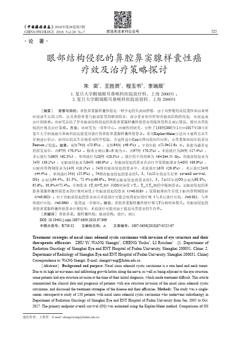
Ž.Applied Surface Science 154–1552000647–658www.elsevier.nl r locate r apsuscEarly stages of pulsed-laser growth of silicon microcolumns andmicrocones in air and SF 6Douglas H.Lowndesa,),Jason D.Fowlkes b ,Antonio J.PedrazabaSolid State Di Õision,Oak Ridge National Laboratory,Oak Ridge,TN,37831-6056,USAbDepartment of Materials Science and Engineering,The Uni Õersity of Tennessee,Knox Õille,TN,37996-2200,USAReceived 1June 1999;accepted 14July 1999AbstractDense arrays of high-aspect-ratio silicon microcolumns and microcones are formed by cumulative nanosecond pulsed excimer laser irradiation of single-crystal silicon in oxidizing atmospheres such as air and SF .Growth of such surface 6microstructures requires a redeposition model and also involves elements of self-organization.The shape of the microstruc-tures,i.e.,straight columns vs.steeply sloping cones and connecting walls,is governed by the type and concentration of the Ž.oxidizing species,e.g.,oxygen vs.fluorine.Growth is believed to occur by a ‘‘catalyst-free’’VLS vapor–liquid–solid mechanism that involves repetitive melting of the tips of the columns r cones and deposition there of the ablated flux of Si-containing vapor.Results are presented of a new investigation of how such different final microstructures as micro-columns or microcones joined by walls nucleate and develop.The changes in silicon surface morphology were systemati-Ž.cally determined and compared as the number of pulsed KrF 248nm laser shots was increased from 25to several thousand in both air and SF .The experiments in air and SF reveal significant differences in initial surface cracking and pattern 66formation.Consequently,local protrusions are first produced and column or cone r wall growth is initiated by different processes and at different rates.Differences in the spatial organization of column or cone r wall growth also are apparent.q 2000Elsevier Science B.V.All rights reserved.Keywords:Silicon;Pulsed laser;Ablation;Deposition;Columns;Cones;Whiskers;Nonequilibrium growth;Surface modification1.Introduction:formation of micron-scale silicon columns and conesRepetitive pulsed-laser irradiation of materials at relatively low laser energy densities,E ,of less than d 1J r cm 2produces gradual changes in surface topog-raphy and the development of ‘‘laser-induced peri-)Corresponding author.Tel.:q 1-423-574-6306;fax:q 1-423-576-3676.Ž.E-mail address:vdh@ D.H.Lowndes .Ž.Žw x odic surface structures’’LIPSS 1;see the series w x .of three papers by Young et al.2a,2b,2c .Such periodic structures have been widely observed in Žmetals,ceramics,polymers and semiconductors see w x .2Ref.3.At the higher E values of 1to 5J r cm d Ž.typically used for pulsed-laser deposition PLD of thin films,columnar or conical structures are formed in laser ablation targets,with the cones or columns w x pointing along the incident laser-beam direction 3,4.Earlier studies have established connections between the laser wavelength,polarization,and incidence an-gle and the spatial period of the near-surface ripple0169-4332r 00r $-see front matter q 2000Elsevier Science B.V.All rights reserved.Ž.PII:S 0169-43329900369-4本页已使用福昕阅读器进行编辑。
福昕软件(C)2005-2009,版权所有,仅供试用。
()D.H.Lowndes et al.r Applied Surface Science154–1552000647–658 648w xstructures2a,2b,2c,while more recent experiments have pointed out the importance of using either aŽ.w x short pulse duration picosecond or femtosecond5Ž.w xor a short deep-UV laser wavelength6for preciseŽand efficient material removal drilling holes or cut-.ting trenches.The conical structures produced by sequential laser irradiation have shapes that vary from circular conesŽw xto straight,high aspect ratio columns see7–10and .w xbelow as well as irregular cone clusters3.Most of the models used to explain the development of coni-cal structures assume that they are formed by prefer-ential remoÕal of material surrounding the cones, with some sort of‘‘cap stone’’present to prevent erosionlocally.The presence of impurities resistant w xto ablation11,or surface modification of polymersw xto produce an ablation-resistant carbon layer12,or surface segregation conducive to a transparent coat-w xing13,all are models proposed to explain coneformation on targets irradiated in vacuum or low-Ž.pressure-1Torr ambient gases.However,we recently reported experiments in which arrays of tall,slender silicon microcolumns were formed by cumulative nanosecond pulsed-ex-w x cimer laser irradiation of a silicon wafer in air7,8.Ž.For example,1000pulses of KrF248nm radiation in air produces;20-m m tall Si columns with both the average column diameter and their mean separa-tion being;2m m,as shown in Fig.1.Experiments were carried out to reveal a succession of surface topographical changes as the number of laser pulses was increased and,through these,it was possible to identify the main features of the mechanism by which high aspect ratio silicon microcolumns are w xgrown7,8.These experiments demonstrate that any explanation of microcolumn formation by cumula-tive pulsed excimer laser irradiation clearly requires()a redeposition not simply erosion model,and also involves elements of within the ar-ray of columns.We note that Sanchez et al.also observed the formation of similar arrays of SiŽw xcolumns in air see Refs.9,10and the Discussion.section below.Additional experiments that we carried out re-cently in various ambient gases have revealed that anŽ. oxidizing atmosphere e.g.,oxygen or fluorine must be present for columns or cones to form at all under our experimental conditions.These experiments alsoŽ.Fig.1.Top SEM image of Si microcolumns formed after10002Ž.Ž. laser shots in air at E s3J r cm scale bar s100m m.BottomdŽDroplets formed at the tips of silicon microcolumns scale bar s10 .m m.show that the shape of the microstructures, e.g.,straight columns or steeply sloping cones connected by walls,is governed by the type and concentrationŽof the oxidizing species e.g.,oxygen-containing air.vs.fluorine-containing SF.Based on these results it6was proposed that,once a sufficient number of laser pulses have succeeded in nucleating some initialŽ.surface features see below,then silicon columns or cones will grow rapidly by a‘‘catalyst-free’’VLS Ž.vapor–liquid–solid growth mechanism.This mech-anism involves both the re-melting of the tips of the columns r cones and the deposition there of the in-tense ablated flux of Si-containing vapor that isw xproduced with each laser pulse7,8.The outline of this paper is as follows.We begin by briefly reviewing earlier results for the formation of fully developed but distinctly different arrays of densely packed micron-scale silicon columns or cones r walls,when cumulative pulsed-laser irradia-tion of silicon is carried out in air or SF,respec-6tively.We then present results of new experiments intended to investigate how such different final sur-face microstructures actually nucleate and develop.本页已使用福昕阅读器进行编辑。