阿洛卡彩超外科探头系列
阿洛卡3500彩超维修

阿洛卡SSD-3500彩色超声(简称彩超)源自阿洛卡高性能的纯净声束成像技术平台,系统采用了大量先进的声学技术优化系统结构,具有全方位M型、实时三维成像技术、组织多普勒成像功能、双幅动态实时显示、血流剖面等功能。
日立阿洛卡前端技术成就新一代超声科技,ProsoundSSD-3500优异的基础图像源于12bit数字声束形成器(DBF)、像素聚焦(PixelFocus)、全层阻抗匹配技术(IMT)、多声束处理技术、纯净谐波成像技术(THE)、血流速度分布图(Flow Profile)、高分辨率放大、DDD模式、超宽视野成像(EFV)、实时三维成像技术(RT-3D)、实时全方位M型(FAM)、心功能综合指数、组织多普勒成像等等。
超宽视野成像:高清晰度的宽景成像,操作简便,成像快捷。
冻结图像后,可任意旋转和逐级放大,可清晰显示病变与毗邻组织结构之间的关系,测量准确,重复性好。
实时三维成像技术(RT-3D):SSD-3500拥有日立阿洛卡具有专利权的实时三维成像技术,它能够利用三维探头快速扫描人体,对回声信号进行实时处理,提供如照片一样真实的高质量图像。
三维图像可以自由地360度旋转,并可显示互相垂直的三个平面。
丰富的处理软件,使您不必考虑胎儿体位,轻松获得任意角度的胎儿三维图像。
RT-3D拓展了三维的临床应用于妇产科,还可应用于人体其他脏器,如肝脏、胆囊、膀胱等。
实时全方位M型(FAM):日立阿洛卡独有的全方位M型,可在任意位置取样并可同时显示3条取样线,取样线可任意旋转,可实时取样也可冻结后调取电影回放中的图像进行取样获取M型曲线。
此功能使M型图像采集更准确、更方便、因而广受临床医生好评。
郑州市大成医疗器械技术服务有限公司是一家大型医疗设备专业维修公司。
公司主要工程师从业时间长,有着丰富的医疗设备维修经验,可以进行芯片级无图纸维修、无局限必。
使用先进的测试仪器,可对在线集成电路进行功能测试、比较及功能模拟。
01全身机高频线阵探头
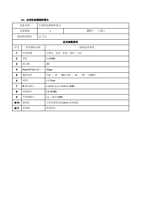
B模式频点
3.0-7.0,4.0-9.0,5.0-11.0MHz
8
谐波频点
8.0,9.0,10.0MHz
9
彩色多普勒频点
4.4,5.0,5.75.5( HR Flow) MHz
10
频谱多普勒频点
4.5,5.0,5.5MHz
11
深度
1.5-28cm
★12
兼容性
与科室现有迈瑞RESONA系列兼容
★13
3
实时三维放大
增加Live 3D Zoom功能
4
三维旋转
增加3D Swivel功能
5
三维切割
增加多种三维切割方式
★6
兼容性
与科室现有飞利浦CX50及食道探头X7-2t相匹配
★7
质保期
终身质保
20:曲柄腔内微凸阵探头
设备名称
曲柄腔内微凸阵探头
设备数量
2
□国产进口
最高投标限价
14万元
技术参数要求
序号
技术指标名称
最高投标限价
14.5万元
技术参数要求
序号
技术指标名称
招标技术要求
1
应用领域
术中、小器官(眼部除外),外周血管
2
带宽
3.5-16.0MHz
3
阵元数
128
4
扫描角度(最大)
25.4mm
5
B模式频点
3.5-9.2;7.6-12.8;9.6-16.0
6
谐波频点
10.0,12.0,14.0MHz
7
彩色多普勒频点
★13
兼容性
与科室现有德国SPINENDOS脊柱内窥镜配套使用
★14
考察报告
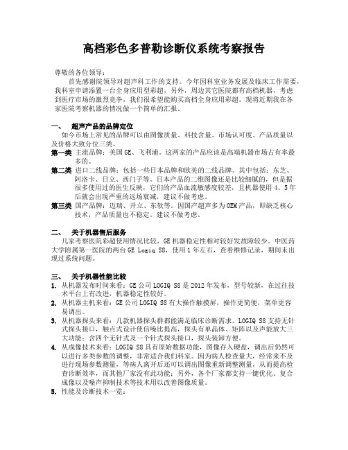
高档彩色多普勒诊断仪系统考察报告尊敬的各位领导:首先感谢院领导对超声科工作的支持。
今年因科室业务发展及临床工作需要,我科室申请添置一台全身应用型彩超,另外,周边其它医院都有高档机器,考虑到医疗市场的激烈竞争,我们很希望能购买高档全身应用彩超。
现将近期我在各家医院考察机器的情况做一个简单的汇报。
一、超声产品的品牌定位如今市场上常见的品牌可以由图像质量、科技含量、市场认可度、产品质量以及价格大致分位三类。
第一类主流品牌:美国GE、飞利浦。
这两家的产品应该是高端机器市场占有率最多的。
第二类进口二线品牌:包括一些日本品牌和欧美的二线品牌。
其中包括:东芝、阿洛卡、日立、西门子等。
日本产品的二维图像还是比较细腻的,但是据很多使用过的医生反映,它们的产品血流敏感度较差,且机器使用4、5年后就会出现严重的远场衰减,建议不做考虑。
第三类国产品牌:迈瑞、开立、东软等。
因国产超声多为OEM产品,即缺乏核心技术,产品质量也不稳定。
建议不做考虑。
二、关于机器售后服务几家考察医院彩超使用情况比较,GE机器稳定性相对较好发故障较少。
中医药大学附属第一医院的两台GE Logiq S8,使用1年左右,查看维修记录,期间未出现过系统问题。
三、关于机器性能比较1.从机器发布时间来看:GE公司LOGIQ S8是2012年发布,型号较新,在过往技术平台上有改进,机器稳定性较好。
2.从机器主机来看:GE公司LOGIQ S8有大操作触摸屏,操作更简便,菜单更容易调出。
3.从机器探头来看:几款机器探头群都能满足临床诊断需求。
LOGIQ S8支持无针式探头接口,触点式设计使信噪比提高,探头有单晶体、矩阵以及声能放大三大功能;含四个无针式及一个针式探头接口,探头装卸方便。
4.从成像技术来看:LOGIQ S8具有原始数据功能,图像存入硬盘,调出后仍然可以进行多类参数的调整,非常适合我们科室。
因为病人检查量大,经常来不及进行现场参数测量,等病人离开后还可以调出图像重新调整测量,从而提高检查诊断效率,而其他厂家没有此功能;另外,各个厂家都支持一键优化、复合成像以及噪声抑制技术等技术用以改善图像质量。
AplioXG技术参数
AplioXG(SSA-790A)超高档彩超技术参数一、设备用途说明:主要用于心脏、腹部、妇产科、外周血管及小器官等方面的临床诊断和科研工作,必须具有世界先进水平,为各进口一流厂家最顶尖机型,具备持续升级能力,能满足开展新的临床应用需求。
二、主要规格及系统概述:4.1彩色多普勒超声波诊断仪包括:4.1.1 智能组件结构设计的超声平台(ICA)*4.1.2 ≥19英寸高分辨率彩色液晶显示器,全方位可调4.1.3 高分辨率二维灰阶成像4.1.4 彩色多普勒血流成像4.1.5 频谱多普勒显示和分析单元4.1.6 组织谐波成像功能,3种以上不同谐波成像技术,并可实现方便切换,可视可调4.1.7 自然组织谐波成像:脉冲翻转或者脉冲减影谐波成像技术*4.1.8 宽带组织谐波成像技术:利用差量谐波技术,同时发射低频/高频基波,接收二次谐波和差量谐波,实现宽带谐波成像,同时提升图像的分辨率和穿透力*4.1.9 梯形拓展成像功能,可应用于线阵、凸阵、相控阵探头,拓展扫查视野,方便完成扫查*4.1.10 先进的空间、频率双模式复合成像技术:空间复合可应用在发射和接收两个方面,提升图像的细节分辨率和加强边界显示;频率复合提升空间分辨率和穿透力;4.1.11 复合成像技术可与谐波、彩色多普勒等成像方式结合使用,可实时完成空间、频率、空间加频率三种复合模式可视切换4.1.12 具有斑点噪声消除技术*4.1.13 先进的宽带多普勒血流成像技术:采用宽带多普勒技术,方向性、高帧频、高分辨率地显示低速血流,可频谱测量,可无外溢的的显示≤0.2mm血管的血流4.1.14 图像智能化一键优化技术,非预设置参数,单键操作,瞬间全场优化。
可应用二维、多普勒模式及造影剂谐波成像*4.1.15支持自动心内膜描记功能:采用最先进的生理定征模型进行心内膜描记,准确计算各种心功能参数,进行时相分析,实现全面左室功能分析。
*4.1.16 支持提供二尖瓣环运动自动跟踪及测量系统:自动跟踪二尖瓣环的运动轨迹,多参数多方向测量二尖瓣的运动,评估心脏运动功能。
ALOKA超声系列技术简介
2D线密度400、扫描帧速率120、彩色显示帧数50f/s、12-bit DBF模数转换器、1024数字化处理通道、多重声束处理技术、HST半球声束技术、多种选频成像技术(QFI)、阻抗匹配技术(IMT)、增强型纯净谐波检测技术(e-PHD)、触摸式液晶显示屏、三个电子探头标准接口、一个CW独立探头接口、心功能定量分析、心肌自动分区运动分析与KI心内膜描迹套件、TDI组织多谱勒成像套件、全方位M模式套件、血流剖面图和组织多普勒测量套件、IMP心功能指数、胎儿心脏成像软件包、DMS数字化图像管理系统
2D线密度400、最新一代的复合脉冲成像技术(CPWG)、动态四维声束处理技术、eFLOW技术、16-bit DBF模数转换器、复合陈列探头技术、多种选频成像技术(QFI)、阻抗匹配技术(IMT)、大屏幕触摸式液晶显示屏、四个电子探头标准接口、数字化图像管理系统、超宽视野成像、造影谐波:包含间歇触发模式和实时监控模式,适用于第一代,第二代和第三代造影剂、血流剖面图和组织多普勒测量套件、IMP心功能指数
负荷超声、血流剖面图、心功能指数TEI指数、实时三维成像技术3D、全方位M型FAM、组织多普勒成像功能TDI
SSD-3500
Prosound
172db系统动态范围.、扫描帧速率90、彩色显示帧数35f/s
应用范围:腹部、心脏、妇产、浅表组织与小器官、颅脑、儿科、泌尿、术中、介入
虽然是超声业的鼻祖,但ALOKA的机器多年来变化不大,一直以优秀的腹部二维图象和稳定的性能著称,售后服务很好,维修价格相对欧美公司要便宜些,我院一个门诊就选择了3500。
SSD-ALPHA5
24cm
dB范围182、扫描帧速率120、彩色显示帧数50f/s、15寸监视器
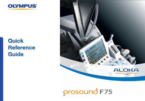
ALOKA F75超声医学仪器操作指南说明书
Quick Reference GuideTable of contents1.General instructions–ALOKA F75 components–Moving and unpacking the unit–Cleaning the unit2.Getting started–B asic setup and shutdown3.Creating a patient file–OPTION 1: Manual entry–OPTION 2: Import from server4.Changing scopes between cases5.Basic image controls– B Mode image brightness–Depth, frequency, contrast and focus6.Doppler modes7.Measurements and comments8.Storing files on ALOKA hard drive–Storing images–Storing video clips9.Searching and copying files–Searching for files on drives–Copying files to USB3214568ALOKA F75 componentsMonitor Touchscreen Operation panel Main bodyMoving and unpacking the unitThe ALOKA F75 should be packed down when moving, to avoid damage to its components. To unpack:Lock front wheels by pressing down on front outer ped al The console (operation panel, touchscreen and monitor) can be raised by holding down the front centr e ped al , and pulling up with the console handleTo adjust the position of the console, hold the lever inside the console handleUnlock the monitor’s handle from the groove above the touchscreen by pushing down on the teethCleaning the unitUse a lint-free cloth dampened with water to clean the touchscreen and monitor . DO NOT use detergent or alcohol. All other parts of the ALOKA F75 can be wiped down as per normal decontamination procedures.1 2 3 45 6787Getting Started – basic setup and shutdownAttach EUS Connector Cable to the scope and to port, located on the front side of the main bodyIf Picture-in-Picture is required, connect image cable from the back of the main body to the “PiP” port on video processor Holding the power button turns the ALOKA on/off. The power switch at the back of the main body can remain on12 3*Settings may differ if your preset has been personalised321Press PROBE PRESETSelect the desired probe and preset on the touchscreen (both should be yellow)Press B GAIN to freeze the image. Ensure image is frozen before changing probes, unplugging EUS cable or turning power off564 4ProbePreset561If storing images on ALOKA, a patient file must be created (see next page ).•Yellow tiles are functions that are “active”•Blue tiles are functions that are available but “inactive”•Multiple presets can be created and linked to a probeCreating a patient file: OPTION 1 Manual entryPress NEW PATIENTUse the trackball and ENTER to navigate cursor on the monitor. Patient ID is the minimum required data. Select OK to save and return to scanning modeMore information can be added later by pressing PATIENT1 231 23Creating a patient file: OPTION 2 Import from serverPress NEW PATIENTUse trackball and ENTER to navigate cursor on the monitor Select “Find” to download the most recent list from the hospital serverSelect “Worklist” to display the list and choose patient1 2 34Specific system setup is required to use this feature. Contact your Olympus Representative for more information.13422Changing scopes between casesPress B GAIN to freeze the imageDisconnect old scope and connect new scopePress B GAIN. This prompts the unit to look for connected probesPress PROBE PRESETSelect the desired probe from the touchscreenPress PROBE PRESET again, and select desired preset from the touchscreen. The unit will only recognise the probe automatically if it i s connected before the unit is turned on1 2 34 13Probe Preset4Confirm the correct probe and preset are activated by checking the bottom left of the image .ProbePreset 2Olympus ultrasonicendoscopes of 180 series or later have a detachable EUS cable as shown .5 6B Mode image brightnessB GAIN adjusts overall brightnessIMAGE OPTIMISER automatically optimises brightness levelSTC slide pots adjust brightness as specific depths (cm) that correspond to the ruler on the left of the image1 23123Depth, frequency, contrast and focusDEPTH/ZOOM adjusts the tissue depth that is displayed on the monitor. This corresponds to the ruler o n the left of the image Image Freq adjusts the frequency of the ultrasound wave Dynamic Range adjusts the contrast level of the image FOCUS allows the trackball to move the point of focus for the ultrasound wave (arrows to the right of the image)12 3 41234Quick Settersautomatically adjust these settings with one touch, ideal for focusing on tissue at specific depth s.Doppler modesFLOW displays directional colour doppler (velocity scale) eFLOW displays directional power doppler (signal strength scale) at high sensitivity for detecting small vesselsPress ENTER to toggle between full-line and dashed doppler box, and trackball to adjust size and location (respectively) MULTI-GAIN dial adjusts the brightness of the doppler signal PW displays the pulse at a defined pointUse the trackball and SELECT to select point of interest on left B Mode image1 234 123 455 66eFLOWPW1231Comments (annotation)Press ARROW to begin annotation functionUse trackball to move cursor on the monitor to point of interestTo add comments, use the keyboard located below Operationpanel or the dictionary located on the touchscreen1231 2 3 Measurement – live or frozen image modePress + key to start measurement functionUse trackball to move caliper to the start pointPress ENTER to start measurementUse trackball and ENTER to define endpoint. Themeasurement will be shown below the image.23To include the arrow on the image, press ENTERafter step 2. Additional labels and a keyboard canbe added to the touchscreen. C ontact yourOlympus Representative for more information.Push keyboardto open14Storing video clips onto the hard driveImage must be in live scan mode (press B GAIN to unfreeze) Press STORE to begin recording video. Recording will automatically stop and save 6 seconds of scan (“Post Time”)These setting can be changed by pressing Store tab on touchscreen To change recording time, adjust the dial below Time Cycle Press Acquire Mode to change to Manual (start/stop) or Pre Time1 2 3 1 23 Storing images onto the hard driveA patient file must be created first ; see page 5Press B GAIN to freeze the imagePress STORE to save a still image onto the hard drive. A thumbnail of the image will appear on the monitor To view a thumbnail in full screen, press ARROW Use trackball to move cursor to the thumbnail, double-click ENTER. Deselect to return to scanA thumbnail of the file will appear on the monitor, with a multi-tiled icon in the top left corner .423Images can also be saved onto a USB. Contact your Olympus Representative for more information.124 5345Copying files onto USB Search for files on hard drive, or press REVIEW to display the current case as thumbnails Use the trackball and ENTER to select files to copy, or click Select All Click Save (USB). This saves the images in a pre-defined format (JPEG or TIFF) to USB. By selecting Copy (USB) the files in original format (DICOM) are copied.1 2 3 1 2 3 Searching for files on drivesPress ARROW and move cursor to the thumbnail area ofthe monitor Click on Find iconDifferent drives can be searched, the local HD is selectedby default. Click search to display all files on this drive12 31 2 3 Save Setup defines the file format that images and videos are saved into the USB. You can also choose“teaching file” format, which maskspatient identifying data.Aloka Prosound F75 Quick Reference GuideFor complete product details see Instructions for Use.OLYMPUS AU S TRALIA PTY LTD3 Acacia Place, Notting Hill VIC 3168, AustraliaCustomer Service: 1300 132 992 | .au OLYMPUS NEW ZEALAND LIMITED28 Corinthian Drive, Albany, Auckland NZ 0632Customer Service: *********** | QR 04.027 V3.0 November 2019。
阿洛卡彩超售后

随着经济的飞速发展,人民生活水平的不断提高,彩超的年销售额以及医院拥有率呈快速上升的趋势。
各级卫生医疗机构的彩超拥有量以每年约3-4%的速度增长。
由于购买彩超后医院效益好,价格适中的中、低档彩超需求量增长较快。
彩超的折旧时间基本为6年。
目前很多医院通过日常的维护保养的方式将彩超设备的有效寿命延长到10-12年,有的使用时间超过了13年,现阶段,越来越多的医院选择将设备托管给专业的公司进行维护保养,以便延长机器的使用寿命,确保了彩超的开机率。
如今,医疗的发展脚步十分迅速,正在向着解决“看病难、就医难”等问题前进,而评价一所医院的标准,其中有一项便是这家医院的设备水平。
目前,三甲医院内的设备等级与一级甲等医院中的是完全不同的,设备的种类、及设备的先进性等都制约着医院的发展,也制约着医疗水平的进步,因而,对于医院来说,想要创二甲或者是创三甲首先是确保设备的正常运行,确保设备的跟进。
除了评等级以外,设备对于医院来说也是至关重要的,它们的正常运行,是确保患者健康、生命的关键,倘若在检查过程中,突然超声机器失灵,导致诊断失误。
发生医疗事故是不利于医院的发展!因此,在机器使用的过程中,要注重机器的保养与检测,做到提前预测,减少损失。
一旦机器故障,就应当及时、快速的维修,还有一点要注重机器的投保,它将为医院带来巨大的利益。
比如说可有效节约人工成本,免去维修所花费的时间与精力,试想一下,如果机器损坏,对于医院来说,约等于将病人健康视为不顾,甚至有可能影响到医院口碑。
有些机器是国外进口,或者是匹配了较为高额的价格,维修起来对原本无利益的医院来说,更是雪上加霜,由此可见,投保具有必要性与实用性。
随着临床技术的提高,以及医院对于设备的重视,投保成了立刻需要进行的事情。
有数据显示,大型医疗设备在整个生命周期内的维修费用,基本略等于购买费用,因此对于医院来说,购买保险要远远地节约成本。
郑州市大成医疗器械技术服务有限公司是一家大型医疗设备专业维修公司。
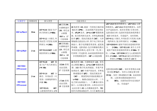
SSD-3500配置
第 1 页,共 5 页
日本阿洛卡全数字化纯净声束彩色多普勒超声诊断系统SSD-3500(Ver3.0) 配 置单
US$2,670.00
Volume Mode Unit 表面三维单元(SSD-3500
Volume-mode Probe 表面三维探头 B:2.5-6MHz, PW/Flow:2.5/3/0/3.8/5.0MHz 扫描角度:电子扇扫90°,机械扇扫60°-30 Convex SHD Probe, Transvaginal SHD妇产科有角手柄端扫式经阴道电子凸阵 探头, 120°14mmR B:4.0/5.0/6.0/7.5MHz, PW/F:3.0/4.0/5.0/6.0MHz
QUANTITY MODEL NO.
MODEL NAME
LIST PRICE(CIF)
1 set SOP-3500-3 1 set UST-9123
1 set UST-5546 1 set UST-5299
独特的照明设计 阿洛卡考虑到医生在较暗的工作环境中撰写 诊断报告的困难,创造性的为本机设计了精 致方便的照明系统。彩灯提示键,可自编触 模式控制面板:操作更方便,完全符合人类 工程学设计。
Convex W-SHD Probe W-SHD腹部/妇产科凸阵探头,60mmR 组织谐波 B:2.5/3.8/6.0MHz, PW/F:2.1/2.5/3.0/3.8MHz, H.E:2.1P,2.5S,2.5R,3.0H
- 1、下载文档前请自行甄别文档内容的完整性,平台不提供额外的编辑、内容补充、找答案等附加服务。
- 2、"仅部分预览"的文档,不可在线预览部分如存在完整性等问题,可反馈申请退款(可完整预览的文档不适用该条件!)。
- 3、如文档侵犯您的权益,请联系客服反馈,我们会尽快为您处理(人工客服工作时间:9:00-18:30)。
UST-9132IMAIN SPECIFICATIONSArray type: Convex Array Scan angle/width: 20mmR / 65°Frequency range:4-10 MHzSystem Compatibility: ProSound a10, ProSound a7Multi-frequency “I”style finger-grip transducer. This transducer design fits comfortably between the index and middle finger which allows for palpating organs and scanning at the same time. Super high density provides superb near field resolution.UST-995-7.5MAIN SPECIFICATIONSArray type: Convex Array Scan angle/width: 20mmR / 65°Frequency range:4-10 MHzSystem Compatibility: P roSound a5, ProSound 4000, ProSound 3500,ProSound 6UST-9132TMAIN SPECIFICATIONSArray type: Convex Array Scan angle/width: 20mmR / 65°Frequency range:4-10 MHzSystem Compatibility: ProSound a10, ProSound a7Multi-frequency “T”style finger-grip transducer that is perfect for sur-geons that prefer a more transverse view in relation to the position of the transducer in the hand. The design fits comfortably between the index and middle finger which allows for palpating organs and scan-ning at the same time. Super high density provides superb near field resolution.UST-MC11-8731MAIN SPECIFICATIONSArray type: Convex Array Scan angle/width: 20mmR / 65°Frequency range:4-10 MHzSystem Compatibility: P roSound a5, ProSound 4000, ProSound 3500,ProSound 6full scalefull scaleFinger-Grip TransducersCourtesy of Ph. Norihiro KokudoDepartment of Surgery, Hepato-Biliary-Pancreatic Surgery Division, University of TokyoT-TransducersUST-5713TMAIN SPECIFICATIONSArray type: Linear Array Scan angle/width: 60mm Frequency range: 3-10 MHz Puncture adapter:MP-2448System Compatibility: ProSound a10, ProSound a7, ProSound a5Multi-frequency “side-fire T”transducer with a wide field of view (60mm) and ability to change frequencies on demand for superficial or deeper penetration. Super high density provides superb near field resolution and detail for needle guidance.UST-579T-7.5MAIN SPECIFICATIONSArray type: Linear Array Scan angle/width: 60mm Frequency range: 4-10 MHz Puncture adapter:MP-2448System Compatibility: P roSound a5, ProSound 4000, ProSound 3500,ProSound 1000, ProSound 900UST-5534T-7.5MAIN SPECIFICATIONSArray type: Linear Array Scan angle/width: 42mm Frequency range:4-10 MHzSystem Compatibility: P roSound a5, ProSound 4000, ProSound 3500,ProSound 1000, ProSound 900Multi-frequency “side-fire T”transducer with a narrow field of view (42mm) and ability to change frequencies on demand for superficial or deeper penetration. Super high density provides superb near field resolution and detail for needle guidance.Multi-frequency micro-surgery transducer that is ideal for tight situ-ations and is choice transducer for cervical spine scanning. With the optional handling tool, the probe can be handled as if holding a pen-cil between your T-533MAIN SPECIFICATIONSArray type: Linear ArrayScan angle/width: 10mmFrequency range: 4-13 MHzSystem compatibility: P roSound a10, ProSound a5, ProSound 4000,ProSound 3500UST-536MAIN SPECIFICATIONSArray type: Linear ArrayScan angle/width: 19mmFrequency range: 4-13 MHzSystem compatibility: P roSound a10, ProSound a7, ProSound a5,ProSound 4000, ProSound 3500Multi-frequency hockey-stick transducer is ideal for cervical spine scanning. Super high density which provides superb near field resolu-tion.full scalefull scaleOptionalT-type handling tool : MP-2749I-type handling tool : MP-2750UST-547MAIN SPECIFICATIONSArray type: Linear Array Scan angle/width: 25mm Frequency range:4-13 MHzSystem compatibility: ProSound a10, ProSound a5Multi-frequency linear transducer provides high-resolution images. This probe is useful for observing cerebral tumors and aneurysms, and is also used in surgery for removing spinal cord tumor, carotid artery intima exfoliation and others. The probe can also be applied to observation of articulations and/or muscles in orthopedic surgery.UST-534MAIN SPECIFICATIONSArray type: Linear Array Scan angle/width: 5mm Frequency range:3-13 MHzSystem compatibility: P roSound a10, ProSound a5, ProSound 4000,ProSound 3500Multi-frequency transsphenoidal transducer for scanning the pituitary gland, cervical spine and other neurosurgical procedures. Super high density provides superb near field resolution.full scaleUST-52109MAIN SPECIFICATIONSArray type: Phased Array Sector Scan angle/width: 90°Frequency range:3-8 MHzSystem Compatibility: ProSound a5, ProSound 4000, ProSound 3500Multi-frequency end-fire laparoscopic transducer that is perfect for CBD scanning and targeting lesions. The point-and-shoot scanning and built-in biopsy channel make it easy to obtain the desired sample the first time. Super high density provides superb near field resolu-tion.UST-5536-7.5MAIN SPECIFICATIONSArray type: Linear Array Scan angle/width: 38mm Frequency range:4-10 MHzS ystem Compatibility: P roSound a5, ProSound 4000, ProSound 3500,ProSound 1000, ProSound 900Multi-frequency flexible laparoscopic transducer built to withstand the rigors of daily laparoscopic scanning and is perfect for renal abla-tions. Super high density provides superb near field resolution.UST-5550MAIN SPECIFICATIONSArray type: Linear Array Scan angle/width: 38mm Frequency range:4-10 MHzSystem Compatibility: ProSound a10, ProSound a7Laparoscopic TransducersMajor Feature ProSounda10ProSounda7ProSounda5ProSound4000ProSound3500ProSound6Image enhancement ExPHDDistortion-free fundamental frequency are transmit-ted to enhance the clarity of harmonic images.PHDOffer clearer edge definition, reduced side lobeartifacts, and less reverbration noise compared tofundamental frequency imaging. Image clarity ofthe obese patients is significantly enhanced. CPWGReduced signal noise for truer, less interpolatedimagingPixel FocusControlled focus at the pixel level provides higherspatial resolutionImage optimizerThis function instantly optimizes overall brightnessof B-mode images, eliminating the need for fre-quent manual adjustment to enhance productivity. SCSOffers enhanced capability for depicting sidewallstructures of tubular cavities and the like by super-posing images created by steering the ultrasoundbeam in multiple directions.Trapezoidal ScanThis provides a wider field of view than with con-ventional displays, to facilitate anatomical under-standing of the region of interest.AIPAdaptive Image Processing (AIP) clearyly displaysdifferencrs intissues, reducing speckle noise whilemaintaing the frame rate. It can also display outlinesmore clearly by selectively emphasizing boundaries. Edge optimizer Provide good smooth image.M.F.I.The M.F.I. is possible to select higher resolutionimaging or higher penetration imaging according tothe situation without changing the probe.D.D.D.D.D.D. is a function to display B-mode images withand without color Flow image simultaneously inreal time.Doppler Directional eFLOWDisplays high-resolution blood flow with directionalinformation. Compared with conventional bloodflow methods, D-eFLOW features enhanced spatialand time resolutions for greater detail.Color Flow andPower Flow Exceptional blood flow analysis.Auto Dopplerangle correlationIt is possible to correct Doppler angle automaticallyto reduce examination time.Storage HDDEasy storage and management of images andpatient information enhances and facilitates theultrasound examination.Cine Memory Cine search and loop displayUSB Data can be stored to USB memory in directory. Raw date Raw data of ultrasound for after-stored analysis DICOMThe patient data and image data can be transmittedto the file server in the network.Other Hi-definition LCDMonitor The flicker -free LCD monitor reduces eye fatigue. Measurement onplayback images It is possible to measure on playback images. Remote ControlUnitSystem functions can be controlled from the sterilefield.。