骨盆及髋臼骨折王满宜
影响髋臼骨折患者术后临床疗效的相关因素分析
影响髋臼骨折患者术后临床疗效的相关因素分析张元勋;董伊隆;钱约男;吴芝芝;柯大观【摘要】目的探讨影响髋臼骨折患者术后临床疗效的相关因素.方法回顾性分析行手术治疗的髋臼骨折患者77例,对患者年龄、性别、骨折类型、手术时机、手术入路、复位质量等临床资料进行统计学分析.结果 77例患者术后临床疗效优良率为80.5%.单因素分析显示性别、手术入路与髋臼骨折患者术后临床疗效无关(均P>0.05),而年龄、骨折类型、手术时机、复位质量、异位骨化、创伤性关节炎与髋臼骨折患者术后临床疗效有关(均P<0.05).多因素分析显示骨折类型、手术时机、复位质量是影响髋臼骨折患者术后临床疗效的独立因素(均P<0.05).结论骨折类型、手术时机、复位质量是影响髋臼骨折患者术后临床疗效的独立因素,而手术时机和复位质量是手术医生可控的,故临床工作中要选择适当手术时机、尽量达到解剖复位,以最大限度地恢复髋臼骨折患者术后髋关节的功能.【期刊名称】《浙江医学》【年(卷),期】2016(038)005【总页数】3页(P352-354)【关键词】髋臼骨折;临床疗效;影响因素;logistic回归分析【作者】张元勋;董伊隆;钱约男;吴芝芝;柯大观【作者单位】325200 温州医科大学附属第三医院;325200 温州医科大学附属第三医院;325200 温州医科大学附属第三医院;温州医科大学信息与工程学院;温州医科大学信息与工程学院【正文语种】中文髋臼骨折是一种严重的关节内骨折,骨折类型多样,局部解剖复杂,治疗难度较大[1]。
虽然目前认为切开复位内固定术是治疗移位的髋臼骨折的最佳方法(能明显缩短骨盆不稳定患者的术后卧床时间、降低髋关节创伤性关节炎的发生率),但是骨折治疗的临床疗效受许多因素的影响,术后仍可能存在髋关节疼痛和活动受限等问题。
本研究通过回顾手术治疗的髋臼骨折患者的临床资料,探讨影响髋臼骨折患者术后临床疗效的相关因素,以期指导临床,现报道如下。
骨盆骨折的急救精品PPT课件

第二步急救措施
• 临时固定骨盆 • 血管造影栓塞 • 要求:1.医生头脑清
醒,判断准确 2.采取有效的止血
措施
一、骨盆容积控制
• 原理:圆柱体积=4/3π×
半径3≈4.2×半径3
• 骨盆骨折移位3cm可使容
积增加2倍
骨盆容积控制方法
• 简单 快速 有效 • 1.骨盆束缚带 • 2.床单 • 3.PASG(冲气式抗休
骨盆骨折的急救
骨盆骨折
• 骨盆骨折——较为常 见的高能量损伤
• 年发病率2-3万 • 占全身骨折3-8% • 死亡率10-20% • C型骨折25-60% • 开放性骨盆骨折更高
骨盆骨折治疗重点
• 早期急救
• 80%骨盆骨折死亡发 生在伤后6小时内--王满
宜,骨盆骨折治疗的研究现状.中华创伤 杂志,2008,24(3)
--救治骨盆治疗经验少 --医护条件有限
3、各医院急救机构设置不一致
王满宜教授总结我国骨盆骨折急救路线
急救路线 生命体征ABCD 物理检查(一次)
不平稳
束带 X光、CT
三步走
上肢输液 2000ml/20min
化验室检查
迅速评估、复苏
外固定架、填塞 平稳
固定、止血
不平稳 外固定架 造影、栓塞 手术2-6天
早期急救成功的关键
• 第一黄金时间--伤后4~6小时内 • 正确的复苏路线 • 合理的早期急救措施 • 有效的控制大出血 • 按损伤控制理论处理合并伤 • 简单 有效 恢复骨盆稳定性
我国骨盆骨折急救现状
1、急救路线流程不统一
--不同医生急救流程不同,或非专业急救医生
2、大多发生在基层——成功率低
复位经皮螺钉内固定治疗跖跗关节损伤
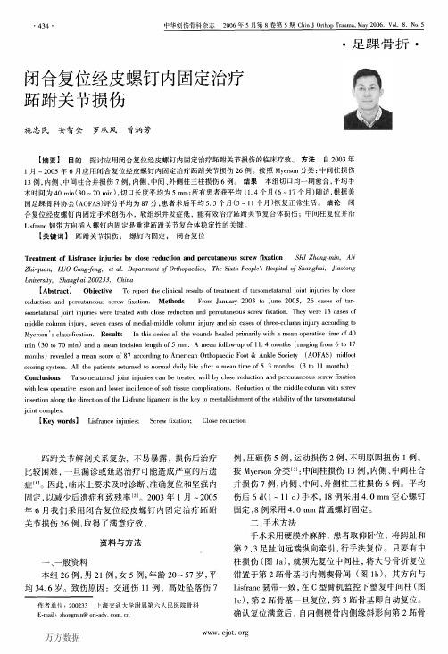
University,Shanghai 200233,China
【Abstract】0bjective
To report the clinical results of treatment of tarsometatarsal joint injuries by close
reduction and percutaneous screw fixation. Methods
Fig.2
Male,30 Y,three columns injury of the right tarsometatarsal joint complicated with fi'actures of the cuboid and the third metatarsal and dislocation of the second metatarsophalangeal joint.The preoperative X—ray of the right foot: AP (1eft)and oblique(right)views(a);the open reduction and internal fixation for the cuboid fracture:picture(1eft)and X—ray film (right)(b);the postoperative roentgenography deulonstrating reduction and internal fixation in place(e);follow—up by the sixth postoperative month:X—ray(1eft)and function picture(right)(d).
资料与方法
一、一般资料 本组26例,男21例,女5例;年龄20~57岁,平 均34.6岁。致伤原因:交通伤1 1例,高处坠落伤7
足部骨肿瘤及瘤样病变的发病特点分析

次 为距 骨 。张从 仁 等 统 计显 示 , 部所 有 的恶 性 骨 足 肿 瘤及 软骨母 细 胞 瘤 均 发 生 于跗 骨 , 而骨 软 骨 瘤 及 内生软 骨 瘤 则 好 发 于前 足 的趾 骨 及 跖 骨 。 H w r o ad
表 2 7 足 部 良 性 骨 肿 瘤 及 瘤 样 病 变 9例
An oa a y i te fau e f n e fb n mo sa d t mo ・i elso so e .Re u t 9 a e i o et mo sa d d t n l ss h t r so s to o e t e o u r n u rl in ff t k e e sl s 2 c s sw t b n u r n h t mo —ie lso sf e a c u t d fr 1 5 o l 4 o et mo a in sa mi e o o rh s i n t es me p r d 9 u rl e in e t c o ne o . % f 2 7 b n u rp t t d t d t u o p t i a e o .7 k l a 6 e t l a h i b n g o e t mo s a d t mo —ie l so so e c o n e o 2 e in b n u r n u rl e in ff ta c u td f r k e 4. % o 1 e i n b n u r n u r1 e l s n f 3 9 b n g o et mo s a d t mo .i e i s 3 k o p t n s a mi e o o rh s i l n t e s mep ro .1 r r l n n o e t mo ain so e c o n e r0 5 ai t d t d t u o p t h a e d e t ai i 1p ma y ma i a tb n u rp t t f e t c u t d f . % i g e f a o
老年心衰合并肺部感染患者的治疗体会
参 考文献 【l】 王满宜,吴新宝与荣国威.髋臼骨折.中华创伤骨科杂志.2001.3(2):
第85一一90页 . 【2】 常敏与苏开荣,髋白骨折的治疗 ;附36例报告.中华骨科杂志.i 996.
286
<求医问药)下半月刊SeekMedicalAndAskTheM edicine 2012年第 10卷 第 5期
其他 组织器 官的损 伤 合并双 跟骨骨折 1例 ,合 并股骨干骨 折1例 ,合并 胫骨 平 台骨折 2例 。 1.2 骨折 分 类 所有 患者 入科 时均 按照Letoumel的分类 方法 ,将髋 臼骨 折 分为简 单 以及复 合骨折 2个大 类 。每一大 类下 又按 照不同 的损伤 特点 进 一 步区分为10小类口】。简单骨折(简单组)即指骨盆髋臼一个壁或柱的全部 或部分骨折,即前柱 、后柱 、前壁、后壁以及横形骨折5种。本组简单骨折患 者 有 loft},其 中 ,前 柱 骨折2例 ,后柱 骨折 3例 ,前 壁骨折2例 ,后 壁骨折 1例 , 横形骨折2例。含有两个以上 简单骨折形式的骨折即归为复合骨折(复合 组 ),包括 双柱骨 折 、横 形骨折 合并 远折段 的纵 形骨折 、横 形骨折 合并 后壁 骨 折 、后柱 合并 后壁骨 折以 及前 柱或前 壁合 并后半横形骨 折5种类型 。本 组 有复合骨折患者14例,其中,双柱骨折3例 ,横形骨折合并远折段的纵形骨 折4例,横形骨折合并后壁骨折l例 ,后柱合并后壁骨折3例,前柱或前壁合 并后半 横形骨 折3例。 1.3 治疗方法 一般来说,骨盆髋臼骨折的治疗方式可以分为2类:手术治 疗和非手术治疗。在本次研究 中,所有的患者均采用后方Kocher— Langenbeck ̄J-J口 ^路进行手术干预治疗0I。术后患者牵引平均时间为4周, 所有患者都接受术后早期功能锻炼 ,以减少并发症的发生。 1.4 评 定标 准及随访 结 果 24例 患者均接 受6个月 2年的随访 。参 照美 国 矫行外科 研究 院评价髋 关节功 能方法评 定术后患 者的恢复情 况 ,评 定为 优 l6例 ,良4例 ,可4例 。 2 讨 论 2.1 骨盆髋臼的生物力学特点 正常生理情况下 ,髋关节的负重较大 ,特 别是受到的剪力较大,再加上髋臼的血供较少,因此 ,髋臼较易发生损伤。 骨盆髋臼骨折多为挤压暴力以及间接暴力引起。当人体一侧或双侧股骨大 粗隆受 到外 力时 ,股骨头可 以撞 击髋 臼造 成髋臼 内壁骨 折或髋 臼无移位 性 骨折 。当 人体 处于屈 髋屈 膝位 时 ,突然 受到沿 股骨纵 轴的作 用力 及可造 成 髋臼 的后缘骨 折 。此外 ,如果人 体下 肢处于 内收位时 ,在 突然受 到外力 的情 况下 ,除了可以引发髋臼骨折之外,还容易引发髋关节后脱位。如若受力 时 ,下肢 处于 外展 位 ,则容 易造 成髋 臼顶部 的粉碎 骨折 。 2.2 骨盆髋 臼的诊断 传统上 ,骨盆髋臼骨折的诊断主要依赖于X线片。 — 般可以通过伤髋闭孔斜位 、伤髋前后位以及伤髋髂骨斜位片三个不同体 位 的X线片 ,对骨 盆的 髋 臼骨 折 加 【 参断 。在X线 片下 ,髋 臼骨 折 的外形 以 及髋 臼的 移位状 况可 以较为清 晰地显 示 出来 。目前 ,CT断面扫描 检查 也广 泛地 应用 于骨盆 髋 臼骨 折 的诊断 工作 当中 。同X线片相 比 ,采 用CT断面 扫 描检 查可 以更加 清楚地 了解骨 折的性 质、方 向 、程度 和复杂骨 折的形 态 。因
桥接组合式内固定系统治疗髋臼骨折疗效分析
桥接组合式内固定系统治疗髋臼骨折疗效分析关翔【摘要】Objective To discuss the methods and advantages of bridge-link combined fixation system in the treatment of acetabular fractures . Methods Totally 27 cases were summarized of their perioperative period management ,essential operative points and treatment effectiveness . Postoperative status of the reduction and the functional recovery of joint were observed through regular X ray plain films and follow -ups.Different approaches were adopted according to the classification of acetabular fractures .Kocher-Langenbeck approach was adopted in 13cases,ilioinguinal approach in 8 cases,and anterior combined with posterior approach ( ilioinguinal approach com-bined with K-L approach) in 6 cases.Fracture sites were fully exposed,reduction was accurately conducted and in-ternal fixation was secured .Results All cases were followed up for 12-18 months with an average of 15 months. The reduction quality was assessed with Matta radiographic grade .Fourteen cases were excellent ,11 good,2 poor, and the excellent and good rate was 92.6%.Traumatic arthritis was found in one case .Conclusion Bridge-link combined fixation system applies to most surgical treatment of acetabular fractures .It can provide effective internal fixation and improve recovery rate .%目的:探讨桥接组合式内固定系统治疗髋臼骨折的手术方法及优势。
中国创伤救治培训(China trauma care training,CTCT)成为国内业内精品继
• 80 •创伤外科杂志2018年第20卷第1期 JTraumaSu>,2〇18,VoL20,N〇.1[32]麦奇光,谷诚,林学智,等.个性化金属三维打印髋臼翼形接骨板结合腹直肌外侧切口人路治疗复杂髋臼骨折[J]•中华外科 杂志,2017,55(3):172 -178.[33]杨光,邓飞,张永弟,等.利用3D打印技术实现骨盆骨折手术导板的设计制造[J]•机械设计与制造,2017,(1 ):201 - 204. [34]王满宜骨盆骨折与髋臼骨折国内治疗现状与将来发展趋势[J].中华创伤骨科杂志,2016,18(2):93 -94.[35]Liia ZJ,Jia J,Zhang YG,et al. Internal fixation of complicated acetabular fractures directed by preoperative surgery witli 3D printingmodels[J].Ortliop Surg,2017,9(2) :257 -260.[36 ]Zhou Y,Kang X,Li C,et al. Application of a 3 - dimensional printed navigation template in Bernese periacetabular osteotomies :acadaveric stiady[J].Medicine#Baltimore),2016,95(50) :e5557.[37]王满宜,杨明辉•重视骨折治疗的并发症[J]•骨科临床与研究杂志,2017,2(3):129.[38]Tennert C,Feldmann K,Haamann E,et al. Effect of photodynamictherapy (PDT)on Enterococcus faecalis biofilm in experimentalprimary and secondary endodontic infections [J ]. BMC OralHealth,2014,14:132.[39 ] Xiang Fu F,Su I,Sharma S,et al. Three - dimensional - printing ofbio -inspired composites [ J ]. J Biomech Eng,2016,138 ( 2 ):021006.[40] Zeng CJ,Huang WH,Huang H J,et al. Laparoscopic acetabular fracture fixation ater three - dimensional modeling and printing [ J ].Indian J Orthiop. 2017,51 (5) :620 -623.[41]赵春鹏,王军强,苏永刚,等.机器人辅助经皮螺钉内固定治疗骨盆和髋臼骨折[J/O L].北京大学学报(医学版),2017,49(2) :274 -280.[42]蒋侃凌,田维,贾健.T io b o t手术机器人辅助经皮骶髂螺钉固定治疗骨盆后环不稳定损伤[J].天津医科大学学报,2017,23(3) :247 -251.[43] Dagnino F,Feorgilas I,Kohler P,et al. Navigation system for robot-assisted intra - articular lower - limb fracture surgery [ J ]. Int J Comput Assist Radiol Surg,2016,11(10 ):1831 - 1843.(收稿日期:2017-10-18)(本文编辑:秦楠)中国创伤救治培训(China trauma care training,CTCT)成为国内业内精品继教项目2017年12月下旬,随着“中国创伤救治培训(C h in a tra u m a c a re t r i n i n g,C T C T)”江苏太仓站降下帷幕,C T C T已经在青海、山东、浙江、贵州、江苏、重庆、福建、天津、海南、新疆、广西、安徽、湖北、山西和河南等15个省、自治区和直辖市,成功举办34 期(附件1),培训学员2000余名。
外固定结合有限切开复位内固定术治疗Chopart关节损伤效果观察
外固定结合有限切开复位内固定术治疗Chopart关节损伤效果观察李绍良;鲁谊;滕星;王满宜【摘要】目的探讨外固定结合有限切开复位内固定术治疗Chopart关节损伤的效果. 方法选择新鲜Chopart关节损伤患者20例,均采用外固定及有限切开复位内固定术治疗. 术后随访12个月,按照美国骨科足踝外科协会( AOFAS)评分标准评估治疗效果. 结果 20例患者术后2~3周拆线,8~12周骨折均愈合. 术后均完成12个月随访,期间均未发生切口感染、外固定针道反应、内固定失效或断裂. AOFAS评分优13例、良5例、可2例,优良率为90%. 结论外固定结合有限切开复位内固定术治疗Chopart关节损伤疗效良好.【期刊名称】《山东医药》【年(卷),期】2016(056)008【总页数】2页(P59-60)【关键词】Chopart关节损伤;外固定术;切开复位;内固定术【作者】李绍良;鲁谊;滕星;王满宜【作者单位】北京积水潭医院,北京100035;北京积水潭医院,北京100035;北京积水潭医院,北京100035;北京积水潭医院,北京100035【正文语种】中文【中图分类】R683Chopart关节包括距舟关节和跟骰关节,这两个关节与距下关节一起,完成足的内外翻动作。
Chopart关节损伤导致关节面破坏,足的内侧柱和外侧柱缩短以及足弓塌陷;如治疗不当,可造成关节不完整,解剖结构紊乱,导致足的不稳定和骨性关节炎,严重影响患者站立、行走等日常生活[1,2]。
切开复位钢板内固定术是目前治疗Chopart关节损伤的常用术式[3~5],但术后常伴有疼痛、创伤性关节炎等并发症[6]。
本研究探讨外固定结合有限切开复位内固定术治疗Chopart关节损伤的临床效果。
1.1 临床资料选择2010年1月~2013年10月我院收治的新鲜Chopart关节损伤患者20例,男13例、女7例,年龄27~57岁、平均38岁,左侧11例、右侧9例,致伤原因:交通事故伤11例、高处坠落伤8例、重物砸伤1例。
- 1、下载文档前请自行甄别文档内容的完整性,平台不提供额外的编辑、内容补充、找答案等附加服务。
- 2、"仅部分预览"的文档,不可在线预览部分如存在完整性等问题,可反馈申请退款(可完整预览的文档不适用该条件!)。
- 3、如文档侵犯您的权益,请联系客服反馈,我们会尽快为您处理(人工客服工作时间:9:00-18:30)。
医学PPT
11
Vertical shear: Mechanisms of injury: Force perpendicular to: Sacral plane SI-joint plane (s)
医学PPT
12
Rotationally and vertically (translationally) unstable fractures: Completely unstable pelvic fractures: Always complete disruption of: Posterior complex Pelvic floor Anterior complex > Completely unstable fracture
医学PPT
8
External rotationally unstable fractures: Partially stable pelvic fracture: Disruption of symphysis, sacrospinous ligament, all sacroiliac ligaments: > Open book injury with: Massive external rotation Flexion > Transition to vertical/translational instability
医学PPT
6
External rotationally unstable fracture: Partially stable pelvic fractures: Disruption of symphysis only: > Slight external rotation > Widening of symphysis max. 2.5 cm = Open book injury
医学PPT
4
Partially stable pelvic fractures: Rotationally unstable: Open book fractures > external rotation Lateral compression fractures > internal rotation
医学PPT
10
Internal rotationally unstable fractures: Partially stable pelvic fracture: Crush of lateral sacrum Disruption of posterior sacroiliac ligament Always anterior lesion: Transsymphyseal Transpubic > Lateral compression injury Always pelvic floor intact!
医学PPT
9
Internal rotation: Mechanisms of injury: Direct pressure to iliac crest Force against greater trochanter > femoral head > acetabulum Force parallel to sacral plane through SI.joint(s)
骨盆及髋臼骨折
Fractures of Pelvis and Acetabulum
王满宜
北京积医水学PP潭T 医院
1
Fractures of Pelvis
医学PPT
2
Pelvic pathology: Undisplaced/minmally displaced/displaced: Stable fracture Partially stable fracture (rotational) Completely unstable (rotational and vertically/translationally)
Isolated iliac wing fractures Undisplaced pubic ramii fractures Transverse fractures of sacrum below pelvic ring Os coccygis fractures All pelvic ligaments intact!
医学PPT
3
Unstable pelvic fractures: Partially stable: Rotationally unstable: External/internal rotation (lateral compression) Flexion/extension Abduction/adduction Completely unstable: Rotationally and vertically (translationally) unstable
医学PPT
7
External rotationally unstable fractures: Partially stable pelvic fracture: Disruption of symphysis, sacrospinous ligament and anterior sacroiliac ligament: > Open book injury Massive external rotation > PSIS abuts sacrum: External rotational instability
医学PPT
5
External rotation: Mechanisms of injury: Posterior crush Direct pressure ASIS External rotation through femur(s) > Rupture symphysis/sacrospinous lig./anterior sacroiliac lig Always posterior and interosses sacroiliac ligaments intact!