Antiproton production in $pp, dp$ and $dd$ collisions close to threshold
production_of_monoclonal_antibodies
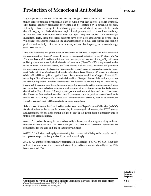
UNIT2.5 Production of Monoclonal AntibodiesHighly specific antibodies can be obtained by fusing immune B cells from the spleen withtumor cells to produce hybridomas,each of which will then secrete a single antibody.The desired antibody-producing hybridoma can be identified by a screening process.If this hybridoma is subjected to a cloning process in which clones are selected,suchthat all progeny are derived from a single cloned parental cell,a monoclonal antibodyis obtained.Monoclonal antibodies have high specificity and can be produced in largequantities.Thus,these biological reagents have been used extensively as probes in awide range of systems including the characterization of novel cell-surface and solubleproteins and carbohydrates,as enzyme catalysts,and for targeting in immunotherapy(see Commentary).This unit describes the production of monoclonal antibodies beginning with protocolsfor immunization(Basic Protocol1)and cell fusion and selection(Basic Protocol2).AnAlternate Protocol describes cell fusion and one-step selection and cloning of hybridomasutilizing a semisolid methylcellulose-based medium(ClonaCell-HY,a registered trade-mark of StemCell Technologies,Inc.;).Methods are providedfor screening primary hybridoma supernatants for antibodies of desired specificity(Sup-port Protocol1),establishment of stable hybridoma lines(Support Protocol2),cloningof these B cell lines by limiting dilution to obtain monoclonal lines(Support Protocol3),recloning of hybridoma cells in semisolid medium(Support Protocol4),and preparationof cloning/expansion medium(thymocyte-conditioned medium;Support Protocol5).Figure2.5.1summarizes these stages and notes the protocols in this and subsequent unitsin which they are detailed.Selection and cloning of hybridomas using the techniquesdescribed in Basic Protocol2require a major commitment of time and labor.However,the Alternate Protocol reduces the overall time necessary to produce monoclonal anti-bodies by18to20days.When successful,the monoclonal antibody may be an extremelyvaluable reagent that will be available in large quantities.Submission of monoclonal antibodies to the American Type Culture Collection(ATCC)for distribution to the scientific community is encouraged.Moreover,the ATCC servesas a repository for cell lines should the line be lost in the investigator’s laboratory due tounforeseen circumstances.NOTE:All protocols using live animals mustfirst be reviewed and approved by an Insti-tutional Animal Care and Use Committee(IACUC)and must conform to governmentalregulations for the care and use of laboratory animals.NOTE:All solutions and equipment coming into contact with living cells must be sterile,and proper aseptic technique should be used accordingly.NOTE:All culture incubations are performed in a humidified37◦C,5%CO2incubatorunless otherwise specified.Some media(e.g.,DMEM)may require altered levels of CO2to maintain pH7.4.Contributed by Wayne M.Yokoyama,Michelle Christensen,Gary Dos Santos,and Diane Miller Current Protocols in Immunology(2006)2.5.1-2.5.25Copyright C 2006by John Wiley&Sons,Inc.Induction of Immune Response2.5.1 Supplement74Production of Monoclonal Antibodies2.5.2Supplement 74Current Protocols in ImmunologyFigure 2.5.1Stages of monoclonal antibody production,with references to the Basic,Alternate,and Support Protocols in this unit (as well as subsequent units)that describe the steps.Induction of Immune Response 2.5.3Current Protocols in Immunology Supplement 74BASIC PROTOCOL 1IMMUNIZATION TO PRODUCE MONOCLONAL ANTIBODIESA wide variety of antigen preparations have been used successfully to produce mon-oclonal antibodies (see Critical Parameters for discussion of antigen preparation).The following protocol provides an immunization schedule for the production of most an-tibodies,although several different schedules can be used.In this protocol,emulsified antigen is injected intraperitoneally into the species of choice.A booster injection is administered 10to 14days after the primary immunization.Three days after the booster injection,the animals’spleens are ready for cell fusion (Basic Protocol 2).MaterialsAntigenComplete Freunds adjuvant (CFA;Sigma)Animal:pathogen-free mouse,hamster,or rat (Armenian hamsters from Cytogen Research are recommended;see Critical Parameters for discussion of animal choice and UNIT 1.1)Incomplete Freunds adjuvant (IFA;Sigma),optional1-to 2-ml glass syringes with Luer-Lok tips,sterile3-way stopcock20-and 22-G needles,sterileAdditional reagents and equipment for handling and restraint of animals (UNIT 1.3)and intraperitoneal injection (UNIT 1.6)CAUTION:CFA is an extremely potent inflammatory agent,particularly if introduced intradermally or into the eyes.Profound sloughing of skin or loss of sight may occur.Self-injection can cause a positive TB skin test and lead to a granulomatous e gloves and protective eyewear when handling CFA.1.Prepare antigen using 2×10⁶to 5×10⁷cells or 1to 50µg protein or peptide per animal to be immunized in normal saline.The antigen may be in several different forms depending on the desired property of the MAb and the method of screening (see Critical Parameters for discussion of antigen preparation and screening assays).If cells are the immunogen,wash three times in serum-free medium before immunization.Plan the immunization of several animals (enough for several fusions)so that primed and boosted animals will be ready 3days before fusion (see Basic Protocol 2).To minimize the risk of introducing a pathogen into the rodent colony,screen cells for pathogens by antibody-production assay (UNIT 1.1).2.Draw up antigen into a sterile 1-to 2-ml glass syringe with a Luer-Lok tip.Connect syringe to a 3-way stopcock.pletely resuspend CFA to disperse the Mycobacterium tuberculosis bacilli,which settle to the bottom of the container with time.Draw up a volume of CFA equal to the antigen volume in a syringe and connect to the antigen-containing syringe.4.Emulsify antigen and CFA by discharging antigen into CFA,then discharging back and forth until a thickened mixture results.Test whether the emulsion is stable—a stable emulsion will not disperse when a drop of it is placed in water.See UNIT 2.4for further discussion of immunization.Figure 2.4.1illustrates the double-syringe device.5.Transfer all of the CFA/antigen emulsion to one syringe and remove the other syringe and stopcock.Attach a sterile 20-G needle to the syringe containing the emulsion.Production ofMonoclonal Antibodies2.5.4Supplement 74Current Protocols in Immunology6.Inject emulsion intraperitoneally into the animal using <0.2ml/mouse,0.5to 1ml/rat,or 0.2to 0.4ml/hamster.Be careful not to force the syringe plunger,since excessive pressure may dislodge the needle and spray the emulsion.Introduce the needle through the skin and tunnel the needle between the skin and peritoneal wall before entering the peritoneal cavity at a site distant from the dermal puncture site.Twirl needle before withdrawal to minimize leakage.Rats are generally anesthetized (UNIT 1.4)whereas mice and hamsters can be manipulated with one hand and do not require anesthetic.7.Boost animal after 10to 14days with approximately the same dose of antigen as in step 5.If cell fusion is planned for 3days after boosting,immunize with antigen alone in aqueous solution,or intact cells in suspension.If a fusion is not immediately planned,boost the animal with antigen emulsified in IFA (which does not contain Mycobacterium tuberculosis bacilli).Do not use CFA for the booster immunizations as this will cause intense inflammation and increased anti-TB antibody response.Blood can be collected from a tail bleed (UNIT 1.7)after the first intraperitoneal boost to obtain antibodies from the blood as a positive control in subsequent detection assays.If desired,antibody titers can be assayed by ELISA (UNIT 2.1)or immunoprecipitation (UNIT8.3),7to 10days after the primary and booster immunizations.BASICPROTOCOL 2CELL FUSION AND SELECTION OF HYBRIDOMAS While animals should be immunized as soon as the decision has been made to produce a monoclonal antibody and the antigen prepared,do not perform cell fusion until the screening assay (Support Protocol 1)has been perfected.Artifactual results that may arise from conditioned media must be identified before cell fusion,because after a fusion there is only a finite amount of time available to assay for the desired monoclonal antibody.Prior to cell fusion,the partner (myeloma)cell line is expanded and a booster injection of antigen is administered to the primed animals.On the day of fusion,the spleens are harvested.Spleen cells and partner cells are washed,harvested,and mixed.Cell fusion is performed at 37◦C in the presence of polyethylene glycol (PEG).The resulting pellet is harvested and plated into tissue culture plates.After incubation with hypoxanthine,aminopterin,and thymidine (HAT)medium and feeding over ∼2weeks,the hybridomas are ready for screening (Support Protocol 1).Materials SP2/0-Ag14myeloma cell line (drug-marked,nonsecretory;ATCC #CRL 1581)Complete DMEM-10and -20media (APPENDIX 2A )with 10mM HEPES and 1mM sodium pyruvate Primed animal;mouse,hamster,or rat (10to 14days after primary immunization;(see Basic Protocol 1)Complete DMEM medium (APPENDIX 2A ),serum-free 50%polyethylene glycol (PEG),sterile Ammonium chloride solution (see recipe)Complete DMEM-20/HEPES/pyruvate/HAT (or HT)medium (see recipe)175-cm 2flasks Fine-mesh metal screen 50-ml conical polypropylene centrifuge tubes Beckman TH-4rotor or equivalent 96-well flat-bottom microtiter platesInduction of Immune Response 2.5.5Current Protocols in Immunology Supplement 74Additional reagents and equipment for animal euthanasia (UNIT 1.8),spleen removal (UNIT 1.10),and counting cells and assessing cell viability by trypan blueexclusion (APPENDIX 3B )Prepare myeloma cells (1week before fusion)1.One week before fusion,begin expansion of SP2/0-Ag14myeloma cell line (the fusion partner cell line)in complete MEM-10/HEPES/pyruvate (see Critical Pa-rameters).By the day cell fusion is to be performed,the following total number of myeloma cells must be available (in multiple 175-cm 2flasks containing 100ml each),depending upon the source of the primed animal:mouse spleen,1×108cells in two or three flasks;hamster spleen,2×108cells in three or four flasks;and rat spleen,5-10×108cells in ten flasks.Two mouse or hamster spleens,or one rat spleen,will provide enough cells for the fusion (see step 7).Boost primed animal (3days before fusion)2.Three days before fusion,boost primed animal(s)according to step 7of Basic Protocol 1.Prepare reagents and split myeloma cells (1day before fusion)3.One day before fusion,prepare all reagents and media,particularly 50%PEG.4.One day before fusion,split SP2/0-Ag14myeloma cells (from step 1)into fresh complete DMEM-10/HEPES/pyruvate medium.Vigorous growth of the SP2/0-Ag14cells is generally required for good fusion.Check myeloma cells and prewarm reagents (day of fusion)e an inverted microscope to check the SP2/0-Ag14myeloma cells to make sure they are growing vigorously (refractile and not pyknotic),they are not contaminated (no obvious bacteria or fungi),and there are enough cells for the fusion.It is better to postpone the fusion than to perform an ill-advised fusion,since the entire selection and screening effort will take ∼3weeks.6.Prewarm the following in a 37◦C water bath:Three 400-and three 600-ml beakers,each containing ∼100ml H 2O20ml sterile complete serum-free DMEM5ml sterile 50%PEG solution.Harvest spleen and prepare cells7.Sacrifice boosted animal(s)(UNIT 1.8)and aseptically harvest spleen(s)(UNIT 1.10).Do not use anesthetics for sacrifice.Instead,use cervical dislocation for mouse,or CO 2asphyxiation for mouse,hamster,or rat to avoid introducing an anesthetic into the bloodstream and therefore into the cultures.8.Transfer spleen to a sterile 100-mm-diameter petri dish filled with 10ml sterile complete serum-free DMEM.Perform all subsequent steps in a laminar flow hood.9.Tease spleen into a single-cell suspension by squeezing with angled forceps or by chopping with fine-tipped dissecting scissors.Remove debris and disperse cells further by passage through a fine-mesh metal screen.10.Transfer spleen cell suspension to a sterile 50-ml conical centrifuge tube and fillwith sterile complete serum-free DMEM.Do not use protein-or HEPES-containing medium because the PEG will precipitate proteins and HEPES can be toxic to cells during fusion.Production ofMonoclonal Antibodies2.5.6Supplement 74Current Protocols in Immunology11.Centrifuge 5min in TH-4rotor at 1500rpm (500×g ),room temperature,and discard supernatant.12.Lyse red blood cells (RBC)by resuspending pellet in 5ml ammonium chloride solution.Let stand 5min at room temperature.13.Add 45ml sterile complete serum-free DMEM,and centrifuge as in step 11.14.Resuspend pellet in 50ml sterile complete serum-free DMEM.Centrifuge as in step 11.Repeat DMEM addition and centrifuging once (each repeat is a wash).15.While spleen cells are being washed,separately harvest the SP2/0-Ag14myeloma cells (from step 5)by transferring the cells to 50-ml conical centrifuge tubes.Centrifuge as in step 11.Resuspend myeloma cells in DMEM and pool all cells into one 50-ml conical centrifuge tube.Wash myeloma cells three times as in step 14.16.Separately resuspend the spleen and myeloma cells in 10ml complete serum-free DMEM.Count cells and assess viability in each cell suspension using a hemacytometer and trypan blue exclusion (APPENDIX 3B );there should be nearly 100%viability of both suspensions.17.On basis of cell counts (from step 16),calculate the amount of complete DMEM-20/HEPES/pyruvate needed to plate cells at ∼2.5×10⁶total cells/ml.Prewarm this amount of complete DMEM-20/HEPES/pyruvate in 37◦C water bath.Prepare 96-well flat-bottom plates by labeling them sequentially:one plate is required for each 10ml of final cell suspension.Perform cell fusion 18.Mix SP2/0-Ag14myeloma and spleen cells at a 1:1ratio in a 50-ml conical cen-trifuge tube.Fill the tube with complete serum-free DMEM.Other cell ratios work.Successful fusions have been performed with a ratio of myeloma/spleen cells as low as 1:20.19.Centrifuge cell mixture 5min at 500×g ,room temperature.20.While cells are in the centrifuge,prepare three 37◦C double-beaker water baths in the laminar flow hood by placing a 400-ml beaker (from step 6)containing 100ml of 37◦C water into 600-ml beaker containing 75to 100ml of 37◦C water.Place the tubes of prewarmed 50%PEG solution and complete serum-free DMEM (from step 6)into two of the 37◦C water baths in the hood.21.Aspirate and discard supernatant from the mixed-cell pellet (from step 19).22.Perform the cell fusion at 37◦C by placing the tube containing the mixed-cell pellet in one of the double-beaker water baths in the laminar flow ing a 1-ml pipet,add 1ml prewarmed 50%PEG to the mixed-cell pellet drop-by-drop over 1min,stirring the cells with the pipet tip after each drop.Stir for an additional ing a clean pipet,add 1ml prewarmed complete serum-free DMEM to the cell mixture drop-by-drop over 1min,stirring after each drop.Repeat once with an additional 1ml of prewarmed complete serum-free DMEM.25.With a 10-ml pipet,add 7ml prewarmed complete serum-free DMEM drop-by-drop over 2to 3min.Macroscopic clumps of cells should be obvious at this point.26.Centrifuge 5min at 500×g ,room temperature.Induction of Immune Response 2.5.7Current Protocols in Immunology Supplement 7427.While the cells are in the centrifuge,rewarm the beaker water baths to 37◦C andplace in the hood.Place prewarmed complete DMEM-20/HEPES/pyruvate (from step 17)in the beaker water bath.28.Discard the supernatant (from step 26).Place tube in the beaker water bath.29.With a clean 10-ml pipet,forcefully discharge 10ml prewarmed complete DMEM-20/HEPES/pyruvate to the cell pellet.30.Repeat step 29until the total volume of prewarmed complete DMEM-20/HEPES(calculated in step 17)is added.If necessary,allow clumps to settle and disrupt with the pipet tip.Further warming of cell suspension is no longer required.If the total volume exceeds 50ml,gently aspirate and transfer to another sterile container such as a tissue culture flask.31.Gently aspirate 10ml of cell suspension with a 10-ml pipet.Add 2drops (100to 125µl)of suspension to each well of a 96-well flat-bottom plate (continue until entire suspension is plated).Incubate overnight in a humidified 37◦C,5%CO 2incubator.Vigorous pipetting of the cell suspension should be avoided at this point,as the newly formed hybrids are unstable.Moreover,the vigorous addition of cells to the wells with repeating micropipettor is not e a pipet aid and hold the 10-ml pipet at a 45◦angle with the tip 1to 2cm above the well,bracing the pipet with a finger from the opposite hand.To avoid introducing contaminants,do not hold hands above the plate.A steady,even flow of drops from the pipet will allow the most efficient delivery of cell sus-pension or medium to the e a fresh pipet to withdraw additional cell suspension.As an optional step to minimize fibroblast overgrowth,permit the fibroblasts in bulk-fused cell suspension to adhere overnight to tissue culture flasks before seeding the 96-well plates.Many investigators select their hybridomas under bulk conditions—i.e.,they incubate large numbers of cells per well in larger plates or flasks.This makes feeding easier,but allows fast-growing hybridomas to overgrow the others.Since nonproducing hybridomas tend to grow faster,especially in the hamster-mouse fusions,hybridomas are isolated initially in multiple small wells in this protocol.The primary hybridomas tend to be monoclonal.This is especially important when screening procedures are used that require differential reactivities,e.g.,to different cell lines by flow cytometry analysis or to different antigen preparations.In those cases,multiple hybridomas per well will obscure the reactivity of the MAb of interest.Monitor and feed cells32.After one day of incubation,check wells under an inverted microscope.If seededwith the appropriate number of cells,there should be a nearly confluent monolayer of highly viable cells on the bottom and obvious clumps of cells.33.Add 2drops complete DMEM-20/HEPES/pyruvate/HAT to each well with a 10-ml pipet (see step 31).Place in humidified 37◦C,5%CO 2incubator.Use a separate pipet for each microtiter plate and keep the same covers with each plate to ensure that each plate remains a separate unit and to avoid spreading contamination.It cannot be overemphasized that it takes practice and meticulous attention to possible sources of contaminants to keep these plates sterile during the subsequent 2-to 3-week feeding and monitoring schedule.If plates become contaminated,discarding them is advised.Alternatively,contamination in one or two wells may be treated by aspirating the contents of the contaminated well with a sterile Pasteur pipet attached to a vacuum flask,rinsing the well with 70%ethanol,and wiping with a sterile cotton swab.Wash twice with ethanol.Finally,blot the well dry with the sterile cotton swab and blot the appropriate area of the cover with a sterile cotton swab soaked in 70%ethanol.Do not open contaminated plates while other plates are in the hood.Production ofMonoclonal Antibodies2.5.8Supplement 74Current Protocols in Immunology34.On days 2,3,4,5,7,9,and 11,aspirate half the volume of each well using a sterile,short Pasteur pipet attached to a vacuum flask,holding pipet at a 45◦angle and touching tip to surface of supernatant at the point where the liquid meets the opposite wall of the well.Feed the cells by adding 2drops complete DMEM-20/HEPES/pyruvate/HAT from a 10-ml pipet (see steps 31and 33)to each well,and return to humidified 37◦C,5%CO e a separate Pasteur pipet for each plate to minimize spreading contamination.Since the frequency of successful viable hybridoma formation is ≤10−5,when HAT is added,profound cell death should be apparent at days 2and 3and the remaining viable cells should not be readily apparent until they have expanded.By day 7to 9for mouse-mouse fusions,day 11for rat-mouse fusions,and day 14for hamster-mouse fusions,clusters of hybridoma cells should become visible under the inverted microscope.If profound cell death is not apparent on days 2and 3,check the medium containing HAT and the parental cell line by incubating an aliquot of the parental myeloma line with the medium containing HAT.The feeding schedule is not rigid except for the first 4days,when it is necessary to remove the toxic products of cell death.Thereafter,feedings will depend on the actual number of cells deposited in the wells,efficiency of fusion,and appearance and growth of hybridomas.Do not allow wells to become yellow (acidic)for more than a day.Examine plates daily,even if cells are not scheduled to be fed,and feed plates if acidic wells are noted.35.On day 14,repeat feeding as outlined in step 34except use complete DMEM-20/HEPES/pyruvate/HT to feed cells.Return to 37◦C,5%CO 2incubator.Cells do not require more than one change of complete DMEM-20/HEPES/pyruvate/HT.After this change,the aminopterin (from prior addition of HAT medium)is apparently diluted out enough so that the cells can survive without additional HT.36.On day 15and subsequently,feed wells as noted using complete DMEM-20/HEPES/pyruvate without HAT or HT.The hybridomas are ready for screening when most of the wells containing growing cells demonstrate 10%to 25%con-fluence and when those with denser populations turn yellow within 2days after feeding (see Support Protocol 1).If the screening assay requires a [3H]thymidine incorporation assay (APPENDIX 3D ),be aware that the large amount of thymidine in complete DMEM-20/HEPES/pyruvate with HAT and HT will serve as a cold-label inhibitor of [3H]thymidine incorporation.At least 3to 4changes of complete DMEM-20/HEPES/pyruvate without HT are required to dilute out excess thymidine.ALTERNATEPROTOCOLCELL FUSION,SELECTION,AND CLONING OF HYBRIDOMAS USING A SEMISOLID MEDIUM (CLONACELL-HY)Traditionally,monoclonal antibody development has involved selecting hybridomas in suspension cultures,identifying cultures that produce antibodies specific for the targeted antigen,and cloning the specific antibody-producing hybridoma(s)by at least one round of culture under limiting-dilution conditions (as described in Basic Protocol 2and Support Protocols 1to 3).This approach is laborious and time consuming and may result in the selection of identical,duplicate clones.This section describes protocols for the use of a methylcellulose-based medium system,ClonaCell-HY ,for cloning and selection of mouse hybridomas.Performing hybridoma selection and cloning simultaneously in ClonaCell-HY reduces the time and reagents necessary to obtain a monoclonal hybridoma producing antibody against the antigen of interest.Cultures do not need feeding or maintenance during the selection process.This approach also allows all daughter cells to remain together during the selection process,decreasing the number of clones that need to be tested for antibody production.Selection of duplicate hybridomas,a common occurrence with hybridoma cloning in suspension cultures,is avoided.An additionalInduction of Immune Response 2.5.9Current Protocols in Immunology Supplement 74advantage is that smaller,slow-growing clones,which can easily be lost due to overgrowth by larger,faster-growing hybridomas in traditional liquid suspension culture,remain physically separated in semisolid medium from the larger,faster-growing clones,and can thus be isolated and screened separately.NOTE:All solutions and media should be prewarmed to 37◦C unless otherwise indicated.MaterialsMyeloma cell line (e.g.,SP2/0,X63Ag8.653;available from ATCC)ClonaCell-HY Monoclonal Antibody Production Kit (StemCell Technologies,Inc.)containing:Medium A—ClonaCell-HY Pre-Fusion Medium and Hybridoma Expansion Medium,500mlMedium B—ClonaCell-HY Fusion Medium,500mlMedium C—ClonaCell-HY Hybridoma Recovery Medium,100mlMedium D—ClonaCell-HY Hybridoma Selection Medium containing HAT,90ml Medium E—ClonaCell-HY Hybridoma Growth Medium containing HT,500ml Polyethylene glycol—ClonaCell-HYPEGSolution,pretestedforcellfusion,1.5ml Immunized mouse,1to 4days after final antigen boost (Basic Protocol 1)3%(v/v)acetic acidLiquid nitrogen (optional)Fetal bovine serum (FBS)containing 20%(v/v)DMSO15-and 50-ml conical polypropylene centrifuge tubes100-mm petri dishesFine-mesh metal screenLow-speed tabletop centrifuge3-ml and 12-ml syringes25-and 75-cm 2tissue culture flasks16-G blunt-ended hypodermic needles96-and 24-well tissue culture platesCryotubes (e.g.,Nunc)Liquid nitrogen freezer (Dewar flask and canes to accommodate cryotubes;optional)Additional reagents and equipment for determining cell viability by trypan blue exclusion (APPENDIX 3B ),animal euthanasia (UNIT 1.8),spleen removal (UNIT 1.10),preparing a single-cell suspension of splenocytes (UNIT 3.1),counting cells using a hemacytometer (APPENDIX 3),assaying for antigen production from hybridoma clones by ELISA (UNIT 2.1),flow cytometry (Chapter 5),or immunoblotting (UNIT 8.10),and cryopreservation of cells (APPENDIX 3G )Prepare myeloma cells1.Culture the parental myeloma cells in 25-cm 2tissue culture flasks with Medium A (Pre-Fusion Medium from ClonaCell-HY kit)for at least 1week prior to fusion to ensure they are well adapted to this medium.Seed cells at a density of ∼5×10⁴cells/ml and passage every 2days.Suggested maximum cell density is 4×10⁵cells/ml.The parental myeloma cells must not secrete any of their own immunoglobulin chains.They should be mycoplasma-free and efficiently fuse to form stable hybridomas that continuously secrete specific monoclonal antibodies.Parental myeloma cells that meet these criteria (such as SP2/0and X63Ag8.653)are widely available.2.Calculate the cell growth rate at every passage (APPENDIX 3A ).The day before the fusion,count the viable cells and split cells so that there will be at least 2×10⁷parental myeloma cells available the next day.The recommended cell density for fusion is 2×10⁵cells/ml.Only 100ml of these cells will be needed,but 200ml should be cultured to ensure an adequate supply.Production of Monoclonal Antibodies2.5.10Supplement 74Current Protocols in Immunology 3.Harvest the required number of parental myeloma cells in a 50-ml conical polypropylene centrifuge tube.Centrifuge 10min at 300×g ,room temperature or 37◦C,and remove the supernatant.Wash three times,each time by adding 30ml of Medium B (Fusion Medium),centrifuging again as before,and removing the supernatant.Resuspend final pellet in 25ml Medium B.This step may be performed simultaneously with or subsequent to spleen cell preparation (steps 6to 10)to ensure that the myeloma cells do not sit for an extended period of time.It is important to remove all the serum adhering to the cells by washing with serum-free Medium B.If the serum is not removed,the PEG will not fuse the cell membranes and the fusion frequency will drop drastically.4.Count live cells using a viability stain (APPENDIX 3B ).Viability of parental myeloma cells should be >95%.5.Calculate the volume of cell suspension that contains 2×10⁷cells,to be used in step 11.Place cells at room temperature or 37◦C.Harvest spleen and prepare spleen cells 6.Sacrifice immunized animal(s)(UNIT 1.8)and aseptically remove spleen(s)(UNITS 1.8&1.10).Place spleen in a sterile 100-mm petri dish containing 5ml Medium A (Pre-Fusion Medium).IMPORTANT NOTE:The ClonaCell-HY kit has been optimized for use with mouse hybridomas.Hybridomas from other species have not been tested.Do not use anesthetics for sacrifice.Instead,use cervical dislocation or CO 2asphyxia-tion to avoid introducing anesthetic into the bloodstream and therefore into the cultures.It is important to collect blood from the animal to obtain serum as a source of antibodies for a positive control in subsequent screening assays.Blood can be collected from a tail bleed (UNIT 1.7)after the first intraperitoneal boost (see Basic Protocol 1,step 7)or from the heart at the time of spleen harvest.To collect blood from heart use a sterile Pasteur pipet and place blood into a 1.5-ml microcentrifuge tube.Let blood stand at room temperature with the cap off to allow blood to clot.After 30min,remove the blood clot with a sterile Pasteur pipet and place tube at 4◦C.The next day,centrifuge tube for 15min at 400×g,4◦C or room temperature.Collect serum and add sodium azide to a final concentration of 0.1%.Store at −20◦C 7.Disaggregate the spleen into a single-cell suspension (UNIT 3.1).Transfer the spleen to a fine-mesh metal screen placed on top of a 50-ml conical centrifuge tube,and use the plunger of a 3-m1syringe to gently grind the cells out of the spleen.Rinse the screen with Medium B to help cells pass through the screen.Pipet the cells up and down in the tube with a 10-ml pipet to break up lumps.Try not to cause the solution to foam.Only the spleen membrane should be left in the screen.See UNIT 3.1for additional dis-cussion of the above procedure.Other spleen disaggregation methods may also be used.8.Centrifuge cell suspension 10min at 400×g ,room temperature or 37◦C,and remove supernatant.Wash the splenocytes three times,each time by adding 30ml Medium B,centrifuging 10min at 400×g ,room temperature or 37◦C,and discarding the supernatant.Resuspend the final cell pellet in 25ml Medium B.It is important to remove all the serum adhering to the cells by washing with serum-free Medium B.If the serum is not removed,PEG will not fuse the cell membranes and the fusion frequency will drop drastically.。
欧盟GMP附录11-计算机系统(中英文对照)

EUROPEAN COMMISSION欧盟委员会HEALTH AND CONSUMERS DIRECTORATE-GENERAL卫生与消费者协会Public Health and Risk Assessment公共卫生与风险评估Pharmaceuticals药品Brussels,SANCO/C8/AM/sl/ares(2010)1064599EudraLexThe Rules Governing Medicinal Products in the European Union欧盟药品生产规范Volume 4卷4Good Manufacturing PracticeMedicinal Products for Human and Veterinary Use人用与兽用药品良好生产管理规范Annex 11: Computerised Systems附件11:计算机系统Legal basis for publishing the detailed guidelines: Article 47 of Directive2001/83/EC on the Community code relating to medicinal products for human use and Article 51 of Directive 2001/82/EC on the Community code relating to veterinary medicinal products. This document provides guidance for the interpretation of the principles and guidelines of good manufacturing practice (GMP) for medicinal products as laid down in Directive 2003/94/EC for medicinal products for human use and Directive 91/412/EEC for veterinary use.依法发布的具体指导方针:2001/83/EC第47条人用药品规范和2001/82/EC第51条兽用药品规范。
蛋白质组学 生物芯片 壁垒

蛋白质组学生物芯片壁垒英文回答:Proteomics and Biochips: Breaking Barriers and Unlocking New Possibilities.Proteomics, the study of proteins, is a vital fieldthat has revolutionized our understanding of biological processes and disease mechanisms. Biochips, miniaturized devices that can analyze multiple proteins simultaneously, have played a critical role in advancing proteomics research. However, the integration of proteomics and biochips has faced several barriers, limiting their full potential.One major barrier has been the complexity of proteomes. With thousands of proteins present in a single biological sample, it has been challenging to develop biochips that can capture and analyze a comprehensive range of proteins. Additionally, post-translational modifications (PTMs) andprotein isoforms can further increase the complexity of proteomes, making it difficult to detect and quantify all relevant protein species.Another barrier has been the lack of standardized protocols and data analysis methods. This has hindered the reproducibility and comparability of proteomics data across different laboratories and studies. As a result, it has been difficult to draw robust conclusions from proteomics studies and translate findings into clinical applications.To overcome these barriers, researchers have explored various innovative approaches. One strategy has been to develop biochips with high capture efficiency andspecificity for target proteins. This has been achieved through the optimization of surface chemistry, the use of affinity ligands, and the incorporation of microfluidic technologies.Advances in mass spectrometry-based proteomics have also played a crucial role in improving the accuracy and sensitivity of biochip-based protein analysis. By couplingbiochips with mass spectrometers, researchers can identify and quantify proteins with high throughput and precision.Furthermore, the development of standardized data analysis pipelines and repositories has facilitated the sharing and comparison of proteomics data. This has enabled researchers to pool their resources and leverage the collective knowledge of the scientific community.As these barriers continue to be addressed, the integration of proteomics and biochips holds immense promise for advancing our understanding of protein functions, cellular processes, and disease mechanisms. By providing high-throughput, multiplexed, and sensitive protein analysis, biochip-based proteomics is poised to revolutionize healthcare, personalized medicine, and drug discovery.中文回答:蛋白质组学与生物芯片,突破障碍,释放新可能。
欧盟GMP附录15:确认与验证(修订版英文+中文)
EUROPEAN COMMISSIONENTERPRISE DIRECTORATE-GENERALSingle market, regulatory environment, industries under vertical legislationPharmaceuticals and cosmeticsBrussels,30 March 2015EudraLex欧盟药品管理法Volume 4EU Guidelines forGood Manufacturing Practice forMedicinal Products for Human and Veterinary Use第四卷欧盟人用和兽用药品GMP指南Annex 15: Qualification and Validation附录15:确认和验证Legal basis for publishing the detailed guidelines:Article 47 of Directive 2001/83/EC on the Community code relating to medicinal products for human use and Article 51 of Directive 2001/82/EC on the Community code relating to veterinary medicinal products. This document provides guidance for the interpretation of the principles and guidelines of good manufacturing practice (GMP) for medicinal products as laid down in Directive 2003/94/EC for medicinal products for human use and Directive 91/412/EEC for veterinary use.发布该细化指南的法律依据:人用药物欧共体法案指令2001/83/EC第47章和兽用药物欧共体法案指令2001/82/EC第51章。
09第九章发酵工程各论3抗生素发酵工艺学.pptx

2
1. 概 述 Introduction
抗生素
是生物在其生产活动过 程中所产生,并能 在低微浓度下有选 择性地抑制或杀灭 其他微生物或肿瘤 细胞的有机物。
Antibiotics
Any organic substances produced by various organisms that have the power of arresting the growth of other microorganisms or of destroying them.
照射 亚硝基胍、亚硝酸、
秋水仙素、氮芥等 诱变剂处理
Conventional strain selection • Physical mutagen
eg. Ultraviolet Radiation • Chemical mutagen
eg. Nitrosoguanidine (NTG) • Improved strain can be
抗生素发酵生产技术 Antibiotics Fermentation Production
韩北忠
中国农业大学 食品学院
Han Bei-zhong, Prof. PhD
College of Food Science & Nutritional Engineering
China Agricultural University
现代分析仪器: • 核磁共振 • 毛细管电泳 • 气相色谱 • 高效液相色谱 • 质谱
Modern analytical instruments:
• Nuclear magnetic resonance (NMR)
• Capillary electrophoresis (CE)
FDA行业指南预防非青霉素β-内酰胺药物交叉污染的CGMP框架-正式版-中英文双语
Guidance for IndustryNon-Penicillin Beta-Lactam Drugs:A cGMP Framework for Preventing Cross-Contamination行业指南:预防非青霉素β-内酰胺类药物交叉污染的cGMP框架U.S. Department of Health and Human Services美国卫生与公共服务部Food and Drug Administration美国食品与药品监督管理局Center for Drug Evaluation and Research (CDER)药品评价和研究中心(CDER)April 20132013.04Current Good Manufacturing Practices (CGMPs)现行质量管理规范(CGMPs)Guidance for IndustryNon-Penicillin Beta-Lactam Drugs:A cGMP Framework for Preventing Cross-Contamination行业指南:预防非青霉素β-内酰胺类药物交叉污染的cGMP框架Additional copies are available from:Office of CommunicationsDivision of Drug Information, WO51, Room 2201Center for Drug Evaluation and ResearchFood and Drug Administration10903 New Hampshire Ave.Silver Spring, MD 20993-0002Phone: 301-796-3400; Fax: 301-847-8714druginfo@/Drugs/GuidanceComplianceRegulatoryInformation/Guidances/default.htmU.S. Department of Health and Human ServicesFood and Drug AdministrationCenter for Drug Evaluation and Research (CDER)April 2013Current Good Manufacturing Practices (CGMP)TABLE OF CONTENTS 目录I. INTRODUCTION简介 (4)II. BACKGROUND背景 (6)III. RECOMMENDATIONS建议 (13)Guidance for Industry1Non-Penicillin Beta-Lactam Drugs:A CGMP Framework for Preventing Cross-Contamination行业指南:预防非青霉素β-内酰胺类药物交叉污染的CGMP框架I. INTRODUCTION 简介This guidance describes the importance of implementing manufacturing controls to prevent cross-contamination of finished pharmaceuticals and active pharmaceutical ingredients (APIs) with non-penicillin beta-lactam drugs. This guidance also provides information regarding the relative health risk of, and the potential for, cross-reactivity in the classes of sensitizing beta-lactams (including both penicillins and non-penicillin beta-lactams). Finally, this guidance clarifies that manufacturers generally should utilize separate facilities for the manufacture of non-penicillin beta-lactams because those compounds pose health risks associated with cross-reactivity.本指南阐述了采取生产控制以预防制剂产品、原料药(APIs)与非青霉素β-内酰胺类药物发生交叉污染的重要性,并且提供了不同类别致敏性β-内酰胺类药物(包括青霉素类和非青霉素β-内酰胺类)发生交叉反应的可能性及其相对健康风险信息。
欧盟GMP附录一 无菌产品生产 2020版_中英文对照版_
9. Viable and non-viable environmental and process monitoring
This section differs from guidance given in section 4 in that the guidance here applies to ongoing routine monitoring with regards to the design of systems and setting of action limits alert levels and reviewing trend data.
microbial contamination such as personnel, materials and the surrounding environment, and assist in the rapid detection of potential contaminants in the environment and product. 厂房设施,设备及工艺设计应根据良好药品生产管理规范(GMP)相关附录中的要求 进行优化,确认和验证。应考虑采用适当技术手段(例如,限制进入屏障系统 (RABS),隔离器,机器人系统,快速微生物测试和监测系统)以加强从潜在外来微 粒和微生物污染(例如人员,物料及周边环境)中对产品进行保护,并实现对环境和 产品中的潜在污染的快速识别。
2 Principle 原则
2.1 The manufacture of sterile products is subject to special requirements in order to minimize risks of microbial, particulate and pyrogen contamination. The following key areas should be considered: 无菌产品的生产应符合特定要求,以减少来自微生物, 颗粒及热原方面的污染风险, 应考虑到 以下关键区域:
靶向捕获试剂英文术语
靶向捕获试剂英文术语1. “Target - Capture Reagent”,这可是靶向捕获试剂最直白的英文术语啦。
就像猎人瞄准猎物一样,这个术语精准地指向了它的功能。
比如说在医学研究中,我们想要抓住特定的癌细胞标记物,就像用这个“Target - Capture Reagent”当神奇的小钩子,把那些狡猾的癌细胞标记物给勾住。
2. “Specific - Binding Agent”,听起来是不是很专业又好理解呢?嘿,这就好比是一把特制的钥匙(试剂),只能开特定的锁(目标分子)。
想象一下,在基因检测里,这个试剂就像个超级专一的小助手,只跟它对应的基因片段结合,就像找到自己唯一的舞伴一样。
3. “Capture Probe”,哇塞,这个词儿就像一个探测小能手呢!就好像我们在一个大宝藏堆里找一颗特别的宝石,这个“Capture Probe”就能像小探针一样,直接扎到我们想要的靶向分子上。
在生物化学实验里,它就专门负责找到那些隐藏起来的特定蛋白,厉害吧!4. “Targeted - Trapping Substance”。
这名字感觉就像设下了一个陷阱,专门逮住特定的目标。
你看啊,就像警察抓小偷,这个物质就是那个聪明的警察,守在特定的路口(细胞环境里的特定位置),等着小偷(靶向分子)出现就立马抓住。
在药物研发中,它可以用来捕捉那些致病的异常分子。
5. “Selective - Capture Molecule”。
选择性捕获分子哦,这就像是一场选美比赛,只选最美的(特定的目标分子)。
比如在环境监测中,如果我们要检测土壤里特定的污染物分子,这个分子就像个挑剔的评委,只对那些特定的污染物分子说“Yes”,然后把它们捕获住。
6. “Target - Locking Reagent”。
这就如同给目标上了一把锁,只有这个试剂能打开。
就像在密码锁的世界里,只有特定的密码(这个试剂)才能打开对应号码(靶向分子)的锁。
USP 1132 HCP
Summary, Conclusions, and References (Guidances)
8
HCP Critical Reagents: the Antigen/Standard Design, Preparation, Characterization
Immunogen = (reformulated) standard
2
HCP Expert Panel Charter and Members
Provide stakeholders with best practices (USP chapter <1132>) for preparation and characterization of assay reagents, and development and validation of Host Cell Protein (HCP) measurement procedures (focus on immunoassays)
5
Draft Chapter Terminology, cont’d.
Process-Specific: A proprietary set of standards and antibodies used for a single product that fall in 2 major classes:
– Upstream process-specific: an assay designed from material where the upstream culture process deviates significantly from the platform. This is prior to any purification and may be applied to more than one product if these parameters are similar.
中华人民共和国反倾销条例(英文)
1. where there is a purchase price that has been paid or a purchase price that is payable for the imported product, that price shall be the export price; or
中华人民共和国反倾销条例(修正) State Council, PRC Anti-dumping Regulations (Revised)1
国务院令第401号
(Promulgated by the State Council on 26 November 2001, revised according to the > Decision> on 31 March 2004, and effective as of 1 June 2004.)
2. where there are no sales of the product that is the same as the imported product in the market of the country (region) of export in the ordinary course of trade, or the price or quantity of such product cannot be compared with that of the imported product on a fair basis, the comparable price for export of the product to a proper third country (region), or the cost of the same product in the original country (region) of its production plus reasonable expenses and profit, shall be the normal value.
- 1、下载文档前请自行甄别文档内容的完整性,平台不提供额外的编辑、内容补充、找答案等附加服务。
- 2、"仅部分预览"的文档,不可在线预览部分如存在完整性等问题,可反馈申请退款(可完整预览的文档不适用该条件!)。
- 3、如文档侵犯您的权益,请联系客服反馈,我们会尽快为您处理(人工客服工作时间:9:00-18:30)。
a rXiv:n ucl-t h /96918v29Sep1996Antiproton Production in pp,dp and dd Collisions close to Threshold ∗G.I.Lykasov,M.V.Rzjanin Joint Institute for Nuclear Research 141980,Dubna,Moscow Region,Russia W.Cassing Institut f¨u r Theoretische Physik Heinrich-Buff-Ring 16,D-35392Giessen,Germany February 9,2008Abstract The production of antiprotons in pp collisions is investigated close to thresh-old where experimental data about the total cross section are not available.We analyze the latter reaction within the LUND string model for inclusive ¯p produc-tion and within the framework of a one-boson exchange model for the exclusive reaction pp →ppp ¯p .The application of our new results to the analysis of sub-threshold antiproton production in d +p and d +d collisions shows cross sections that are much lower than expected before.Nevertheless,the comparison of ex-perimental ¯p differential cross sections from d +p and d +d is expected to provide valuable information about a nonnucleonic component in the deuteronwavefunc-tion.The production of particles at energies below the free nucleon-nucleon threshold is one of the most promising sources of information about the properties of nuclear matter at high densities or about nucleon-nucleon correlations at short N−N distances[1]. Apart from heavy meson(η,K+,K−,ρ,ω,Φ)production,the investigation of antipro-tons is of particular interest since they involve a much larger production threshold and can be more easily identified with magnetic spectrometers due to their large mass and negative charge.In fact,antiprotons have been detected at far subthreshold energies in both,p+A and A+A collisions[2,3,4,5,6].The actual magnitude of the cross sections observed indicate strong in-medium modifications of the antiprotons as found from independent transport theoretical studies[7,8,9,10].Although the proper magnitude of the¯p potential and its annihilation cross section in the medium is still a matter of debate,it is clear that the dominant production mechanism in nucleus-nucleus collisions proceeds via intermediate baryon resonances since the latter act as short-time energy reservoirs for the¯p production[8,10,11].On the other hand the N∆or even∆∆channels play a minor role in proton-nucleus and deuteron-nucleus reactions since the resonances on average decay before colliding with another nucleon due to the much lower densities involved[12].However,the poor knowledge of the elementary production cross section pp→¯p+X especially close to threshold leads to large ambiguities in the interpretation of the experimental data about subthreshold¯p production in p+A and A+A collisions and thus to sizeable uncertainties for the antiproton potential or selfenergy in the nucleus. It is thus mandatory to analyse the elementary production in situations where the kinematical conditions are more clearly defined and where in-medium potentials as well as intermediate pion induced production channels as well as antiproton annihilation can approximately be neglected.This is clearly the case for pp collisions and also quite well fulfilled for d+p and d+d reactions according to the analysis in[12,13].In this paper we thus concentratefirst on the inclusive process pp→¯p X,which in comparison to the data from[14]is quite well described within the LUND string√formation and fragmentation model[15]at invariant energies aboveWe start with the inclusive¯p production in pp collisions and show in Fig.1a) the available experimental data from[14](full squares)in comparison to the results from the LUND string model(LSM)[15](open circles)as a function of the invariant energy above threshold.Since the description of the inclusive data within the LSM is quite good,we use the same approach to compute the relative fraction of events corresponding to the exclusive channel pp→ppp¯p which is represented in Fig.1a)by the crosses.Whereas at higher energies the inclusive antiproton production corresponds to events with3baryons and further mesons,below about√128(2π)5λ(s,m2,m2) s+3m2ds3 t+3t−3dt3 t+1t−1dt1 s+2s−2ds2 t+2t−2(1)|T NN→¯p pNN(t1,t2,t3,s2)|2dt22s3(s3+m2−t3)(s3+m2−s2)∓λ1/2(s3,m2,t3)λ1/2(s3,m2,s2)); t±2=t1+m2−1s±2=s3+m2−1s(s+m2−s3)∓λ1/2(s,m2,m2)λ1/2(s,m2,s3)).2sThe matrix element T NN→¯p pNN(t1,t2,t3,s2)can be calculated within the framework of the one-meson exchange model taking into account both pseudoscalar,scalar and vector mesons[18].Neglecting the higher order terms in t2/m4caused by the tensor part of the NNρ-vertex,which is legitimate at not too large transfers t1and t3(cf. [18,19]),it can be written in the form:|T NN→¯p pNN(t1,t2,t3,s2)|2= i g2iNN|t1|F2i(t1)(t3−m2i)2|f ii→¯p p(s2,t2)|2,(2) where f ii→¯p p(s2,t2)is the amplitude for the process ii→¯p p and the index i stands for the exchanged meson(cf.Fig.2)while F i is the corresponding formfactor and g iNN is the iNN coupling constant corresponding to the exchanged meson(i=π0,ρ0,ω).The coupling constants and formfactors are taken from refs.[18,19,20].We incoherently sum the contributions of theπ0,ρ0,ω-exchange graphs(cf.Fig.2) because the amplitudes for the processes ii→¯p ppp are not known sufficiently well and more reliable theoretical approaches are not available so far.Furthermore,assuming off-mass shell effects in the amplitudes f ii→¯p p(s2,t2)to be small,they can be related to the differential cross sections of the reactions ii→¯p p by|f ii→¯p p(t1,t2,s2)|2=16πλ(s2,m2i,m2i)dσii→¯p p=σii→¯p p(s2)φ(t2),(4)dt2whereσii→¯p p(s2)is the cross section for the production of the¯p p pair in the annihilation of the mesons of type i(π,ρ,ω,etc.).In(4)φ(t2)is a function normalized to1that determines the t2-dependence of the differential cross section dσii→¯p p/dt2.In our actual computation it was choosen to be of exponential formφ(t2)=C1exp(Bt2),(5) where the constant C1is determined by the normalization ofφ(t2)and B≈6−9 GeV2.We note that our results for the total¯p cross section will be without noticablesensitivity to the actual value of B in the threshold regime where the cross section is dominated by phase space(see below).In order to calculateσii→¯p p(s2)we address to the experimental data for the cross sectionσ¯p p→π−π+,ρ0ρ0[21]which can be related to the cross sectionσπ−π+→¯p p(s2)or σρ0ρ0→¯p p(s2)using the detailed balance principle1.The s2-dependence of theπ+π−cross section now can be approximated by the following expression:M4(¯h c)2σπ−π+→¯p p(s2)=C2Λ2i+|t1,3|2(7) involving a cut-offparameterΛi.The actual parameters used are:Λπ=0.7GeV/c,Λρ=2.0GeV/c,Λω=1.5GeV/c,g2πNN/4π=14.7,g2ρNN/4π=40.8and g2ωNN/4π=20 [18,19];the values forΛπ,g2πNN were taken from[20]where they were found to yield a good description ofπ−N scattering.Note,that the cut-offparametersΛi and the coupling constants g iNN forρ-andω-mesons correspond to the relativistic(energy-independent)one-boson-exchange potential as considered in ref.[18,19].√Calculating the cross sectionσ4(1)as a function ofs−4m≈0.5GeV.The dotted line in Fig.1a)shows the result when including onlyπ0exchange which,however, is suppressed as compared to the vector meson exchange contributions due to the lower restmass and cut-offparameter.Qualitatively,a similar result was found for p¯p production from meson-meson annihilation in ref.[22].One might worry about the validity of the boson exchange model for the p¯p pro-duction close to threshold.In this respect we additionally employ a simple phase-spacemodel assuming that the ¯p cross section is proportional to the 4-body phase-space in-tegral R4(√s −4m ≈1GeV.The result of this simple approximation is displayed in Fig.1b)by the dotted line and practically coincides with the result from the OBE model up to√s )≈R 4(√4((√s −4m ≤40MeV [23].Before exploring the consequences for ¯p cross sections in subthreshold hadron-nucleus and nucleus-nucleus reactions employing our new ’elementary’cross section,we consider the reactions d +p and d +d where medium effects can approximately be neglected as discussed above.Recently,these reactions were studied [13]to extract some new information about the nuclear structure at short N −N distances employing the extrapolation from Batko et al.[17].Since the latter cross section now sizeably overestimates our new results by more than an order of magnitude close to threshold (cf.Fig.1b)the sensitivity of the Lorentz invariant ¯p cross section due a non-nucleonic component in the deuteron wavefunction has to be reexamined.Our model for antiproton production in d +p and d +d is described in detail in ref.[13]and doesn’t have to be repeated here.The only modification introduced is to replace the parametrized form of the elementary ¯p cross section by our new results(8).The deuteron wavefunction (d.w.f.)employed is that obtained from the Paris potential [24]transformed to a relativistic version that only depends on the light cone variable x and the transverse momentum k t (cf.[16]).The results of our calculation (with the Paris d.w.f.)for the Lorentz invariant cross section E ¯p d 3σ/dp 3¯p (√2In [13]for the calculation of the ¯p spectra in the dp →¯p X reaction a factor 4πwas missing in the normalization of the d.w.f.factor of7,the(d+d)cross section decreases up to a factor of36.The maximum in the differential cross section for(d+d)of about7pb c3/GeV2at P d=7GeV/c now will be hard to measure experimentally.We follow ref.[13]and additionally consider the possibility that the deuteron has a3%admixture of a non-nucleonic component as described by Eqs.(5)-(8)in[13]in line with refs.[16,25].The result for the Lorentz invariant¯p cross section in this case is also shown in Fig.3using the extrapolation from Batko et al.[17](dot-dashed lines) and our new’elementary’cross section(dotted lines).Here wefind a very pronounced enhancement of the¯p yield when including the non-nucleonic component;the relative enhancement is even larger for the new cross section than for the parametization used previously.Thus by measuring antiproton production in pp collisions and comparing relative to the d+p reaction the existence of a non-nucleonic component or its relative strength should be clearly measurable.In ref.[13]it was,furthermore,suggested that the ratio of the antiproton cross section from d+d to d+p reactions might provide some information on the non-nucleonic component of the d.w.f.itself because ratios of cross sections are less sensitive to the actual magnitude of the elementary cross section.In fact,within the parametrization from ref.[13]a relative sensitivity up to a factor of1.5-2has been found(cf.Fig. 3of[13]).Our reanalysis of this suggestion with the new’elementary’cross section is shown in Fig.4for a deuteron momentum of9.5GeV/c.Here the solid line reflects a calculation including a3%admixture of the non-nucleonic component while the dashed line is obtained with the Paris d.w.f.,only.Contrary to ref.[13]wefind that the relative ratio dd/dp changes only slightly with the¯p momentum such that the ratio itself does no longer qualify for determining the deuteron structure.In summary,we have reexamined the production of antiprotons in pp,dp and dd reactions close to threshold energies.Our results are based on a combined analysis within the LUND string model[15],an effective OBE model for the exclusive channel as well as on4-body phase space and clearly indicate that the estimates for¯p production at subtreshold energies in p+A and A+A collisions within the extrapolation of Batko et al.[17]are severely overestimated.In d+p reactions at10GeV/c the relative reduction is about a factor of7whereas in d+d collisions at7GeV/c wefind a reduction by a factor of about36as compared to previous estimates.These reduced cross sections,on the other hand,will require much more attractive antiproton selfenergies in the nuclear medium than estimated before in p+A and A+A reactions.The comparison of pp,dp and dd collisions will,however,still provide valuable infor-mation about a non-nucleonic component of the deuteron wavefunction itself.Contrary to our previous analysis[13]the dd/dp ratio is no longer promising in this respect.Thenext step in the clarification of this problem is clearly related to experimental data that e.g.can be taken at KEK.The authors like to thank A.A.Sibirtsev for valuable hints and inspiring discussions. References[1]W.Cassing,V.Metag,U.Mosel and K.Niita,Phys.Rep.188(1990)363.[2]O.Chamberlain et al.,Nuovo Cimento3(1956)447.[3]T.Elioffet al.,Phys.Rev.128(1962)869.[4]D.Dorfan et al.,Phys.Rev.Lett.14(1965)995.[5]J.Chiba et al.,Nucl.Phys.A553(1993)771c.[6]A.Schr¨o ter et al.,Z.Phys.A350(1994)301.[7]W.Cassing et al.,Nucl.Phys.A545(1992)123c.[8]S.Teis et al.,Phys.Lett.B319(1993)47;Phys.Rev.C50(1994)388.[9]G.Batko et al.,J.Phys.G20(1994)461.[10]G.Q.Li,C.M.Ko,X.S.Fang and Y.M.Zheng,Phys.Rev.C49(1994)1139.[11]S.W.Huang et al.,Nucl.Phys.A547(1992)653.[12]W.Cassing,G.Lykasov and S.Teis,Z.Phys.A348(1994)247.[13]Yu.L.Dorodnykh,G.I.Lykasov,M.V.Rzjanin,and W.Cassing,Phys.Lett.B346(1995)227.[14]P.Danielewicz,Phys.Rev.C42(1990)1564.[15]B.Nilsson-Almqvist and E.Stenlund,m.43(1987)387.[16]G.I.Lykasov,Phys.Part.Nucl.24(1993)59.[17]G.Batko,W.Cassing,U.Mosel,K.Niita and Gy.Wolf,Phys.Lett.B256(1991)331.[18]R.Machleidt,K.Holinde and Ch.Elster,Phys.Rep.149(1987)1.[19]get,F.Wellers and J.F.Lecolley,Phys.Lett.B257(1991)254.[20]M.Brack,D.O.Riska and W.Weise,Nucl.Phys.A287(1977)425.[21]A.Baldini et al.,in Landolt B¨o rnstein,vol.12,ed.by H.Schopper,Springer-Verlag,Berlin1988.[22]C.M.Ko and X.Ge,Phys.Lett.B205(1988)195.[23]H.Calen et al.,Phys.Lett.B366(1996)39.[24]combe et al.,Phys.Rev.C21(1980)861;Phys.Lett.B101(1981)139.[25]A.V.Efremov et al.,Z.Phys.A338(1991)247.Figure CaptionsFig.1:The antiproton cross section as a function of the invariant energy above √thresholds−4m≈0.7GeV.The solid line represents thefit from eq.(8)while the dashed line shows the approximation from Batko et al.[17].Fig.2:The one-boson exchange model for the exclusive process pp→¯p ppp.Fig.3:a)The Lorentz invariant differential cross section E d3σ/dp3in(mb c3GeV−2) atθlab=0o for the reaction d+p→¯p+X at a deuteron momentum P d=10GeV/c. The solid and dashed lines are the calculations with the Paris d.w.f.[24]using the parametrization of the elementary cross section from[17]and eq.(8),respectively. The dashed-dotted and dotted lines are the calculations with the Paris d.w.f.and a 3%admixture of a non-nucleonic component[13]using the parametrization from[17] and eq.(8),respectively.b)Lorentz invariant differential cross section within the same units and notations as in a)for the reaction dd→¯p X at P d=7GeV/c.Fig.4:Ratio of Lorentz invariant antiproton spectra atθlab=00from d+d and d+p reactions at P d=9.5GeV/c for the Paris d.w.f.(dashed line)and the sum of the contribution from the Paris d.w.f.and a3%admixture of a non-nucleonic component according to ref.[13].。