肾脏小肾癌的影像学特征
肾癌影像学表现

肾癌影像学表现肾癌是泌尿系统最常见的恶性肿瘤之一,其影像学表现具有一定的特征性。
本文将详细介绍肾癌的影像学表现,包括超声、CT、MRI等检查方法的表现。
一、超声超声是肾癌诊断的常用方法之一,可以显示肾脏的形态、大小、结构以及肿瘤的位置、大小、形态等。
肾癌在超声图像上通常表现为肾实质内不均质的肿块,形态不规则,边界不清,内部回声不均匀,有时可见钙化或出血。
肿瘤较大时,可引起肾盂或肾盏的扩张。
彩色多普勒超声可以显示肿瘤内部的血流情况,有助于判断肿瘤的良恶性。
二、CTCT是肾癌诊断的重要方法之一,可以清晰地显示肾脏的形态、结构以及肿瘤的位置、大小、形态、与周围器官的关系等。
肾癌在CT图像上通常表现为肾实质内不规则的肿块,形态不规则,边界不清,密度不均匀,有时可见钙化或出血。
增强扫描时,肿瘤通常表现为不均匀强化。
CT还可以显示肾癌转移的情况,如淋巴结转移、肺转移等。
三、MRIMRI是肾癌诊断的另一种方法,具有更高的软组织分辨率和多方位成像的优点。
肾癌在MRI图像上通常表现为肾实质内不均质的肿块,形态不规则,边界不清,信号不均匀。
增强扫描时,肿瘤通常表现为不均匀强化。
MRI还可以显示肾癌侵犯周围器官的情况以及肿瘤内部的细节。
四、PET-CTPET-CT是利用PET和CT技术相结合的一种检查方法,可以显示肿瘤内部的代谢情况以及肿瘤转移的情况。
肾癌在PET-CT图像上通常表现为高代谢的肿块,同时可以显示肿瘤转移的情况,如淋巴结转移、肺转移等。
肾癌的影像学表现具有一定的特征性,超声、CT、MRI等检查方法都可以用于诊断肾癌。
不同类型的影像学表现可以帮助医生判断肿瘤的性质、位置、大小以及与周围器官的关系等,为肾癌的诊断和治疗提供重要的依据。
肾囊肿的影像学表现肾囊肿是肾脏疾病中较为常见的一种,其影像学表现对于诊断和治疗具有重要意义。
本文将介绍肾囊肿的影像学表现,包括超声、CT和MRI等检查方法的表现。
一、肾囊肿的超声表现超声检查是诊断肾囊肿的首选方法,其表现如下:1、肾实质内出现圆形或类圆形的无回声区,边界清晰,壁薄而光滑。
小肾癌的螺旋CT诊断

2 结果
CT平扫表现:12例中,均呈圆形或类圆形,直径1.2 cm~2.8 cm,平均1.8 cm。8例呈等密度,3例呈略低密度,1例呈略高密度,其中4例密度欠均匀、有不规则低密度区,其中2例伴小斑点状钙化。癌灶轮廓局限于肾轮廓内者8例,突出肾轮廓外者4例,位于肾下极7例、肾中极3例及肾上极2例,肾周脂肪间隙均清楚。增强CT表现:皮质期8例呈明显强化,CT值增加68 Hu~102 Hu,平均增加85 Hu,其中6例与肾皮质增强程度相近,4例呈轻中度强化,CT值增加23 Hu~55 Hu,6例可见斑点状或裂隙状低密度影,病理证实为癌灶内有出血、坏死或囊变。实质期8例癌灶增强程度明显下降,癌灶境界模糊,4例癌灶增强程度小幅度下降。肾盂期所有癌灶增强程度均较实质期有所下降,癌灶境界显示清晰,其中7例邻近肾盏或肾盂有受压征象,4例癌灶与正常肾实质可见环形低密带,病理显示癌灶周边有厚薄不均的假包膜形成。
1 资料和方法
本组12例,男9例,女3例,年龄23岁~74岁,平均年龄52.8岁。3例有血尿症状,9例无明显症状,10例先经B超发现病变, 2例经CT检查发现。所有癌灶均经手术病理证实,其中,10例透明细胞癌,2例混合细胞癌。使用GE ProSpeed AI螺旋CT扫描仪,先作平扫,范围包括两侧肾区,然后分别作肾脏皮质期、实质期及肾盂期螺旋CT增强扫描,扫描期间患者均应在适量吸气状态下屏气。造影剂用碘海醇(300 MGI/ml),肘前静脉高压注射器注射,剂量95 ml,速度2.5 ml/s,皮质期延迟时间25 s~30 s,实质期70 s~90 s,肾盂期超过4 min。扫描参数:层厚7 mm~10 mm,重建间隔7 mm~10 mm,螺距1.0,120 kV,160 mA。患者常规空腹,扫描前30 min口服1.5%阳性对比剂或水800 ml~1 000 ml充盈胃肠道。
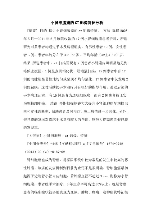
肾癌分型及影像表现[仅供参考]
![肾癌分型及影像表现[仅供参考]](https://img.taocdn.com/s3/m/ee90cc5ced630b1c59eeb5e8.png)
• 1 肾上腺
• 2 肾小盏
• 3 肾大盏
2
• 4 肾皮质
3
• 5 肾柱
4
• 6 肾髓质
5
6
• 7 肾窦内的肾周脂肪 7
• 8 纤维囊
8
• 9 输尿管
• 10 肾盂
1 10
医疗模板
9
3
肾癌的病理分型
1997 WHO 肾癌病理分型
2004 WHO 肾癌病理分型
透明细胞癌
75-85%
易染细胞癌(乳头状细胞癌) 15%
医疗模板
9
医疗模板
10
• (右肾肿物)透明细胞性肾细胞癌(Fuhrman分级III级),侵及周围肾组织。
IHC:CD10、Vimentin:(+)、S-100(+);CD34(血管+);CK、 HMB45、Melan-A、Actin:(-);Ki-67(+、10%)。
• (左肾肿瘤1)透明细胞癌(Fuhrman核级II级),(左肾肿瘤2)透明细胞癌 (Fuhrman核级III级),局限于肾被膜内。IHC:Vimentin(+)、CD10 (+)、P504S(+)、CK(+)、CK7(-)、TFE3(-)、Ki-67(+、 10%)、CD34血管(+)。
医疗模板
25
Bellini集合管癌
肾集合管癌细胞中等大小 ,部分胞质嗜酸,部分透 亮,部分核呈空泡状,有 小核仁。
医疗模板
26
4
医疗模板
(左肾)符合Bellini 集合管癌,伴广泛 坏死及脉管内癌栓 形成,肿瘤侵犯肾 盂及周围肾组织。
27
(左肾)符合Bellini集合管癌伴大片坏死, 灶区伴肉瘤样分化,侵犯肾被膜及肾门处脂
医疗模板
肾癌的CT、磁共振诊断

邻近器官受侵及术后评价 与CT相似
肾癌淋巴结转移、对侧肾积水
肾癌淋巴结转移、对侧肾积水
囊性肾癌淋巴结转移
囊性肾癌淋巴结转移
囊性肾癌淋巴结转移
左肾癌、下腔静脉癌栓
左肾癌、下腔静脉癌栓
左肾癌、下腔静脉癌栓
肾 癌 术 后 的 CT 评 价
MR
T1WI 为等低信号 , T2WI 多为等低信号 ; 透明细胞癌T2WI为高信号; MR 强化方式与 CT相似;
治疗及预后
目前,乳头状肾细胞癌的治疗采用肾癌根治术,但 对于小肾癌也可以采用保留肾单位的肿瘤切除术
与非乳头状肾细胞癌相比 ,乳头状肾细胞癌就诊时 大部分在早期 ,恶性度低 ,5 年生存率为 82 %~ 90 % ,前者仅为 44 %~54 %。
匀,少数有钙化和出血 增强后多血供者增强明显(>20 Hu),少血供
者10 ~20 Hu 动脉期肿瘤等于或高于肾实质,下降快,实质
期病灶呈相对低密度,实质后期及排泄期多为 低密度,分界清晰
小肾癌的CT、MRI表现
CT平扫 大多数小肾癌呈等密度,部分 稍低密度,少数可呈高密度(出 血?),偶见钙化,位于肿瘤中央。
小肾癌缺乏临床症状,常在体检或做其它
检查偶然发现,当肾癌逐渐增大时,常见
临
的临床表现是: 血尿,疼痛和肿块(三联征):间歇性无
床
痛性血尿表明肿瘤已侵犯肾盂、肾盏。肿 瘤侵犯邻近结构或牵张包膜引起疼痛。
表
发热,高血压,血沉快等肾癌的肾外表 现,易与全身其他疾病混淆,发热可能是
现
由于肿瘤坏死、出血、毒性物质吸收引起; 高血压可能是瘤内动-静脉瘘或肿瘤压迫
假包膜与标本对照
小肾细胞癌的CT影像特征分析
小肾细胞癌的CT影像特征分析[摘要] 目的探讨小肾细胞癌的ct影像特征。
方法选择2003年5月—2011年6月该院收治的17例小肾细胞癌患者资料,所选研究对象患者均通过手术及病理证实。
有男性患者12例,女性患者5例。
患者年龄分布于33~77岁,平均年龄(42±4.12)岁。
结果所选患者中,ct扫描发现有7例患者小肾癌内可明显地见到略低密度区,1例呈点状钙化状。
经增强扫描,15例患者中有12例的动脉期显著性地均匀或呈现不均匀强化。
17例患者中仅发现2例假包膜,这对后续的手术治疗具有很好的指导作用。
通过后续的手术病理证实,有15例患者为透明细胞癌,而有2例患者被证实为颗粒细胞癌。
结论多期扫描能够大大提升小肾细胞癌早期检出率和定性诊断率,帮助患者及时治疗,防止病情进一步恶化。
另外,假包膜的发现对临床手术具有较大的帮助,应努力提高患者假包膜的发现率。
[关键词] 小肾细胞癌;ct影像;特征[中图分类号] r445 [文献标识码] a [文章编号] 1674-0742(2013)02(c)-0187-02肾细胞癌也成为肾癌,是泌尿系统中较为常见的发生率较高的恶性肿瘤。
该病的发病机制到目前为止还不是很明确。
肾细胞癌最初起源于近端肾小管内皮细胞,若肿瘤直径不超过3 cm,则称为小肾细胞癌,患者经手术治疗,5年生存率可高达80%以上。
晚期肾癌患者的临床症状较多地表现为血尿、肿块、疼痛,这种症状特征很少在早期的诊断中出现。
随着影像技术的不断提高,以及人们对健康管理的逐渐重视,提高了肾细胞癌早期诊断发现率。
通过分析小肾细胞癌的ct影像特征,能够在一定程度上增强对其影像诊断的认识。
为了探讨小肾细胞癌的ct影像特征,该院通过回顾性分析2003年5月—2011年6月该研究收治的17例小肾细胞癌的ct平扫和多期扫描影像,提高了对小肾细胞癌的早期诊断发现率,对后续的临床手术治疗具有较好的指导作用。
具体的过程现报道如下。
肾脏肿瘤的影像诊断

AML
囊肿
几个提示RAML的CT征象
病灶突出比:大于1/2,即病灶的主体部分位于肾 轮廓外的以RAML 为多; 皮质掀起征:是在皮质内生长的肿瘤缓慢向肾外 膨胀生长, 最终突破皮质并将相邻的皮质掀起所 致。RCC 呈侵袭性生长,该征象少见, 即使早期 肿瘤因生长迅速掀起皮质, 但随着肿瘤的侵袭性 生长, 掀起的皮质逐渐被破坏。 肿瘤内血管影:RAML 的畸形血管粗大,CT表现为 粗圆点状或条状高密度影。RCC肿瘤血管管径较细, 表现为细点状影或线状影。 钙化:RCC 内出现的几率为10%,是其特征性征象, 而RAML 肿瘤内钙化罕见。
低 信 号
MR
平扫
强化
平扫T2和T1均为低信号,高血供:皮 质期等信号,小管期低信号,不均匀
MRI
CT
小 肾 癌 的 影 像
CT和MR有相同的增强方式
小肾癌与AML的鉴别
一、小肾癌[1]:1,平扫病灶呈等或稍低均匀密度, 2,增强后皮质期明显强化,密度高于或等于皮质, 3,髓质期表现为相对低密度,4,排泄期,呈相对 低密度或等密度,5,有假包膜。呈“快进快 退”可诊断之。 二、AML:1,平扫病灶呈脂肪密度或不均匀密度 (注意薄层),2,增强后平滑肌轻微强化,血管 明显强化,脂肪不强化,使病灶呈不均匀密度,3, 无包膜。可诊断之。 三、随访观察[2]:对于不典型小肾癌及无法确定脂 肪成分的AML要随访,小肾癌生长可快可慢,A ML生长慢,如病灶增长较快则高度怀疑。
影像学检查在小肾癌诊断中的临床应用及比较
1 2 影 像 学 检 查 6 . 8例 均 常 规 行 B 超 检 查 , 中 其
6 5例 行 多 层 螺 旋 C T描 , 9例 行 MR 检 查 。各 项 3 I
. g wk t m z mn . o
ht t } tp {w
40 8
JM o o , 11 . e . 0 2 d Ur l Vo . 7 No 5 S p 2 1
影像检查 均 由第 一作 者 结合 报告 逐 一进 行 判读 、 分析 。 1 3 治疗 方 法 6 . 8例 均 给予 手 术 治 疗 , 中 5 其 9例 行 肾部 分 切 除术 , 9例 行 。 根 治 术 , 后 均送 病 理 肾癌 术
收 稿 日期 : 0 20 — 5 2 1—41 修 回 日期 : 0 2 0 - 1 2 1 — 5 3 作 者 简 介 : 红 ( 9 6)女 ( 族 )副 主任 医 师 . 究 方 向 : 床 超 声 付 1 6一 , 汉 , 研 临 E malf. 1 6 wa 1 6 @ sn . n - i b : wk 9 5 wi9 6 ia c
时多 属 中晚期 , 后 较 差 。小 肾 癌 指 直径 ≤ 3c 的 预 m
1 资 料 与 方 法
1 1 一般资 料 .
本组 6 8例 , 中男 4 其 4例 (4 7 %) 6. 1 ,
单 发 病灶 肾癌 , 期 诊 断 对 于 早期 手 术 治疗 、 高 预 早 提 后 具有 重要 价 值 。近 年 来 随 着 影 像 学 技 术 的快 速 发 展 , 肾癌 的诊断 取 得 了 明显 进 展 _3。作 者对 2 0 小 2¨ _ 07 年 6月 至 2 1 0 1年 8月 陕西 省 宝鸡 市第 二人 民医院 收 治的6 8例术 后经 病理 确诊 为小 肾癌患 者 的影像 学 资 料 进 行 回顾性 分 析 , 以探讨 各影 像 学检 查在 小 。 肾癌诊 断 中的应 用价 值 。
肾脏及肾上腺疾病影像学表现
常见疾病
1,肾癌
2,肾错构瘤(肾血管平滑肌脂肪瘤)
3,单纯性肾囊肿
4,多囊肾
5,肾结石
6,肾结核 7,肾脏先天性变异
2,临床表现 尿频、尿急、尿痛、血尿、脓尿、腰痛。
1. 3, CT表现 (1)平扫 早期可无阳性发现。当肾皮质脓肿形成时表现为肾脏增大,肾实质内有单发或多发大小不等,形态不一囊腔,CT值接近于水,囊腔内或周边可有钙化斑。病变侵及输尿管可引起同侧肾盂肾盏积水,当对侧输尿管受侵时,可同时合并对侧肾盂肾盏积水。晚期肾自截表现为肾脏缩小,弥漫性钙化。 (2)增强扫描 脓腔周边可见环状强化。
(六) 肾结核
1,病理 肾结核是全身结核病变的一部分,绝大部分继发于肺结核,。结核杆菌经血行播散进入肾髓质引起乳头炎,进一步发展至溃疡,坏死,皮质内脓肿形成,肾盂肾盏积水积脓,最后发展成肾脏弥漫性钙化、肾自截。肾结核菌常经尿液蔓延到膀胱,导致结核性膀胱炎,常引起对侧输尿管下端狭窄,继发对侧肾积水。
肾结核(CT图)
(六) 肾外伤
1.包膜下血肿:平扫紧贴肾实质表面的新月型高密度影,肾脏受压变形,增强后肾实质明显强化,而血肿密度相对较低。 2.肾内血肿:平扫肾内局限性高密度影无强化。 3.肾撕裂 平扫肾实质内线样低密度影。增强扫描,损伤处肾实质不强化。
腹腔肾(图)
肾形态异常 包括融合肾和分叶肾 2.融合肾 [临床与病理] 常见是马蹄肾(Horseshoe kidney),其特点是两肾上极或下极相互融合,以下极多见。马蹄肾发现率0.01%到2%。以腹部肿块就诊,部分有尿路梗阻、感染表现
马蹄肾(图2)
(二)肾盂、输尿管先天性异常 1.肾盂、输尿管重复畸形 [临床与病理] 肾盂、输尿管重复畸形较为常见,为一个肾脏分上、下两部,各有一套肾盂、输尿管。上部小,下部较大,两者表面有一浅沟。重复或部分重复输尿管,也分别汇入膀胱
肾癌影像学表现
肿瘤生长比较快可以侵犯肾实质和肾周脂肪囊, 同时可以侵犯到后腹壁和腰大肌。下图可见肿瘤血 供丰富,有高密度结节状表现,为肿瘤血管。肿瘤 血管形成动静脉瘘,导致对比剂排泄很快,另外肾 周脂肪囊里可见迂曲的肿瘤血管。这可以解释肿瘤 在肾实质期表现100%密度低于肾实质。
小于10%的肿瘤为浸润状生长,肾实质内 肿块边界不明显,侵犯整个肾脏后导致肾脏 变形。下图为浸润生长肿瘤,可见正常肾结 构完全消失,肾盂明显扩张和积水。
(2)血管瘤拴
肾癌可以发生血管瘤拴,见下图。左侧两幅图 为CT图像,中间为核磁图象,右侧为超声图象。 CT图象上可见肿瘤发生在右肾后侧,软组织肿物突 向肾盂,形成软组织结节,相互融合在一起,即肾 癌和肾静脉瘤拴。成长条状。核磁上T2全纵图象可 见肿瘤信号不均匀,和肾实质没有分界,肿瘤侵犯 肾盂,两者界限不清;增强核磁图象可见肿瘤明显 强化,瘤拴信号密度稍低。超声图象可见肾皮质边 缘部位低回声团块,边界较清楚,肾盂存在鹿角状 低回声区域,即肾静脉瘤拴。
乳头状癌CT上往往表现为均匀密度的结 节,肿瘤密度均匀,边界清楚,增强后有轻 度延迟强化。核磁图象T1全纵表现为稍高均 匀信号结节,T2全纵表现为高信号且不均匀 的囊性区域,冠状位清楚可见多囊状改变。 增强扫描后信号不均匀,稍许强化,见下图。 因此,平扫时CT上表现为均匀密度而在 核磁上表现为不均匀信号时,应考虑乳头状 癌可能。标本上可见肿瘤呈多囊状改变。镜 下可见乳头的分布情况。
5%-7%肾癌为多囊性生长。囊性肾癌包括两 种:①囊性肾癌:肿瘤坏死后形成囊性改变, 或囊肿内侧壁发生肿瘤,形成软组织结节; ②肿瘤本身以多房囊性方式生长。 下图为由囊肿癌变后形成的囊性肾癌,图中 可见肾下极巨大囊性区,里边有多个结节, 边界不清楚,密度不均匀。标本白色的区域 为肿瘤结节区域,囊壁弥漫增厚。
肾癌影像学表现
(4)集合管癌 集合管癌是非常少见的类型,发病率不足1%。集合 管癌好发在肾髓质部分,可沿肾盂边缘呈浸润生长。 下图为集合管癌。T2全纵图象可见肾髓质部分有软 组织影,包绕肾盂,冠状位可见肾盂边缘充满软组 织影。增强后可见边缘的肾实质强化,中心部位肾 盂周围的软组织呈轻度强化,肾盂表现为低信号, 冠状位可见肾皮质强化,肾髓质部分轻度强化,肾 盂部分的软组织有轻度强化,表明肿瘤在肾盂呈浸 润性生长。最后一幅图象可见输尿管和肾盂肾盏周 围被肿瘤组织包绕。
第一幅图为平扫CT,左肾上极有一软组织结节, 表现为低密度。动脉期增强后可见结节边缘明显不 规则强化,左肾下极可见两个囊性区,中间有房间 隔。此时无法判断是肾癌还是肾囊肿。矢状位可见 左肾上极病灶表现为多房性囊性结节,房间隔显著 增厚,下极存在多房性低密度区,房间隔的后部有 强化结节。此时可做成明显诊断。标本可见两个病 灶,在左肾上极病灶中心变性,周围有囊性区域。 左肾下极病灶为多房囊性改变,里面有点、片状红 色区域,表明肿瘤出血。
(2)不同生长方式的表现
肾癌呈膨胀性外突生长。如果肿瘤位于肾实质边缘 时,外突生长表现为侵犯肾周肪囊,也可类似肾膜 肿瘤外突生长,需注意鉴别诊断。 下图示肾癌外突生长。第一幅图可见肾实质压迫侵 蚀改变;第二幅图为CT增强扫描,静脉期可见肿瘤 强化,与肾实质边界不清楚;第三幅图即超声图象, 清晰可见肿瘤大部分呈外突生长,一小部分突入到 肾实质里面。标本上可见肿瘤外突生长,侵犯肾实 质。
(2)受侵器官及转移表现
肾脏形态:正常或失常。 肾盂肾盏:正常或受压移位、拉长、扩张或破坏。 肾静脉、IVC瘤栓:管腔增粗、腔内有异物等。 淋巴结转移:肾门及腹膜后淋巴结肿大。
(3)远处转移灶特征性表现 骨的单发性、溶骨性、膨胀性生长。 肺内的大结节、棉团状转移。
- 1、下载文档前请自行甄别文档内容的完整性,平台不提供额外的编辑、内容补充、找答案等附加服务。
- 2、"仅部分预览"的文档,不可在线预览部分如存在完整性等问题,可反馈申请退款(可完整预览的文档不适用该条件!)。
- 3、如文档侵犯您的权益,请联系客服反馈,我们会尽快为您处理(人工客服工作时间:9:00-18:30)。
肾脏小肾癌的影像学特征作者:赵艳王成林袁知东冯飞余宏建罗莉丽刘鹏程【摘要】目的探讨肾脏小肾癌的影像学特征和鉴别诊断。
方法对2003年至今我院经手术和病理证实的直径≦3CM的小肾癌和小良性病变的影像学表现进行分析,总结其影像学特征,并进行其鉴别诊断。
结果本组小肾癌8例,全部行CT检查,其中一例小肾癌还做了MRI 和DSA检查。
小肾癌CT三期扫描,8例小肾癌10个病灶,动脉期明显强化2个病灶,轻度强化7个病灶,实质期病灶内造影剂退出明显,其中一例两个病灶中的1个病灶各期均未见强化,MRI动脉期两个病灶也是一个强化,一个不强化,这与其DSA表现一个多血供,一个少血供吻合。
1例感染性病变密度各期均低于正常肾实质,1例不典型囊性病变未见强化。
本组8例小肾癌10个病灶,病理诊断均为肾透明细胞癌。
结论 1.小肾癌螺旋CT三期扫描中多数动脉期呈轻度-明显强化,实质期强化明显减退,呈典型“快进快出”特点,少数少血病灶不强化。
2.同一肾脏多发病灶,且影像学表现相同或不同,病理确相同。
【关键词】小肾癌诊断螺旋CT 鉴别诊断Imageologic characteristic of small renal carcinoma[Abstract] Objective :To investigate the imageologic characteristic and diferential diagnosis of small renal carcinoma. Methods: Retrospectively analyze the imageologic characteristic of small renal carcinoma and small benign pathological changes (diameter less than or equal to 3cm) proved by surgery and pathology, summarize its imageologic characteristic and make diferential diagnosis. Results:This group has 8 patients and 10 small renal carcinomas proved by surgery and pathology, all of them have three phase dynamic enhanced CT image and one case has MRI and DSA image also.2 carcinomas enhanced markedly and 7 enhanced lightly during the cortical phase, all the carcinomas wash-out apparently during the parenchyma phase. One of patients has 2 carcinomas, 1 carcinomas enhanced and another showed no enhancement during all phases on CT, 1 enhanced and another did mot enhance during the cortical phase on MRI, this characteristic was confirmed by DSA. One infective pathological change showed hypo-attenuation during all phases. One atypical cystal pathological change showed no enhancement. Conclusion:1.Major of small renal carcinoma showed light to marked enhancement during the cortical phase and the enhancement wash-out during the parenchyma phase, presented typical characteristic of“quick staining and quick fainting”. Minor of small renal carcinoma lack of blood supply showed no enhancement. 2. The imageologic characteristic of multiple carcinomas grew in one renal are same or different, but pathology confirmed them.[Key words] small renal carcinoma ,Diagnosis,Spiral CT,diferential diagnosis肾癌病灶直径小于3CM时诊断困难,一般称之为小肾癌,但随着医学影像技术日新月异的发展和人们健康意识的提高,肾脏小占位病变(直径≦3CM)的发现率逐年提高,而肾癌和肾良性占位的治疗方法和预后迥异,所以对肾小占位病变作出早期准确的定性诊断非常重要。
现将我院2003年至今经手术和病理证实的直径≦3CM的小肾癌和小良性病变的影像学表现进行分析,总结其影像学特征,并进行其鉴别诊断。
材料与方法一、一般资料本组10例,8例小肾癌,1例感染性病变,1例不典型囊性病变。
男8例,女2例。
年龄28-56岁,平均41.3岁。
腰痛4例,腰痛伴血尿1例,无任何症状于体检时发现6例。
二、仪器和方法仪器为9例行螺旋CT三期扫描(动脉期、静脉期、实质期),1例同时行MRI和DSA检查。
结果本组10例,小肾癌8例和2例良性病变,全部行CT检查,其中一例小肾癌还做了MRI和DSA检查。
小肾癌CT三期扫描,8例小肾癌10个病灶,动脉期明显强化2个病灶,轻度强化7个病灶,实质期病灶内造影剂退出,其中一例两个病灶中的1个病灶各期均未见强化,MRI动脉期两个病灶也是一个强化,一个不强化,这与其DSA表现一个多血供,一个少血供吻合(如图1)。
1例感染性病变密度各期均低于正常肾实质,1例囊性病变未见强化(如图)。
病理诊断,本组8例小肾癌10个病灶均为肾透明细胞癌。
一例为感染性病变,治疗后病灶吸收消失(如图),一例病理为囊性病变。
讨论一、小肾癌小肾癌是指肿瘤直径≤3.0cm的病变,微小肾癌是指病变直径≤1.5cm的病变【1】。
随着B超、CT的普及和健康普查的广泛开展,小肾癌诊断率逐年上升。
潘柏年等【2】报道小肾癌占同期肾癌的9.66%,本组小肾癌的诊断率占同期的12.90%(8/62)。
本组还发现微小肾癌1例,且为多发病灶。
二、小肾癌的影像学特征郭燕【3】等报道,小肾癌的影像学表现为平扫常为等密度,部分为稍低密度,少数因出血可呈高密度。
多数癌肿稍突出肾轮廓外,部分局限于肾轮廓内,边界多模糊不清。
增强扫描动脉期呈轻中度和明显强化,实质期和肾盂期病灶强化迅速减低,呈“快进快退”改变。
本组8例小肾癌10个病灶,动脉期明显强化2个病灶,轻中度强化7个病灶,实质期病灶内造影剂退出明显,其中一例两个病灶中的1个病灶各期均未见强化,MRI动脉期两个病灶也是一个强化,一个不强化,这与其DSA表现一个多血供,一个少血供吻合。
由此说明,小肾癌多血供病灶,多表现为典型“快进快退”增强模式,而少血供病灶,动脉期和各期则不强化。
三、同一肾脏多发小肾癌的特点本组可见2例多发病灶,这在以往病例中非常少见,一例2个病灶,CT平扫发现:右肾下极肾皮质外缘处见一小低密度病变,边界欠清,病灶局部膨隆,大小约8mm*11mm病灶,其余肾脏呈等密度。
增强扫描,动脉期右肾实质见两个病灶,一个位于上极皮质区呈高密度,一个位于下极皮质区呈低密度,平扫所见病灶呈低密度(与正常肾实质比)见轻度强化,另一个位于肾上极病灶呈高密度强化,大小约8mm*8mm病灶,但平扫为等密度,与正常肾脏无密度差异。
实质期平扫所见下极病灶为低密度,边界尚清,另一个位于上极病灶呈等密度,无分界。
MRI所示T1WI上两个病灶呈等低信号,其中CT平扫所见下极皮质区等密度灶环以稍高信号环,T2WI上肾上极皮质区病灶呈等低信号,下极皮质区病灶为略高信号。
增强扫描动脉期右肾上极病灶强化,但略低于肾实质,下极病灶无明显强化,信号明显低于肾实质。
静脉期上极病灶周边环形强化,病灶实质内造影剂消退。
下极病灶与正常肾实质对比明显,呈低信号,边界清晰。
实质期两个病灶为等低信号,下极病灶见稍高信号环。
DSA右肾上极皮质区病灶血供丰富,实质期染色明显,直径8mm。
实质期右肾下极皮质区病灶为乏血管病灶,直径约1.1mm。
剖开病灶,病灶呈鱼肉样。
病理:肉眼见直径7mm和8mm肿物,镜下为肾透明细胞癌,尚未突破被膜(如图)。
另外一例,左肾中极实质内可见两个稍低密度结节影,稍向肾轮廓外突出,密度尚均,边界不清,大小分别为2.2cm×2.0cm和1.3cm×1.2cm,增强后动脉期轻度强化,静脉期和实质期未见明显强化,表现相同(如图),病理两个病灶均为透明细胞癌。
由此看出,同一肾脏多发病灶,且影像学表现相同或不同,病理确相同,因此表明肾癌表现是多样的,诊断时应提高警惕。
【参考文献】1. 孔垂泽,王毅,刘同才,等.微小肾癌(附31例报告).中华泌尿外科杂志,2001,22:139-141.2. 潘柏年,王田,杨勇,等.小肾癌(附31例报告). 中华泌尿外科杂志,1998,19:32-34.3. 郭燕,黄兆民,刘明娟,等.螺旋CT在小肾癌诊断中的应用. 中华放射学杂志,2001,35:627-629.. .。