颈静脉孔区解剖培训课件
颈内静脉的解剖(PPT课件)

• 缺点:
• 由于距下腔静脉较远,故置管的位置不易达到中心静脉,所测得的压力受腹腔内 压力的影响,往往高于实际中心静脉压;
• 由于导管在血管内的行程长,留置时间久时,难免引起血栓性静脉炎; • 处于会阴部,易被污染; • 易发生局部水肿;
• 置管深度:
• 约40cm,如仅用与输液,置管深度以进入股静脉为宜。
小量气胸。
•
2020-12-09
• 导丝的尾端必须超出导管的尾部,并保留于患者体外
2020-12-09
颈内静脉的解剖
27
(六)置管注意事项
• 中心静脉在吸气时可能形成负压,穿刺过程中,更换输液器及导 管和接头脱开时,尤其是头高半卧位的病人,容易发生空气栓塞。 病人应取头低位穿刺,插管时嘱病人不要大幅度呼吸,可避免空 气栓塞的可能。
2020-12-09
颈内静脉的解剖
8
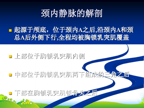
2)颈内静脉
• 体位:
• 病人仰卧,头低位,右肩部垫起,头后仰使颈部充分仲展,面部略转向对侧。
2020-12-09
点:胸锁乳突肌前缘中点(锁骨上5cm),向内推开颈总动脉, 旁开0.5~1.0cm • 操作者以左手示指和中指在中线旁开3cm,于胸锁乳突肌的中点前缘相当于甲状软骨上缘水
的顶点,锁骨上缘约3cm • 进针:针干与皮肤呈30°角,紧靠胸锁乳突肌锁骨头内侧缘
进针,直指同侧乳头 • 进针2~3cm即可进入颈内静脉 • 一般选用中路穿刺。因为此点可直接触及颈总动脉,可以避开
颈总动脉,误伤动脉的机会较少。另外此处颈内静脉较浅,穿 刺成功率高
2020-12-09
颈内静脉的解剖
13
• 后路法
• 穿刺点:胸锁乳突肌外侧缘中、下1/3交点作为进 针点(锁骨 上缘2~3横指)
颈静脉孔的应用解剖学

#应用解剖#颈静脉孔的应用解剖学肖 明, 丁 炯, 韩群颖, 王鹤鸣, 左国平(南京医科大学解剖学教研室,江苏南京210029) =摘要>目的:为与颈静脉孔相关的影像诊断和临床治疗提供解剖学资料。
方法:从颅底内、外面,对80具成年颅骨的颈静脉孔进行观测;并对20具成人尸头进行解剖,观察该区域神经血管解剖关系。
结果:¹62.3%右侧颈静脉孔较左侧大,15.9%左侧较大,21.8%两侧大小一致;º14.38%的颈静脉孔有骨桥,85.62%无骨桥;»颈静脉孔内、外侧缘距正中矢状面两侧的平均距离颅外均较颅内大:颅外分别为26.11mm 和33.41mm,颅内分别为22.29mm 和27.52mm 。
¼Ù脑神经多沿颈静脉孔前上缘,Ú、Û脑神经沿内侧缘出颅,两者被纤维索(占87.5%)或骨桥(占12.5%)隔开。
½Ù脑神经多经颈静脉孔外口前上缘向前下越过颈内动脉表面;Ù脑神经经颈内静脉深面(占57.5%)或其浅面(42.5%)行向后下。
结论:右侧颈静脉孔通常较左侧大,左右不对称;影像学观测该区域血管、神经应选择恰当的层面。
=关键词>颈静脉孔; 颈内静脉; 脑神经; 应用解剖学=中图分类号>R323.1 =文献标识码>A =文章编号>1001-165X(2001)02-0159-03Applied anatomy of jugular foramen XIAO Ming,DING J ong ,HAN Qun -ying,et al.De p a rtment o f A natom y ,Nan j ing Medical University ,Nan j ing 210029,China=Abstract >Objective:To provide anatomic data for imaging diagnosis and microsurgical treatment of jugular fora -men (JF)lesions.Methods:The JF was observed and measured from internal and external aspects in 80adult skulls.The anatomic relationships between the nerves and vessles in this region were observed i n detail by dissecting 20adult cephalic specimens.Results:¹In 62.3%of all these cases the right JF was larger than the left.In 15.9%the left was larger and in 21.8%they were equal in size.ºBone bridges could be seen in 14.38%and could not in 85.62%.»Measured from extracranial aspect,the average distance from midsagi ttal plane to the medial and lateral border of the JF (medial 26.11mm,lateral 33.41mm),was larger than those measured from intracramial aspect (medial 22.29mm,lat -eral 27.52mm).¼Ùcranial nerve made its ex i t through the anterior superior border of the JF in most cases,meanwhile Úand Ûcranial nerve through the medial border,they were distinctly separated from each other by a band of fibrous tissue (account for 87.5%)or a bone bridge (12.5%).½Jus t outside the J F,Ùcranial nerve appeared at the anter-ior border and made a loop downward and forward superficial to the internal carotid artery.Ùcranial nerve run down ward and back ward deep (account for 57.5%),or superficially (42.5%)to the internal jugular vei n.Conclusions:T he right J F is usually larger than the left,and not symmetry on both sides.The key to imaging diagnosi s of nerves and ves -sels in the JF region is to select the sectional plane correctly.=Key w ords >Jugular foramen; Internal jugular vein; Cranial nerve; Applied anatomy颈静脉孔为枕骨与颞骨岩部之间的一骨性孔道,位于岩枕缝的后端,被颈内静脉结节分为二部或三部[1]。
颈内静脉应用解剖 PPT

➢ 颈内动脉起自颈总动脉, 自颈部向上至颅底,经颈 动脉管外口入颅,分为颅 外段和颅内段。
➢ 颅外(又称颈段)行程 直,位置深,全程无分支;
➢ 颅内段行径弯曲,毗邻复 杂,分支多。
1颈内动脉终段 2颈内动脉膝段 3颈内动脉海绵窦段 4颈内动脉岩骨段 5颈内动脉颅外段 6颈外动脉 7颈总动脉 8颈动脉颅外部 9椎动脉颅内 部 10基底动脉 11后交通动脉 12大脑前动脉
海绵窦段、膝段和前床突上段又合称颈内动脉虹吸部,其形 态多为“U”型和“v”型,少数为“C’型和“S”型。 颈内动脉虹吸部的形态通常随年龄的增长而发生变化,年龄 越大,其迂曲度也越大。 颈 内动脉闭塞,其狭窄段多数在颈内动脉起始部或颈内动脉 "虹吸部"。
大家应该也有点累了,稍作休息
大家有疑问的,可以询问和交流
颈静脉孔区解剖

颈静脉孔区解剖
HELLO NOVEMBER
颈静脉孔由枕骨基底部和颞骨岩部下面围成,近似呈尖向前内的三角形,内侧壁为枕骨基底部,后外侧壁为颈静脉窝,前外侧壁为颈静脉内突和颈动脉管内侧壁。
内主要有岩下窦、颅神经及乙状窦等重要结构。
Hovelague将颈静脉孔分为两部分:前内侧部较小,内含有舌咽神经,也叫作神经部;后外侧部较大,内含有颈静脉球体、迷走神经和副神经,叫作静脉部。
这两部分通常被纤维桥或骨桥分隔。
在颈静脉孔上覆盖有硬脑膜,在硬脑膜上有两个孔洞,一个孔为舌咽神经经过的通路,此神经经孔进入神经部;另一个是迷走神经孔,迷走神经和副神经由此孔进入静脉部。
上述诸结构在颈静脉孔内的位置与相互毗邻关系, 国内外有关文献描术不一。
岩下窦在颈静脉孔注入颈内静脉。
舌咽神经、迷走神经及副神经经颈静脉孔出颅。
乙状窦经颈静脉孔延续为颈内静脉。
颈静脉孔区解剖课件

04
术后护理:预 防感染、保持 1 诊断方法:CT、MRI、超声等影像学检 查
02 治疗方法:药物治疗、手术治疗、介入治 疗等
03 颈静脉孔区病变的临床表现:头痛、头晕、 耳鸣、视力下降等
04 颈静脉孔区病变的预防:保持良好的生活 习惯,避免长时间低头、颈部受凉等。
03
颈内静脉的主 要功能是收集 头部和颈部的 血液,并将其 输送到心脏
04
颈内静脉在颈 部的位置和走 行与颈动脉相 似,但比颈动 脉更靠近中线
迷走神经
01
02
03
04
迷走神经损伤可能导致心脏、 呼吸、消化等功能障碍,需 要及时治疗。
迷走神经在颈静脉孔区与颈 动脉、颈静脉等血管紧密相 邻,容易受到损伤。
热等症状
颈静脉孔区损伤: 如外伤、手术损 伤等,可引起局 部出血、神经损
伤等症状
颈静脉孔区畸形: 如先天性颈静脉 孔区畸形、颈静 脉孔区发育不良 等,可引起局部 畸形、功能障碍
等症状
颈静脉孔区的手术
01
手术目的:治 疗颈静脉孔区 病变,如肿瘤、 畸形等
02
手术方式:微 创手术、开放 手术等
03
手术风险: 损伤神经、 血管等
颈静脉孔是颈部的重要结构,位于颈部前外 侧,是颈部血管、神经和淋巴管的通道。
颈静脉孔是颈部血管的重要通道,包括颈内 静脉、颈外静脉和颈总动脉。
颈静脉孔是颈部神经的重要通道,包括颈神 经、臂神经和胸神经。
颈静脉孔是颈部淋巴管的重要通道,包括颈 淋巴管、臂淋巴管和胸淋巴管。
颈静脉孔区的神经 血管
颈内动脉
谢谢
颈静脉孔区解剖课件
演讲人
目录
01
颈静脉孔区解剖结 构
《颈内静脉应用解剖》课件

通过影像学检查如CT、MRI等可确诊颈内静 脉压迫综合征。
症状
颈内静脉压迫综合征可能出现头颈部肿胀、 疼痛,以及颅内压增高等症状。
治疗
治疗方式包括去除病因、改善血液循环等, 严重时可考虑手术治疗。
颈内静脉的损伤与处理
病因
症状
颈内静脉损伤通常由创伤、手术操作等因 素导致。
颈内静脉损伤可能出现出血、肿胀、瘀斑 等症状,严重时可能导致休克。
04
CATALOGUE
颈内静脉的解剖变异
颈内静脉的解剖变异类型
颈内静脉位置变异
颈内静脉的位置可能发生改变,偏离 正常位置,如高位、低位或中位。
颈内静脉口径变异
颈内静脉属支变异
颈内静脉的属支(如甲状腺下静脉、 胸廓内静脉等)可能存在变异,包括 属支的数量、起源和汇入位置等。
颈内静脉的口径大小存在差异,有的 可能较粗,有的可能较细。
是颈内静脉的属支,引流甲状腺区域的血液。
副半奇静脉
右侧副半奇静脉汇入右颈内静脉,左侧汇入左颈内静脉。
奇静脉
奇静脉引流胸腹壁、食管、胃等部位的血液,右侧奇静脉汇入右 颈内静脉,左侧汇入左颈内静脉。
02
CATALOGUE
颈内静脉的生理功能
颈内静脉的血流动力学
颈内静脉是颈部主要的静脉通道 ,负责收集头面部、颈部的血液
颈内静脉是临床上常用的血管通路之一,其临床意义在于 为患者提供长期的血液透析、药物治疗、营养支持等治疗 手段。颈内静脉具有管径粗、变异小、位置表浅等优点, 便于穿刺和置管,且并发症发生率较低。
颈内静脉的临床应用需根据患者的具体情况选择合适的操 作技术和护理措施,以确保治疗的安全和有效性。同时, 医护人员应具备扎实的解剖知识和操作技能,以应对可能 出现的并发症和意外情况。
最新颈静脉孔区解剖

• posterosuperior view of the intrajugular process and ridge, which separate the sigmoid and petrosal parts of the jugular foramen
Rembrandt van Rijn (Dutch, 1606-1669). ห้องสมุดไป่ตู้his painting is called "The Anatomy Lecture of Dr. Nicolaes Tulp", painted in 1632
• the vestibular aqueduct opens onto the posterior surface of the temporal bone superolateral to the jugular foramen
• The inferior petrosal sinus extends along the petroclival fissure and enters the petrosal part of the foramen
• cochlear aqueduct opens above the petrosal part of the foramen , where the glossopharyngeal nerve enters the intrajugular part of the foramen on the medial side of the intrajugular process.
• larger lateral part, the sigmoid part, which receives the drainage of the sigmoid sinus, and a smaller medial part, the petrosal part, which receives the drainage of the inferior petrosal sinus
颈内静脉的解剖-课件
缺点:
由于距下腔静脉较远,故置管的位置不易达到中心静脉,所测 得的压力受腹腔内压力的影响,往往高于实际中心静脉压;
由于导管在血管内的行程长,留置时间久时,难免引起血栓性 静脉炎;
处于会阴部,易被污染; 易发生局部水肿;
置管深度:
约40cm,如仅用与输液,置管深度以进入股静脉为宜。
(六)置管注意事项
少量可予观察,大量须行胸腔闭试引流
1)插管时并发症
液胸:
无论是颈内静脉还是锁骨下静脉穿刺时,在送管时将穿透静脉 而送入胸腔内,此时液体都输入胸腔内。
从此路给药均无效 测量中心静脉压时出现负压 此路输液通畅但抽不出回血 拔管,引流
1)插管时并发症
动脉及静脉损伤
在锁骨下,颈内,股静脉穿刺时均可能引起动静脉损伤,可致穿 刺局部出血,形成血肿,应立即拔除导针或导管,局部加压。 如果血肿较大,必要时要行血肿清除术。
另一类则与导管感染有关,所以插管前、 中、后均应严格遵守无菌操作原则,这是 减少感染并发症的重要措施。
1)插管时并发症
气胸
气胸是常见的插管并发症之一,偶可发生张力性气胸。 无论是锁骨上或锁骨下径路,均有并发气胸的可能。一般均因
穿刺针撕裂了顶部胸膜所致。 在锁骨下静脉置管后应多次听诊呼吸音或作胸部X片检查,以
在导管拔除同时,空气偶可经皮肤静脉隧道进入静脉,故拔管 后,应按压加揉擦进皮点至少20min,然后严密包扎24h。
3)导管感染后败血症
导管败血症:是指接受胃肠外营养或液体治 疗的患者出现临床败血症,而全身各组织 器官又未能发现明确的感染源,且败血症 的症状和体征,在拔除中心静脉导管后得 以控制或缓解。
导管阻塞
防止导管扭曲、受压;输血前后用生理盐水充分冲 洗;用稀释肝素液封管,可防止导管阻塞情况发生。 疑有管腔堵塞时不能强行冲注,只能拔除,以防血 块栓塞。
- 1、下载文档前请自行甄别文档内容的完整性,平台不提供额外的编辑、内容补充、找答案等附加服务。
- 2、"仅部分预览"的文档,不可在线预览部分如存在完整性等问题,可反馈申请退款(可完整预览的文档不适用该条件!)。
- 3、如文档侵犯您的权益,请联系客服反馈,我们会尽快为您处理(人工客服工作时间:9:00-18:30)。
3/11/2021
颈静脉孔区解剖
5
• hypoglossal canal passes above the middle third of the occipital condyle and opens laterally into the interval between the jugular foramen and carotid canal
dra3i/1n1/2a02g1 e of the inferio颈r静p脉孔e区tr解o剖sal sinus
8
• enlarged view
3/11/2021
颈静脉孔区解剖
9
• intrajugular process projects into the interval between the sigmoid and petrosal parts of the foramen
• intrajugular ridge, extends forward from the intrajugular proc3/1e1/s20s21 along the medial 颈si静d脉e孔区o解f剖the jugular bulb 10
• cochlear aqueduct opens above the petrosal part of the foramen , where the glossopharyngeal nerve enters the intrajugular part of the foramen on the medial side of the intrajugular process.
• the vestibular aqueduct opens onto the posterior surface of the
temp3/o11r/2a02l1 bone superolateral to颈静th脉e孔区ju解g剖ular foramen
11
• The inferior petrosal sinus extends along the
• 颈静脉孔由颞骨岩部和枕骨颈突围成。 颞骨和枕骨向孔内的突起分别被称为颞 突和枕突,二者以纤维或骨桥连接,构 成孔内神经和血管的分隔。由颞突下方 沿颈静脉球内侧缘伸向后方的骨性隆起 称为颈内嵴,舌咽神经行于其内侧。
• 颈静脉孔为一自颅后窝通向前、外、下方的 骨性管道 。
• 颈静脉管(jugular canal)。
3/11/2021
颈静脉孔区解剖
1
osseous relationships, superior view
3/11/2021
颈静脉孔区解剖
2
• osseous relationships, posterosuperior view. The jugular foramen is best seen in a posterosuperior view oriented perpendicular to the clivus.
颈静脉孔的硬膜结构及分部
• Hovelacque将颈静脉孔分为前内侧的神经部和后外 侧的血管部两部分。
• Katsuta根据通过颈静脉孔的结构将其分为岩部、颈 内部(或神经部)和乙状窦部。
• 神经部的硬膜形成舌咽道和迷走道,分别有舌咽神 经和迷走神经及副神经穿过。舌咽道和迷走道位于 颈内突内侧,二者间隔以0.5-4.9mm宽的硬膜。
3/11/2021
颈静脉孔区解剖
7
• larger lateral part, the sigmoid part, which receives
the drainage of the sigmoid sinus, and a smaller
medial part, the petrosal part, which receives the
• 神经部上外侧缘的硬膜返折增厚并伸向下内覆于舌 咽道和迷走道上方,称颈静脉孔硬膜返折,是辨认颅 神经的重要标志。
of the jugular foramen
3/11/2021
颈静脉孔区解剖
13
Rembrandt van Rijn (Dutch, 1606-1669). This painting is called "T3h/11e/20A21natomy Lecture of D颈静r.脉N孔区ic解o剖laes Tulp", painted in 1164 32
• stylomastoid foramen is located lateral and the anterior half of th3/1e1/20o21ccipital condyle m颈e静d脉i孔a区l解t剖o the jugular foramen 6
• anterior and backward reveals the shape of the jugular foramen
3/11/2021
颈静脉孔区解剖
• the jugular foramen is located between the temporal and occipital bones
• sigmoid groove descends along the mastoid and crosses the
petroclival fissure and enters the petrosal part of
3t/1h1/2e021foramen
颈静脉孔区解剖
12
• posterosuperior view of the intrajugular process and
ridge, which separate the sigmoid and petrosal parts
occipitomastoid suture , turns forward on the upper surface
of th3/1e1/20j2u1 gular process , en颈t静e脉rs孔区t解h剖e foramen
4
• from posterior and superior shows the shape of the foramen