MOLECULAR BIOLOGY OF GLIOMA
labinvest2009126a
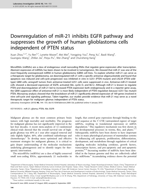
Downregulation of miR-21inhibits EGFR pathway and suppresses the growth of human glioblastoma cells independent of PTEN statusXuan Zhou 1,2,7,Yu Ren 3,7,Lynette Moore 4,Mei Mei 3,Yongping You 5,Peng Xu 1,Baoli Wang 6,Guangxiu Wang 1,Zhifan Jia 1,Peiyu Pu 1,Wei Zhang 4and Chunsheng Kang 1MicroRNAs (miRNAs)are a class of endogenous small noncoding RNAs that regulate gene expression after transcription.Aberrant expression of miRNAs has been shown to be involved in tumorigenesis.We showed that miR-21was one of the most frequently overexpressed miRNA in human glioblastoma (GBM)cell lines.To explore whether miR-21can serve as a therapeutic target for glioblastoma,we downregulated miR-21with a specific antisense oligonucleotide and found that apoptosis was induced and cell-cycle progression was inhibited in vitro in U251(PTEN mutant)and LN229(PTEN wild-type)GBM cells;xenograft tumors from antisense-treated U251cells were suppressed in vivo .Antisense-miR-21-treated cells showed a decreased expression of EGFR,activated Akt,cyclin D,and Bcl-2.Although miR-21is known to regulate PTEN and downregulation of miR-21led to increased PTEN expression both endogenously and in a reporter gene assay,the GBM suppressor effect of antisense-miR-21is most likely independent of PTEN regulation because U251has mutant PTEN.Microarray analysis showed that the knockdown of miR-21significantly altered expression of 169genes involved in nine cell-cycle and signaling pathways.Taken together,our studies provide evidence that miR-21may serve as a novel therapeutic target for malignant gliomas independent of PTEN status.Laboratory Investigation (2010)90,144–155;doi:10.1038/labinvest.2009.126;published online 4January 2010KEYWORDS:miR-21;glioma;PTEN;Akt;EGFRMalignant gliomas are the most common primary brain tumors with high mortality and morbidity.The prognosis for malignant gliomas has not significantly improved in the last four decades.A recent meta-analysis of 12randomized clinical trials showed that the overall survival rate of high-grade gliomas was 40%at 1year after surgical removal and only slightly higher,46%,after combined radiotherapy and chemotherapy.1To develop more optimized and effective treatment strategies for malignant gliomas,it is critical to gain deeper understanding of the molecular mechanisms underlying gliomagenesis and to identify targets for ther-apeutic intervention.The microRNAs (miRNAs)are a class of highly conserved small non-coding RNAs,approximately 22nucleotides inlength,that control gene expression through binding to the seed sequence at the 30-UTR (untranslated region)of target mRNAs,resulting in translational repression or mRNA degradation.2This regulatory mechanism was first shown in the developmental processes in worms,flies,and plants.3–5Subsequently,miRNAs have been shown to have important roles in many physiological processes of mammalian systems by influencing cell apoptosis,proliferation,differentiation,development,and metabolism through regulation of critical signaling molecules including cytokines,growth factors,transcription factors,and pro-apoptotic and anti-apoptotic proteins.6–8Increasing number of miRNAs have been iden-tified in the human genome and they are collectively called the miRNome.9Accumulating evidence shows the potentialReceived 8June 2009;revised 27August 2009;accepted 6September 20091Department of Neurosurgery,Tianjin Medical University General Hospital,Laboratory of Neuro-oncology,Tianjin Neurological Institute,Tianjin,China;2Department of Head and Neck Cancer,Tianjin Medical University Cancer Institute and Hospital,Tianjin,China;3Tianjin Research Center of Basic Medical Science,Tianjin Medical University,Tianjin,China;4Department of Pathology,University of Texas,MD Anderson Cancer Center,Houston,TX,USA;5Department of Neurosurgery,Nanjing Medical University Affiliated Hospital,Nanjing,China and 6Key Lab of Hormone and Development,Institute of Endocrinology,Tianjin Medical University,Tianjin,China Correspondence:Dr C Kang,MD,Department of Neurosurgery,Tianjin Medical University General Hospital,Laboratory of Neuro-oncology,Tianjin Neurological Institute,Tianjin 300052,China.E-mail:kang97061@ or Dr W Zhang,PhD,Department of Pathology,University of Texas,MD Anderson Cancer Center,Houston,TX 77030,USA.E-mail:wzhang@ 7These authors contributed equally to this work.Laboratory Investigation (2010)90,144–155&2010USCAP,Inc All rights reserved 0023-6837/10$32.00involvement of altered regulation of miRNAs in initiation and progression in a wide range of human cancers.Altered expression profiles of miRNAs are associated with genetic and epigenetic alterations including deletion,amplifica-tion,point mutation,and aberrant DNA methylation.10 The miRNA cluster miR-15a–16,including miR-15a and miR-16–1,is located near13q14,a region that is character-ized by a high frequency of deletion in chronic lymphocytic lymphoma.11The expression of miR-15a and miR-16–1were inversely correlated with the anti-apoptotic Bcl-2gene expression in chronic lymphocytic leukemia and both miR-NAs negatively regulated Bcl-2at the post-transcriptional level,resulting in the potentiation of the normal apoptotic response.12In a mouse model of lymphoma,increased expression of the miR-17–92cluster strongly accelerated lymphomagenesis and was the first functional evidence of an miRNA acting as a mammalian oncogene.13Another example of oncogenic miRNAs is miR-29.Enforced miR-29reduced the expression level of Mcl-1,an anti-apoptotic factor, to facilitate oncogenesis of cholangiocarcinoma.14Some miRNAs function as tumor suppressor genes.Let-7was reported to negatively regulate Ras and c-Myc expression after the let-7a-1precursor construct was transfected into human colon tumor cell lines and expression of miR-34a induced apoptosis in neuroblastoma cells.15–17Recent reports suggested that miR-21functions as an oncogene in human cancers.Ciafre`et al18profiled the expression of245miRNAs in10glioblastoma(GBM)cell lines and nine freshly resected GBM samples and observed that miR-21was overexpressed in human brain tumors.18 It was shown that when miR-21was suppressed,cell growth inhibition and caspase-dependent apoptosis were observed in A172,U87,LN229,and LN308cells.19Multiple target genes of miR-21have been reported.It has been shown that miR-21 modulates breast cancer cell anchorage-independent growth through suppressing TMP1expression.20In human color-ectal,breast cancer,and renal cell carcinoma,miR-21con-tributes to invasion and metastasis cell by inhibiting Pdcd4 mRNA at the post-transcription level.21–23A recent study showed that miR-21targets PTEN gene through a binding site on the30-UTR in hepatocellular carcinoma.24PTEN has been shown to be a critical tumor suppressor gene that is commonly inactivated in GBM by deletion,mutation, or attenuated expression.25Thus,increased expression of miR-21may contribute to the attenuated expression of PTEN in GBM.To further characterize the potential of miR-21as a target for treating GBM and to clarify the role of PTEN in the response,we used both in vitro and in vivo systems to study the effect of miR-21suppression in the PTEN wild-type (LN229)and PTEN mutant(U251)GBM cell lines.26 We found for the first time that downregulation of miR-21 inhibited the EGFR and Akt pathways and the anti-GBM effect was independent of PTEN status,suggesting that miR-21is a broader therapeutic target for GBM.MATERIALS AND METHODSCell Culture ConditionsThe human U251and LN229glioblastoma cell lines were purchased from the Institute of Biochemistry and Cell Biology,Chinese Academy of Science.Human glioblastoma cell lines,TJ861,TJ905,and TJ899,were established and characterized in Laboratory of Neuro-oncology,Tianjin Neurological Institute.27Human glioblastoma cell line,A172, and astrocytoma cell line,H4,were provided by Professor Jinhuan Wang(Tianjin First Central Hospital,China).All cell lines were maintained in Dulbecco’s modified Eagle’s medium(DMEM)(Invitrogen,Carlsbad,CA,USA)supple-mented with10%fetal bovine serum(Invitrogen),2mM glutamine(Sigma,St Louis,MO,USA),100units of peni-cillin/ml(Sigma),and100m g of streptomycin/ml(Sigma), at371C with5%CO2.Oligonucleotides and Cell TransfectionThe20-O-methyl(20-OMe-)oligonucleotides were chemically synthesized by SBS Genetech(Beijing,China).The20-O-Methyl oligos were composed entirely of20-O-methyl bases with the following sequences:scramble sequence50-AAGGC AAGCUGACCCUGAAGU-30and20-O-Me-miR-2150-GUCA ACAUCAGUCUGAUAAGCUA-30.19Oligonucleotides(50nm/l) were transfected into U251cells at70%confluence using Oligofectamine according to the manufacturer’s instructions (Invitrogen).MicroRNA Array and Reverse Transcription(RT)-Real-Time PCRTotal RNA was extracted from GBM cells(U251,TJ866, TJ905,TJ899,and A172)and human astrocytoma cell line H4using Trizol reagent(Invitrogen)for miRNA profile examination.Two separate total RNA samples of anti-sense-miR-21-treated U251cell and control U251cell were prepared for mRNA expression profiling.The miRNA microarray was obtained from CapitalBio Corporation (Beijing,China).The individual oligonucleotide probes were printed in triplicate on chemically modified glass slides in a21Â21 spot configuration of each subarray for miRNA microarray. All of the oligonucleotide probes were printed in triplicate on one microarray,and each of the four subarrays contained16 controls(Zip5,Zip13,Zip15,Zip21,Zip23,Zip25,Y2,Y3, U6,New-U2-R,tRNA-R,hsa-let-7a,hsa-let-7b,hsa-let-7c, 50%dimethyl sulphoxide(DMSO),and Hex).Total RNA(FirstChoice Total RNA,Ambion,Austin,TX, USA)from normal brain was used as a control.The miRNAs were enriched from total RNA using a mirVana miRNA Isolation Kit(Ambion)and labeled with mirVana Array La-beling Kit(Ambion).Labeled miRNAs were used for hy-bridization on each miRNA microarray containing509 probes in triplicate at421C overnight.A double-channel laser scanner(LuxScan10K/A,CapitalBio)was used to scan the arrays and quantified using image analysis software Downregulation of miR-21suppresses glioblastomaX Zhou et al(LuxScan3.0,CapitalBio).Raw data were normalized to the result of normal brain RNA and analyzed using the sig-nificance analysis of microarrays(SAM,version2.1,http:// /B tibs/SAM,Stanford University,CA, USA)software.For transfected cells,reverse transcription(RT)reaction was conducted with the mirVana t qRT-PCR miRNA detec-tion kit(Ambion).The real-time PCR was carried out with the mirVana qRT-PCR miRNA detection kit(Ambion). Amplification reaction was performed using MJ-real-time PCR(Bio-Rad,Hercules,CA,USA)and the protocol was performed for40cycles at951C for3min,951C for15s,and 601C for30s.Both RT and PCR primer were purchased from Ambion.5S RNA was used for normalization.Relative quantification was conducted using amplification efficiencies derived from cDNA standard curves and obtained relative gene expression.Data were shown as fold change(2ÀDD Ct) and analyzed initially using Opticon Monitor Analysis Software V2.02software(MJ Research,Waltham,MA,USA). Microarray AnalysisTwo separate total RNA samples were extracted from control and miR-21knocked down U251cells using Trizol reagent (Invitrogen).Samples were then sent to Beijing CapitalBio Corporation for processing.A total of500ng of total RNA was processed for use on the microarray by using the Affymetrix GeneChip30IVT Express Kit(Affymetrix,Santa Clara,CA, USA)according to the manufacturer’s recommended proto-cols.In brief,a first-stranded cDNA was synthesized from the total RNA sample using Superscript II Reverse Transcriptase (Invitrogen)and an oligo d(T)primer was linked with a T7 RNA polymerase binding site sequence.The amplified,labeled cRNA was produced using T7RNA polymerase and biotiny-lated nucleotides.After removal of free nucleotides,cRNA yield was measured by UV260absorbance with Nanodrop spectrophotometer(Gene,Carlsbad,CA,USA).The labeled cRNA was incubate with the5Âarray fragmentation buffer at 941C for35min for fragmentation reaction and combined with hybridization internal positive control(ACTB,GAPD, LDHA,and RPS9)and positive control(50-Hex–GTCAC ATGCGATGGATCGAGCTCCTTTATCATCGTTCCCACCTTA ATGCA-30)for DNA immobilization.After the signal am-plified according to the standard protocols,arrays were scanned with the Affymetrix Model3000scanner and data were analyzed using GSEABase software(http://www. /).To identify differentially expressed genes,sig-nal values were transformed to logarithm base2and z-scores were calculated.A significant P-value was assigned to each z-score by z-test.Genes with P-value of r0.01were considered differentially expressed.Statistical analysis was conducted using MATLAB7.0(The MathWorks,Inc.).Gene Ontology and KEGG Pathway AnalysisGenes with P-value of o0.01were selected as differentially expressed genes and used for further analysis.Expression Analysis Systematic Explorer(EASE)was used to analyze gene ontology and KEGG pathways.28Overrepresentation of genes are present if a larger fraction of genes within that pathway is differentially expressed compared with all genes in the genome.An EASE score of r0.05was used as a cutoff. MiR-21Detection by In Situ HybridizationUsing antisense locked nucleic acid(LNA)-modified oligo-nucleotides probe,in situ hybridization was performed with In situ hybridization kit(Boster,Wuhan,China).LNA/DNA oligos contained locked nucleic acids at eight consecutive centrally located bases(indicated by the underline)and had the following sequences:LNA-miR-2150-TCAACATCAGT CTGATAAGCTA-30.At72h after transfection,U251cells were fixed with freshly prepared4%paraformaldehyde (containing0.1%DEPC).The in situ hybridization detection of miR-21in U251GBM cells were conducted according to the protocol of the manufactures.The fixed U251cells were incubated with20m l LNA-miR-21hybridization solution at 421C for16h,and Cy3-avidin was used to label miR-21at a concentration of0.5mg/ml.Nuclei were counterstained with DAPI karyotyping kit(Genmed,Boston,MA,USA)and visualized using FluoView Confocal Laser Scanning Micro-scopes-FV1000(Olympus,Tokyo,Japan)and analyzed using IPP5.1(Olympus).Cell Proliferation AssayU251and LN229cells were seeded into96-well plates at4000 cells per well.After transfection as described previously,20m l of MTT(5g/l)was added into each well at each day of consecutive6days after treatment and the cells were incubated for additional4h,and the supernatant was then discarded.Finally,200m l of DMSO was added to each well to dissolve the precipitate.Optical density(OD)was measured at the wavelength of570nm.The data are presented as the mean±s.d.,which are derived from triplicate samples of at least three independent experiments.Cell-Cycle AnalysisFor cell-cycle analysis using FCM(flow cytometry),trans-fected and control cells in the log phase of growth were harvested,washed with PBS,fixed with90%ethanol over-night at41C,and then incubated with RNase at371C for 30min.Nuclei of cells were stained with propidium iodide for additional30min.A total of104nuclei were examined in a FACS Calibur flow-cytometer(Becton Dickinson,Franklin Lakes,NJ,USA).Samples were analyzed using flow cytometry for FL-2area and DNA histograms were analyzed using Modifit software.Experiments were performed in triplicate. Results are presented as percentage of cell in phase. Apoptosis Assays Using Annexin Stainingand TUNEL MethodParental and transfected cells in the log phase of growth were harvested and collected by centrifugation for5min at500gDownregulation of miR-21suppresses glioblastoma X Zhou et aland cells were then resuspended at a density of1Â106cells/ml. For the Annexin V assay,the annexin V-Cy3-labeled Apoptosis Detection Kit(Abcam,Cambridge,MA,USA)was used.The apoptotic cells were detected and quantified using FACSCalibur(Becton Dickinson,San Jose,CA,USA).The data obtained were analyzed using CellQuest software.The apoptotic cell death in the tumor specimens of mouse models from the in vivo study was examined by TUNEL method using an in situ cell death kit(Roche,Indianapolis,IN, USA).For detecting miRNA-21expression and apoptotic cells simultaneously,in situ hybridization was performed as de-scribed above,and then apoptosis was detected using TUNEL method.Positive cells were visualized using fluorescence mi-croscopy.The reaction mixture was incubated without enzyme in a control coverslip to detect nonspecific staining.Nuclei were counterstained with DAPI karyotyping kit(Genmed)and visualized using FV-1000laser scanning confocal biological microscopes and analyzed using IPP5.1(Olympus). Western Blot AnalysisParental and transfected cells were thrice washed with pre-chilled phosphate-buffered saline(PBS).The cells were then solubilized in1%Nonidet P-40lysis buffer(20mM Tris,pH 8.0,137mM NaCl,1%Nonidet P-40,10%glycerol,1mM CaCl2,1mM MgCl2,1mM phenylmethylsulfonyl fluoride, 1mM sodium fluoride,1mM sodium orthovanadate,and a protease inhibitor mixture).A total of40m g lysates were subjected to SDS-PAGE on8%SDS-acrylamide gel.Separate proteins were transferred to PVDF membranes(Millipore, Bedford,MA,USA)and incubated with primary antibodies against Akt-2(Santa Cruz,Santa Cruz,CA,USA),29pAKT (for Ser473,Santa Cruz),PTEN,EGFR,STAT3,and Bcl-2 (Zhongshan Bio Corp.,Beijing,China)followed by incu-bation with an HRP-conjugated secondary antibody(Zymed, San Francisco,CA,USA).The specific protein was detected using a SuperSignal protein detection kit(Pierce,Rockford, IL,USA).The membrane was stripped and re-probed with a primary antibody against b-actin(Santa Cruz).DNA Constructs and Luciferase AssayLuciferase activity assay-related constructs were made by ligating PTEN30-UTR fragments(approximately500bp) containing the predicted binding sites into luciferase reporter vector(pGL3).PCR was performed to amplify a fragment containing the miR-21target sequence using human DNA as the template. The primers used for PTEN30-UTR were50-ACTCTAGA GTCGACACCACTGACTCTGATC-30and50-ACTCTAGACA TGACACAGCTACACAACC-30.The product was digested using Xba I enzyme and afterward ligated with Xba I-treated pGL3-control vector containing the SV40promoter.The ligated product was transformed into E.coli JM109and colony PCRs were used to screen for the clones harboring the forwardly oriented insert using the antisense primer of PTEN and a sense primer of the vector(50-AGGAGTTGTGTT TGTGGACG-30).The desired construct was subsequently sequenced by Invitrogen.For luciferase reporter experiments,the pGL3-PTEN-3-UTR construct,which contains the putative binding site for miR-21of the3-UTR of the PTEN,was amplified by PCR from human genomic DNA and inserted into the pGL3 control vector(Promega,Madison,WI,USA),using the Xba I site immediately downstream from the stop codon of luci-ferase in the reporter gene vector.U251,A172,and H4cells were plated(2Â106cells per well)in six-well plates.After transfection,the cells were split into96-well plates in duplicate and harvested for luciferase assays24h later using a luciferase assay kit(Promega).U251Xenograft Tumor AssayFemale immune-deficient nude mice(BALB/C-nu),5weeks old,were purchased from the animal center of the Cancer Institute of Chinese Academy of Medical Science,and were bred at Compare Medicine Center,Tianjin Medical University.All experimental procedures were performed according to Tianjin Medical University policies.In all,four mice were injected subcutaneously with1Â106 of U251GBM cells,in a volume of50m l of PBS pre-mixed with equal volume of matrigel matrix(Becton Dickinson). Mice were monitored daily and three out of four mice formed tumors subcutaneously.When the tumor size reached approximately5mm in length,the tumors were surgically removed,cut into pieces of1–2mm3and re-seeded into the left inguinal region of30mice.When the diameter of the subcutaneous tumor reached7mm the mice were divided into three groups(10mice per group)randomly:U251 control group,U251scramble oligonucleotide(ODN)-trea-ted group,and U251As-miR-21-treated group.A mixture of20m l oligofectamine and ODN(50nmol/l)mixture was injected into the xenograft tumor model in a multi-site injection manner.Mice in the U251control group received 10m l of PBS only.A second administration was conducted on day3.The tumor volume was measured with a caliper every3days,using the formula volume¼lengthÂwidth2/2. At the end of the22-day observation period,the mice bearing xenograft tumors were killed and the tumor tissues were removed for formalin fixation and preparation of paraffin-embedded sections.Immunofluorescence and Immunohistochemistry StainingThe paraffin-embedded tissue sections were used for examination of PTEN,EGFR,p-AKT,AKT-2,Ki67,cyclin D1,Bcl-2expression,and HE staining.For immuno-fluorescence staining,transfected cells were seeded on cov-erslips and fixed with4%paraformaldehyde(PFA,Sigma), treated with3%H2O2for10min and incubated with the antibodies described above overnight at41C.FITC-or TRITC-labeled secondary antibody(1:200dilutions)was added for2h at371C.DAPI reagent was used to stain the Downregulation of miR-21suppresses glioblastomaX Zhou et alU251cell nuclei and the cells was visualized using FV-1000 laser scanning confocal microscopes and analyzed using IPP5.1(Olympus).For immunohistochemistry study,sec-tions were incubated with primary antibodies(1:200dilu-tions)overnight at41C,followed by biotin-labeled secondary antibody(1:100dilutions)for1h at room temperature. Sections were then incubated with ABC-peroxidase and DAB (diaminobenzedine),counterstained with hematoxylin,and visualized using light microscope.Statistical AnalysisData were expressed as means±s.e.Statistics was determined using ANOVA,w2test,or Student’s t-test using SPSS11.0 (Windows).Statistical significance was determined as P o0.05(*)or P o0.01(**).RESULTSMiR-21was Overexpressed in Human Glioblastoma Cell LinesWe first profiled miRNA expression in five glioblastoma cell lines,one astrocytoma cell line,and one normal brain tissue. Our analysis showed that8of the435human miRNAs (1.84%)were overexpressed with a greater than twofold increase and18of453(3.68%)showed a greater than twofold reduction in all glioma cell lines(Figure1a and b).Among the increased miRNAs,miR-21showed the most significant increase relative to normal brain tissue(7.0-fold).Other increased miRNAs include miR-221,miR-222,and miR-23a. The miR-21expression results from microarrays were con-firmed with quantitative RT-PCR assays(Figure1c).MiR-21 was chosen for further study because it was the most pro-minently overexpressed miRNA in glioma.Antisense-miR-21Suppressed U251Glioma Cell Proliferation and Induced ApoptosisTo evaluate the significance of miR-21overexpression in glioma cells,we used a loss-of-function antisense approach. An As-miR-21oligonucleotide(ODN)was used to knock down miR-21expression in U251and LN229cells.RT-real-time PCR results determined that the relative expression level of miR-21in As-miR-21ODN-treated U251cell was6.25% (P o0.01)and12.5%for LN229cells(P o0.01)compared with their control cells,respectively(Figure2a).In addition, LNA-based in situ hybridization showed that transfection of a scrambled ODN had no effect on miR-21expression. In contrast,the cy3red fluorescence signal in As-miR-21-transfected U251cells was lower(Figure2b).Thesedata TJ899TJ95U251A172H4hsa-miR-128ahsa-miR-128bhsa-miR-132hsa-miR-125ahsa-miR-323hsa-miR-219hsa-miR-1hsa-miR-223hsa-miR-126hsa-miR-329hsa-miR-495hsa-miR-330hsa-miR-137hsa-miR-95hsa-miR-497hsa-miR-127hsa-miR-368hsa-miR-335hsa-miR-338hsa-miR-451hsa-miR-342hsa-miR-124ahsa-miR-451hsa-miR-124hsa-miR-495hsa-miR-223hsa-miR-329hsa-miR-126hsa-miR-219hsa-miR-1hsa-miR-330hsa-miR-342hsa-miR-323hsa-miR-127hsa-miR-128hsa-miR-128hsa-miR-132hsa-miR-132hsa-miR-95hsa-miR-23bhsa-miR-137hsa-miR-23ahsa-miR-222hsa-miR-221hsa-miR-106hsa-miR-15bhsa-miR-21RelativeFoldTJ899TJ905TJ866A172H4NormalU2511.21.00.80.60.40.2Log2 (Expression in Glioma and Glioblastoma cell line/Ambion normal brain)-10-8-6-4-202468hsa-miR-108ahsa-miR-130ahsa-miR-17-5phsa-miR-22hsa-let-7ahsa-miR-106bhsa-miR-16hsa-miR-24hsa-miR-27ahsa-miR-221hsa-miR-222hsa-miR-15bhsa-miR-23bhsa-miR-21hsa-miR-23aFigure1miR-21is overexpressed in glioma cells.(a)Cluster analysis of miRNA expression profile across human glioblastoma cell lines.Glioma cell lines were presented in columns,miRNAs in rows.(b)The26most upregulated(right)and downregulated(left)miRNAs in six human glioma and globlastoma cell lines with respect to Ambion normal brain total RNA.The relative expression of miRNAs level is present in log2transformed for each glioblastoma cell line.(c)RT-real-time PCR shows miR21overexpression in glioma cell lines.U6snRNA was used as a loading control.Downregulation of miR-21suppresses glioblastomaX Zhou et alsuggested that As-miR-21can specifically inhibit the endogenous miR-21expression in U251and LN229cells. Cell viability was measured in ODN-transfected cells in up to4days after treatment.As-miR-21ODN-treated cells showed a significant decrease in viability compared with control ODN-treated cells or untreated cells(Figure2c).We found that the growth-inihibitory effect of decreased miR-21 reached maximum at3days after transfection.The lowest survival rate was57±16%for U251cell and49±9%for LN229cell.****Control200160120804001282563845122001601208040012825638451220016012080400128256384512200160120804001282563845122001601208040012825638451220016012080400128256384512U251LN229DNA content1201008060402012010080604020G/G1S G2/MControl Scramble As-miR-21G/G1S G2/MControl Scramble As-miR-21CellCycleDistribution(%)U251Control Scramble As-miR-212.62%3.91%252015105********U251LN229Control Scramble As-miR-21ApoptosisPercentage(%)252015105Control Scramble As-miR-21ApoptosisPercentage(%)5.30%FL1-H18.74%3.59%11.88%104103102101100104103102101100104103102101100104103102101100104103102101100104103102101100104103102101100104103102101100104103102101100104103102101100104103102101100104103102101100FL2-HFL2-HLN229CellNumberCellNumberScramble As-miR-21Control Scramble As-miR-2110080604020Day After T ransfection01234LN229CellSurvivalRate(%)10080604020Day After Transfection01234U251CellSurvivalRate(%)Control Scramble As-miR-21U251LN229Control Scramble As-miR-21miR-21relativeexpression(fold) 1.21.00.80.40.20.6Figure2The effect of miR-21knockdown on U251and LN229GBM cell proliferation.(a)miR-21expression was suppressed in U251and LN229cells using RT-real-time PCR.(b)In situ examination of miR-21expression in U251cells.Arrows highlight miR-21in situ expression in U251cells.Bar¼20m m.(c)MTT cell proliferation assay.miR-21knockdown in U251and LN229GBM inhibits cell proliferation.(d)Cell-cycle profiles after PI staining.miR-21 knockdown induced G1arrest in both U251and LN229GBM cells.(e)Graphical representation of the cell-cycle profiles in(d).As-miR-21and scramble ODN-transfected U251and LN229GBM cells were analyzed using FCM to determine cell-cycle status.w2Test was performed.**P o0.01.(f)Analysis of apoptosis using Annexin V staining.MiR-21knockdown led to U251and LN229GBM cell apoptosis.(g)Graphical representation of(f).As-miR-21and scramble ODN-transfected U251and LN229GBM cells were analyzed using FCM to determine cell apoptosis.One-way ANOVA was performed.**P o0.01.Downregulation of miR-21suppresses glioblastomaX Zhou et al。
PI3K和MAPK信号通路调节细胞增殖
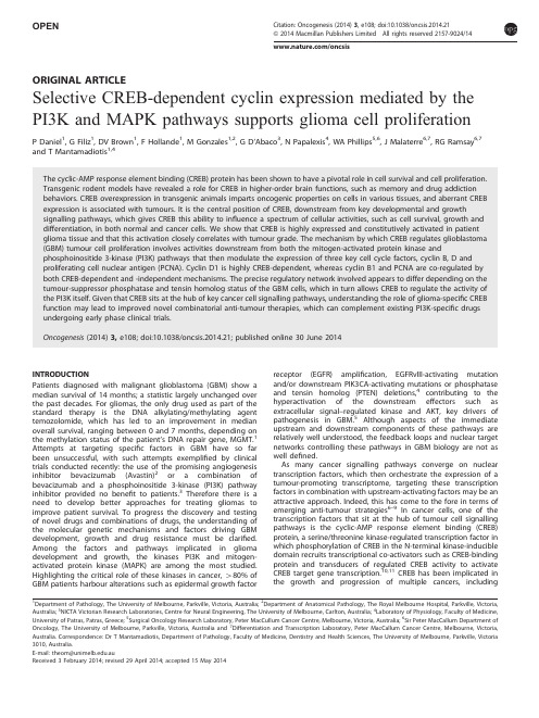
OPENORIGINAL ARTICLESelective CREB-dependent cyclin expression mediated by the PI3K and MAPK pathways supports glioma cell proliferationP Daniel 1,G Filiz 1,DV Brown 1,F Hollande 1,M Gonzales 1,2,G D’Abaco 3,N Papalexis 4,WA Phillips 5,6,J Malaterre 6,7,RG Ramsay 6,7and T Mantamadiotis 1,4The cyclic-AMP response element binding (CREB)protein has been shown to have a pivotal role in cell survival and cell proliferation.Transgenic rodent models have revealed a role for CREB in higher-order brain functions,such as memory and drug addiction behaviors.CREB overexpression in transgenic animals imparts oncogenic properties on cells in various tissues,and aberrant CREB expression is associated with tumours.It is the central position of CREB,downstream from key developmental and growthsignalling pathways,which gives CREB this ability to influence a spectrum of cellular activities,such as cell survival,growth and differentiation,in both normal and cancer cells.We show that CREB is highly expressed and constitutively activated in patient glioma tissue and that this activation closely correlates with tumour grade.The mechanism by which CREB regulates glioblastoma (GBM)tumour cell proliferation involves activities downstream from both the mitogen-activated protein kinase andphosphoinositide 3-kinase (PI3K)pathways that then modulate the expression of three key cell cycle factors,cyclin B,D and proliferating cell nuclear antigen (PCNA).Cyclin D1is highly CREB-dependent,whereas cyclin B1and PCNA are co-regulated by both CREB-dependent and -independent mechanisms.The precise regulatory network involved appears to differ depending on the tumour-suppressor phosphatase and tensin homolog status of the GBM cells,which in turn allows CREB to regulate the activity of the PI3K itself.Given that CREB sits at the hub of key cancer cell signalling pathways,understanding the role of glioma-specific CREB function may lead to improved novel combinatorial anti-tumour therapies,which can complement existing PI3K-specific drugs undergoing early phase clinical trials.Oncogenesis (2014)3,e108;doi:10.1038/oncsis.2014.21;published online 30June 2014INTRODUCTIONPatients diagnosed with malignant glioblastoma (GBM)show a median survival of 14months;a statistic largely unchanged over the past decades.For gliomas,the only drug used as part of the standard therapy is the DNA alkylating/methylating agent temozolomide,which has led to an improvement in median overall survival,ranging between 0and 7months,depending on the methylation status of the patient’s DNA repair gene,MGMT.1Attempts at targeting specific factors in GBM have so far been unsuccessful,with such attempts exemplified by clinical trials conducted recently:the use of the promising angiogenesis inhibitor bevacizumab (Avastin)2or a combination of bevacizumab and a phosphoinositide 3-kinase (PI3K)pathway inhibitor provided no benefit to patients.3Therefore there is a need to develop better approaches for treating gliomas to improve patient survival.To progress the discovery and testing of novel drugs and combinations of drugs,the understanding of the molecular genetic mechanisms and factors driving GBM development,growth and drug resistance must be clarified.Among the factors and pathways implicated in glioma development and growth,the kinases PI3K and mitogen-activated protein kinase (MAPK)are among the most studied.Highlighting the critical role of these kinases in cancer,480%of GBM patients harbour alterations such as epidermal growth factorreceptor (EGFR)amplification,EGFRvIII-activating mutation and/or downstream PIK3CA-activating mutations or phosphatase and tensin homolog (PTEN)deletions,4contributing to the hyperactivation of the downstream effectors such as extracellular signal–regulated kinase and AKT,key drivers of pathogenesis in GBM.5Although aspects of the immediate upstream and downstream components of these pathways are relatively well understood,the feedback loops and nuclear target networks controlling these pathways in GBM biology are not as well defined.As many cancer signalling pathways converge on nuclear transcription factors,which then orchestrate the expression of a tumour-promoting transcriptome,targeting these transcription factors in combination with upstream-activating factors may be an attractive approach.Indeed,this has come to the fore in terms of emerging anti-tumour strategies 6–9In cancer cells,one of the transcription factors that sit at the hub of tumour cell signalling pathways is the cyclic-AMP response element binding (CREB)protein,a serine/threonine kinase-regulated transcription factor in which phosphorylation of CREB in the N-terminal kinase-inducible domain recruits transcriptional co-activators such as CREB-binding protein and transducers of regulated CREB activity to activate CREB target gene transcription.10,11CREB has been implicated in the growth and progression of multiple cancers,including1Department of Pathology,The University of Melbourne,Parkville,Victoria,Australia;2Department of Anatomical Pathology,The Royal Melbourne Hospital,Parkville,Victoria,Australia;3NICTA Victorian Research Laboratories,Centre for Neural Engineering,The University of Melbourne,Carlton,Australia;4Laboratory of Physiology,Faculty of Medicine,University of Patras,Patras,Greece;5Surgical Oncology Research Laboratory,Peter MacCullum Cancer Centre,Melbourne,Victoria,Australia;6Sir Peter MacCallum Department of Oncology,The University of Melbourne,Parkville,Victoria,Australia and 7Differentiation and Transcription Laboratory,Peter MacCallum Cancer Centre,Melbourne,Victoria,Australia.Correspondence:Dr T Mantamadiotis,Department of Pathology,Faculty of Medicine,Dentistry and Health Sciences,The University of Melbourne,Parkville,Victoria 3010,Australia.E-mail:theom@.auReceived 3February 2014;revised 29April 2014;accepted 15May 2014Citation:Oncogenesis (2014)3,e108;doi:10.1038/oncsis.2014.21&2014Macmillan Publishers Limited All rights reserved 2157-9024//oncsisleukemia,12,13colorectal cancer 14and breast cancer.15CREB lies at the hub of multiple oncogenic signalling pathways initiated by ligand-growth factor receptor interactions such as the platelet-derived growth factor receptor and EGFR receptor tyrosine kinases and is activated by numerous downstream kinase pathways,including PI3K and MAPK,and deactivated by tumour suppressors,including PTEN.In melanoma cells harbouring BRAF(V600E)mutations,CREB is a key factor involved in MAPK pathway-mediated drug resistance 16In GBM,CREB function appears to regulate growth of tumour cells via transcriptional control of miRNA-23a 17and Nf-1.18Previous work has shown that loss of CREB function during brain development leads to neuronal death 19,20and that CREB is required for efficient neuronal stem and progenitor cell proliferation 21,22Based on these findings,we hypothesized that CREB activation is a critical step in gliomagenesis and that CREB function sits downstream of key cancer signalling pathways.We show that CREB expression and activation correlates with glioma grade and that activation occurs via both the PI3K and MAPK pathways and that CREB promotes glioma cell proliferation.Furthermore,we show that the mechanism by which CREB promotes glioma cell proliferation includes the regulation of key cell cycle factors cyclin B1,D1and proliferating cell nuclear antigen (PCNA).RESULTSCREB is constitutively activated in human GBM cells lines and human brain tumours exhibit grade-dependent pCREB expression Although CREB is expressed throughout the normal brain during all stages of life,constitutively phosphorylated CREB (pCREB)is progressively restricted to neurogenic zones.It is within these zones that neural stem/progenitor cell (NSPC)populations reside and CREB gene/expression disruption studies in mice and zebrafish show that NSPC proliferation and survival depends on CREB function.21,22Based on data showing that CREB has oncogenic roles in the hemopoietic system 12and other tissues 23and as the CREB functions required by NSPCs may be important in GBM biology,we explored CREB expression,activation and function in brain tumour tissue and cells.Assessment of CREB expression and activation in human GBM tumour cell lines using CREB and pCREB antibodies revealed that GBM tumour lines showed robust CREB and pCREB expression.Non-tumour brain tissue exhibited weaker total CREB expression and undetectable levels of pCREB (Figure 1a).Using whole GBM surgical tumour specimens prepared from formalin-fixed,paraffin-embedded tissue,we examined the mor-phological landscape of a further 15patient specimens diagnosed with grade IV GBM.GBM tumour morphologies associated with pCREB expression are shown in Figures 1b-i–vi.pCREB expression was evident in most tumour cells examined with a clustering of strong pCREB expression in highly vascularized tumour regions (Figure 1b-i)and in regions with strong nestin expression (Figure 1b-vi).pCREB-positive cell density and expression was high in hypercellular pseudopalisading regions adjacent to necrotic regions,where migrating proliferating tumour cells are seen (Figure 1b-iii,arrows).Of note,tumour morphologies corresponding to GBM regions associated with less malignant characteristics exhibited weaker pCREB expression.Giant cells in GBM showed relatively weaker pCREB expression (Figure 1b-ii,arrows),and cells in tumour regions with oligodendroglial features,characterized by the so-called ‘chicken wire’or ‘fried egg’morphology showed the weakest pCREB expression (Figures 1b-iv and -v,arrows).Interrogation of glioma tissue microarray specimens harbouring 40patient glioma tissue cores (3mm diameter)in duplicate showed that while ‘normal’non-tumour and ‘normal tissue adjacent to tumour’showed little-to-no CREB activation.By contrast,tumour tissue exhibited robust grade-dependent levels of pCREB expression (Figure 1c).Grade II tumours showed the lowest levels of pCREB,and WHO grade IV tissues exhibited the strongest pCREB positivity.Both pCREB intensity and the number of cells expressing pCREB correlated with tumour grade,where grade IV GBM showed the most intense and highest density of pCREB-positive ing a ‘þ’scale where ‘þ’is just detectable staining/1%to o 5%of cell/field positive and ‘þþþþþ’is strong dark staining/almost 100%cells/field positive,grade II tumours showed between zero and ‘þþ’;grade III tumours showed between ‘þ’and ‘þþþ’and grade IV/GBM showed between ‘þþþ’and ‘þþþþþ’.Three oligodendroglioma specimens showed ‘þ’,‘þ’and ‘þþ’,while all ‘adjacent to tumour’and ‘normal’brain tissue showed zero staining.Using the GBM cell line T98G to examine CREB activation,we show that pCREB coincides with cells in cycle.Specifically,pCREB is present in cells undergoing S-phase of the cell cycle as shown by co-labelling with PCNA and newly divided cells labelled withtheFigure 1.CREB is hyperactivated in glioma cell lines and tissue.(a )Cells from a range of human glioma cell lines were lysed and analysed from CREB expression and activation.All GBM cell lines tested showed an abundance of CREB protein,which was phosphorylated/activated.Non -tumour mouse brain tissue shows expression of CREB but no detectable pCREB.(b )Immunohisto-chemical analysis of human GBM tissue specimens show differential pCREB expression.(i)pCREB-positive tumour cells surround a tumour blood vessel;(ii)Giant cells in GBM show moderate-to-strong pCREB labelling;(iii)pseudopalidaing areas (arrows)in GBM show dense pCREB-positive tumour cells;(iv)and (v)two different patient tumours featuring oligodendroglial features show weak pCREB expression (arrows indicate oligodendroglial tumour cells);and (vi)double-labelling immunohistochemistry showed dense pCREB-positive cells (brown)surrounding foci with strong nestin expression (blue).(c )Brain tumour tissue microarray (US Biomax)with pCREB immunostaining.Top image shows the complete array with tumour grade layout indicated and cores magnified in the lower panel.CREB-mediated cell cycle factor expression in GBM cellsP Daniel et al2Oncogenesis (2014),1–10&2014Macmillan Publishers Limitednucleotide analogue bromodeoxyuridine (BrdU;Figure 2).In contrast,cells labelled with the M-phase marker phospho-histone 3(pH3)demonstrated poor colocalization to pCREB-positive cells.PI3K and MAPK but not protein kinase A (PKA)signalling pathways modulate CREB activity in GBM cellsMany signalling pathways relevant to GBM converge onto CREB,suggesting that constitutive CREB activation as observed in GBM is the result of cooperation between multiple upstream factors.To determine which pathways activate CREB,we evaluated cells grown in stimulatory and non-stimulatory conditions for differ-ences in kinase activation.Several serine/threonine kinases that are known to phosphorylate CREB at Serine133were investigated.Analysis of cell lines T98G,LN18and U118demonstrated activation of CREB,AKT and MAPK upon serum stimulation.By contrast,we did not observe activation of PKA,an activator of CREB in other cell types (Figure 3a),showing that there is a selective use of upstream components regulating CREB activity.To link the specificity of AKT and MAPK activation with CREB activation,the PI3K pathway inhibitor LY294002and the MAPK pathway inhibitor U0126were used on T98G and U118(Figures 3b and c).These cell lines were chosen for further analysis for two reasons.First,they showed the greatest CREB-dependent change in proliferation (Figures 4b and c),and second,these cell lines differ in the tumour-suppressor PTEN status,where T98G has intact PTEN function and U118is PTEN deficient.One of the PTEN’s major functions is to act as the major phosphatase that inhibits the activation of the PI3K pathway 24Significant variability was seen between the cell lines tested with respect to the contribution of either the PI3K or MAPK pathways on CREB activation.In U118cells,inhibition of the PI3K pathway resulted in no change in CREB activation,while inhibition of the MAPK pathway resulted in knockdown of CREB activation to basal ‘serum-deprived’levels,suggesting that MAPK signalling is the predominant CREB activation pathway in U118cells (Figures 3c and e).By contrast,inhibition of either or both the PI3K or MAPK pathways in T98G cells did not result in significant changes in CREB activation (Figures 3b and d),highlighting the diversity and complex interdependence between signalling pathways involved in CREB activation in GBM cells.As a further and independent measure of the contribution of the PI3K pathway to CREB activation,we used a mutant NSPC that has a constitutively activated PI3K pathway,due to the combined conditional mutation of the Pik3ca gene,which encodes for the major catalytic p110alpha subunit of PI3K,and has a conditional loss of tumour-suppressor PTEN.These NSPCs were derived from a conditional mutant mouse shown to develop tumours in various tissues tested.25–27CREB activation (pCREB)was significantly elevated along with AKT activation (pAKT),compared with parental NSPCs from the same mouse in which the PI3K pathway mutations were not activated (Figure 3f).CREB function contributes to GBM cell proliferationAs CREB is strongly associated with proliferative zones within the normal adult brain,we examined the effect of CREB knockdown on GBM cell proliferation.We tested siRNA-mediated CREB knockdown in five human GBM cell lines (T98G,U118,U373,ANGM-CSS,U87),which represent cells with an array of GBM-associated genetic alterations.The combined use of three CREB siRNAs targeting different mRNA regions allowed robust CREB expression knockdown (490%)over at least 120h (Figure 4a)in T98G cells.All five cell lines tested showed a reduction in cell proliferation (n ¼1,not shown).Further experiments were performed with selected cell lines as indicated.The greatest reduction in proliferation was seen in cell lines T98G and U118,with a 58%and 52%reduction compared with scrambled siRNA-transfected controls after 120h (Figures 4b and c).As wehaveFigure 2.CREB is activated in dividing cells.T98G cells were analysed for the colocalization of pCREB(Ser 133)with markers of cell proliferation:(a )PCNA,(b )phospho -histone (pH3),nd (c )BrdU.CREB-mediated cell cycle factor expression in GBM cells P Daniel et al3&2014Macmillan Publishers LimitedOncogenesis (2014),1–10Figure 3.The PI3K and MAPK -dependent signalling pathways but not the PKA pathway activate nuclear CREB in glioma cells.(a )GBM cell lines T98G,LN18and U118were serum starved for 24h then exposed to serum and protein lysate immunoblotted for the indicated antibodies.(b ,c )GBM cell lines T98G and U118were pretreated with a PI3K inhibitor LY294002,a MAPK inhibitor U0126or both inhibitors,and then exposed to serum,protein lysate collected after 4h and analysed for the presence of the indicated antibodies (d ,e )Quantification of the effects of inhibitor combinations on pCREB levels in T98G and U118GBM cells.*P o 0.05,**P o 0.005.(f )Western blotting showing the level of pCREB and pAKT in mouse NSPCs grown as neurospheres,with an activated Pik3ca H1047R mutation and PTEN deletion (M)or parental NSPCs with ‘wild-type’Pik3ca and PTEN (C).All western blottings were performed at least three times,and where applicable,total CREB,AKT and MAPK were imaged,then membranes were stripped and reprobed for pCREB,pAKT and pMAPKdetection.Figure 4.CREB is required for efficient human glioma cell line proliferation.(a )T98G cells were transfected with either CREB1-specific siRNA (siCREB)or scramble siRNA and then lysate analysed by western blotting for efficiency of CREB knockdown.*P ¼0.05,**P ¼0.005,***P ¼0.0005.(b ,c )Scrambled or siCREB was transfected into GBM cell lines T98G and U118,and proliferation was analysed every 24h using an MTT assay.(d ,e )Scrambled or siCREB was transfected into GBM cell lines T98G and U118,and apoptosis was determined by FACs determination of AnnexinV uptake.CREB-mediated cell cycle factor expression in GBM cellsP Daniel et al4Oncogenesis (2014),1–10&2014Macmillan Publishers Limitedpreviously shown that loss of CREB function in zebrafish and mouse brain as well as mouse neural NSPCs leads to compromized cell survival,we tested whether this was the case in GBM cells.Notably,cell survival was not affected by CREB knockdown using two different assay systems,measuring lactate dehydrogenase released from dead cells and measuring early apoptosis by measuring appearance and accumulation of Annexin V (Figures 4d and e)following CREB knockdown.Thus reduced cell numbers can be attributed to reduced proliferation.CREB knockdown leads to aberrant GBM cell cycle kinetics due to inhibition of cell cycle factor expressionTo determine the mechanism by which CREB regulates GBM cell proliferation,cell cycle parameters were measured using DNA content by flow cytometry in T98G and U118cells,which were the cell lines showing the greatest CREB-dependent decrease in proliferation.CREB knockdown increased the proportion of cells in G0/G1phase in both cell lines tested (Figures 5a and b).siCREB T98G cells also showed significantly reduced proportions in cell cycle phase distribution in S-and G2/M phases compared with control cells,whereas siCREB U118cells exhibited increased G0/G1and fewer G2/M phase cells only.To explore the molecular basis underlying disruption of the cell cycle,we measured the expression of cell cycle/proliferation factors previously reported to be transcriptionally regulated by CREB,including cyclins B1,D128and PCNA.14,29Protein expression of cyclins B1,D1and PCNA was assessed in T98G and U118cells in CREB knockdown or scrambled control siRNA-treated GBM cell lines (Figures 5c and d).Cells were serum deprived for 24h before the addition of serum to synchronize cell cycle activation,and cyclin and PCNA protein levels were determined every 12h,over 48h.The dynamics of expression seenwas consistent with the reported cyclin and PCNA expression profiles for mammalian cells,where PCNA and cyclin D1are consistently expressed throughout the cell cycle while cyclin B1expression peaks at G2/M.30Upon treatment with serum (triggering cell cycle entry),we observed inhibition of protein expression of cyclin B1,cyclin D1and PCNA in both T98G and U118cell lines as early as 12h (not shown)with maximal inhibition reached at 24h,compared with no-serum cells (Figures 5c and d).T98G cells showed a consistent and almost complete block in cyclin D1,cyclin B1and PCNA expression over 48h in CREB knockdown cells,in contrast to U118cells,which exhibited a more modest inhibition of cyclin D1,over 48h.In U118cells,cylin B1protein expression was maximally inhibited at 24h,but little effect was seen in PCNA expression (Figure 5d).Moreover,in U118cells cyclin B1inhibition was not sustained,with expression approaching control levels beyond 24h.Given that cyclin B1,D1and PCNA harbour cAMP-resonsive elements (CREB-binding sequences)in their promoters (see Supplementary Data),we tested whether CREB exerted its influence on these target genes directly;we performed reverse transcriptase–PCR (RT–PCR)at 24h following siCREB treatment to measure mRNA levels of cyclin B1,D1and PCNA.CREB knockdown robustly inhibited cyclin D1mRNA expression in both T98G and U118cells,while cyclin B1mRNA expression was significantly reduced in T98G cells only.Surprisingly,PCNA mRNA expression was unaf-fected by CREB knockdown in both cell lines (Figures 5e and f),implying that CREB exerts its affect on PCNA protein expression indirectly.The transcriptional influence CREB exerts in the expres-sion of these genes reflects the context of the cAMP-resonsive elements in their respective promoters (see Supplementary Data),with cyclin D1showing the best context with a full (8-base pair consensus)cAMP-resonsive element positioned closest to the transcription startsite.Figure 5.Knockdown of CREB alters human GBM cell cycle kinetics through regulation of cyclin B1,cyclin D1and PCNA.(a ,b )Synchronized GBM cell lines T98G and U118were treated with either siCREB or scramble control siRNA and then stimulated with serum for 24h before being analysed for cell cycle proportions using DNA-content analysis.(c ,d )Synchronized GBM cell lines T98G and U118were treated with either siCREB or scramble control siRNA,stimulated with serum and harvested every 12h for analysis for cyclin B1,cyclin D1and PCNA expression.(e ,f )Synchronized GBM cell lines T98G and U118were treated with either siCREB or scrambled control siRNA and then stimulated with serum for 24h before harvesting and analysis using qRT–PCR for CCNB1,CCND1and PCNA mRNA expression.***P o 0.0005,**P o 0.005,*P o 0.05.All western blottings were performed at least three times,and blottings for total CREB assay were imaged,then membranes were stripped and reprobed for pCREB detection.CREB-mediated cell cycle factor expression in GBM cells P Daniel et al5&2014Macmillan Publishers LimitedOncogenesis (2014),1–10Selective CREB regulation of cell cycle factors by canonical and non-canonical interations with the PI3K pathwayTo investigate the mechanism of the differential regulation of cyclin D1,cyclin B1and PCNA by CREB in T98G compared with U118cells,we looked at whether CREB knockdown affected upstream signalling components,given that previous studies have shown a link between CREB and upstream signalling components of the PI3K and MAPK pathways,such as insulin growth factor receptor (IGFR)and insulin receptor substrate-1/2(IRS-1/2).31,32Crucially,T98G and U118differ in PTEN status (T98G PTEN þandU118PTEN À),which may account for the differential CREB-mediated regulation of cell cycle factors via interaction with the MAPK and PI3K pathways.33,34Treatment of T98G cells with siCREB resulted in an almost complete block of AKT activation,which was not rescued by the addition of serum (Figure 6a).In comparison,CREB knockdown in U118cells showed reduced AKT activation only in serum-free conditions (Figure 6b).Upon the addition of serum,AKT activation in U118cells increased to levels identical to control cells (Figure 6b).To determine whether the cell-specific regulation of cyclin B1and PCNA by CREB could be due to effects on AKT activation and/or PTEN status,we used the PI3K inhibitor LY294002in conjuction with siCREB in U118cells (Figure 6c).This experiment confirmed the dependence of cyclin D1expression on CREB,with siCREB treatment alone showing almost complete attenuation of cyclin D1expression.Furthermore,the expression of cyclin B1and PCNA were found to be dependent on both CREB and PI3K signalling.Neither siCREB nor the PI3K inhibitor LY294002alone could block cyclin B1or PCNA expression in U118cells.DISCUSSIONCREB-dependent brain functions have been extensively explored with most previous studies focussed on neurological functions ranging from learning and memory,35opiate withdrawal 36to feeding behavior.37Other work has shown that CREB is critical for neuronal survival and that CREB disruption in vivo leads to a neurodegenerative phenotype 20and reduction in neural stem cell survival and proliferation 21,22In parallel,a body of evidence has accumulated showing that CREB imparts oncogenic properties on cells outside the central nervous system.23,38We focussed our investigations on defining CREB-dependent mechanisms orchestrating malignant GBM cell proliferation.Analysis of a panel of human GBM cell lines shows that CREB is not only highly expressed but also constitutively hyperactivated (Figure 1a).This is in contrast to non-tumour brain tissue,which show highly regulated and transient activation,dependent on external stimuli.11Cells in adult mouse brain showing robust constitutive phosphorylated CREB expression are enriched in the neurogenic zones,22,39so the constitutive expression of pCREB in GBM tumour cells is consistent with the generally held view that tumour cells exhibit immature stem/progenitor cell-like characteristics.In GBM tissue examined,pCREB was evident in many GBM hallmark morphological features,including near tumour angiogenic regions (Figure 1b-i),peripheral to necrotic foci characterized by pseudopalisading regions (Figure 1b-iii).This is interesting as GBM regions with pseudopalisading features harbour migrating tumour cells adjacent to hypoxic necrotic regions with high levels of pro-angiogenic factors,including hypoxia-inducible factor-1,vascular endothelial growth factor and interleukin-8,which are all CREB target genes.40–42CREB may therefore be intimately involved in regulating the angiogenic tumour niche in GBM.In one GBM tumour examined,strong pCREB-positive cells were clustered in foci exhibiting strong nestin co-labelling (Figure 1b-vi).Nestin,an intermediate filament protein,is highly expressed in immature NSPCs and,similar to pCREB,is associated with brain tumour grade 43–45This suggests that CREB may contribute to immature tumour cell biology by regulating CREB target genes involved in tumour cell growth and suppression of differentiation/promotion of immaturity;this is supported by studies showing that PI3K regulation of CREB is crucial for NSPC function.46Our data also shows that less aggressive GBM tumour subtypes,known to be associated with better survival,express lower levels of activated CREB.Giant cell GBM and oligodendroglial regions in GBM tissue show low to almost no pCREB labelling in most tumour cells (Figures 1b-iv and -v).It may be that pCREB is a biomarker associated with GBM malignancy and may also actively promote malignant properties in the more aggressive types of braincancer.Figure 6.Selective dependence of cyclin expression on CREB is determined by the PI3K pathway and tumour-suppressor PTEN status in glioma cells.(a ,b )T98G and U118cells were treated with either scramble or siCREB for 24h before exposure to serum-free or serum conditions for 12h.Cell lysate was then analysed for markers of PI3K activation.(c )U118cells were treated with either LY294002,siCREB or both,and cell lysate was then immunoblotted for cyclin B1,cyclin D1and PCNA expression changes.All western blottings were performed at least three times,and where applicable,total CREB and AKT were imaged,and then membranes were stripped and reprobed for pCREB and pAKT detection.CREB-mediated cell cycle factor expression in GBM cellsP Daniel et al6Oncogenesis (2014),1–10&2014Macmillan Publishers Limited。
谷氨酰胺合成酶(GS)活性检测试剂盒说明书

BC0910 -- 第 1 页,共 3 页谷氨酰胺合成酶(GS )活性检测试剂盒说明书可见分光光度法注意:本产品试剂有所变动,请注意并严格按照该说明书操作。
货号:BC0910 规格:50T/24S产品组成:使用前请认真核对试剂体积与瓶内体积是否一致,有疑问请及时联系索莱宝工作人员。
试剂名称 规格 保存条件 提取液 液体30 mL×1瓶 2-8℃保存 试剂一 液体12 mL×1瓶 -20℃保存 试剂二 液体12 mL×1瓶 -20℃保存 试剂三 粉剂×2瓶 -20℃保存 试剂四液体15 mL×1瓶2-8℃保存溶液的配制:试剂三:临用前取一瓶加入5mL 蒸馏水充分溶解备用,用不完的试剂-20℃分装可保存4周,避免反复冻融。
产品说明:GS (EC 6.3.1.2)主要存在于植物中,是生物体内氨同化的关键酶之一,催化铵离子和谷氨酸合成谷氨酰胺,不仅可以防止过多的铵离子对生物有毒性,而且谷氨酰胺也是氨的主要储存和运输形式。
GS 在ATP 和Mg 2+存在下,催化铵离子和谷氨酸合成谷氨酰胺;谷氨酰胺进一步转化为γ-谷氨酰基异羟肟酸,在酸性条件下与铁形成红色的络合物;该络合物在540nm 处有最大吸收峰。
Glutamic Acid + NH 4+GlutamineGlutamine γ- Glutamyl Hydroxamic Acid Iron Hydroxamic注意:实验之前建议选择2-3个预期差异大的样本做预实验。
如果样本吸光值不在测量范围内建议稀释或者增加样本量进行检测。
需自备的仪器和用品:可见分光光度计、水浴锅、台式离心机、可调式移液器、1 mL 玻璃比色皿、研钵/匀浆器/细胞破碎仪、冰和蒸馏水。
操作步骤:一、样本处理(可适当调整待测样本量,具体比例可以参考文献)细菌或培养细胞:先收集细菌或细胞到离心管内,离心后弃上清;按照细菌或细胞数量(104个):提取液体积(mL )为500~1000:1的比例(建议500万细菌或细胞加入1mL 提取液),超声波破碎细菌或细胞(冰浴,功率200W ,超声3s ,间隔10s ,重复30次);8000g 4℃离心10min ,取上清,置冰上待测。
Elabscience

(本试剂盒仅供体外研究使用,不用于临床诊断!)Elabscience®乳酸脱氢酶(LDH)比色法测试盒Lactate Dehydrogenase (LDH) Activity Assay Kit产品货号:E-BC-K046-M产品规格:96T(40 samples)检测仪器:酶标仪(440-460 nm)使用前请仔细阅读说明书。
如果有任何问题,请通过以下方式联系我们:销售部电话************,************技术部电话131****6790具体保质期请见试剂盒外包装标签。
请在保质期内使用试剂盒。
联系时请提供产品批号(见试剂盒标签),以便我们更高效地为您服务。
用途本试剂盒适用于检测血清、血浆、胸水、组织、细胞等样本中LDH活力。
检测原理以辅酶I为递氢体,LDH催化乳酸产生丙酮酸,丙酮酸与2,4-二硝基苯肼作用生成丙酮酸二硝基苯腙,后者在碱性溶液中呈棕红色,颜色的深浅与丙酮酸的浓度呈正比,通过测定OD值,可计算LDH的活力。
本试剂盒测组织和细胞样本时,需测定总蛋白浓度,推荐使用BCA法(货号:E-BC-K318-M)。
提供试剂和物品说明:试剂严格按上表中的保存条件保存,不同测试盒中的试剂不能混用。
对于体积较少的试剂,使用前请先离心,以免量取不到足够量的试剂。
所需自备物品仪器:酶标仪(440-460 nm)、37℃恒温箱、微量移液器(1000 μL,200 μL,100 μL,10 μL)、多道移液器(300 μL)。
耗材:枪头(1000 μL,200 μL,10 μL )。
试剂:双蒸水、生理盐水(0.9% NaCl)或PBS(0.01 M,pH 7.4)。
试剂准备①试剂盒中试剂平衡至室温(试剂二除外)。
②试剂二应用液的配制:取试剂二置于冰盒上,一支粉剂加1.33 mL双蒸水溶解,混匀,2-8℃可保存15天。
如需多次检测,可分装后-20℃保存1个月。
③试剂四应用液的配制:按试剂四:双蒸水为1:9的体积比混匀即可,现用现配,2-8℃可保存7天。
医学ppt-丁健分子靶向抗肿瘤药物十年中文终稿

GDC-0449治疗晚期基底细胞癌
Von Hoff et al, N Engl J Med, 2009
Phase I study 33例原位或转移的晚期基底细胞癌患者 GDC-0449单药口服给药 PR(16/33) , CR(2/33), SD (11/33), PD (4/33)
联合用药:增敏 烷化剂: temozolomide Topo I抑制剂: Irinotecan DNA交联剂: Cisplatin 离子辐射
PARP1抑制剂特异作用于同源重组修复通路 缺陷的肿瘤病人
正常细胞
肿瘤细胞
修复通路
协同致死
BSI-201*** Triple negative breast cancer Phase III Brain cancer(malignant glioma); uterine Phase II (endometrial)cancer;ovarian cancer AZD2281*** Metastatic breast cancer Phase III advanced ovarian cancer; Ovarian cancer Phase I ABT-888 Metastatic breast cancer, Phase II metastatic melanoma, brain cancer AG-14699 Solid tumors, metastatic breast cancer Phase II advanced ovarian cancer INO-1001 Malignant melanoma Phase II MK-4827 Ovarian cancer; advanced solid tumors Phase I CEP-9722 advanced solid cancer Phase I
经典Wnt_通路与胶质瘤
Advances in Clinical Medicine 临床医学进展, 2023, 13(3), 3037-3041 Published Online March 2023 in Hans. https:///journal/acm https:///10.12677/acm.2023.133431经典Wnt 通路与胶质瘤苏 瑜1,靳 峰21济宁医学院临床医学院,山东 济宁 2济宁医学院附属医院,山东 济宁收稿日期:2023年2月3日;录用日期:2023年2月28日;发布日期:2023年3月6日摘要 脑胶质瘤(glioma)源于胶质细胞的胶质瘤是中枢神经系统最常见的原发性肿瘤。
由于中国的胶质瘤患者预后差,死亡率高,在人群中进行有效的胶质瘤筛查和危险因素研究是实现早期诊断和治疗的有效手段。
胶质瘤细胞中经典的Wnt 通路的异常激活使Wnt/β相关蛋白通路被用作精确治疗胶质瘤的目标。
经典的Wnt 通路是调控胶质瘤细胞增殖和凋亡的关键通路,也是调控胶质瘤发病机制的重要通路。
因此,研究经典的Wnt 通路在胶质瘤中的机制对该疾病的诊断、治疗和预后具有重要的临床意义。
关键词胶质瘤,经典Wnt 通路,β-连环蛋白,卷曲蛋白,低密度脂蛋白受体相关蛋白Canonical Wnt Pathway and GliomaYu Su 1, Feng Jin 21School of Clinical Medicine, Jining Medical University, Jining Shandong 2Affiliated Hospital of Jining Medical University, Jining ShandongReceived: Feb. 3rd , 2023; accepted: Feb. 28th , 2023; published: Mar. 6th , 2023AbstractGliomas derived from glial cells are the most common primary tumours of the central nervous system. Due to the poor prognosis and high mortality rate of glioma patients in China, effective glioma screening and risk factor studies in the population are an effective means to achieve early diagnosis and treatment. Aberrant activation of the classical Wnt pathway in glioma cells has led to the use of the Wnt/β-related protein pathway as a target for the precise treatment of gliomas. The classical Wnt pathway is a key pathway in the regulation of glioma cell proliferation and apoptosis, and an important pathway in the regulation of glioma pathogenesis. Therefore, study-苏瑜,靳峰ing the mechanisms of the classical Wnt pathway in glioma has important clinical significance for the diagnosis, treatment and prognosis of this disease.KeywordsGlioma, Canonical Wnt Pathway, β-Catenin, FZD, Lrp Array Copyright © 2023 by author(s) and Hans Publishers Inc.This work is licensed under the Creative Commons Attribution International License (CC BY 4.0)./licenses/by/4.0/1. 引言脑胶质瘤(glioma)源于胶质细胞的胶质瘤是中枢神经系统最常见的原发性肿瘤[1]。
上海交通大学医学院学者介绍
上海交通大学学报(医学版),2018, 38 (2)MENG Guo-yu born in 1976, professor of Ruijin Hospital, Shanghai Jiao Tong University School of Medicine. He got his bachelor’s degree from Sun Yat-Sen University in 1999 and received his Ph.D at Brimingham University in 2003. He worked as a postdoctoral associate and co-PI in Birkbeck College, London University from 2003 to 2009. At present, he is specially-appointed professor of Eastern Scholar, PI of State Key Laboratory of Medical Genomics, New Century Talent of the Education Ministry, first selectee of Shanghai Municipal Education Commission—Gaofeng Clinical Medicine Grant Support and first batch PI in Systems Biomedicine Collaborative Innovation Center of Shanghai Jiao Tong University.·Dr. Meng’s team apply techniques including bioinformatics, structural biology, molecular biology, cell biology and animal model to study oncogenic drivers in leukemia such as PML-RARA in acute promyelocytic leukemia (APL), DUX4-IGH in acute lymphoblastic leukemia (ALL) (Nature Communications , 2018; Leukemia , 2018; Blood , 2013). Besides, Dr. Meng is also interested in the molecular mechanism underpinning the bacterial infection (eLife , 2017; EMBO J , 2011; EMBO J , 2006; Nature Structure Biology , 2003). Dr. Meng has been supported by nine grants including provincial and ministry level research projects and National Natural Science Foundation of China. He led the group to win Excellence Award of Mingzhi Life Science Award and third prize of Shanghai Medical Science Award.(1976—),上海交通大学医学院附属瑞金医院教授。
TCR测序,怎么哪哪都有你?
TCR测序,怎么哪哪都有你?T细胞受体,即T细胞抗原受体(T cell receptor,TCR): 是T细胞特异性识别和结合抗原肽-MHC分⼦的分⼦结构,通常与CD3分⼦呈复合物形式存在于T细胞表⾯。
⼤多数T细胞的TCR由α和β肽链组成,少数T细胞的TCR由γ和δ肽链组成。
β链的CDR3区的多样性使得T细胞能识别各种各样的抗原。
CDR3多样性主要是T细胞在重组酶RAG1和RAG2催化作⽤下,通过体细胞V(D)J基因重排产⽣。
TCRα链通过VJ重组产⽣,⽽β链通过VDJ重组产⽣。
V、D、J基因⽚段本⾝具有多样性,此外在重排的过程中,在V-D及D-J的连接区经常有⾮模板的核苷酸随机插⼊或删除,进⼀步增加了CDR3区的多样性,以识别不同的抗原。
TCR的复杂度和多样性不仅影响肿瘤细胞的识别、与免疫治疗相关,异常的TCR多样性还影响⾃⾝免疫疾病、免疫缺陷疾病、过敏反应、器官移植排斥等多种疾病的发⽣、发展。
今天我们就来说⼀说TCR测序的应⽤。
对治疗的反应监控、评估· 监控联合使⽤免疫检查点抑制剂(PD-1和CTLA-4)对治疗爆发性⼼肌炎、⿊⾊素瘤(Fulminant Myocarditis)效果。
Fulminant Myocarditis with Combination Immune Checkpoint Blockade.November, 2016TCR beta sequencing to determine clonal T-cell populations in melanoma patients undergoing immunotherapy. ICI 2016 | August, 2016· 监测免疫检查点抑制剂CTLA-4治疗前列腺病( prostate cancer)、早期乳腺癌(breast cancer)的效果。
Clonal expansion of CD8 T cells in the systemic circulation precedes development of ipilimumab-induced toxicities. PNAS | October, 2016 Deep Sequencing of T-Cell Receptor DNA as a biomarker of clonally expanded TILs in breast cancer after immunotherapy. Cancer ImmunologyResearch | September, 2016· 评估PD-1抑制剂对免疫系统副作⽤反应。
分子生物学简介
Schematic drawing of a prokaryotic cell
Cell wall: to prevent cell lysis in environments of low osmolarity Plasma membrane: lipid bilayer and embedded proteins for small molecule exchange Genetic materials: nucleiod (single and circular chromosome), plasmid Ribosomes: protein synthesis machinery Pili: to allow the cell to attach to other cells and surface Flagella: cell movement
Phillip Sharp
Richard Roberts
Joan Steitz determined that the 5’ end of snRNA is partially complementary to the consensus sequence of 5’ splice junctions.
一个新的erm家族分子的鉴定及对细胞形态的影响
分类号密级国际十进分类号(UDC)第四军医大学学位论文一个新的ERM家族分子的鉴定及对细胞形态的影响(题名和副题名)徐清(作者姓名)指导教师姓名张斌副教授指导教师单位第四军医大学生物化学与分子生物学教研室申请学位级别医学硕士专业名称生物化学与分子生物学论文提交日期2009.05答辩日期2009.05论文起止时间2006年8月至2009年5月学位授予单位第四军医大学独创性声明秉承学校严谨的学风与优良的科学道德,本人声明所呈交的论文是我个人在导师指导下进行的研究工作及取得的研究成果。
尽我所知,除了文中特别加以标注和致谢的地方外,论文中不包含其他人已经发表或撰写过的研究成果,不包含本人或他人已申请学位或其他用途使用过的成果。
与我一同工作的同志对本研究所做的任何贡献均已在论文中作了明确的说明并表示了致谢。
申请学位论文与资料若有不实之处,本人承担一切相关责任。
论文作者签名:日期:保护知识产权声明本人完全了解第四军医大学有关保护知识产权的规定,即:研究生在校攻读学位期间论文工作的知识产权单位属第四军医大学。
本人保证毕业离校后,发表论文或使用论文工作成果时署名单位仍然为第四军医大学。
学校可以公布论文的全部或部分内容(含电子版,保密内容除外),可以采用影印,缩印或其他复制手段保存论文。
学校有权允许论文被查阅和借阅,并在校园网上提供论文内容的浏览和下载服务。
同意学校将论文加入《中国优秀博硕士学位论文全文数据库》和编入《中国知识资源总库》,同意按《中国优秀博硕士学位论文全文数据库出版章程》规定享受相关权益。
论文作者签名导师签名:日期:一个新的ERM家族分子的鉴定及对细胞形态的影响研究生:徐清学科专业:生物化学与分子生物学所在单位:第四军医大学导师:张斌副教授辅导教师:陈南春高级实验师资助基金项目:国家自然科学基金(30770420)全军医药卫生科研基金(06H036)关键词:人Ermin(hErmin);少突胶质细胞;细胞骨架;胶质瘤;多克隆抗体中国人民解放军第四军医大学2009年05月目录缩略语表 (1)中文摘要 (2)ABSTRACT (4)前言 (6)文献回顾 (7)一、少突胶质细胞相关临床疾病 (7)(一)脱髓鞘疾病 (7)(二)胶质瘤 (8)二、少突胶质细胞的发育和分化 (10)(一)少突胶质细胞的起源和分化 (10)1、神经元和胶质细胞共同的前体细胞:多能干细胞 (11)2、VZ中的少突胶质前体细胞(precursor cell) (12)3、SVZ中的少突胶质细胞祖细胞 (12)4、少突胶质细胞迁移的机制 (13)5、少突胶质细胞的成熟阶段 (16)(二)少突胶质细胞发育所需的营养因子 (16)1、少突胶质细胞存活和成熟所必需的因子 (16)2、影响髓鞘形成的因子 (17)(三)少突胶质细胞的形态 (17)(四)少突胶质细胞的特异组分 (20)1、少突胶质细胞系成熟过程中的Marker (21)2、髓鞘标志物 (22)3、其它少突胶质细胞蛋白标志物 (23)(五)少突胶质的其它细胞生物学特性 (23)1、接触抑制作用 (23)2、调节神经元和胶质细胞的存活与功能 (24)3、构成神经网络 (24)三、本文研究的HERMIN分子 (25)正文 (27)实验一 (27)1 材料 (27)1.1主要仪器 (27)1.2主要试剂 (27)1.3实验动物 (27)2 方法 (28)2.1 hErmin160、hErmin 148原核表达载体的构建以及hErmin160蛋白的诱导和纯化 (28)2.2 hErmin160抗血清的制备和纯化 (30)3 结果 (31)3.1 hErmin160、hErmin148编码序列的扩增 (31)3.2表达载体pET41-b(+)-hErmin160、 pET41-b(+)-hErmin148的酶切鉴定和测序 (32)3.3 hErmin160融合蛋白的表达和纯化 (35)3.4 hErmin160融合蛋白的Western-blot鉴定 (35)3.5抗血清和纯化的hErmin160抗体的Western-blot 检测 (36)4 讨论 (37)4.1 hErmin160融合蛋白的表达 (37)4.2 hErmin160融合蛋白的纯化 (37)4.3抗hErmin160多克隆抗体的纯化 (38)实验二 (39)1 材料 (39)1.1主要仪器 (39)1.2主要试剂 (39)1.3组织标本 (39)2 方法 (40)2.1用纯化的抗体鉴定正常人脑组织中表达的hErmin分子 (40)2.2细胞免疫荧光法观察hErmin过表达对细胞形态的影响 (41)2.3免疫组织化学法检测胶质瘤石蜡切片中hErmin分子表达的特点413 结果 (41)3.1 hErmin真核表达载体的构建 (41)3.2 hErmin在正常人脑组织中的鉴定 (42)3.3 hErmin真核表达载体的转染和免疫荧光检测 (43)3.4 hErmin在人胶质瘤中的表达特点 (44)4 讨论 (45)4.1人脑少突胶质瘤中hErmin和Olig2表达的关系 (45)4.2 hErmin和其它ERM家族成员 (45)小结 (48)参考文献 (50)个人简历和研究成果 (57)致谢 (58)缩略语表缩略词英文全称中文全称CNP 2ˊ, 3ˊ-Cyclic Nucleotide 3ˊ-Phosphodiesterase2ˊ, 3ˊ-环核苷3ˊ-磷酸二酯酶DAB diaminobenzidine 二氨基联苯胺E.coli Escherichiacoli 大肠杆菌GFAP Glial fibrillary acidicprotein 胶质纤维酸性蛋白IPTG Isopropylthio-β-D-Galactoside 异丙基硫代β-D半乳糖苷MBP Myelin Basic Protein 髓鞘碱性蛋白Olig2 Oligodendrocyte lineage transcriptionfactor 2少突胶质细胞系转录因子2PCR polymerase chain reaction 聚合酶链式反应SDS-PAGE Sodium dodecyl sulphatepolacrylamine gel electrophoresis聚丙烯酰胺凝胶电泳SVZ subventricularzone 室下层VZ ventricularzone 室层-1-一个新的ERM家族分子的鉴定及对细胞形态的影响硕士研究生:徐清导师:张斌副教授第四军医大学生化教研室,西安 710032中文摘要目的制备抗hErmin氨基端160个氨基酸(hErmin160)多克隆抗体,鉴定hErmin分子在正常成人脑组织中的表达,以及研究hErmin对细胞形态的影响,为深入研究hErmin分子的功能奠定基础。
- 1、下载文档前请自行甄别文档内容的完整性,平台不提供额外的编辑、内容补充、找答案等附加服务。
- 2、"仅部分预览"的文档,不可在线预览部分如存在完整性等问题,可反馈申请退款(可完整预览的文档不适用该条件!)。
- 3、如文档侵犯您的权益,请联系客服反馈,我们会尽快为您处理(人工客服工作时间:9:00-18:30)。
CHAPTER 1MOLECULAR BIOLOGY OF GLIOMA Tomotoshi Marumoto1 and Hideyuki Saya*,21Department of Molecular Genetics, Division of Molecular and Clinical Genetics, Medical Instituteof Bioregulation, Fukuoka, Japan; 2Division of Gene Regulation, Institute for Advanced Medical Research,Keio University School of Medicine, Tokyo, Japan*Corresponding Author: Hideyuki Saya—Email: hsaya@a5.keio.jpAbstract:Glioblastoma (GBM) is the most aggressive form of glioma. Despite ceaseless efforts by researchers and physicians to find new therapeutic strategies, there havebeen no significant advances in the treatment of GBMs for several decades and mostpatients with GBM die within one and half years of diagnosis. Undoubtedly, onereason for this is the insufficient understanding of the initiation and progression ofGBMs at the molecular level. However, recent information regarding the geneticand epigenetic alterations and the microRNAs that are aberrantly activated orinactivated in GBMs has helped elucidate the formation of GBM in more detail.Here, we describe recent advances in the understanding of the biology of GBMs. INTRODUCTIONGliomas are the most common central nervous system tumors in adults. They are histologically classified as astrocytomas, oligodendrogliomas and oligoastrocytomas. Gliomas are graded on the World Health Organization (WHO) grading system according to their degree of malignancy. The most malignant Grade IV gliomas are termed glioblastomas (GBMs) and exhibit advanced features of malignancy, which include necrosis, vascular proliferation and pleomorphism. Patients with GBMs generally die within one and half years from the time of diagnosis because of their strong resistance to conventional therapies, which include surgery, chemotherapy and irradiation. Although until recently chemotherapy for GBMs did not substantially improve disease outcome when combined with other treatment methods, a randomized clinical trial showed that combination therapy using temozolomide and radiation is superior to radiation therapy Glioma: Immunotherapeutic Approaches, edited by Ryuya Yamanaka.©2012 Landes Bioscience and Springer Science+Business Media.23 MOLECULAR BIOLOGY OF GLIOMAalone.1 The median survival of the patients who received this combination treatment was 14.6 months.1 This is 2.5 months longer than the median survival time of patients who received radiation therapy alone. Therefore, combination therapy using temozolomide and radiation is now widely used worldwide; however, physicians keep seeking for a more effective treatment for GBMs. An understanding of the genetic and epigenetic alterations and resulting aberrant molecular pathways leading to the initiation and progression of GBMs is essential and indispensable for the identification of new therapeutic strategies for GBM.PRIMARY AND SECONDARY GBMsGBMs have been subdivided into primary or secondary GBM subtypes on the basis of their clinical presentation. Primary GBMs are generally found in older patients, while secondary GBMs are found in younger patients (below the age of 45). Primary GBMs are not associated with prior symptoms or with evidence of detectable antecedent lower grade tumors. In contrast, secondary GBMs arise from lower grade gliomas within 5-10 years of diagnosis.2 Although they have distinctive clinical histories, primary and secondary GBMs exhibit features that are histologically similar and are generally associated with an equally poor prognosis.Several genes, such as Tumor protein 53 (TP53), p16Ink4a, Phosphatase and Tensin Homolog Deleted from Chromosome 10 (PTEN) and Epidermal Growth Factor Receptor (EGFR), are altered in both primary and secondary GBMs.3-7 These alterations are thought to occur in a sequential order during the malignant progression of gliomas (Fig. 1). Loss or mutation of the PTEN tumor suppressor gene and amplification or overexpression of the EGFR oncogene are thought to be a characteristic of primary GBMs, whereas mutation of the TP53 tumor suppressor gene appears to be an early event during the development of secondary GBMs8-10 (Fig. 1). Most importantly, abnormal activation of growth factor signaling and dysregulation of cell cycle checkpoints are found in both types of GBMs (Figs. 1 and 2).ACTIVATED GROWTH FACTOR SIGNALING PATHWAYSVarious growth factors, such as platelet-derived growth factor (PDGF)-A, PDGF-B, epidermal growth factor (EGF), transforming growth factor (TGF)-_, insulin growth factor (IGF)-1 and fibroblast growth factor (FGF) are often produced and secreted in GBMs. In addition, the various growth factor receptors corresponding to each specific growth factor, such as EGFR, PDGFR-A, PDGFR-B and FGFR2, are often overexpressed in GBMs. Consequently, the autocrine/paracrine loops between ligand and receptor enhance their impact in gliomagenesis.3,9,11-13Several important signaling pathways, such as Ras-mitogen-activated protein kinase (MAPK), phosphoinositide 3-kinase (PI3K)—AKT and Phospholipase-C gamma (PLC a)—Protein Kinase C (PKC) are activated downstream of growth factor receptors. Recent reports using genetically engineered mice suggest that these growth factor signaling pathways cause the formation of gliomas directly. Weissenberger et al reported that the astrocyte-specific expression of v-src, which is one of the oncogenes expressed in GBMs, causes the formation of gliomas.14 In their experiments, abnormal astrogliosis was found in4GLIOMA: IMMUNOTHERAPEUTIC APPROACHES all the astrocyte-specific v-src expressing mice; however, only 14.4% of mice developed astrocytomas within 65 weeks. Therefore, the authors speculated that factors other than v-src are needed for the malignant transformation of astrocytes.DYSREGULATION OF CELL CYCLE CHECKPOINTSThe eukaryotic cell cycle is composed of four distinct phases: G1 phase, S phase (DNA synthesis), G2 phase and M phase (mitosis). Through the cell cycle, the genetic materials are correctly replicated and transmitted to the two daughter cells. Accurate regulation of the cell cycle is crucial for the survival of cells. Each phase has to progress in a sequential fashion and, to activate each phase, proper progression and completion of the previous phase is required 15-17 (Fig. 2).The engine of the cell cycle is the cyclin—cyclin-dependent kinase (CDK) complex. Cyclins constitute the regulatory subunit and CDK is the catalytic subunit of an activated heterodimeric kinase complex. CDKs are inactive in the absence of cyclin. When activated, CDKs bound to cyclin catalyze protein phosphorylation reactions that mediate the coordination of the progression of the cell cycle.15-17 When cells receive promitotic extracellular signals, G1 cyclin—CDK complexes, such as cyclinD/cdk4,6, are activated and promote the expression of transcription factors, which leads to the increase of S-phase cyclin expression.15-17The disruption of cell cycle checkpoints caused by the dysregulation of major tumor suppressors appears to be crucial for gliomagenesis. Firstly, the TP53 gene is either mutated or deleted in certain gliomas, particularly in secondary GBMs.3-5Secondly, the Figure 1. Distinct molecular alterations between primary and secondary GBMs. Molecular alterations during the malignant progression of gliomas between primary GBMs and secondary GBMs are different. It should be noted that amplification or overexpression of EGFR is more frequently found in primary GBMs whereas mutation of TP53 is an early event in secondary GBMs. MDM2: murine double minute 2, LOH : loss of heterozygosity, RB: retinoblastoma, DCC: deleted in colorectal carcinoma.5MOLECULAR BIOLOGY OF GLIOMA retinoblastoma (RB ) gene is mutated in 10-25% of high-grade astrocytomas. In addition, RB is functionally silenced in another 15% of astrocytomas via the amplification of its antagonist CDK4.3,5,18 Lastly, the CDKN2A locus encoding p16INK4A and p14Arf (which positively regulate the RB and TP53 pathways, respectively) is deleted in approximately half of high-grade astrocytomas.3,5,6 Similar effects on the RB and TP53 pathways are also frequently found in anaplastic oligodendrogliomas.19ISOCITRATE DEHYDROGENASE 1 (IDH1) AND IDH2 MUTATIONS IN GBMsRecently, a somatic mutation at amino acid 132 of the isocitrate dehydrogenase 1 (IDH1) protein was identified in more than 70% of WHO Grade II and III astrocytomas, oligodendrogliomas and secondary GBMs.5,20 Surprisingly, tumors expressing an intact IDH1 gene often have mutations at the analogous amino acid 172 of the IDH2 protein. Interestingly, the R132 residue of IDH1 and the R172 residue of IDH2 are located in the active site of these enzymes and form hydrogen bonds with the isocitrate substrate.21A recent report showed that forced expression of a mutated IDH1 gene in cultured cells results in the reduction of the enzyme product _-ketoglutarate (_-KG), which leads to the increase of the levels of the hypoxia-induced factor-1_ (HIF-1_), the stability of which is regulated by _-KG.22 Because HIF-1_ is thought to promote tumor growth at low oxygen levels, mutation of the IDH1 gene may facilitate tumor progression in GBMs.It should be noted that mutations in the IDH1 or IDH2 genes are identified in the vast majority of WHO Grade II or III gliomas and secondary GBMs.20,23 It has been pointed out that approximately 80% of anaplastic astrocytomas and GBMs with mutated IDH1Figure 2. RB and TP53 pathways controlling G1-S transition. Phosphorylation of RB by CDK4,6/CyclinD activated by mitogenic signals leads to the activation of transcription factor E2F, which results in the activation of S phase genes. Due to the inactivation of tumor suppressors such as TP53 and RB, G1-S transition is dysregulated in GBMs.6GLIOMA: IMMUNOTHERAPEUTIC APPROACHES or IDH2 genes also express mutated TP53, whereas only 3% express mutated PTEN, EGFR, or p16Ink4a.20 On the other hand, only 18% of anaplastic astrocytomas and GBMs with intact IDH1 or IDH2 genes express mutated TP53; however, more than 70% of these tumors exhibit mutations in PTEN, EGFR, or p16Ink4a.20 These findings suggest that gliomas with mutated IDH genes are genetically different from gliomas with intact IDH genes.ABERRANT FUNCTIONS OF MicroRNAS IN GBMIt has been speculated that at least one-third of all protein-coding genes in the human genome are partially regulated by microRNAs.24 MicroRNAs, which are single-stranded RNA molecules that comprise approximately 20-22 nucleotides, have been shown to play a central role in biological processes and distinct patterns of microRNA expression are seen in different normal cells and tissues.25-27MicroRNAs are noncoding RNAs from various chromosomal origins, which include intergenic, intronic, or exonic regions. The primary transcripts of microRNAs (Pri-miR) are transcribed from the genome by RNA polymerase II and are processed through a conserved mechanism to yield mature microRNAs (Fig. 3). Mature microRNAs recognize partially complementary sequences in the 3v—UTR of their target genes and decrease the mRNA levels of the target by cleavage or by inhibiting translation initiation directly.28 Other reported mechanisms of microRNA-mediated translational repression are the destabilization of mRNA via deadenylation of the poly-A tail and recruitment of theFigure 3. Maturation of microRNAs in normal cells. The primary transcripts of microRNAs (Pri-miR) are transcribed from the genome by RNA polymerase II and are processed through a conserved mechanism to yield mature microRNAs. Initially, the hairpin—loop sequences in Pri-miRs are cleaved by a nuclear protein complex that includes Drosha, which is a Type III RNase and Pre-miRs 60-80 base pairs in length are formed. After being exported to the cytoplasm by the Exportin 5 mediated mechanisms, Pre-miRs are further processed by a protein complex containing Dicer, a Type III RNase, which leads to the generation of mature microRNAs. The mature microRNAs are then loaded onto the RNA-induced silencing complex (RISC) and target mRNAs, thus reducing protein translation.7 MOLECULAR BIOLOGY OF GLIOMAribosome inhibitory protein eLF6.29,30 Surprisingly, it has also been reported that certain types of microRNAs upregulate the translation of their target genes.31 These reports on the functions of microRNAs suggest that microRNAs have hundreds of potential targets and regulate multiple signaling pathways.24Various levels of microRNA expression have been observed in cancers, including GBMs.32-34 It has been reported that microRNA-21 (miR-21) is highly expressed in GBMs.35 Inhibition of miR-21 in glioma cell lines results in the increase of caspase-dependent apoptosis, which suggests that miR-21 acts as an oncogene by inhibiting apoptosis.35 MiR-21 has also been reported to promote the cellular invasion by directly downregulating metalloprotease inhibitors, such as tissue inhibitor of metalloproteinases-3 (TIMP3) and Reversion-inducing cysteine-rich protein with Kazal motifs (RECK).36 A computer-based analysis suggested that miR-21 targets a network of TP53, TGF-` and mitochondrial apoptosis factors in GBMs.37 MiR-7 is downregulated in GBMs and targets EGFR.38 MiR-7 has also been reported to target insulin receptor substrate-2 (IRS-2), which is an adaptor protein that mediates PI3K signaling downstream of receptor tyrosine kinases and suppresses AKT activation.38 These facts suggest that the oncogenes responsible for GBM formation may be regulated by microRNAs. MiR-124 and miR-137, which target CDK6, are also downregulated in GBMs when compared with normal brain tissues.39 MiR-128 is downregulated in GBMs and targets B-cell-specific Moloney murine leukemia virus integration site 1 (Bim-1), which is thought to be one of the oncogenes responsible for GBM formation.36DRUG RESISTANCE IN GBMDrug resistance in GBM is one of the major factors that render this type of tumor incurable. EGFR is frequently amplified, overexpressed, or mutated in GBMs.9,11-13 An activated form of EGFR mutant caused by the deletion of exons 2-7 (known as EGFRvIII) occurs in 20-30% of GBMs; EGFRvIII reduces apoptosis and increases the proliferation of GBM cells. In addition, EGFRvIII has the capacity to malignantly transform murine Ink4a/Arf null neural stem cells (NSCs) or astrocytes.40,41 Therefore, EGFR is a strongly validated molecular target for GBM treatment and the use of EGFR inhibitors is a reasonable therapeutic strategy for this disease. However, only 10-20% of GBM patients respond to EGFR inhibitors.42 Although the mechanism of the resistance of GBMs to EGFR inhibitors is not known, the loss of PTEN is suggested to be associated with this mechanism. PTEN is commonly lost in GBMs and is an inhibitor of the PI3K signaling pathway, which is activated downstream of EGFR. It is suggested that loss of PTEN may dissociate EGFR inhibition from the PI3K pathway inhibition, which may result in the resistance of GBMs to EGFR inhibitors.42Alkylating agents, which include the chloroethylnitrosoureas (nimustine (ACNU), carmustine (BCNU) and lomustine (CCNU)), procarbazine and temozolomide, are commonly used for the treatment of GBMs.43-45 These drugs cause DNA damage by adding alkyl groups to DNA (i.e., alkylation) and induce apoptosis in tumor cells. O6-methylguanine-methyltransferase (MGMT) is a DNA repair protein that can reverse alkylation at the O6 position of guanine. The neutralization of the cytotoxic effects of alkylating agents by the MGMT has been demonstrated in cell lines, in xenograft models and in an MGMT transgenic mouse.46-518GLIOMA: IMMUNOTHERAPEUTIC APPROACHES High levels of MGMT activity in tumor tissues are associated with resistance to alkylating agents and the extent of methylation of the promoter region of the MGMT gene, which results in the decreased expression of MGMT in GBMs, correlates with the outcome of treatment with alkylating agents.52-54Despite the information described above, there has been no significant improvement in overcoming the drug resistance in GBM for several decades. Therefore, the drug resistance mechanism in GBMs still warrants investigation.CONCLUSIONAs described above, there is accumulating information regarding the molecular mechanisms responsible for the initiation and progression of GBMs (Fig. 1); however, the prognosis of GBMs remains dismal. Some crucial questions remain unsolved. For example, it is well known that the loss of chromosomes 1p and 19q is frequently associated with the prognosis of the patients with oligodendrogliomas.55 The deletion of chromosome 1p is most commonly combined with loss of chromosome 19q (“whole arm” deletion). Oligodendrogliomas carrying the codeletion of 1p/19q are sensitive to both chemotherapy and radiotherapy, which leads to a better prognosis in patients with this type of tumor.56,57 However, this genetic abnormality has not been modeled in mice, which is partly because of the lack of knowledge regarding the precise genes involved in these regions.55 The elucidation of the molecular mechanisms leading to the high sensitivity of tumors lacking chromosomes 1p/19q to chemotherapy and radiotherapy may provide new and improved therapies for oligodendrogliomas.Another crucial question that remains unsolved is the cellular origin of GBMs. It is now believed that brain tumors, including GBMs, arise from brain tumor stem/ initiating cells (BTSCs).58,59 BTSCs have been identified as a rare population of cells in tumor tissues and have a robust tumor initiating capability when injected into immunocompromised mice.58,59 BTSCs are positive for CD133, which is a marker of stem or progenitor cells in various organs.60,61 CD133 cells from GBMs have the ability to form neurospheres in vitro and as few as 100 CD133 cells are able to form tumors in immunocompromised mice, while most human GBM cell lines require 105 to 106 cells for tumor engraftment and formation.59,62,63 Several reports from different laboratories support this contention; however, more recent evidence questions the reliability of CD133 as a marker for BTSCs. Wang et al showed that transplantation of CD133< cells from human GBMs results in the formation of tumors in nude rats and that those cells give rise to CD133 cells.58,64 Another report indicates that the expression of CD133 in GBMs may just reflect a response of the tumor cells to environmental stress.65 Therefore, it may be necessary to identify additional markers that more consistently represent BTSCs.As described above, knowledge on the molecular signaling pathways responsible for GBM formation and the BTSC hypothesis have significantly advanced our understanding of CNS malignancies. H owever, as several key questions remain unanswered, the continuous efforts of researchers and physicians are necessary to improve the management of GBMs.MOLECULAR BIOLOGY OF GLIOMA9 REFERENCES1. Stupp R, Mason WP, van den Bent MJ et al. Radiotherapy plus concomitant and adjuvant temozolomidefor glioblastoma. N Engl J Med 2005; 352:987-996.2. Kleihues P, Ohgaki H. Primary and secondary glioblastomas: from concept to clinical diagnosis. NeuroOncol 1999; 1:44-51.3. Comprehensive genomic characterization defines human glioblastoma genes and core pathways. Nature2008; 455:1061-1068.4. Louis DN. The p53 gene and protein in human brain tumors. J Neuropathol Exp Neurol 1994; 53:11-21.5. Parsons DW, Jones S, Zhang X et al. An integrated genomic analysis of human glioblastoma multiforme.Science 2008; 321:1807-1812.6. Ueki K, Ono Y, Henson JW et al. CDKN2/p16 or RB alterations occur in the majority of glioblastomas andare inversely correlated. Cancer Res 1996; 56:150-153.7. Wong AJ, Bigner SH, Bigner DD et al. Increased expression of the epidermal growth factor receptor genein malignant gliomas is invariably associated with gene amplification. Proc Natl Acad Sci USA 1987;84:6899-6903.8. Ohgaki H, Kleihues P. Genetic pathways to primary and secondary glioblastoma. Am J Pathol 2007;170:1445-1453.9. Furnari FB, Fenton T, Bachoo RM et al. Malignant astrocytic glioma: genetics, biology and paths to treatment.Genes Dev 2007; 21:2683-2710.10. Weber RG, Sabel M, Reifenberger J et al. Characterization of genomic alterations associated with gliomaprogression by comparative genomic hybridization. Oncogene 1996; 13:983-994.11. Huse JT, Holland EC. Genetically engineered mouse models of brain cancer and the promise of preclinicaltesting. Brain Pathol 2009; 19:132-143.12. Di Rocco F, Carroll RS, Zhang J et al. Platelet-derived growth factor and its receptor expression in humanoligodendrogliomas. Neurosurgery 1998; 42:341-346.13. Hesselager G, Holland EC. Using mice to decipher the molecular genetics of brain tumors. Neurosurgery2003; 53:685-694; discussion 95.14. Weissenberger J, Steinbach JP, Malin G et al. Development and malignant progression of astrocytomas inGFAP-v-src transgenic mice. Oncogene 1997; 14:2005-2013.15. Hartwell L. Defects in a cell cycle checkpoint may be responsible for the genomic instability of cancercells. Cell 1992; 71:543-546.16. Nurse P. Checkpoint pathways come of age. Cell 1997; 91:865-867.17. Kastan MB, Bartek J. Cell-cycle checkpoints and cancer. Nature 2004; 432:316-323.18. Henson JW, Schnitker BL, Correa KM et al. The retinoblastoma gene is involved in malignant progressionof astrocytomas. Ann Neurol 1994; 36:714-721.19. Watanabe T, Yokoo H, Yokoo M et al. Concurrent inactivation of RB1 and TP53 pathways in anaplasticoligodendrogliomas. J Neuropathol Exp Neurol 2001; 60:1181-1189.20. Yan H, Parsons DW, Jin G et al. IDH1 and IDH2 mutations in gliomas. N Engl J Med 2009; 360:765-773.21. Xu X, Zhao J, Xu Z et al. Structures of human cytosolic NADP-dependent isocitrate dehydrogenase reveala novel self-regulatory mechanism of activity. J Biol Chem 2004; 279:33946-33957.22. Zhao S, Lin Y, Xu W et al. Glioma-derived mutations in IDH1 dominantly inhibit IDH1 catalytic activityand induce HIF-1alpha. Science 2009; 324:261-265.23. Watanabe T, Nobusawa S, Kleihues P et al. IDH1 mutations are early events in the development ofastrocytomas and oligodendrogliomas. Am J Pathol 2009; 174:1149-1153.24. Lim LP, Lau NC, Garrett-Engele P et al. Microarray analysis shows that some microRNAs downregulatelarge numbers of target mRNAs. Nature 2005; 433:769-773.25. Lee RC, Feinbaum RL, Ambros V. The C. elegans heterochronic gene lin-4 encodes small RNAs withantisense complementarity to lin-14. Cell 1993; 75:843-854.26. Wightman B, Ha I, Ruvkun G. Posttranscriptional regulation of the heterochronic gene lin-14 by lin-4mediates temporal pattern formation in C. elegans. Cell 1993; 75:855-862.27. Baulcombe D. DNA events. An RNA microcosm. Science 2002; 297:2002-2003.28. Doench JG, Sharp PA. Specificity of microRNA target selection in translational repression. Genes Dev2004; 18:504-511.29. Wu L, Fan J, Belasco JG. MicroRNAs direct rapid deadenylation of mRNA. Proc Natl Acad Sci USA2006; 103:4034-9.30. Chendrimada TP, Finn KJ, Ji X et al. MicroRNA silencing through RISC recruitment of eIF6. Nature2007; 447:823-838.10GLIOMA: IMMUNOTHERAPEUTIC APPROACHES 31. Vasudevan S, Tong Y, Steitz JA. Switching from repression to activation: microRNAs can up-regulatetranslation. Science 2007; 318:1931-1934.32. Volinia S, Calin GA, Liu CG et al. A microRNA expression signature of human solid tumors defines cancergene targets. Proc Natl Acad Sci USA 2006; 103:2257-2261.33. Calin GA, Ferracin M, Cimmino A et al. A MicroRNA signature associated with prognosis and progressionin chronic lymphocytic leukemia. N Engl J Med 2005; 353:1793-1801.34. Lawler S, Chiocca EA. Emerging functions of microRNAs in glioblastoma. J Neurooncol 2009; 92:297-306.35. Corsten MF, Miranda R, Kasmieh R et al. MicroRNA-21 knockdown disrupts glioma growth in vivo anddisplays synergistic cytotoxicity with neural precursor cell delivered S-TRAIL in human gliomas. Cancer Res 2007; 67:8994-9000.36. Godlewski J, Nowicki MO, Bronisz A et al. Targeting of the Bmi-1 oncogene/stem cell renewal factor bymicroRNA-128 inhibits glioma proliferation and self-renewal. Cancer Res 2008; 68:9125-9130.37. Papagiannakopoulos T, Shapiro A, Kosik KS. MicroRNA-21 targets a network of key tumor-suppressivepathways in glioblastoma cells. Cancer Res 2008; 68:8164-8172.38. Kefas B, Godlewski J, Comeau L et al. microRNA-7 inhibits the epidermal growth factor receptor and theAkt pathway and is down-regulated in glioblastoma. Cancer Res 2008; 68:3566-3572.39. Silber J, Lim DA, Petritsch C et al. miR-124 and miR-137 inhibit proliferation of glioblastoma multiformecells and induce differentiation of brain tumor stem cells. BMC Med 2008; 6:14.40. H olland EC, H ively WP, DePinho RA et al. A constitutively active epidermal growth factor receptorcooperates with disruption of G1 cell-cycle arrest pathways to induce glioma-like lesions in mice. Genes Dev 1998; 12:3675-3685.41. Bachoo RM, Maher EA, Ligon KL et al. Epidermal growth factor receptor and Ink4a/Arf: convergentmechanisms governing terminal differentiation and transformation along the neural stem cell to astrocyte axis. Cancer Cell 2002; 1:269-277.42. Mellinghoff IK, Wang MY, Vivanco I et al. Molecular determinants of the response of glioblastomas toEGFR kinase inhibitors. N Engl J Med 2005; 353:2012-2024.43. Kreisl TN. Chemotherapy for malignant gliomas. Semin Radiat Oncol 2009; 19:150-154.44. Sarkaria JN, Kitange GJ, James CD et al. Mechanisms of chemoresistance to alkylating agents inmalignant glioma. Clin Cancer Res 2008; 14:2900-2908.45. Engelhard HH. The role of interstitial BCNU chemotherapy in the treatment of malignant glioma. SurgNeurol 2000; 53:458-464.46. Dumenco LL, Allay E, Norton K et al. The prevention of thymic lymphomas in transgenic mice by humanO6-alkylguanine-DNA alkyltransferase. Science 1993; 259:219-222.47. Gerson SL. Clinical relevance of MGMT in the treatment of cancer. J Clin Oncol 2002; 20:2388-2399.48. Kaina B, Fritz G, Mitra S et al. Transfection and expression of human O6-methylguanine-DNAmethyltransferase (MGMT) cDNA in Chinese hamster cells: the role of MGMT in protection against the genotoxic effects of alkylating agents. Carcinogenesis 1991; 12:1857-1867.49. Baer JC, Freeman AA, Newlands ES et al. Depletion of O6-alkylguanine-DNA alkyltransferase correlates withpotentiation of temozolomide and CCNU toxicity in human tumour cells. Br J Cancer 1993; 67:1299-1302.50. Redmond SM, Joncourt F, Buser K et al. Assessment of P-glycoprotein, glutathione-based detoxifyingenzymes and O6-alkylguanine-DNA alkyltransferase as potential indicators of constitutive drug resistance in human colorectal tumors. Cancer Res 1991; 51:2092-2097.51. Hermisson M, Klumpp A, Wick W et al. O6-methylguanine DNA methyltransferase and p53 status predicttemozolomide sensitivity in human malignant glioma cells. J Neurochem 2006; 96:766-776.52. Watts GS, Pieper RO, Costello JF et al. Methylation of discrete regions of the O6-methylguanine DNAmethyltransferase (MGMT) CpG island is associated with heterochromatinization of the MGMT transcription start site and silencing of the gene. Mol Cell Biol 1997; 17:5612-5619.53. H egi ME, Diserens AC, Godard S et al. Clinical trial substantiates the predictive value ofO-6-methylguanine-DNA methyltransferase promoter methylation in glioblastoma patients treated with temozolomide. Clin Cancer Res 2004; 10:1871-1874.54. H egi ME, Diserens AC, Gorlia T et al. MGMT gene silencing and benefit from temozolomide in glioblastoma.N Engl J Med 2005; 352:997-1003.55. Reifenberger J, Reifenberger G, Liu L et al. Molecular genetic analysis of oligodendroglial tumors showspreferential allelic deletions on 19q and 1p. Am J Pathol 1994; 145:1175-1190.56. Cairncross JG, Ueki K, Zlatescu MC et al. Specific genetic predictors of chemotherapeutic response andsurvival in patients with anaplastic oligodendrogliomas. J Natl Cancer Inst 1998; 90:1473-1479.57. Cairncross G, Berkey B, Shaw E et al. Phase III trial of chemotherapy plus radiotherapy compared withradiotherapy alone for pure and mixed anaplastic oligodendroglioma: Intergroup Radiation Therapy Oncology Group Trial 9402. J Clin Oncol 2006; 24:2707-2714.。