Fermentas-Marker说明书
Roche MagNA Lyser 产品说明书

Ordering Information Cat. No. Product ***********MagNA Lyser Instrument (230 Volt)***********MagNA Lyser Instrument (110 Volt)(Instruments supplied with rotor and rotor cooling block)***********MagNA Lyser Green Beads (100 tubes)Related Products Cat. No. Product***********MagNA Pure LC DNA Isolation Kit II (Tissue)***********MagNA Pure LC mRNA Isolation Kit II (Tissue)03 330 591 001MagNA Pure LC RNA Isolation Kit III (Tissue)***********MagNA Pure LC DNA Isolation Kit III (Bacteria, Fungi)***********MagNA Pure LC RNA Isolation Tissue Lysis Buffer – Refill (70 ml)System DescriptionHomogenize up to 16 samples in just a few seconds.Save valuable lab space with a small benchtop instrument.Reduce hands-on time by replacing the mortar and pestle and other manual methods.Integrate your workflow with the automated nucleic acid isolation of the MagNA Pure LC Instrument.Perform consistent and reproducible sample disruption.Process many different sample types.Prevent nucleic acid degradation with the benchtop cooling unit.Ease your setup with a removable rotor and prefilled disposable vials.Automate with an easy-to-use instrumentVersatile, efficient, and rapid pre-preparationFigure 71. Add your sample and lysis buffer to the MagNA Lyser Green Beads.2. Homogenize with the MagNA Lyser Instrument.3. Centrifuge to pellet the debris.4. Proceeed with the supernatant to prepare nucleic acids or proteins.For detailed information,visit or contact your local representative.Trademarks:MagNA Pure, MagNA Lyser, LightCycler, and the MagNA Pure Logo are trademarks of a member of the Roche Group.The technology used for the LightCycler System is licensed from Idaho Technology Inc., Salt Lake City, UT, USA.Fully automated sample preparationon the PCR Workflow SystemRoche Diagnostics GmbH Roche Applied Science Nonnenwald 282372 Penzberg Germany0000Roche Applied Science Part of Roche DiagnosticsMagNA Lyser InstrumentStart the Ball Rollingwith Automated Tissue HomogenizationᕤᕣᕢᕡFigure 6Components of the system.The MagNA Lyser InstrumentAutomated tissue homogenizationProcessing conditionsRefer to the following tables for guidelines on setting up your homogenizationSample material(10 mg)*Time settings(seconds)Cooling(between the runs)Speed Average yield(µg)***Average purity(OD 280/260 nm)***Spleen 2 x 25 906,00030–40 1.9Liver 25-6,00016–18 1.8Lung 2 x 25906,00025 1.8Kidney25-6,000201.8Maize leaves **20-5,00010n.d.Maize polenta **20-5,0008n.d.Tortilla chips **20-5,0001n.d.*Aliqout containing 10 mg sample material (here mouse and food samples) was taken for the DNA purificationusing the MagNA Pure LC DNA Isolation Kit II (Tissue), (see pack insert)**Centrifugation after the homogenization for 5 minutes at 2,200 x g*** Yield and purity strongly depend on the condition of the sample material n.d.not determinedData kindly provided by Dr. Peterhänsel, RWTH Aachen, GermanyFigure 1Gel electrophoresis from genomic DNA isolated from tissue homogenized with the MagNA LyserInstrument, using the MagNA Pure LC DNA Kit II (Tissue).Marker: DNA Marker III*Aliquot containing 10 mg sample material (here mouse and human research samples) was taken to purify RNAeither with the MagNA Pure LC RNA Isolation Kit III (Tissue) or the MagNA Pure LC mRNA Isolation Kit II (Tissue) homogenized with the MagNA Lyser Instrument.** Yield and purity strongly depend on the condition of the sample material. The yield for mRNA was not determined.Sample material(10 mg)*Time settings(cycles/seconds)Cooling(between/afterthe runs in seconds)SpeedAverage yield (mg)(total RNA)**Average purity(OD 280/260 nm)**RNA/mRNARarely expressed targets in small numbers of target cells,as seen in experiments about minimalresidual diseases,are difficult to detect.Increasing the cell number can improve sensitivity and lead to accurate results.Without the MagNA Lyser pre-processing,the MagNA Pure mRNA HS Kit can efficiently obtain mRNA from a maximum of 1 x 10⁷white blood cells (WBCs),as shown in research studies with human samples.However,using greater cell numbers results in a saturation effect with quantitative assays (Figure 3).Homogenization of the lysate with the MagNA Lyser Instrument prior to the purification eliminatesthe amplification saturation at 1 x 10⁷cells and allows the use of up to 2.5 x 10⁷WBCs (Figure 4 and 5),enhancing the analytical sensitivity of the assay.Eliminate sensitivity barriers with increased sample inputFigure 3mRNA was purified from different amounts of human white blood cells with the MagNA Pure mRNA HS Kit. G6PDH was amplified using the LightCycler t(9;22) Quantification Kit (see text beside).Figure 4mRNA was purified from different amounts of human white blood cells with the MagNA Pure mRNA HS Kit. The lysates from 2.5 x 10⁷cells and 5 x 10⁷cells were homogenized with the MagNA Lyser Instrument (2x50 seconds with 90 seconds cooling in between) prior to the mRNA purification. G6PDH was amplified using the LightCycler t(9;22) Quantification Kit (see text beside).Figure 5Scalability from 1 x 10⁶cells to 2.5 x 10⁷cells is represented in the graph and the table of the relationship between crossing points and cell numbers. The limitation of cell input is indicated by no change in crossing point with increased cell number (see text beside).Cell number 5 x 1072.5 x 1071 x 1075 x 1061 x 106Log (cell number)7.77.47.06.76.0Crossing point 20.320.321.822.424.4crossingpointLog(cell number)252423222120195.86.36.87.37.8Figure 2Gel electrophoresis from total RNA isolated from tissue homogenized with the MagNA Lyser Instrument, using the MagNA Pure LC RNA Kit III (Tissue).Ma r k e rS p l e e nL i v e rL u n gK i d n e yM a r k e rMa i z e l e a v e sMa i z e l e a v e sS p l e e nL i v e r11 kb5 kb5 kb28 S rRNA 18 S rRNASpleen 2 x 50 90 6,500–7,000 30–40 1.9Liver 50 - 6,500–7,000 13–17 2.0Thymoid tissue60906,500n.d.n.d.Heart 60 90 6,500 n.d. n.d.Abdominal fat 60 90 6,500 n.d. n.d.Aorta 60 90 6,500 n.d. n.d. Other samples1+n x 50 90 6,500–7,000- -1 x 105 x 101 x 10- 5 x 101 x 105 x 105 x 10- 5 x 102.5 x 10 5 x 10。
fugene 4k 转染试剂 中文说明书

2022版 CTM694原英文技术手册TM694中 文 说 明 书适用产品目录号:E5911和E5912FuGENE ®4K TransfectionReagent普洛麦格(北京)生物技术有限公司Promega (Beijing) Biotech Co., Ltd 地址:北京市东城区北三环东路36号环球贸易中心B座907-909电话:************网址:技术支持电话:400 810 8133技术支持邮箱:*************************CTM 6942022制作1所有技术文献的英文原版均可在/ protocols 获得。
请访问该网址以确定您使用的说明书是否为最新版本。
如果您在使用该试剂盒时有任何问题,请与Promega 北京技术服务部联系。
电子邮箱:*************************1. 描述 (2)2. 产品组分和储存条件 (2)3. 一般注意事项 (2)3. A. 转染试剂与DNA的比例 (3)3. B. DNA (3)3. C. 时间 (3)3. D. 血清 (3)3. E. 细胞培养条件 (3)3. F. 稳定转染 (3)4. 推荐操作步骤 (4)4. A. 细胞铺板 (5)4. B. FuGENE® 4K Transfection Reagent准备 (5)4. C. 一般转染操作步骤 (6)4. D. 稳定转染操作步骤 (8)4. E. 转染优化 (9)4. F. 报告基因活性和细胞健康的多重检测方案 (11)5. 疑难解答 (12)FuGENE® 4K Transfection Reagent普洛麦格(北京)生物技术有限公司Promega (Beijing) Biotech Co., Ltd 地址:北京市东城区北三环东路36号环球贸易中心B座907-909电话:************网址:技术支持电话:400 810 8133技术支持邮箱:*************************CTM 6942022制作21. 描述FuGENE® 4K Transfection Reagent是一个多组分,非脂质体试剂,用于将DNA高效、低毒地转染至多种哺乳动物细胞系中,无需在加入试剂-DNA复合物后更换培养基。
考马斯亮蓝染色套装使用说明
考马斯亮蓝染色套装使用说明货号:P1305保存:室温保存,至少一年有效。
规格:100ml+500ml产品简介:本考马斯亮蓝染色套装(Commassie Blue Staining Kit)采用了最经典的考马斯亮蓝染色和脱色方法,以考马斯亮蓝G-250为染料,可用于SDS-PAGE或非变性PAGE等蛋白电泳凝胶的常规染色和脱色,或Western 转膜后PAGE胶上残余蛋白的检测。
产品内容:考马斯亮蓝染色液100ml考马斯亮蓝脱色液500ml说明书1份使用方法:1、电泳结束后,取凝胶放入适量考马斯亮蓝染色液中(至少5倍胶体积),确保染色液可以充分覆盖凝胶。
2、将凝胶置于摇床上缓慢摇动,室温染色至少2小时,低丰度蛋白染色需4小时以上。
3、染色结束后移除染色液,染色液通常可重复利用2-3次,但染色效果会略有影响。
4、将凝胶浸泡于和染色液等量体积的脱色液中,摇床上脱色4-8小时,期间更换3-5次脱色液。
注意事项:1.染色时间4小时以上效果最佳。
2.脱色可根据目的蛋白丰度和脱色效果随时终止,或延长脱色时间。
3.脱色越彻底,检测的蛋白量越低,脱色24小时一般可以检测出0.1ug蛋白量。
4.脱色后,将凝胶保存在水中,拍照或扫描保存图像。
或制成干胶保存。
相关试剂:P1300-500考马斯亮蓝蛋白胶快速染色液PC0020BCA蛋白浓度测定试剂盒PR1400低分子量蛋白MARKERPR1700预染次高分子量蛋白MARKERI1020IPTG溶液(50mg/ml)A101030%丙烯酰胺(29:1)T10705×Tris-甘氨酸电泳缓冲液P1300SDS-PAGE凝胶制备试剂盒。
蛋白标准品(Marker)知识汇总
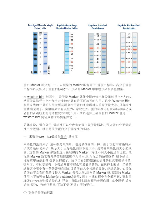
蛋白Marker可分为:一、未预染的Marker即宽分子量蛋白标准、高分子量蛋白标准以及低分子量蛋白标准;二、预染的Marker即单色预染和多色预染。
在western blot 过程中,分子量Marker就像个螺丝钉一样没虽然是个小细节,然而就是这样一个小细节对实验结果有着不可忽视的作用。
这个Western Blot 参照家族的一员的作用主要是用来指示蛋白条带所对应的分子量大小,只有标准量精确无误了,实验结果才有说服力,除此之外,蛋白标准还有表示转移成功或者蛋白在凝胶上的电泳程度等等的作用,所以选择正确的蛋白Marker也是western blot实验成功的必要条件之一。
总体来说,蛋白分子量标准可以分成未染蛋白分子量标准、预染蛋白分子量标准二个级别。
以下是关于蛋白分子量标准的小叙:一. 未染色(pre mixed)蛋白分子量标准未染色的蛋白分子量标准是最简单,也是最准确的一种。
由于没有附带染料分子或者是标记分子,所示大小正好是蛋白原本的大小,是精确判断蛋白大小必须的。
现在的Marker多数都选用预混和的Marker,方便不同大小的蛋白比较。
预混的Marker通常有几条带加倍浓度作为指示,因为混合的条带越多,越不好记,谁知道哪条是那条!数到眼都花了。
所以当看到特别浓的那几条标志带就记得是哪里了。
不过要记得,小带通常都不那么容易看清楚的。
在选择上来说,当然是选择其中至少有一条条带和自己的目的蛋白大小相近的最好,越近越好。
如果你的蛋白不幸在两条跨度较大Marker条带之间,选别的Marker吧。
预混的Marker 使用上不如预染Marker(pre-stained)好用,因为电泳过程中完全看不到,要和目标蛋白一起等到最后染色才“开蛊”,无法对实验起预示参照作用。
完全属于“后知后觉”型的,当然还是比“不知不觉”不做对照的要好。
①宽分子量蛋白标准如果从实验室总体考虑的,就选范围尽量宽,条带分布比较均匀的,这样你的蛋白无论在哪个区间都容易判断,避免正好落在一段空白区的危险。
MarkerⅢ DNA Ladder说明书

北京索莱宝科技有限公司
MarkerⅢDNA Ladder说明书
货号:M1600
规格:50T(250μl)/100T(500μl)
保存:-20℃。
有效期1年。
产品简介:
本产品是由7条带状双链DNA条带组成,适用于琼脂糖凝胶电泳中DNA条带的分析。
本产品为即用型产品,已含有1×loading buffer,直接取5μl电泳,使用方便,电泳图像清晰。
本产品中7条带分为200,500,800,1500,2000,3000,5000bp,其中1500bp条带约为20ng/μl,其余条带浓度大约10ng/μl。
储存液成份:
10mM Tris-HCl(pH8.4)
10mM EDTA
0.02%溴酚蓝
5%甘油
使用方法:
1.取5μl本产品加入到琼脂糖凝胶的加样孔中(每1mm加样孔宽度加1μl,如果加样孔较宽,可适当增加上样量)进行电泳。
2.建议凝胶浓度为1%琼脂糖凝胶,电泳电压4-10v/cm,电泳时间30-40分钟。
3.核酸染色,在紫外灯下观察电泳条带。
第1页共2页
北京索莱宝科技有限公司
第2页共2
页5μl 上样,1.0%琼脂糖凝胶电泳示意图
注意事项:
及时更换电泳缓冲液并使用新配制的琼脂糖凝胶,以免影响电泳结果。
marker说明书
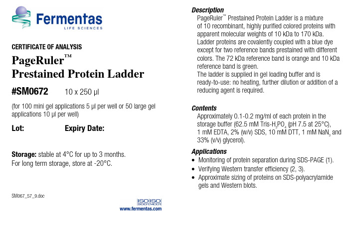
CERTIFICATE OF ANALYSISPageRuler ™Prestained Protein Ladder#SM067210 x 250 µl(for 100 mini gel applications 5 µl per well or 50 large gel applications 10 µl per well)Lot: Expiry Date:Storage: stable at 4°C for up to 3 months. For long term storage, store at -20°C.SM067_57_9.docDescriptionPageRuler ™Prestained Protein Ladder is a mixture of 10 recombinant, highly purified colored proteins with apparent molecular weights of 10 kDa to 170 kDa. Ladder proteins are covalently coupled with a blue dye except for two reference bands prestained with different colors. The 72 kDa reference band is orange and 10 kDa reference band is green.The ladder is supplied in gel loading buffer and isready-to-use: no heating, further dilution or addition of a reducing agent is required.ContentsApproximately 0.1-0.2 mg/ml of each protein in the storage buffer (62.5 mM Tris-H 3PO 4 (pH 7.5 at 25°C), 1 mM EDTA, 2% (w/v) SDS, 10 mM DTT, 1 mM NaN 3 and 33% (v/v) glycerol).Applications∙ Monitoring of protein separation during SDS-PAGE (1). ∙ Verifying Western transfer efficiency (2, 3).∙ Approximate sizing of proteins on SDS-polyacrylamidegels and Western blots.Instruction for Use❶ Thaw the ladder at room temperature for a few minutes to dissolve precipitated solids. DO NOT BOIL!❷ Mix gently, but thoroughly, to ensure the solution is homogeneous.❸ Load the following volumes of the ladder on an SDS-polyacrylamide gel:– 5 µl per well for mini gel,– 10 µl per well for large gel.Use the same volumes for Western blotting.❹ After the run is complete, stain the gel or perform Western transfer procedure as desired.Note•Each lot of the PageRuler™ Prestained Protein Ladder is calibrated against a precisely sized, PageRuler™ Unstained Protein Ladder and calculated apparent molecular weights are reported in the picture.•For precise molecular weight determinations use PageRuler™ Unstained Protein Ladder, #SM0661, see.•In 8 or 10% gels low molecular weight proteins may migrate with the dye front.•Loading volumes are intended for use in gels with a thickness of 0.75 mm. For thicker gels, the recommended loading volume should be increased.•PageRuler™ Prestained Protein Ladder could be used in Western blotting with all common membranes: PVDF, nylon and nitrocellulose.•Longer transfer times or higher transfer voltages may be required for Western blotting of large (>100 kDa) proteins.Lot specific MW, kDa4-20% Tris-glycine SDS-PAGEcontinued on back pageQUALITY CONTROL5 µl of PageRuler™ Prestained Protein Ladder resolves 10 bands of equal intensities in 4-20% SDS-PAGE (Tris-glycine buffer) and after Western blotting onto PVDF membrane.Quality authorized by: Jurgita Zilinskiene References1. Laemmli, U.K., Cleavage of structural proteins during the assembly of the head of bacteriophage T4, Nature, 227, 680-685, 1970.2. Burnette, W.N., "Western blotting": electrophoretic transfer of proteins from sodium dodecyl sulfate – polyacrylamide gels to unmodified nitrocellulose and radiographic detection with antibody and radioiodinated protein A, Anal. Biochem., 112 (2), 195-203, 1981.3. Towbin, H., et al., E lectrophoretic transfer of proteins from polyacrylamide gels to nitrocellulose sheets: procedure and some applications, Proc. Natl. Acad. Sci. USA, 76, 4350-4354, 1979.This product is manufactured under the license forStrep-tag® technology covered by US patents Nos.5,506,121, 6,103,493 and foreign counterparts. Related Products∙DualColor™ Protein Loading Buffer Pack #R1011∙Loading Buffer Pack #R0891∙Spectra™ Multicolor Broad Range Protein Ladder #SM1841∙PageRuler™ Unstained #SM0661∙PageRuler™ Plus Prestained Protein Ladder #SM1811∙PageSilver™ Silver Staining Kit #K0681∙PageBlue™ Protein Staining Solution #R0571∙10X Tris-glycine-SDS Buffer #B46∙10X Tris-tricine-SDS Buffer #B48∙DTT #R0861 ∙ProteoJET™ Mammalian Cell Lysis Reagent #K0301∙ProteoJET™ Cytoplasmic and Nuclear ProteinExtraction Kit #K0311∙Bradford Reagent, ready-to-use #R1271∙Bovine Serum Albumin Standard Set, ready-to-use #R1281∙Bovine Gamma Globulin Standard Set, ready-to-use #R1291PRODUCT USE LIMITATION.This product is developed, designed and sold exclusively for research purposes andin vitro use only. The product was not tested for use in diagnostics or for drugdevelopment, nor is it suitable for administration to humans or animals.Please refer to for Material Safety Data Sheet of the product.。
Bradford蛋白质浓度定量试剂盒产品说明书中文版主要
Bradford蛋白质浓度定量试剂盒产品说明书(中文版)主要用途Bradford蛋白质浓度定量试剂是一种旨在使用考马斯亮蓝染色剂G-250与可溶性蛋白质特异性结合产生色彩差异变化,通过比色测定其最大吸收转换来定量蛋白质浓度的权威而经典的技术方法。
该技术由大师级科学家精心研制、成功实验证明的。
其适用于各种蛋白质(动物、人体、植物、微生物等)的含量检测。
产品即到即用,操作简捷,性能稳定,检测敏感,定量精确。
技术背景考马斯亮蓝染料G-250(Coomassie Brilliant Blue G-250)是一种与蛋白质结合的化学染料。
它的三苯甲烷(triphenylmethane)基团,主要与蛋白质的非极性结构结合。
而它的阴离子磺酸盐(anion sulfonate)基团与蛋白质分子的阳离子和芳香类氨基酸,尤其是精氨酸和赖氨酸侧链的结合。
在酸性环境下,其最大吸收波长从465nm转换为595nm。
Bradford提出的方法的最大优点在于不受样品中各种化学成分的干扰。
较之Lowry,Hartree-Lowry和 BCA技术,更为敏感。
产品内容反应液(Reagent A)毫升标准液(Reagent B)毫升补充液(Reagent C)毫升产品说明书1份保存方式保存标准液(Reagent B)在-20℃冰箱里,其余的保存在4℃冰箱里,反应液(ReagentA),避免光照;有效保证6月用户自备48孔板:用于样品比色的容器比色杯:用于样品比色的容器分光光度仪或酶标仪:用于比色分析实验步骤一、48孔板微量测定A.建立标准样品曲线1.将-20℃冰箱里试剂盒中的标准液(Reagent B)置入冰槽里融化2.准备1个48孔板,做好相应的标记3.按下表配制标准样品反应液1)分别移取适量的标准液(Reagent B)到48孔板里指定序号的样品孔里2)分别移取适量的补充液(Reagent C)到48孔板里指定序号的样品孔里3)最后分别加入100微升反应液(Reagent A)序号标准液(Reagent B)补充液(Reagent C)反应液(Reagent A)标准样品蛋白质含量(微克)1 25微升 375微升 100微升252 12.5微升 387.5微升 100微升12.53 10微升 390微升 100微升104 5微升 395微升 100微升 55 2.5微升 397.5微升 100微升 2.56 1.25微升 398.75微升 100微升 1.257 0 400微升 100微升04.轻轻摇动48孔板,混匀反应物5.室温下暗室里孵育5分钟6.即刻放进分光光度酶标仪测定:波长为595nm7.绘制蛋白质浓度标准曲线:纵座标(Y)为吸光值OD595,横座标(X)为蛋白质含量(微克)B.样品测定1.将待测样品置入冰槽里融化2.分别移取100微升待测样品到48孔板里指定序号的样品孔里(注意:参见注意事项4)3.分别移取300微升补充液(Reagent C)到48孔板里指定序号的样品孔里4.最后分别加入100微升反应液(Reagent A)5.轻轻摇动48孔板,混匀反应物6.室温下暗室里孵育5分钟7.即刻放进分光光度酶标仪测定:波长为595nm8.根据上述标准曲线,测出待测样品的检测含量,再除以100微升(样品量),获得待测样品的实际浓度(微克/微升)(注意:参见注意事项5)二、比色杯标准测定A.建立标准样品曲线1.将-20℃冰箱里试剂盒中的标准液(Reagent B)置入冰槽里融化2.准备好1毫升比色杯3.按下表配制标准样品反应液1)分别移取适量的标准液(Reagent B)到1毫升比色杯2)分别加入适量的补充液(Reagent C)3)最后分别加入200微升GENMED反应液(Reagent A)序号标准液(Reagent B)补充液(Reagent C)反应液(Reagent A)标准样品蛋白质含量(微克)1 50微升 750微升 200微升502 25微升 775微升 200微升253 20微升 780微升 200微升204 10微升 790微升 200微升105 5微升 795微升 200微升 56 2.5微升 797.5微升 200微升 2.57 0 800微升 200微升04.轻轻上下倾倒比色杯,混匀反应物5.室温下暗室里孵育5分钟6.即刻放进分光光度酶标仪测定:波长为595nm7.绘制蛋白质浓度标准曲线:纵座标(Y)为吸光值OD595,横座标(X)为蛋白质含量(微克)B.样品测定1.将待测样品置入冰槽里融化2.移取200微升待测样品到1毫升比色杯里(注意:参见注意事项4)3.加入600微升补充液(Reagent C)4.加入200微升反应液(Reagent A)5.轻轻上下倾倒比色杯,混匀反应物6.室温下暗室里孵育5分钟7.即刻放进分光光度酶标仪测定:波长为595nm8.根据上述标准曲线,测出待测样品的检测含量,再除以200微升(样品量),获得待测样品的实际浓度(微克/微升)(注意:参见注意事项5)三、玻璃管大量测定(注意:参见注意事项2)A.建立标准样品曲线1.将-20℃冰箱里试剂盒中的标准液(Reagent B)置入冰槽里融化2.准备好5毫升玻璃管3.按下表配制标准样品反应液1)分别移取适量的标准液(Reagent B)到5毫升玻璃管2)分别加入适量的补充液(Reagent C)3)最后分别加入1毫升反应液(Reagent A)序号标准液(Reagent B)补充液(Reagent C)反应液(Reagent A)标准样品蛋白质含量(毫克)1 4毫升0 1毫升 42 2.5毫升 1.5毫升1毫升 2.53 2毫升2毫升1毫升 24 1毫升3毫升1毫升 15 0.5毫升 3.5毫升1毫升0.56 0.25毫升 3.75毫升1毫升0.257 0 4毫升1毫升0 4.涡旋震荡,混匀反应物5.室温下暗室里孵育30分钟6.即刻放进分光光度酶标仪测定:波长为595nm7.绘制蛋白质浓度标准曲线:纵座标(Y)为吸光值OD595,横座标(X)为蛋白质浓度(毫克)B.样品测定1.将待测样品置入冰槽里融化2.移取250微升待测样品到5毫升玻璃管里30030.23.加入3.75毫升补充液(Reagent C ) 4.加入1毫升反应液(Reagent A )5.涡旋震荡,混匀反应物 6.室温下暗室里孵育30分钟7.即刻放进分光光度酶标仪测定:波长为595nm8.根据上述标准曲线,测出待测样品的检测含量,再除以0.25毫升(样品量),获得待测样品的实际浓度(毫克/毫升)注意事项1. 本产品为200次(96孔板测定),100次操作(48孔板测定),50次操作(比色杯测定)包括标准曲线 2. 玻璃管大量测定须另购 Bradford 大量蛋白质浓度定量试剂盒()3. 操作时,须戴手套4. 建议用户使用48孔板微量测定时样品和试剂的用量,以及在最后浓度计算时的除数 序号待测样品补充液(Reagent C )反应液(Reagent A )蛋白浓度计算的除数 1 5微升 395微升 100微升 5 2 50微升 350微升 100微升 50 3 100微升 300微升 100微升 100 4250微升 150微升 100微升250如果是96孔板测定:待测样品、补充液(Reagent C )和反应液(Reagent A )的用量是48孔板测定的二分之一如果是比色杯测定:待测样品、补充液(Reagent C )和反应液(Reagent A )的用量是48孔板测定的2倍 5. 样品浓度计算公式:标准曲线求得蛋白含量(微克)÷样品量(微升)=微克/微升6. 加样后即刻比色测定7. 比色测定后,比色杯须清洗彻底 8. 强碱性样品将会干扰比色检测9. 下列化学成分和浓度范围不会干扰比色检测:Acetate0.6 M KCI1.0 M AcetoneMalic acid0.2 MAdenosine 1 mM MgCl 2 1.0 M Amino Acids Mercaptoethanol 1.0 M Ammonium sulfate 1.0 M MES 0.7 M Ampholytes Methanol 0.5% AcidMOPS 0.2 M ATP 1 mM NaCl 5 M Barbital NAD 1 mM BES2.5 M NaSCN3 MBoric acidPeptonesCacodylate-Tris 0.1 M Phenol 5% CDTA 0.05 M Phosphate 1.0 MCitrate 0.05MM PIPES 0.5acid 1mM Deoxycholate 0.1% Polyadenylic(MW<3000)PolypeptidesMDithiothreitol 1M0.2mg/ml PyrophosphateDNA 1mg/ml EDTA 0.1M rRNA 0.25mg/mlM tRNA 0.4 EGTA, 0.05mg/ml0.30RNAEthanol totalEagle’s MEM SDS 0.1%phosphatesolution SodiumsaltEarle’s20%sulfateacid 1.0M StreptomycinFormicX-100 0.1% FructoseTritonGlucose TricinemM Glutathione Tyrosine 1mMThymidine 1 Glycerol99%Glycine 0.1 M Tris 2.0 MGuanidine-HCI Urea 6 Msaltsolution VitaminsHank'sHEPES buffer 0.1 M10.建立微量测定的标准曲线,建议使用的蛋白质浓度范围为0至50微克11.本公司提供系列蛋白定量检测产品质量标准1.本产品经鉴定性能稳定2.本产品经鉴定检测敏感。
λ-Hind ⅢdigestDNAMarker使用说明书(D3403A-Hind III)
λ-Hin dⅢdigest DNA Marker使 用 说 明 书Code No.:D3403A●包 装 量:50 µg(500 µl×2支;内含1×Loading Buffer)●浓 度:50 ng/µl●制品说明:λ-Hin d III digest DNA Marker是由Bacteriophage λc I857Sam7 DNA用Hin d III酶切反应后配制而成的。
本制品浓度为50ng/µl,内含1×Loading Buffer,可根据实验需要,每次取5µl~20 µl电泳,使用方便,电泳图像清晰。
●保存温度:-20℃●使用注意:1.λDNA digest DNA Markers的原始末端之间经常由COS末端结合在一起,在电泳前进行热处理(60℃, 5分钟),能使Marker的电泳图像变得更为清晰。
以λ-Hin d III digest DNA Marker为例,原始末端之间的COS末端的结合,影响23,130 bp和4,361 bp的DNA条带。
如果电泳以后的图像中的4,361 bp的DNA条带亮度低于比其小的DNA片段时,那说明COS末端的结合情况比较严重。
2. 电泳时的加样孔宽度小于6 mm时,每次取5µl制品电泳便可得到清晰条带。
如果加样孔增宽,须适当增加Marker制品的加样量。
3.对DNA电泳而言,Agarose的纯度对DNA条带的清晰度影响很大。
因此,电泳时应尽量选用质量好的Agarose。
Agarose电泳图像示意图。
考马斯亮蓝快速染色液使用说明
考马斯亮蓝快速染色液产品编号产品名称包装P0017 考马斯亮蓝快速染色液 250ml产品简介:考马斯亮蓝快速染色液(Commassie Blue Fast Staining Solution)是以考马斯亮蓝G250为染料,可用于SDS-PAGE或非变性PAGE等蛋白电泳胶的快速、高灵敏染色,或Western转膜后PAGE胶上残余蛋白的检测。
本考马斯亮蓝快速染色液是一种无毒,无刺激性气味的高度环保型染色液。
普通的常规方法需使用剧毒的甲醇及强刺激性的乙酸,而碧云天生产的考马斯亮蓝快速染色液则采取全新配方,实现了无毒和无刺激性气味。
本考马斯亮蓝快速染色液只需一小时或更短时间就可以检测到低达100ng的蛋白电泳条带,而常规方法需3小时以上。
本试剂盒可以进行高灵敏染色,最低可以清楚检测到10ng的蛋白电泳条带,参考图1。
蛋白上样量: 10μg 5μg 2μg 1μg 500ng 200ng 100ng 50ng 20ng 10ng 5ng 2ng 1ng图1. 使用碧云天的考马斯亮蓝快速染色液对不同上样量的BSA蛋白样品用10% SDS-PAGE胶电泳后的两次染色结果。
无需对蛋白和凝胶进行固定,可以不进行脱色,操作更加简单。
本染色液和质谱分析兼容,即经过本染色液染色的蛋白条带或蛋白点可以用于后续的质谱分析。
包装清单:产品编号产品名称包装P0017 考马斯亮蓝快速染色液250ml—说明书1份保存条件:4℃保存,一年有效。
注意事项:本染色液呈酸性,有轻微腐蚀性,使用时请作必要防护。
如果使用微波炉加热,请特别注意避免沸腾。
以免因暴沸而导致凝胶碎裂。
为了您的安全和健康,请穿实验服并戴一次性手套操作。
使用说明:1. 电泳结束后,取胶放入约50-100ml蒸馏水中,在摇床上摇动5分钟,倒弃液体,再重复两次,共洗涤三次。
注:本步骤的目的是为了洗去凝胶中的SDS等干扰物质。
如果不能充分去除SDS会导致背景较高,如果胶非常厚,每次的洗涤时间需适当延长并且洗涤次数也需适当增加。
TaKaRa BioMasher Standard 产品手册说明书
Cat. #9790A9791A Product ManualTaKaRa BioMasher Standard (Non-sterile)TaKaRa BioMasher Standard (Sterile)For Research UseTable of ContentsI. Description (3)II. Components (3)III. Storage (3)IV. Materials (3)V. Protocol (3)VI. Experimental Examples (4)VII. Related Products (4)I. DescriptionTaKaRa BioMasher Standard is a disposable microtube homogenizer designed to efficiently crush small amounts of biological samples for nucleic acid and/or protein extraction. This product is a set of a 1.5-ml microtubes and micro stir bars. The inner wall of the tube and the tip of the stir bar are textured allowing effective disruption of the sample. In addition, there is a lid to prevent splashing of the sample and reagents during sample processing. After the sample has been homogenized, the tube can be centrifuged to minimize sample loss.The TaKaRa BioMasher Standard is available either sterilized (Cat. #9791A) or nonsterilized (Cat.# 9790A).II. Components(1) TaKaRa BioMasher Standard (micro stir bar and microtube) 50(2) Stir bar grip (PESTLE GRIP) 1III. Storage Room temperatureKeep out of sun- and UV light.IV. MaterialsTaKaRa BioMasher StandardPolypropylene microtubes AutoclavablePolyacetal stir bar Not autoclavable*Silicon rubber stir bar grip Autoclavable* : The stir bar can not be autoclaved. If you require sterilization, please use Cat. #9791A. V. ProtocolNOTE: Wear a mask, gloves, lab coat, and safety goggles. If necessary, perform in a safety cabinet.1. Insert the micro stir bar into the end of the PESTLE GRIP.2. Add the sample to the microtube. Use less than 100 mg of sample. If necessary, add theextraction reagent (less than 250 μl).3. Insert the micro stir bar into the microtube. With the PESTLE GRIP, rotate and move themicro stir bar up and down, crushing the sample. If necessary, perform on ice.4. After crushing, disconnect the PESTLE GRIP and discard the micro stir bar. The PESTLEGRIP can be used repeatedly; store for future use.5. Close the lid of the microtube. The sample is ready for extraction.VII. Related ProductsRNAisoPlus (Cat. #9108/9109)*NucleoSpin® RNA II (Cat. #740955.10/.50/.250)NucleoSpin® RNA XS (Cat. #740902.10/.50/.250)NucleoSpin® Tissue (Cat. #740952.10/.50/.250)NucleoSpin® Tissue XS (Cat. #740901.10/.50/.250)Wide-Range DNA Ladder (100 - 2,000 bp) (Cat. #3427A/B)* : Not available in all geographic locations. Check for availability in your region.VI. Experimental ExampleTotal RNA extraction from mouse liver 100 mg of mouse liver was crushed using the TaKaRa BioMasher Standard (Sterile). As a comparison, 100 mg frozen mouse liver tissue was crushed with a mortar or with crusher beads. For all samples, total RNA was extracted using 1 ml RNAiso Plus (Cat. #9108/9109) according to the recommended protocol. Total RNA was quantified and analyzed on an 1% agarose gel.Results: Processing the samples using the TaKaRa BioMasher Standard had similar extraction efficiency as the mortar and bead crushing protocols.Crushing method total RNA (μg) A 260/A 2801.Mortar 360 1.702.Mortar 424 1.803.Crusher beads 530 1.924.Crusher beads 494 1.935.TaKaRa BioMasher Standard 411 1.896.TaKaRa BioMasher Standard4311.90M 125364(bp)2,000750Lane 1-6 Equal amounts of RNA M Wide-Range DNA Ladder (100 - 2,000 bp)NOTE : This product is for research use only. It is not intended for use in therapeutic or diagnostic procedures for humans or animals. Also, do not use this product as food, cosmetic, or household item, etc.Takara products may not be resold or transferred, modified for resale or transfer, or used to manufacture commercial products without written approval from TAKARA BIO INC.If you require licenses for other use, please contact us by phone at +81 77 543 7247 or from our website at .Your use of this product is also subject to compliance with any applicable licensing requirements described on the product web page. It is your responsibility to review, understand and adhere to any restrictions imposed by such statements.All trademarks are the property of their respective owners. Certain trademarks may not be registered in all jurisdictions.。