小鼠早期胚胎发育共57页文档
小鼠早期胚胎发育ppt课件

7 Development 139, 3-14, 2012
Two possible mechanisms for the generation of multiple cell types and polarity
8 Development 139, 3-14, 2012
Mouse trophectoderm distinguish from inner cell mass regulated by Hippo signaling pathway
9 Dev Cell 16, 398-410, 2009
Cell position-dependent differentiation mediated by Hippo signaling
10 Development 139, 3-14, 2012
Mouse embryo development from fertilization to gastrulation
?
15 Development 139, 3-14, 2012
Emerging asymmetry and establishment of anteriorposterior polarity in the E5.75 mouse embryo
Axis of Proximal & Distal
Axis of Anterior-Posterior
23 Development 139, 3-14, 2012
Mouse embryo development from fertilization to gastrulation
24 Development 139, 3-14, 2012
Early mouse embryo development and lineage segregation
实验小鼠和大鼠各胚胎发育时期的变化
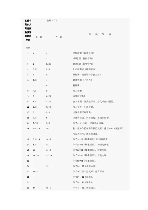
实验小鼠和大鼠各胚胎发育时期的变化时期胎龄(日)胚胎发育小鼠大鼠1 1 1 单核细胞(输卵管内)2 2 2细胞期(输卵管内)3 2 3.25 4细胞期(输卵管内)4 2.5 3.5 8-12细胞期(输卵管内)5 3 3. 桑椹期(输卵管—子宫上部)6 3.5 4 囊胚初期(子宫内)7 4 5 囊胚期8 4.5 6 植入开始9 5 6.75 内外胚层分化10 5.5 7.25 植入末期,卵黄腔形成,分化成内外胚层,11 6.5 7.75 植入完毕,出现羊膜。
12 7 8.5 出现中胚层和原条。
13 7.5 9 出现神经板,头部突起,出现尿膜褶。
14 7.75 9.5 体节1-4(头部)完成外层胎盘。
15 8~8.5 10 腔,胚外体腔内和羊膜腔发育。
体节5-12(颈椎部)形成神经沟,胚回转开始。
16 8.5~9 10.5 体节13-20(胸椎前部)胚回转结束。
17 9.5 11 体节21-25(胸椎后部),神经沟闭锁。
18 10 11.5 体节26-28(腰椎前部),前肢出现。
19 10.25 11.75 体节29-31(腰椎后部),后肢出现。
20 体节32-33(荐椎后部)。
21 12 体节34、35(荐椎后部)。
22 10.5 体节36(第一层尾椎)鼻窝形成23 体节37、38(尾椎)。
24 体节39、40(尾椎)。
25 11 12.5 体节41、42,前肢变大。
26 体节43-45(尾椎)手指分化。
27 12 13 体节46-48(尾椎)脐疝明显。
28 12.5 13.5 体节49-51(尾椎),鼻上颌闭锁。
29 14 体节52-55(尾椎)。
30 13 14.5 体节56-60(尾椎),耳孔开,耳壳形成31 13.5~14 15 体节61-63(尾椎),口盖板闭锁32 14.5 15.5 体节64(尾椎)、体表出现毛囊。
33 15 16 体节65(最终尾椎)34 16-16.5 17-18 脐疝消失,眼脸、耳壳闭锁。
小鼠早期胚胎发育和胚胎移植
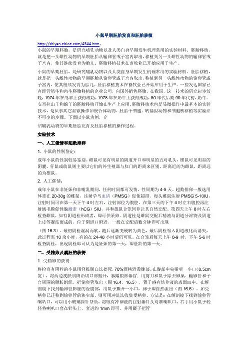
小鼠早期胚胎发育和胚胎移植/4544.htm、小鼠的早期胚胎,是研究哺乳动物以及人类自身早期发生机理常用的实验材料。
胚胎移植,就是把一头雌性动物的早期胚胎从输卵管或子宫内取出,移植到另一头雌性动物的输卵管或子宫内,使其继续发育为胎儿。
胚胎移植技术在畜牧业已开始应用于生产。
小鼠的早期胚胎,是研究哺乳动物以及人类自身早期发生机理常用的实验材料。
胚胎移植,就是把一头雌性动物的早期胚胎从输卵管或子宫内取出,移植到另一头雌性动物的输卵管或子宫内,使其继续发育为胎儿。
胚胎移植技术在畜牧业已开始应用于生产。
一些发达国家已有经营奶牛和肉牛胚胎移植的企业公司,向国外销售胚胎。
在我国,这一技术的研究起步较晚,1974年在绵羊上获得成功,1978年在奶牛上获得成功,80年代后期90年代初,奶牛、安哥拉山羊和绵羊的胚胎移植开始在生产上应用。
胚胎移植术也是显微操作中最基本的实验技术,是从事其它显微操作如嵌合体动物、胚胎干细胞、转基因动物和细胞核移植等实验必不可少的步骤。
下面以小鼠为例,介绍哺乳动物的早期胚胎发育及胚胎移植的操作过程。
实验技术一、人工催情和超数排卵1. 小鼠的性别鉴定:成年小鼠的性别较易鉴别。
雌鼠可见有明显的阴道开口和明显的五对乳头,雄鼠可见明显的阴囊,仔鼠或幼鼠则主要以它们的外生殖器与肛门的距离来区别,距离近的为雌鼠,距离远的为雄鼠。
2. 人工催情:成年小鼠在非妊娠和非哺乳期间,任何时间都可发情,性周期为4-5天。
超数排卵一般选用体重在20-30g的雌鼠,注射孕马血清(PMSG)促使超排。
每头雌鼠注射PMSG 5-10IU,注射时间可在第一天下午4时左右,注射部位为腹腔。
在第三天的下午4时左右腹腔再注射绒毛膜促性腺激素(hCG)5IU,并和雄鼠合笼饲养让其自然交配。
第四天上午8时左右检查雌鼠,如有阴道栓形成者,即可供采卵。
阴道栓是雌鼠交配后精液与阴道分泌物及阴道上皮等凝结而成的,位于阴道口附近,一般在交配后数分钟即可出现(图16.3)。
小鼠早期胚胎发育的研究 )

小鼠早期胚胎发育的研究(实验方案)基础生物学课程设计所在学院生物科学与工程学院专业班级生物科学学生姓名学生学号指导教师XXXX年XX月XX日小鼠早期胚胎发育的研究一、引言胚胎发育一词通常是指从受精卵起到胚胎出离卵膜的一段过程。
而无脊椎动物胚胎学家则常把其概念扩展到胎后发育直到性成熟,甚至整个生活史。
动物胚胎的发育过程虽然动物的种类繁多,但是胚胎的发育依然拥有相似的过程,能够分成受精、卵裂、桑葚胚、囊胚、原肠胚与器官形成等阶段。
此外脊椎动物的胚胎发育过程中,各种动物共同拥有的特征会首先出现(如皮肤),之后才逐渐发展出特化的构造(如鱼鳞),而且较复杂的物种与较原始的物种之间一开始相当类似,之后才随着发育的时间而慢慢增加变异。
受精:卵子和精子融合为一个合子的过程。
它是有性生殖的基本特征,普遍存在于动植物界,卵裂:精子与卵子结合之后会形成受精卵,由于卵黄的分布具有不对称的特性,因此受精卵可以分为动物极(会发展成外胚层)和植物极(会发展成中胚层与内胚层)。
在卵裂时期,卵子会先分裂成两个细胞,之后细胞通常会逐次倍增,但是对哺乳类而言,有时候会有不同时分裂并造成只有奇数个细胞的现象。
在这个阶段,胚胎的总体积大致不变。
不同的物种具有不同的卵裂方式,可以分为完全卵裂(holoblastic cleavege)和不完全卵裂(meroblastic cleavage),分别又可以细分成许多不同方式。
如无脊椎动物的辐射卵裂与螺旋卵裂、哺乳动物的旋转式分裂等。
原肠胚:当细胞分裂成为囊胚之后,会经过一段称为原肠形成的型态发生过程,之后形成原肠胚。
原肠形成过程有许多不同方式,能够大致分成5种:内陷式(Invagination)、衰退式(Involution)、进入式(Ingression)、脱层式(Delamination)、包覆式(Epiboly),动物的胚胎利用这5种方式形成了外胚层、中胚层与内胚层的组合,而这三种胚层在之后会形成各种细胞。
小鼠早期胚胎发育

Axis of Dorsal-Ventral
Myocardin
Cell lineages in early mouse embryo
Embryo culture system for time-lapse movie acquisition of node morphogenesis
in vitro embryo culture
GFP knock-in tracing
Dev Cell 13, 884-896, 2007
Time-lapse images of node morphogenesis
Noto-
Dev Cell 13, 884-896, 2007
The displacement model of endoderm morphogenesis in the mouse gastrula
Primitive streak formation
VE: visceral endoderm AVE: anterior VE DVE: distal VE
PS: Primitive Streak
EMT at mouse primitive streak
EMT at mouse primitive streak
E6.5
Dye injection
The distal cap epiblast cells migrate to the anterior neuroectoderm
M ag a zi ne
105
ne
ht yol k Epi bl ast nd
en
pxm l m ht
Yol k cel l s
?
Development 139, 3-14, 2012
老鼠胚胎发育时期示意图
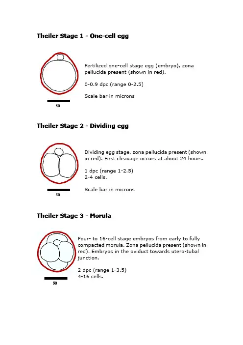
Theiler Stage1-One-cell eggFertilized one-cell stage egg(embryo),zonapellucida present(shown in red).0-0.9dpc(range0-2.5)Scale bar in micronsTheiler Stage2-Dividing eggDividing egg stage,zona pellucida present(shownin red).First cleavage occurs at about24hours.1dpc(range1-2.5)2-4cells.Scale bar in micronsTheiler Stage3-MorulaFour-to16-cell stage embryos from early to fullycompacted morula.Zona pellucida present(shown inred).Embryos in the oviduct towards utero-tubaljunction.2dpc(range1-3.5)4-16cells.All scale bars in microns.Theiler Stage4-Blastocyst(ICM apparent)Embryo progresses from morula to the blastocyststill within the zona pellucida(shown in red).There isearly evidence of the blastocoelic cavity.3dpc(range2-4)16-40compacted cells.All scale bars in microns.In the blastocyst stage(zona-intact)there is adistinct inner cell mass and an outer layer oftrophectoderm ually located in the uterinelumen.Theiler Stage5-Blastocyst(zona-free)Blastocyst(zona-free),invariably located withinthe uterine lumen.4dpc(range3-5.5)Scale bar in micronsTheiler Stage6-Attachment of blastocystBlastocysts implants,first evidence ofembryonic endoderm cells coveringthe blastceolic surface of the inner cellmass.4.5dpc(range4-5.5)Witschi Stage8(rat)Carnegie Stage4(human)Scale barTheiler Stage7-Implantation and formation of Egg CylinderEctoplacental cone appears.Rapid increasein the number of inner cell mass cellsleading to the formation of the epiblast withsubsequent growth to form the eggcylinder.The proximal or visceral cells(opposite side from the trophoblastic cap)are cuboidal in shape.Primary endodermlines the mural trophectoderm.5dpc(range4.5-6)Witschi Stage10(rat)Carnegie Stage5(human)ScaleTheiler Stage8-Differentiation of egg cylinderImplantation site2x3mm.The maternal tissue isinvaded by trophoblast(primary)giant cells andthe ectoplacental cone is invaded by maternalblood.Differentiation of the egg cylinder intoembryonic and extraembryonic regions and theformation of the proamniotic cavity.Reichert'smembrane,which is non-cellular and secreted bythe distal endoderm first appears.6dpc(range5-6.5)Witschi Stage10-11(rat)Carnegie Stage5(human)Scale bar in micronsTheiler Stage9-Advanced Endometrial ReactionPS:Advanced egg-cylinder stage with the firstevidence of of an embryonic axis.There is clearmorphological distinction between the embryonicand extraembryonic ectoderm.The ectoplacentalcone is further invaded by maternal blood and theoriginal lumen of the uterine crypt hasdisappeared.ES:Late in this stage gastrulation begins,producing the first mesodermal cells.6.5dpc(range6.25-7.25)Downs&Davies PS-ES:prestreak to earlystreakWitschi Stage11(rat)Carnegie Stage6-7(human-stage table)Scale bar in microns.Theiler Stage10-AmnionGastrulation continues throughout allsub-divisions of this stage and:MS:Tissue at the posterior end of the primitivestreak bulges into the proamniotic cavity andforms the posterior amniotic fold.LSOB:In the mesoderm of the posterior amnioticfold small cavities coalesce to form a single cavity,the exocoelom.LSEB:The allantoic bud first appears,gastrulationcontinues and the node becomes visible.7dpc(range6.5-7.5)Downs&Davies MS-LS midstreak to latestreakWitschi Stage12(rat)Carnegie Stage8(human-stage table)Scale bar in microns.Theiler Stage11-Neural Plate,Presomite StageNP:The amniotic cavity is sealed off with nowthree cavities-amniotic,exocoelom andectoplacental cleft.The neural plate is definedanteriorly and the head process is developing.Inthe midline,sub-adjacent to the neural groove,the notochodal plate is visible.LNP:The allantoic bud elongates.EHF:The rostral of the neural plate begins toenlarge to form the head folds.LHF:Head folds continue to enlarge and theforegut pocket begins to form.7.5dpc(range(7.25-8)Downs&Davies OB-EB/LB-EHF-LHF:-no All.bud to late All.bud to early head fold to late headfoldWitschi Stage12-13(rat)Carnegie Stage9(human-stage table)Scale bar in microns.Theiler Stage12-First somitesUnturned embryo with first appearenceof somite pairs.1-4somites:The allantois extendsfurther into the exocoelom and themaxiliary components of the1stbranchial arch become prominent.Thepreotic sulcus is visible in the2-3somite embryo.The cardiogenic platebegins to form and the foregut pocket isclearly visible.5-7:The headfolds are particularlyprominent and neural closure occurs inthe region of the4th and5th somites,extending in both directions from thissite.The optic placodes are first evidentand become indented to form the opticpits.The heart rudiment developsrapidly.The allantois contacts thechorion at the end of this stage.Absent:The2nd branchial arch and>7somites.8dpc(range7.5-8.75)1-7somite pairsWitschi Stage14-15(rat)Carnegie Stage9(human-stage table)All scale bars in micronsClick on an image for a larger version Theiler Stage13-Turning of the embryoThis is a short period with turning initiated inembryos with6-8pairs of somites and usuallycompleted in embryos with14-16pairs ofsomites.The first branchial arch hasmaxillary and mandibular components butthe maxillary process is not visible until later(TS16).A second branchial arch is nowevident.There is evidence of regionalisationof the heart and the neural tube is closedfrom a point opposite the outflow tract to theproximal part of the tail.Absent:3rd branchial arch.8.5dpc(range8-9.25)8-12somite pairsWitschi Stage15(rat)Carnegie Stage10(human-stage table)All scale bars in microns.Click on an image for a larger version.Theiler Stage14-Formation and closure of anterior neuroporeThe rostral extremity of the neural tube closes inembryos with usually about15-18somite pairsand defines this stage.The otic pit becomesprogressively more indented but not closed,themandibular process of the1st branchial arch isclearly visible.The3rd branchial arch becomesvisible late in the stage.An increasinglyprominent ridge on the lateral body wall,approximately at the level of the8th-12th somite,indicates the site of the future forelimb bud.Absent:forelimb bud.9dpc(range8.5-9.75)13-20somite pairsWitschi Stage16(rat)Carnegie Stage11(human-stage table)All scale bars in microns.Click on an image for a larger version.Theiler Stage15-Formation of posterior neuropore; forelimb budThe posterior neuropore form and thecondensation of the forelimb bud becomesapparent near the8th-12th somite pairs.A distinct condensation of the hind limbbud appears just at the end of the stage.The forebrain vesicle subdivides intotelencephalic and diencephalic vesicles.Absent:hindlimb bud,Rathke's pouch.9.5dpc(range9-10.25)21-29somite pairsWitschi Stage17-19(rat)Carnegie Stage12(human-stage table)All scale bars in microns.Click on an image for a larger versionTheiler Stage16-Closure of posterior neuropore;hind limb bud and tail budThe hind limb bud becomes visible atthe level of the23rd-28th somites.Thetail bud appears as a short stump andthe3rd and4th branchial arches aredistinctly concave.Rathke's pouch andthe nasal processes start to form.At theend of this stage the posteriorneuropore begins to close.Absent:thin and long tail.10dpc(range9.5-10.75)30-34somite pairsWitschi Stage20-21(rat)Carnegie Stage13(atlas),14(atlas),15(atlas)(human-stage table)All scale bars in microns.Click on an image for a larger version. Theiler Stage17-Deep lens indentationThe most obvious distinguishingfeatures are the deepening of the lenspit,with a narrowing of it's outerpore-like opening,and the firstappearence of the physiologicalumbilical hernia.The1st branchialarch is conspicuously divided into itmaxillary and mandibularcomponents.There is advanceddevelopment of the brain tube and thetail elongates and thins.Absent:nasal pits.10.5dpc(range10-11.25)35-39somite pairsWitschi Stage24-25(rat)Carnegie Stage13(atlas),14(atlas),15(atlas)(human-stage table)All scale bars in microns.Click on an image for a larger version.Theiler Stage18-Closure of lens vesicleThe primary externally recognisable feature isthe progressive closure of the lens vesicle.Thesomites in the cervical region are no longervisible and the rapid growth of the brain isstriking.The nasal pits start to form.Absent:auditory hillocks,anterior footplate.11dpc(range10.5-11.25)40-44somite pairsWitschi Stage25-26(rat)Carnegie Stage13(atlas),14(atlas),15(atlas)(human-stage table)All scale bars in microns.Click on an image for a larger versionTheiler Stage19-Lens vesicle completely separated from surfaceThe lens vesicle becomes completely closed and detached from the ectoderm.The peripheral margins of the eye become well defined.The forelimbs are seen to be divided into two regions, the proximal part consisting of the futurelimb-girdle and arm and the more peripheral part which forms a circular or paddle-shaped handplate(anterior footplate).The medial and lateral margins of the otic pit are coming together reducing the entrance to a narrow slit and the auditory hillocks become visible.Absent:retinal pigmentation,signs of fingers.11.5dpc(range11-12.25)45-47somite pairsWitschi Stage26-27(rat)Carnegie Stage16(atlas),(human-stage table) All scale bars in microns.Click on an image for a larger version.The handplate(anterior footplate)is no longer circular but develops angles which correspond to the future digits.The posterior footplate is also distinguishable from the lower part of the leg.It is possible to see the pigmentation of the pigmented layer of the retina through the transparent cornea.The tongue and brain vesicles are clearly visible.Absent:5rows of whiskers,indented handplate.12dpc(range11.5-13)48-51somite pairsWitschi Stage28(rat)Carnegie Stage17(atlas),(human-stage table)All scale bars in microns.Click on an image for a larger versionThe distal borders of the anterior and posterior footplates are now indented and the digit widths and locations can be discerned.The elbow and wrist are now identifiable.The pinna rapidly develops and forms a crest at right angles to the head.Five rows of vibrissae are visible as well as a prominant hair follicle over the eye and another over the ear.The lens vesicle has lost it's lumen. The physiological umbilical hernia is prominent. Absent:hair follicles,distally separate fingers.13dpc(range12.5-14)52-55somite pairsWitschi Stage29-30(rat)Carnegie Stage18(atlas),19(atlas),(human-stage table)All scale bars in microns.Click on an image for a larger version.Individual fingers are visible in the anterior footplate and there are deep indentations between the toes which are not yet separated. The long bones of the limbs are present and there are hair follicles in the pectoral,pelvic and trunk regions.The pinna is turned forwards and the umbilical hernia is conspicuous. Absent:hair follicles in the cephalic region.14dpc(range13.5-15)56-~60somite pairsWitschi Stage31(rat)Carnegie Stage20(atlas),21(atlas),22(atlas),23(atlas),(human-stage table)All scale bars in microns.Click on an image for a larger version.The toes separate and are clearly divergent,not becoming parallel until later.Hair follicles are present in the cephalic region but not at the periphery of the vibrissae.The pinna covers more than half of the external auditory meatus and theeyelids are still open.Absent:nail primordia,fingers2-5parallel.15dpc>60somite pairsWitschi Stage32(rat)Human foetal period.All scale bars in microns.Click on an image for a larger version.Fingers2-5are nearly parallel.Nail primordia are visible on the toes.The eyelids have fused in most cases by the end of the stage and the pinna almost completely covers the external auditory meatus.The umbilical hernia isdisappearing and there is a correspondingincrease in the size of the peritoneal sac.Absent:fingers and toes joined together.16dpc>60Somite pairsWitschi Stage33(rat)All scale bars in microns.Click on an image for a larger version.The skin has thickened and formed wrinkles and the subcutaneous veins are less visible.The fingers and toes have become parallel and the umbilical hernia has disappeared.The eylids have fused.Whiskers are just visible. Absent:ear extending over auditory meatus,long whiskers.17dpcWitschi Stage34(rat)All scale bars in microns.Click on an image for a larger version.The whiskers visible at stage25are definitely longer and the skin has thickened.The pinna is larger and such that virtually none of the lumen of the auditory meatus is visible.The eyes are barely visible through the closed eyelids18dpcWitschi stage35(rat)All scale bars in microns.Click on an image for a larger version.Newborn19-20dpcAll scale bars in microns.Click on the image for a larger version.Mouse embryos can be staged according to a variety of criteria,the most general of which are those described by Theiler in"The House Mouse:Atlas of Mouse Development"(Springer-Verlag, New York,1989).Theiler's criteria are too broad to distinguish many of the important phases of early development and must therefore be supplemented by others,for example,cell number, somite number or those charcteristics used by Downs and Davies(1993)Development,118,1255. We have therefore combined these different criteria in the table1below which defines a new set of stages based on the numbered Theiler series,but with intermediate divisions indicated by non-integer stage numbers.Embryos of the same gestational age may differ in their stage of development.We have therefore included in the table an indication of the expected range of gestational ages(days of gestation,dpc)over which each developmental stage may be found. Different mouse strains develop at different rates and,in some cases show differences in the relative rates of development of different organs.Strictly,the stages recognised by Downs and Davies apply to outbred mice of the PO strain.The data in the remainder of the table below refer to embryos of crosses between F1hybrid(C57BL X CBA)mice.Each Theiler stage is linked(click on the number)to its corresponding diagram,with more details of the defining features for that stage.A brief text or pictorial index to the diagrams is also provided.Theiler Stage dpc(range)2SomiteNo.3Cellnumber(C57BLxCBA)F1mice4POmice521(1-2.5)2-4Dividing egg4316-40Blastocyst,Inner cell mass apparent(2-4)6 4.5(4-5.5)Attachment of blastocyst ,primary endoderm covers blastocoelic surface ofinner cell mass86(5-6.5)Differentiation of egg cylinder .Implantation sites 2x3mm.Ectoplacentalcone region invaded by maternal blood,Reichert's membrane and proamnioticcavity form10a 7(6.5-7.75)Mid streak (MS),amniotic fold starts to formMS 10b Late streak,no bud (LSOB ),exocoelomLS 10c Late streak,early bud (LSEB),allantoic budfirst appears,node,amnion closing12a 8(7.5-8.75)1-41-4somites ,allantois extends,1st branchial arch,heart starts to form,foregut pocket visible,preotic sulcus (at 2-3somitestage)12b 5-75-7somites ,allantois contacts chorion atthe end of TS12Absent 2nd arch,>7somites2nd arch presentAbsent 3rd branchial arch149(8.5-9.75)13-20Formation &closure of ant .neuropore,otic pit indented but not closed,3rd branchial arch visibleAbsent forelimb bud1610(9.5-10.75)30-34Posterior neuropore closes ,Formation ofhindlimb &tail buds,lens plate,Rathke'spouch;the indented nasal processes startto formAbsent thin &long tail1811(10.5-11.25)40-44Closure of lens vesicle ,nasal pits,cervicalsomites no longer visibleAbsent auditory hillocks,anterior footplate2012(11.5-13)48-51Earliest sign of fingers (splayed-out),posterior footplate apparent,retinapigmentation apparent,tonguewell-defined,brain vesicles clearAbsent 5rows of whiskers,indentedanterior footplate2214(13.5-15)56-~60Fingers separate distally ,only indentationsbetween digits of the posterior footplate,long bones of limbs present,hair follicles inpectoral,pelvic and trunk regionsAbsent open eyelids,hair follicles incephalic region2416Reposition of umbilical hernia ,eyelidsclosing,fingers 2-5are parallel,nailprimordia visible on toesAbsentwrinkled skin,fingers &toes joinedtogether2618Long whiskers ,eyes barely visible throughclosed eyelids,ear covers auditory meatus28Postnatal development 1.Bard,J.B.L.,Kaufman,M.H.,Dubreuil,C.,Brune.R.M.,Burger,A.,Baldock,R.A.,Davidson,D.R.(1998).An internet-accessible database of mouse developmental anatomy based on a systematic nomenclature.Mechanisms of Development 74,111-20.2.Days post conception,with the morning after the vaginal plug is found being designated0.5dpc (or E0.5).For detailed discussion see Kaufman (1994).The Atlas of Mouse Development (2nd printing),pp.515-525.London:Academic Press.3.The figure given refers to the number of the most caudal somite.No account is taken ofsomites partitioning into dermomyotomes and sclerotomes,nor of their subsequent differentiation.4.Adapted from Theiler (1989)[The House Mouse:Atlas of Embryonic Development .NewYork:Springer-Verlag]and Kaufman (1994);detailed staging for Theiler stages 9-12courtesy of wson [personal communication].5.From Downes,K.M.and Davies,T.(1993).Staging of gastrulating mouse embryos bymorphological landmarks in the dissecting microscope.Development ,118,mentsGeneral comment on timing (dpc):In judging the lower and upper ranges of dpc equivalent to a particular Theiler stage,we have generally followed Theiler's book and,in most cases,have given a wider range than Theiler,because the numbers of embryos he cites are small.We have given a larger range at the maximum than the minimum because,in general,embryos are more likely to be retarded by their environment or genetic constitution than made to proceed more quickly through development.In most cases,however,the resulting dpc range is an estimate that is consistent with the results of Theiler,but not based on additional evidence.Comment on somite numbers:The range of somite numbers for each stage is given only as a guide to what might be expected of typical embryos.As can be seen from Theiler (1989)4the truerange can be much wider.Therefore,for all stages after TS12,the somite number should not be taken as a reliable global indicator of the overall embryo stage.Richard Baldock,Jonathan Bard,Duncan Davidson and Kirstie Lawson,7th May1998Please mail comments to Prof.Jonathan Bard。
C57BL6J小鼠生长周期
C55混凝土配合比设计
小鼠的生长周期
1、胚胎期
小鼠卵在输卵管壶腹部受精后开始分裂发育,至桑椹胚(约3天)进入子宫,形成囊胚(约第5 天)开始着床,妊娠期为19~21天。
2、生后早期及哺乳期
1)新生小鼠赤裸无毛,皮肤肉红色,不开眼,双耳与皮肤粘连。
2)4~6日龄双耳张开耸立。
3)7~8日龄四肢发育开始爬动游走,被毛逐渐浓密,下门齿长出。
4)9~10 日龄有听觉,被毛长齐。
5)12~14日龄睁眼,长出上门齿,开始采食及饮水。
3、断奶期:出生后3 周龄可离乳独立生活。
4、青春早期及生长期
1)4周龄,雌鼠阴腔张开。
2)5周龄,雄鼠睾丸降落至阴囊,开始生成精子。
3) 出生后4-8周为生长期。
5、青春后期出生后5-8周
6、性成熟期:出生后45~60日龄性发育成熟。
性周期:4~5天
7、体成熟期出生后60~90天,此即为成年期。
8、中老年期:出生后120天,逐渐进入中年期,至出生后180天,随后进入老年期。
9、健康小鼠寿命可达18~24个月,最长可达3年。
页脚内容1。
小鼠早期胚胎发育的研究 )
小鼠早期胚胎发育的研究(实验方案)基础生物学课程设计所在学院生物科学与工程学院专业班级生物科学学生姓名学生学号指导教师XXXX年XX月XX日小鼠早期胚胎发育的研究一、引言胚胎发育一词通常是指从受精卵起到胚胎出离卵膜的一段过程。
而无脊椎动物胚胎学家则常把其概念扩展到胎后发育直到性成熟,甚至整个生活史。
动物胚胎的发育过程虽然动物的种类繁多,但是胚胎的发育依然拥有相似的过程,能够分成受精、卵裂、桑葚胚、囊胚、原肠胚与器官形成等阶段。
此外脊椎动物的胚胎发育过程中,各种动物共同拥有的特征会首先出现(如皮肤),之后才逐渐发展出特化的构造(如鱼鳞),而且较复杂的物种与较原始的物种之间一开始相当类似,之后才随着发育的时间而慢慢增加变异。
受精:卵子和精子融合为一个合子的过程。
它是有性生殖的基本特征,普遍存在于动植物界,卵裂:精子与卵子结合之后会形成受精卵,由于卵黄的分布具有不对称的特性,因此受精卵可以分为动物极(会发展成外胚层)和植物极(会发展成中胚层与内胚层)。
在卵裂时期,卵子会先分裂成两个细胞,之后细胞通常会逐次倍增,但是对哺乳类而言,有时候会有不同时分裂并造成只有奇数个细胞的现象。
在这个阶段,胚胎的总体积大致不变。
不同的物种具有不同的卵裂方式,可以分为完全卵裂(holoblastic cleavege)和不完全卵裂(meroblastic cleavage),分别又可以细分成许多不同方式。
如无脊椎动物的辐射卵裂与螺旋卵裂、哺乳动物的旋转式分裂等。
原肠胚:当细胞分裂成为囊胚之后,会经过一段称为原肠形成的型态发生过程,之后形成原肠胚。
原肠形成过程有许多不同方式,能够大致分成5种:内陷式(Invagination)、衰退式(Involution)、进入式(Ingression)、脱层式(Delamination)、包覆式(Epiboly),动物的胚胎利用这5种方式形成了外胚层、中胚层与内胚层的组合,而这三种胚层在之后会形成各种细胞。
小鼠胚胎发育及生长的基因组学研究
小鼠胚胎发育及生长的基因组学研究小鼠是生物学实验中最常用的实验动物之一,其因为生命过程相对短、繁殖能力较高、易于繁殖以及基因组与人类相似等众多特性而成为生物科学家们研究各种类疾病的理想实验模型。
其中,小鼠胚胎发育及生长的基因组学研究更是当前研究的热点之一。
一、小鼠胚胎发育小鼠胚胎发育是指小鼠从受精卵到出生这一过程中的发育过程。
该过程涉及到各种类型的细胞按照各自的命运轨迹进行依次分化,最终形成成熟的组织和器官。
小鼠的胚胎发育过程大致分为三个阶段:早期胚胎发育(0-6天)、胚体分化(6-12天)和胚胎膜发育(12-17天)。
早期胚胎发育是指小鼠受精卵到囊胚阶段的发育过程。
该阶段的发育过程非常复杂,涉及到细胞分裂、形态变异、羧化、基因表达及相关调控等诸多细胞和分子行为。
而在该阶段,母体信息在基因组水平得到严格的调控,其中包括针对基因组的转录、剪切调节等相关行为。
胚体分化阶段涉及小鼠囊胚到内胚层形成的整个发育过程,该阶段涉及到多巴胺培养前神经前体细胞在发育过程中的分化过程、胚胎干细胞分化、干细胞分化、提供与环境相适应的恒定胚体和神经组织反应的相关基因、基因组学调控、多发性硬化及其他神经退化疾病的机制等相关研究。
最后,胚胎膜发育阶段涉及小鼠成熟度国的发育过程,该阶段中包含中胚层、外胚层、内胚层等相关成分的形成。
而在该阶段,相关的生长和发育调节基因如血管生成相关因子和胚胎成长实证相关因子等在基因组水平上也得到了相应的调控。
二、小鼠生长的基因组学研究小鼠的生长过程包含了从胚胎发育到成年的全过程。
小鼠生长的过程中涉及到多类细胞类型的增殖、分化和功能等相关过程,其中包括骨骼、心脏、肌肉、内分泌、神经和免疫系统等重要组织和器官的发育过程。
同时,小鼠生长的过程还牵涉到相关的代谢和调节等机制。
小鼠的生长过程中,相关基因的表达在基因组水平上扮演着十分重要的角色。
诸如IGF-I、IR、TNFα、IL-1β、COX-2等多个相关基因均能影响小鼠的整体生长过程,而这些基因的表达量的变化在质量和代谢水平上也会有很大的变化,而这些也与小鼠生长过程中各个组织和器官的发育过程有着紧密的联系。
小鼠早期胚胎发育
06
结论与展望
研究结论
小鼠早期胚胎发育是一个高度 复杂的过程,涉及到多个基因
和蛋白质的相互作用。
研究发现,某些基因在胚胎发 育过程中起着至关重要的作用 ,如Oct4、Sox2和Nanog等
。
这些基因通过调控细胞增殖、 分化和迁移等过程,影响胚胎 的正常发育。
此外,某些外部因素如环境激 素和营养物质也会对胚胎发育 产生影响。
通过共聚焦显微镜获取胚胎的高 分辨率图像,观察细胞内部结构 和动态变化。
分子生物学技术
基因表达分析
利用分子生物学技术检测胚胎发育过程中特 定基因的表达情况,了解基因在胚胎发育中 的作用。
蛋白质组学分析
通过蛋白质组学分析研究胚胎中蛋白质的表达和功 能,揭示胚胎发育的分子机制。
表观遗传学分析
研究胚胎发育过程中表观遗传学修饰的变化 ,如DNA甲基化和组蛋白乙酰化等,探讨 它们对胚胎发育的影响。
细胞行为研究
通过显微观察和成像技术,研究胚胎细胞在早期发育过程中的迁移、 增殖和分化等行为。
信号转导通路
利用分子生物学和药理学方法,研究参与胚胎发育的信号转导通路及 其调控机制。
02
小鼠早期胚胎发育过程
受精卵的形成
受精
精子和卵子结合形成受精卵,标志着新生命的开始。
卵子激活
受精后,卵子开始激活,发生一系列细胞内变化,为后续发育做准备。
胚胎培养技术
01
02
03
体外受精技术
通过体外受精技术获得早 期胚胎,并进行培养和观 察,研究胚胎发育过程中 的影响因素。
胚胎移植技术
将早期胚胎移植到代孕母 体中,观察胚胎在母体中 的发育过程,研究母体环 境对胚胎发育的影响。