Isolation and Characterization of Novel Chlorpyrifos Degrading Fungus Isaria Farinosa
农业食品化学投稿要求

Scope, Policy, and Instructions for Authors(Revised May 2014)Contents (click on the topic)Submission of Manuscripts | Journal Scope | Manuscript Types | Ethics, Conflict of Interest | Author List | Editorial Peer Review Process |Manuscript Preparation – Title and Authorship – Abstract and Keywords – Introduction – Materials andMethods – Results and Discussion – Abbreviations and Nomenclature – Acknowledgment – References – Tables and Artwork – Table of Contents Graphics – Supporting Information – Currently Acceptable Word-Processing Packages – Word-Processing DetailsRevisions and Resubmissions | Journal Publishing Agreement | Proofs and Reprints | ReportingSpecific DataIMPORTANT MANUSCRIPT SUBMISSION REQUIREMENTSManuscripts and revised manuscripts must be submitted via the ACS Paragon Plus Web site (/login). E-mailed submissions and hardcopy submissions will not be processed. An overview of and complete instructions for the Web submission process are available at the ACS Paragon Plus Web site.When submitting, please be aware of the following requirements.•All manuscripts must be accompanied by a cover letter that includes an explanation of the manuscript’s significance, including its originality, its contribution to newknowledge in the field, and its relevance to research in agricultural and food chemistry.•The system requires authors to supply the names, e-mail addresses, and affiliations of at least four recommended reviewers. The recommended reviewers should be experts in the subject matter of the manuscript and not be anyone who is or has been a formeradviser/advisee, colleague in the same institution, research collaborator, and/or coauthor of papers and patents or in any other way has a conflict of interest.•The author’s preference for manuscript category is indicated during the submission process. However, the final decision on the category under which the manuscript will be listed lies with the Editor.•The manuscript abstract and text must appear in a single, double-spaced column; lines in the abstract and text must be numbered consecutively from beginning to end in a separate column at the left.•All coauthors listed on the title page of the manuscript must be entered into the Paragon Plus System at step 2 in the manuscript submission process. Only one correspondingauthor is allowed for each manuscript in Paragon Plus. Additional corresponding authors may be designated on the manuscript title page.•Authors selecting the Just Accepted manuscript option when submitting should be sure that the form of author and coauthor names as entered into the Paragon Plus System isidentical to the form on the manuscript title page.•References must be numbered in the order in which they appear in the text.•All of the text (including the title page, abstract, all sections of the body of the paper, figure captions, scheme or chart titles and footnotes, and references) and tabular material should be in one file, with the complete text first followed by the tabular material.• A separate conclusion section is not to be used. Conclusions should be incorporated into the results and discussion section.•If the manuscript is one of a series of companion manuscripts that will be published sequentially, please describe the planned series in the cover letter, mentioning previously published parts and giving an estimate of when subsequent parts will be submitted. Complete instructions for manuscript preparation and the Journal Publishing Agreement form are updated frequently and are available at the Journal’s Web site. Please conform to these instructions when submitting manuscripts.Authors whose manuscripts are published in Journal of Agricultural and Food Chemistry will be expected to review manuscripts submitted by other researchers from time to time.JOURNAL SCOPEThe Journal of Agricultural and Food Chemistry publishes high-quality, cutting edge original research representing complete studies and research advances dealing with the chemistry and biochemistry of agriculture and food. The Journal also encourages papers with chemistry and/or biochemistry as a major component combined with biological/sensory/nutritional/toxicological evaluation related to agriculture and/or food. As a general rule, manuscripts dealing with herbal remedies or those testing specific compounds in cell-based assays related to disease states (e.g., “anticancer” activity) will no longer be considered within the scope of the Journal and should be submitted elsewhere. Manuscripts describing properties of extracts, without detailing the chemical composition of the extracts responsible for the described properties, will generally not be accepted for review.The Journal is organized into the following sections:Agricultural and Environmental Chemistry: crop protection chemistry, addressing synthesis combined with testing of new materials, environmental behavior and fate, residues, and mechanism of effects on both target and nontarget organisms.Analysis and Chemosensory Perception of Flavor: chemistry of flavor (i.e., smell, aroma, taste, texture, and color) of foods and associated with plant and animal production of foods.Analytical Methods: new analytical method development using chemical, physical, and biological methods. When a manuscript describes the application of an existing method, even when modified, the category selected should be driven by the application (e.g., Agricultural and Environmental Chemistry, Analysis and Chemosensory Perception of Flavor, Bioactive Constituents and Functions, etc.).Bioactive Constituents and Functions: identification and characterization of bioactive constituents (e.g., antioxidants and other phytonutrients and nutraceuticals) in foods and how they function to affect health status of consumers, including molecular nutrition aspects. Manuscripts describing work with traditional medicines, herbal remedies, etc., will not normally be considered. We also urge authors to evaluate bioactives in animal models or clinical human trials and not rely solely on cell-based or other in vitro assays.Biofuels and Biobased Products: chemistry of biofuel and biobased products, including feedstocks, conversion, refining, waste utilization, disposal, and sustainability, and environmental emissions and effects associated with these processes.Chemical Aspects of Biotechnology and Molecular Biology: processes and underlying chemical science involving classical and modern aspects of biotechnology applicable to food and agricultural systems; to include proteomic and genomic techniques applicable to measurement and evaluation in food production and metabolism.Food and Beverage Chemistry/Biochemistry: chemistry and biochemistry associated with food and beverage production, processing, preparation, composition, packaging and storing, including both naturally occurring and added components.Food Safety and Toxicology: chemical aspects of toxicology of crop protection, contaminants, and related chemicals and the design and action of chemically related processes that enhance food safety.Targeted Metabolomics Applied to Agriculture and Food (see more details on p 18): applications of metabolomics as related to research topics in agriculture, food, and nutrition, in particular metabolite-targeted analysis and progress in the development of analytical platforms for metabolomics approaches.MANUSCRIPT TYPESResearch articles must report original research that is expected to have a definable impact on the advancement of science and technology, incorporating a significant component of innovative chemistry. Originality will be documented by novel experimental results, theoretical treatments, interpretations of data, and absence of prior publications on the same/similar topics. Fragmentation of work into an incremental series of manuscripts is not acceptable.Letters are manuscripts describing results deemed to be highly important and urgent in a field of research. Only manuscripts reporting complete research,as opposed to preliminary results, will be considered. The cover letter for these manuscripts must clearly describe why the results are timely and urgent enough to justify the Letters format. In addition, the abstract must also make this plain to the reader. If deemed appropriate by the editors for the format, these manuscripts will be subject to the normal, but expedited, peer review process.Review articles will be considered that summarize information in a field in which the literature is scattered and/or treat published data or other information so as to provide a new approach or stimulate further research. Authors considering the preparation of a review should submit a synopsis to the Editor before submission to establish whether the manuscript will meet these guidelines.Perspectives, which explore needs and opportunities in agricultural and food chemistry in a less technical format than a review article, will be considered. Authors should contact the Editor to outline the area to be covered before submitting a Perspectives manuscript. For an example, see J. Agric. Food Chem.2008, 56, 7587–7592.Comments related to published papers will be considered from readers if the correspondence is received within six months of the date of publication of the original paper; the authors of the original paper will be given the opportunity to reply to such comments within two months, if they so desire. Both comments and replies should not exceed 1000 words each, including citations, and will be published consecutively in the same issue of the Journal after peer review. For examples, see J. Agric. Food Chem.2007, 55, 7213–7214 and J. Agric. Food Chem.2007, 55, 7215–7216; J. Agric. Food Chem.2011, 59, 464–465 and J. Agric. Food Chem.2011, 59, 466.Symposia or Topical Collections. The Editor will consider publication of a series of manuscripts reporting or synthesizing original research that are presented in a symposium or otherwise clustered around a single topic. Prospective organizers should contact the Editor well in advance to determine whether the subject matter conforms to the Journal’s goals, criteria, and available space and to obtain specific instructions for submission of the manuscripts. For an example, see J. Agric. Food Chem.2008, 56, 5983–6184. Each manuscript will be subject to the normal peer-review process.Additions/Corrections. Corresponding authors wishing to submit a correction to a paper already published in print should submit the item via the Paragon Plus Web site. In your cover letter, include the manuscript number of the paper to be corrected. In the correction document, include the full title of the original publication, all author names, the volume and page numbers of the print publication, the original manuscript number, and a brief description of the correction(s) needed. If a figure is to be corrected, please include the figure in the correction document. Please note that the Editor has final approval as to whether an addition/correction will be published.ETHICS, CONFLICT OF INTERESTAuthors and coauthors are responsible for the integrity of their manuscripts. The Editor may impose a two year submission moratorium on authors and coauthors that are found to be in violation of the ethical guidelines.Authors and coauthors should familiarize themselves by reading the entire Ethical Guidelines to Publication of Chemical Research, which is available at the ACS Publications Web site.A statement describing any financial conflicts of interest or lack thereof is published with each manuscript. During the submission process, the corresponding author must provide this statement on behalf of all authors of the manuscript.The statement should describe all potential sources of bias, including affiliations, funding sources, and financial or management relationships, that may constitute conflicts of interest (please see /ethics,ACS Ethical Guidelines). The statement will be published in the final paper. If no conflict of interest is declared, the following statement will be published in the paper: “The authors declare no competing financial interest.”In publishing only original research, ACS is committed to deterring plagiarism, including self-plagiarism. ACS Publications uses CrossCheck's iThenticate software to screen submitted manuscripts for similarity to published material. Note that your manuscript may be screened during the submission process. Further information about plagiarism can be found in Part B of the Ethical Guidelines to Publication of Chemical Research.AUTHOR LISTDuring manuscript submission, the submitting author must provide contact information (full name, e-mail address, institutional affiliation and mailing address) for all of the co-authors. Because all of the author names are automatically imported into the electronic Journal Publishing Agreement, the names must be entered into ACS Paragon Plus in the same sequence as they appear on the first page of the manuscript. (Note that co-authors are not required to register in ACS Paragon Plus.) The author who submits the manuscript for publication accepts the responsibility of notifying all co-authors that the manuscript is being submitted. Deletion of an author after the manuscript has been submitted requires a confirming letter to the Editor-in-Chief from the author whose name is being deleted. For more information on ethical responsibilities of authors, see the Ethical Guidelines to Publication of Chemical Research. EDITORIAL PEER REVIEW PROCESSPeer review is used to help ensure the highest possible quality in published manuscripts. For a discussion of this, see“The Importance of Peer Review” by H. L. Wheeler and W. B.Wheeler, J. Agric. Food Chem. (Editorial) 2006, 54,8983–8983. Scientists with expertise in the subject matter being treated will evaluate the manuscript for validity of the experimental design and results, originality, significance, and appropriateness to the Journal. The Editors may exercise their prerogative to decline a manuscript without external peer review if that paper is judged to be outside the scope of the Journal (lacks significant chemistry/biochemistry), poorly written or formatted, fragmentary and marginally incremental, or lacking in significance.All manuscripts submitted are reviewed and handled by the Editor-in-Chief or assigned to one of the Associate Editors. The Associate Editor and Editorial Assistant are then responsible for the assigned manuscripts, including evaluating the content and format of the paper, selecting reviewers, monitoring the progress of the review process, evaluating the comments of reviewersand forwarding them to the authors for their response, communicating ultimate acceptance or rejection to the corresponding author, and carrying out a final check of accepted manuscripts for appropriate format and style.Typically, three reviewers are selected per paper on the basis of the subject matter, available expertise, and the Editor’s knowledge of the field. Potential reviewers for each paper are identified by various means, including a computerized search of the subject area. Authors must submit the names and addresses (including e-mail addresses) of at least four potential reviewers who do not have conflicts of interest with the authors or manuscript content; however, the Editors are under no obligation to use specific individuals. Reviewers are normally asked to provide their assessments within two to three weeks. Anonymous copies of the reviews and the Editor’s decision regarding the acceptability of the manuscript are sent to the corresponding author. If the reviewers’ evaluations of the manuscript disagree, or if reviewer’s and Editor’s comments are not satisfactorily addressed by the authors, the Editor may reject the manuscript or select additional reviewers. These additional reviews are used by the Editor to assist in reaching the final decision regarding disposition of the manuscript.The obligations of the Editors and Reviewers are outlined in the Ethical Guidelines. Aids for reviewers titled “A Guide to a Review” and “Components of a Manuscript to be Considered in a Review” are available at the Reviewer Information Web site (/4authors).Just Accepted Manuscripts. Just Accepted manuscripts are peer-reviewed, accepted manuscripts that are published on the ACS Publications Web site prior to technical editing, formatting for publication, and author proofing—usually within 30 minutes to 24 hours of acceptance by the editorial office. During the manuscript submission process, authors can choose to have their manuscript published online as a Just Accepted manuscript. Authors choosing this option must ensure that all intellectual property/patent issues are resolved. To ensure rapid delivery of the accepted manuscript to the Web, authors must adhere carefully to all requirements in the journal’s Scope, Policy, and Instructions for authors. For further information, please refer to the Just Accepted FAQ, at /pubshelp/passthru.cgi?action=kb&item=244. Note that publishing a manuscript as Just Accepted is not a means by which to comply with the NIH Public Access Mandate.ASAP Publication. Accepted manuscripts will be published on the “Articles ASAP” page on the Journal’s Web site as soon as page proofs are corrected and all author concerns are resolved. Publication on the Web usually occurs within 4 working days of receipt of page proof corrections, and this can be anywhere from 2 to 6 weeks in advance of the cover date of the issue. Manuscripts assigned to a special issue often remain published ASAP for several months. Authors should take this schedule into account when planning intellectual and patent activities related to a manuscript. The date on which an accepted paper is published on the Web is recorded on the Web version of the manuscript and on the first page of the PDF version. MANUSCRIPT PREPARATIONManuscript Format. Manuscripts must be prepared using accepted word-processing software, and all parts must be double-spaced. All pages must be numbered consecutively starting with the title page and including tables and figures. Lines in the abstract and text should be numbered consecutively from beginning to end in a separate column at the left. Do not put line numbers on pages with tables or figures. A standard font, in a size of 12 points or greater, must be used. The Journal has a 20 typed page limit, not including references, tables, and figures. Authors must request approval to submit manuscripts exceeding 20 typed pages.Standard American English usage is required. Authors who are not familiar with standard American English are urged to seek assistance; deficiencies in grammar may be a serious hindrance during the review process.Assistance with English Language Editing. Authors may want to have their manuscripts edited professionally before submission to improve clarity. The ACS ChemWorx English Editing Service can assist you in improving and polishing the language in your manuscript. You can learn more about the services offered, at .The ACS Style Guide(3rd ed., 2006; ISBN 0-8412-3999-1), available from Oxford University Press, Order Department, 201 Evans Road, Cary, NC 27513, provides a detailed treatment of the fundamentals of manuscript preparation. Refer to a current issue of the Journal for general style. The style guide is also available at the Journal’s Web site and through ACS ChemWorx.The various sections of the manuscript should be assembled in the following sequence: Title and authorship (single page)Abstract and keywords (single page)IntroductionMaterials and Methods (including Safety information)Results/DiscussionAbbreviations UsedAcknowledgmentSupporting Information descriptionReferencesFigure captionsTablesFigure graphicsGraphic for table of contentsTITLE AND AUTHORSHIPThe title, authorship, and institutional affiliations should be included on a single page.Title. The title should be specific, informative, and concise. Keywords in the title assist in effective literature retrieval. If a plant is referred to in the title or elsewhere in the text by its common or trivial name, it should be identified by its scientific name in parentheses immediately following its first occurrence. This term should also be provided as one of the keywords. If trade names are mentioned, give generic names in parentheses.Authorship. Be consistent in authorship designation on the manuscript and on all correspondence. First name, middle initial, and last name are generally adequate for correct identification, but omit titles. Give the complete mailing address of all institutions where work was conducted and identify the affiliation of each author. If the current address of an author is different, include it in a footnote on the title page. The name of the author to whom inquiries about the paper should be addressed must be marked with an asterisk; provide the telephone and fax numbers and e-mail address of this correspondent.ABSTRACT AND KEYWORDSAbstract. Authors’ abstracts are used directly for Chemical Abstracts. The abstract should be a clear, concise (100–150words), one-paragraph summary, informative rather than descriptive, giving scope and purpose, experimental approach,significant results, and major conclusions. Write for literature searchers as well as journal readers.Keywords. Provide significant keywords to aid the reader in literature retrieval. The keywords are published immediately before the text, following the abstract.INTRODUCTIONDiscuss relationships of the study to previously published work, but do not reiterate or attempt to provide a complete literature survey. Use of Chemical Abstracts/Scifinder and other appropriate databases is encouraged to ensure that important prior publications or patents are cited and that the manuscript does not duplicate previously published work. The purpose or reason for the research being reported, and its significance,originality, or contribution to new knowledge in the field,should be clearly and concisely stated.Do not include or summarize current findings in this section.MATERIALS AND METHODSAuthors are required to call special attention in their manuscripts to safety considerations such as explosive tendencies, special precautionary handling procedures, and toxicity.Apparatus, reagents, and biological materials used in the study should be incorporated into a general section. List devices of a specialized nature or instruments that may vary in performance, such that the model used may affect the quality of the data obtained (e.g., spectroscopic resolution).List and describe preparation of special reagents only. Reagents normally found in the laboratory and preparations described in standard handbooks or texts should not be listed.Specify the source, vendor [city and state (or city and country if non-U.S.)], and availability of special equipment, reagents, kits, etc. Do not include catalog numbers.Biological materials should be identified by scientific name (genus, species, authority, and family) and cultivar, if appropriate, together with the site from which the samples were obtained. Specimens obtained from a natural habitat should be preserved by deposit of samples in an herbarium for plants or in a culture collection for microorganisms, with a corresponding collection or strain number listed.Manuscripts describing studies in which live animals or human subjects are used must include a statement that such experiments were performed in compliance with the appropriate laws and institutional guidelines and also name the institutional committee that approved the experiments. Authors are encouraged to note the approval code or number or give the name of the approving office or official.(See Reporting Specific Data: Animal or Human Studies.) Manuscripts reporting data from inhumane treatment of experimental animals will be rejected.Specific experimental methods should be sufficiently detailed for others to repeat the experiments unequivocally. Omit details of procedures that are common knowledge to those in the field. Brief highlights of published procedures may be included, but details must be left to the References, and verbatim repeat of previously published methods, even if done by the authors, will not be permitted unless a quotation from a published work is included, and placed in quotation marks, with the reference to the source included at the end of the quotation. Describe pertinent and critical factors involved in reactions so the method can be reproduced, but avoid excessive description. For information on the reporting of certain types of data see Reporting Specific Data.Describe statistical design and methods in this section.RESULTS/DISCUSSIONResults and discussion may be presented in separate sections or combined into a single section, whichever format conveys the results in the most lucid fashion without redundancy. Be complete but concise in discussing findings, comparing results with previous work and proposing explanations for the results observed.All data must be accompanied by appropriate statistical analyses, including complete information on sampling, replication, and how the statistical method employed was chosen. Avoid comparisons or contrasts that are not pertinent, and avoid speculation unsupported by the data obtained.A separate summary or conclusion section is not to be used; any concluding statements are to be incorporated under Results and Discussion.ABBREVIATIONS AND NOMENCLATUREStandard abbreviations, without periods, should be used throughout the manuscript.Refer to The ACS Style Guide for the preferred forms of commonly used abbreviations. Specialized abbreviations may be used provided they are placed in parentheses after the word(s) for which they are to substitute at first point of use and are again defined in this section. Avoid trivial names and “code” abbreviations (e.g., NAR for naringenin) unless such codes are in common usage (e.g., MTBE for methyl tert-butyl ether).If trade names are used, define at point of first use. If nomenclature is specialized, include a “Nomenclature” section at the end of the paper, giving definitions and dimensions for all terms. Use SI units insofar as possible. Refer to The ACS Style Guide for lists of SI units and a discussion of their use.Write all equations and formulas clearly and number equations consecutively. Place superscripts and subscripts accurately; avoid superscripts that may be confused with exponents. Identify typed letters and numbers that might be misinterpreted, such as “oh” for zero or “ell” for one. Chemistry numbering requiring primes should be identified as such (i.e., 3,3´-dihydroxy-), not by an apostrophe (e.g., 3,3´-dihydroxy- ).It is the authors’ responsibility to provide correct nomenclature. Structures should be included for uncommon chemicals, particularly when the systematic or common name is too complex or unclear to readily denote the structure. Such structures should be included as a figure or table. All nomenclature must be consistent and unambiguous and should conform with current American usage. Insofar as possible, authors should use systematic names similar to those used by Chemical Abstracts Service, the International Union of Pure and Applied Chemistry, and the International Union of Biochemistry and Molecular Biology. Chemical Abstracts (CA) nomenclature rules are described in Appendix IV of the Chemical Abstracts Index Guide. For CA nomenclature advice, consult the Manager of Nomenclature Services, Chemical Abstracts Service, P.O. Box 3012, Columbus, OH 43210-0012. A name generation service is available for a fee through CAS Client Services, 2540 Olentangy River Road, P.O. Box 3343, Columbus, OH 43210-0334 [telephone (614) 447-3870; fax (614) 447-3747; e-mail answers@]. In addition, the ACS Web site has links to nomenclature recommendations at . ACKNOWLEDGMENTInclude essential credits but hold to an absolute minimum. Omit academic and social titles. Meeting presentation data and acknowledgment of financial support of the work should not be。
非酒精性脂肪性肝病代谢组学研究进展

机制尚未完全明确,1998 年Day 等[12]提出“二次打击”学说。 开。同时NAFLD 肝硬化患者与酒精性肝硬化患者也可有效区
随后Tilg 等[13 -14]提出“多重平行打击”理论,包括遗传因素、 分开(AUC =0. 83)。他们认为此方法可作为区分NAFLD 纤维
IR、氧化应激、脂毒性、慢性炎症、纤维化、免疫和肠道菌群等, 化程度及诊断的无创生物标志物,且可以显著减少对肝活检的
黄酯和13 - cisRA 呈正相关。他们在人类组织中首次检测到 验证;单不饱和TAG 的增加可能是NAFLD 和CHB 患者NASH
atRA 的活性代谢物4 - oxo - atRA,表明这种类维生素A 可能 的特异性标志物。
有助于人体类维生素A 的信号传导。肝脏维生素A 的稳态平 2. 3 代谢组学对NAFLD 药物作用与疗效研究的推动作用
录组学、蛋白质组学为代表的系统生物学技术提供了新的技术 展的新学科,代谢组学较为全面的展示了机体的代谢结果,为
与思路。区别于其他组学技术,以内源性小分子代谢物为研究 临床医学提供了新的技术和方法。
对象的代谢组学可以很好的揭示机体变化的最终代谢结果。因 2 非酒精性脂肪性肝病(NAFLD)
收 基 作DO稿 金 者I:日 项 简10期 目 介. 3:::912上 栾研6709)2海究雨/0j.中婷-is医1s(n1药.1-19大090006学1—;修-附)5回,属2女5日第6,.期七主20:人2要210民.2从00医4事-.院01慢42人7-性才1肝7培病养计的划基(础XX与20临19床- 通信作者:顼志兵,xzb6160@ 163. com
和遗传易感密切相关的代谢应激性肝损伤,包括非酒精性单纯 1 代谢组学概述
性肝脂肪变(NAFL)、非酒精性脂肪性肝炎(NASH)、肝硬化和 1. 1 代谢组学含义 代谢组学最初于1999 年由Nicholson
用温和的胰蛋白酶消化法分离小胶质细胞
High-Yield Isolation of MurineMicroglia by Mild TrypsinizationJOSEP SAURA,1*JOSEP MARIA TUSELL,2AND JOAN SERRATOSA1 1Department of Pharmacology and Toxicology,Institut d’Investigacions Biome`diques deBarcelona,IIBB-CSIC,Barcelona,Spain2Department of Neurochemistry,Institut d’Investigacions Biome`diques de Barcelona,IIBB-CSIC,Barcelona,SpainKEY WORDS in vitro;trypsin;CD11b;M-CSF;lipopolysaccharide;interferon␥ABSTRACT Microglia can be isolated with high purity but low yield by shaking off loosely adherent cells from mixed glial cultures.Here we describe a new technique for isolating microglia with an average yield close to2,000,000microglial cells/mouse pup, more thanfive times higher than that of the shaking method.Confluent mixed glial cultures are subjected to mild trypsinization(0.05–0.12%)in the presence of0.2–0.5mM EDTA and0.5–0.8mM Ca2ϩ.This results in the detachment of an intact layer of cells containing virtually all the astrocytes,leaving undisturbed a population offirmly at-tached cells identified asϾ98%microglia.These almost pure microglial preparations can be kept in culture for weeks and show proliferation and phagocytosis.Treatment with macrophage colony-stimulating factor and lipopolysaccharide,alone or in the presence of interferon␥,induces typical microglial responses in terms of proliferation, morphological changes,nuclear factor-B translocation,NO,and tumor necrosis␣release and phagocytosis.This method allows for the preparation of highly enriched mouse or rat microglial cultures with ease and reproducibility.Because of its high yield, it can be especially convenient when high amounts of microglial protein/mRNA are required or in cases in which the starting material is limited,such as microglial cultures from transgenic animals.The Supplementary Video Clip1referred to in this article can be found at the GLIA website(/jpages/0894-1491/suppmat/ 2003/v44.html©2003Wiley-Liss,Inc.INTRODUCTIONMicroglial cells,the resident immune cell population in the CNS,are derived from cells of mesodermal origin that enter the CNS during development.In the healthy adult brain,microglial cells are small,highly ramified cells with a low-profile phenotype.As a response to alterations in the environment,particularly neuronal damage,microglial cells exhibit marked morphological changes,proliferate,become phagocytic,and upregu-late the expression of a large number of molecules such as cytokines,adhesion molecules,membrane receptors, and transcription factors.This process,called micro-glial activation,is a physiological response aimed at protecting the affected neural tissue.However,due to their capacity to produce highly neurotoxic species, chronically activated microglial cells may participate in the pathogenesis of neurodegenerative disorders such as Alzheimer’s disease.Many features of microglial activation can be repro-duced in culture.Isolation of microglial cells for cultur-ing can be obtained by several methods,including iso-lation from CNS tissue by Percoll gradient(Ford et al., 1995),isolation from primary cultures by nutritional deprivation(Hao et al.,1991)or by collectingfloating cells(Ganter et al.,1992),and preparation of microglia-enriched primary cultures with horse serum(Colton et al.,1991).By far the most popular protocol is the shak-ing method described simultaneously by Giulian and Grant sponsor:the Spanish Ministerio de Ciencia y Tecnologı´a;Grant number: SAF2001-2240.*Correspondence to:Dr.Josep Saura,Department of Pharmacology and Tox-icology,IIBB-CSIC Rossello´161,6a planta,08036Barcelona,Spain.E-mail:jsafat@iibb.csic.esReceived19February2003;Accepted14April2003DOI10.1002/glia.10274GLIA44:183–189(2003)©2003Wiley-Liss,Inc.Baker(1986)and Frei et al.(1986).With this method, microglial cells are separated from confluent primary mixed glial cultures from newborn rodent cerebral cor-tex by agitation on a rotary shaker.This method allows for the preparation of highly enriched(Ͼ95%)micro-glial cultures;unfortunately,the amount of microglial cells obtained is low.In the present study,we describe a new method to isolate microglial cells from primary mixed glial cul-tures of rodent brain by a mild trypsinization protocol. This method is simple,reproducible,and allows for the preparation of microglial cultures of high purity(Ͼ98%)with a much higher yield than the shaking method.Microglial cells obtained with this method are functional as assessed by their capacity to proliferate, phagocyte,change morphology,release NO and tumor necrosis factor␣(TNF␣),or translocate NF-B in re-sponse to specific stimuli.MATERIALS AND METHODSTrypsin-EDTA solution(0.25%trypsin,1mM EDTA in HBSS;25200-072),Dulbecco’s modified Eagle medi-um-F-12nutrient mixture(DMEM-F12;31330-038), macrophage serum-free medium(M-SFM;12065-074), fetal bovine serum(FBS),and penicillin-streptomycin were from Invitrogen.Deoxyribonuclease I,trypsin in-hibitor,mouse anti-␣smooth muscle actin(␣SMA; clone1A4),biotin-labeled tomato lectin,extravidin per-oxidase,Harris hematoxylin,macrophage colony-stim-ulating factor(M-CSF;M-9170),sulfanilamide,phos-phoric acid,N-1-naphtylenediamine,bisbenzimide (Hoechst no.33258),and lipopolysaccharide(LPS; L-2654)from Escherichia coli were from Sigma.Inter-feron␥(IFN␥;585-IF)was from R&D Systems.Rabbit anticow glialfibrillary acidic protein(GFAP)was from Dako.Rat antimouse CD11b(clone5C6)was from Se-rotec.Goat anti-p65was from Santa Cruz Biotechnol-ogy.Biotinylated antirabbit IgG was from Pierce.Bio-tinylated antimouse IgG and biotinylated antirat IgG were from Vector Laboratories.FluoSpheres carboxy-late microspheres(F-8826)and Alexa Fluor488goat antimouse were from Molecular Probes.Fluorescein (FITC)-conjugated streptavidin was from Chemicon In-ternational.5-bromo-2Ј-deoxyuridine(BrdU)and Mowiol were from Calbiochem.BrdU staining kit (HCS24)was from Oncogene.Mouse TNF␣enzyme-linked immunosorbent assay(ELISA)kit(EM-TNFA) was from Endogen.Cell CulturesMixed glial cultures were prepared from cerebral cortices of1-day-old Bl/C57mice(Charles River, France)according to the method of Giulian and Baker (1986).After mechanical and chemical dissociation, cortical cells were seeded in DMEM-F12with10%FBS at a density of250,000cells/ml(ϭ62,500cells/cm2)and cultured at37°C in humidified5%CO2/95%air.Me-dium was replaced every4–5days and confluency was achieved after10–12days in vitro(DIV).Microglial cultures were prepared by two methods: mild trypsinization and shaking.For the mild trypsinization method,various parameters,such as trypsin,Ca2ϩ,and EDTA concentrations,were studied and the optimal protocol obtained is described below. For comparison,the shaking method of Giulian and Baker(1986)was applied as follows.Mouse primary mixed glial cultures were prepared on25cm2flasks at different seeding densities in the60,000–200,000cells/ cm2range.The highest microglia yield was obtained when cells were plated at100,000–120,000cells/cm2. Microglial cells were obtained by shaking theflasks overnight at200rpm.Floating cells were pelleted and subcultured at400,000cells/ml(ϭ100,000cells/cm2) on mixed glial-conditioned medium.Immunocytochemistry and Lectin Staining Cells werefixed with4%paraformaldehyde(60min, 22°C)for tomato lectin,GFAP,or CD11b immunode-tection,and with methanol(8min,Ϫ20°C)for␣SMA, phosphorylated H3histone,and p65immunodetection. When immunocytochemistry was revealed with diami-nobenzidine(DAB),a standard protocol was used (Casal et al.,2001).Dilutions of primary antibodies/ lectins were anti-GFAP,1:1,000;anti-CD11b,1:3,000; anti-␣SMA,1:1000;biotinylated tomato lectin,1:500. Dilutions of secondary biotinylated antibodies were an-tirabbit,1:1,000;antirat,1:300;and antimouse,1:200. For immunofluorescence,cells were cultured on glass coverslips,a mouse anti-p65(1g/ml)was the primary antibody and Alexa Fluor488-labeled goat antimouse antibody(1:1,000)was the secondary antibody.Cover-slips were mounted with Mowiol and stored at4°C.Cell CountsAfter immunocytochemistry,nuclei were counter-stained with Harris hematoxylin.Underϫ20objective, 15fields of0.135mm2were photographed per well. Total cells,immunopositive and immunonegative cells were counted for GFAP,tomato lectin,CD11b,and ␣SMA.Nitrite AssayNitric oxide production was assessed by the Griess reaction,a colorimetric assay that detects nitrite (NO2Ϫ),a stable reaction product of NO and molecular oxygen.Briefly,100l of conditioned medium were incubated with100l of Griess reagent at22°C for10 min.The optical density of the samples was measured at540nm.The nitrite concentration was determined from a sodium nitrite standard curve.184SAURA ET AL.TNF ␣The amount of TNF ␣released in 100l of the con-ditioned medium was determined using an ELISA kit specific for mouse TNF ␣.ELISA measurements were performed using the standard and instructions sup-plied by the manufacturer.BrdU IncorporationCells were labeled with 10M BrdU for 2h and fixed for 10min in 70%ethanol.After blocking endogenous peroxidases with 3%hydrogen peroxide,the protocol supplied by the manufacturer was followed.PhagocytosisCells were cultured on glass coverslips and incubated for 10,30,or 90min at 37°C in the presence of 2m diameter FluoSpheres at 0.01%solid mass.Cells were then washed with PBS,fixed in methanol (8min,Ϫ20°C),labeled with biotinylated tomato lectin 1:500,and visualized with FITC-conjugated streptavidin (1:100).After 5-min incubation with Hoechst-33258(2.5g/ml in PBS),coverslips were mounted with Mowiol.Statistical AnalysisResults are expressed as mean ϮSD from at least three independent cultures.Statistical analysis of TNF ␣release in treated cells vs.their respective con-trols was performed using Student’s t -test.Statistical analysis of nitrite production in treated cells vs.their respective controls was performed using one-way ANOVA and the Turkey posthoc test.RESULTSThe starting point of this study was the serendipi-tous observation that incubation of mixed glial cultures with a trypsin solution (0.25%trypsin,1mM EDTA in HBSS;named henceforth trypsin 0.25%)diluted 1:4in DMEM-F12resulted in the detachment of an upper layer of cells in one piece,whereas a number of cells remained attached to the bottom of the well (Fig.1and Video Clip 1(available at the Glia Web site at:http://Fig. 1.Photographic sequence of isolation of microglia by mild trypsinization of mixed glial cultures.A shows a confluent murine mixed glial culture at DIV20prior to mild trypsinization.The same field in A is shown in B and C 23and 24min,respectively,since the beginning of trypsinization performed as indicated in text.Note the progressive and rapid detachment of the mainly astrocytic layer of cells and the presence of a population of microglial-looking cells at-tached to the surface of the well.D shows the isolated microglial cells after trypsinization is terminated (30min).The process is also illus-trated as a supplementary material Video Clip 1found at the GLIA website ( /jpages /0894-1491/suppmat /2003/v44.html ).Magnification bar,40m.Video Clip 1.This movie shows the temporal sequence of mild trypsinization of a murine mixed glial culture.It illustrates the detachment of a layer of cells containing virtually all the astrocytes and some microglia.A highly enriched micro-glial population remains attached to the bottom of the well.The sequence lasted 30min and consists of 23individual photographs.185MURINE MICROGLIA CULTURES BY TRYPSINIZATION/jpages/0894-1491/suppmat/2003/v44.html)).The detachment started in the pe-riphery of the well after approximately 15min in tryp-sin solution and was typically completed after 25–35min.The effect was inhibited in the presence of serum (7%)or soybean trypsin inhibitor (0.3mg/mg trypsin),indicating that trypsin was indeed responsible for the detachment.A similar detachment of an intact layer of cells was observed when trypsin 0.25%was diluted 1:3,1:2,or 1:1in DMEM-F12but not with undiluted tryp-sin 0.25%,in which case all the cells detached,individ-ually or in small clumps.Interestingly,whereas tryp-sin 0.25%diluted 1:4in PBS containing 1mM CaCl 2induced the effect seen with trypsin 0.25%:DMEM-F121:4,trypsin 0.25%diluted 1:4in Ca 2ϩ-free PBS did not.Note that the Ca 2ϩconcentration in DMEM:F12is 1mM.These results indicate that the presence of Ca 2ϩis necessary for the trypsin-induced separation of a de-tached layer of cells from a population of attached cells.Twenty-four hours after trypsinization,the isolated cells were stained for various cellular markers.A great majority of cells (98.4–99.2%;range of four experi-ments)were positive for CD11b (Fig.2),and the same figure was obtained with tomato lectin histochemistry.In contrast,0.8–1.7%were positive for ␣SMA,which is present in pericytes and astrocytes but not in micro-glia,and 0.2–0.5%for GFAP,an astroglial marker.Cells negative for CD11b and tomato lectin were large,flat,and polygonal,and this was the morphology of ␣SMA-positive cells.These observations indicate that a large majority (Ͼ98%)of cells isolated by trypsiniza-tion were microglia.After isolation by trypsinization,adherent microglial cells could be grown in various culture media.For short-term culture (Ͻ3days),DMEM-F12or M-SFM with or without 10%FBS could be used but resulted in poor viability for long-term culture.DMEM-F12with 10%FBS conditioned by mixed glial cultures allowed cells to be cultured for at least 2weeks.Except for experiments requiring no serum,this was the medium selected for all the experiments.Cell density of cultures obtained by trypsinization was 31,160Ϯ8,524cells/cm 2(n ϭ7experiments).Since the plating density was 62,500cells/cm 2,this represents a 50%yield.Considering that on average we obtain 3,500,000cortical cells/mouse pup,this results in a yield of 1,750,000microglial cells/pup.In contrast,with the shaking method,225,000Ϯ92,000microglial cells were obtained from one 25cm 2flask initially plated with 2,500,000cells (9%yield or 315,000micro-glial cells/pup).Cells obtained by shaking were mainly microglia (Ͼ98%),as estimated by CD11b and tomato lectin staining.Time-course experiments revealed that the age of the mixed glial culture determines the yield of the trypsinization method (Fig.3).A high yield was ob-tained between DIV15and DIV35,peaking at DIV20to DIV25,whereas nonconfluent cultures (DIV10)re-sulted on a much lower yield.As shown in Figure 3,purity of the microglial cultures was low when obtained from mixed glial cultures at DIV10(78%),peaked be-tween DIV15and DIV30(Ͼ97%),and declined at DIV35(94%).Since confluency is reached before (or after)when primary glial cultures are plated at a higher (or lower)density,the graphs in Figure 3canbeFig.3.Microglial cell density and purity as a function of the age of the murine primary mixed glial culture.Bars show cell density and solid line %microglia 24h after trypsinization of mixed glial cultures of 10–35DIV.Preparation of microglial cultures by trypsinization of mixed glial cultures of DIV15–30results in both high density (Ͼ20,000cells/cm 2)and high purity (Ͼ97%).Data were obtained from three independent experiments and bars showSD.d trypsinization of murine primary cortical mixed glial cultures results in the isolation of highly enriched microglial cultures.Images show staining for CD11b in mixed glial cultures (A )or 24h after mild trypsinization (B ).After trypsinization,virtually all cellsare positive for CD11b.Note also the ramified morphology and high abundance of microglial cells in mixed glial cultures.Magnification bar,50m.186SAURA ET AL.somewhat shifted to the left or right depending on the seeding cell density.From these experiments,the following protocol was established for24-well plates.First,prepare primary mixed glial cortical cultures as described e them between DIV15and DIV30for isolation of micro-glia.It is important that mixed glial cultures have been confluent for at least3days.Second,wash mixed glial cells for1min in DMEM:F12to eliminate serum.Keep the conditioned medium for step 5.Third,incubate cells at37°C with500l per well of trypsin0.25%: DMEM-F121:3until intact layer is detached.This step takes20–45min.However,longer incubations(up to 6h)do not affect microglial yield or viability.Fourth, add500l of DMEM-F12with10%FBS for trypsin inactivation.Fifth,aspirate medium containing the layer of detached cells and replace with mixed glial-conditioned medium from step2.This protocol has been successfully used for isolating microglial cells from mixed glial cultures of rat and mouse growing on uncoated or polylysine-coated6-, 24-,or48-well plates or glass coverslips.After step5, isolated microglial cells can be recovered by a5-min incubation with trypsin0.25%with vigorous pipetting and replated.The morphology of microglial cells obtained by this method was typically elongated,either bipolar or unipolar.Cells with ameboid morphology were also abundant,whereas round refringent cells often seen in mixed glial cultures were extremely rare.Treatment with IFN␥(10ng/ml),and especially with LPS(1g/ ml)or LPSϩIFN␥,increased the number of cells with ameboid morphology.In contrast,M-CSF(200ng/ml) reduced the proportion of ameboid cells and induced an even more elongated morphology.When cultured in M-SFM for72h,microglial cells isolated by this method showed a low proliferation rate (Fig.4A).In these conditions,a24-h exposure to M-CSF(200ng/ml)strongly increased the number of pro-liferating,BrdU-positive cells(Fig.4B).We also as-sessed the ability of various factors to induce NF-B nuclear translocation in these cells.By immunocyto-chemistry,the NF-B subunit p65was localized in control cells throughout the cytoplasm with a weaker signal in the nucleus(Fig.4C).Six hours after treat-ment with LPS(1g/ml;Fig.4D)or LPSϩIFN␥(10 ng/ml;not shown),p65immunoreactivity was predom-inantly localized in the nucleus.In order to test the capacity of microglial cells iso-lated by this method to release NO,nitrite concentra-tion was estimated in the conditioned medium24h after treatment with various factors.Nitrite levels in control medium were low(4.5Ϯ0.9M)and were not affected by IFN␥(10ng/ml)or M-CSF(200ng/ml).In contrast,LPS(1g/ml)induced a modest but signifi-cant release of NO(8.2Ϯ1.4M;PϽ0.05),which was markedly potentiated by IFN␥(19.1Ϯ2.4;PϽ0.001). We also studied whether trypsinization-isolated micro-glial cells were able to release TNF␣upon stimulation. TNF␣levels in medium conditioned by control cells were barely detectable(8Ϯ20pg/ml),whereas a sig-nificant increase in TNF␣concentration was observed in the conditioned medium of microglial cells treated for6h with LPSϩIFN␥(1,277Ϯ174pg/ml;PϽ0.001).Trypsinization-isolated microglial cells were incu-bated withfluorescein-labeled latex beads in order to ascertain their phagocytic capacity.Since latex beads are easily seen by phase-contrast microscopy,we could follow the phagocytic process“live.”Beadsfloating in the medium had a Brownian movement even when located on top of a microglial cell.This movement abruptly stopped,indicating that the cell had captured the particle.This was followed by a rapid transport (5–10min)of the latex bead to a perinuclear location. After90min,microglial cells were packed with latex beads(Ͼ200beads per cell)concentrated around the nucleus.In contrast,flat,large,polygonal cells resem-bling contaminating␣SMA-positive cells did not phagocyte latex beads.Exposure to LPS(1g/ml,24h) enhanced microglial phagocytic capacity as shown by the increased number of beadsincorporated after10or 30min(Fig.4E and F).DISCUSSIONTrypsinization of primary mixed glial cultures fol-lowed by replating at low density is a common practice for preparing highly enriched astroglial cultures. Trypsinization is done at0.05–0.25%in calcium-free medium generally in the presence of0.5mM EDTA and results in the rapid detachment of all cells in the cul-ture.In the present study,we have observed that trypsinization at0.05–0.12%in the presence of0.2–0.5 mM EDTA did not induce the individual detachment of all cells but the detachment of an intact layer of cells provided Ca2ϩwas present in the medium at concen-trations between0.52and0.84mM.Since this Ca2ϩconcentration is higher than the chelating capacity of the EDTA present,our hypothesis is that the nonse-questered free Ca2ϩpartially inhibits trypsin and this results in the observed partial trypsinization.The detachment of an intact cell layer left a popula-tion of cellsfirmly attached to the bottom of the well. With histochemical markers,these cells were identified as predominantly(Ͼ98%)microglia.Therefore,this trypsinization step can be used as a new method to isolate microglia from mixed glial cultures.The process resembles an accelerated form of that occurring when confluent mixed glial cultures are nutritionally de-prived,which results in a progressive retraction of astrocytes and the appearance of increasing number of ameboid microglial cells(Hao et al.,1991).The method here presented for the isolation of mi-croglial cells is simple,reproducible,and versatile.It can be used for isolating mouse or rat microglia from mixed glial cultures growing in a variety of supports.If needed,microglial cells can be recovered after isolation and replated at a different density or in a different187MURINE MICROGLIA CULTURES BY TRYPSINIZATIONsupport.A major advantage of this method with re-spect to the shaking method is its higher yield.Thus,with the optimal conditions for the shaking method,we obtain a yield of 315,000microglial cells/pup,which is in the range of that reported for the shaking method (Sawada et al.,1990;Hassan et al.,1991;Abromson-Leeman et al.,1993;Fischer et al.,1993;Pinteaux et al.,2002)or other methods (Colton et al.,1991;Slepko and Levi,1996).In contrast,with the novel trypsiniza-tion method,an average yield of 1,750,000microglial cells/pup was obtained.Therefore,the trypsinization method provides a greater than fivefold increase in microglial yield when compared with the shaking method.A critical point for validating the trypsinization pro-tocol as an alternative method for preparing microglial cultures was to show that microglial cells obtained were functionally equivalent to microglial cells isolated by shaking.To this end,we studied their response to two activating stimuli such as M-CSF and LPS (ϩIFN ␥).In microglial cells isolated by the shaking method,M-CSF,a hematopoietic cytokine,induces pro-liferation (Suzumura et al.,1990;Casal et al.,2001)and morphological elongation (Sawada et al.,1990;Suzumura et al.,1991).We have reproduced both ob-servations in microglial cells isolated by trypsinization.On the other hand,treatment of microglial cells iso-lated by shaking with the bacterial endotoxinLPSFig.4.Cells isolated by mild trypsinization show typical microglial responses.In A and B ,isolated microglial cells were allowed to incor-porate BrdU as indicated in text.A:Control cells.B:Cells treated with M-CSF (200ng/ml,24h).M-CSF induces a marked increase in the number of proliferating,BrdU-positive cells as well as an elonga-tion of microglial cells.Arrows with (ϩ)and (Ϫ)point to examples of BrdU-positive and –negative microglial cells,respectively.Magnifica-tion bar,50m.In C and D ,the NF-B subunit p65was immunode-tected in isolated microglia.C:Control cells.D:Cells treated for 6h with LPS (1g/ml).In control cells p65is localized throughout the cytoplasm,whereas LPS induces the nuclear translocation of theprotein.Magnification bar,30m.In E and F ,microglial cells were exposed to fluorescein-labeled latex beads for 30min.E:Control cells.F:Microglial cells treated with LPS (1g/ml)for 24h.Control micro-glial cells internalize latex beads showing the phagocytic activity of these cells in basal conditions.Pretreatment with LPS enhances microglial phagocytic activity as shown by the increased amount of internalized latex beads.LPS also induces transformation into an ameboid morphology.Arrows show examples of fluorescent latex beads.Microglial cells are labeled with tomato lectin.Magnification bar,30m.188SAURA ET AL.alone or in the presence of the proinflammatory cyto-kine IFN␥results in an in vitro model of microglial activation with a characteristic ameboid morphology (Suzumura et al.,1991;Wollmer et al.,2001),NF-B activation(Heyen et al.,2000;Wollmer et al.,2001), NO release(Boje and Arora,1992;Chao et al.,1992b), TNF␣release(Sawada et al.,1989;Chao et al.,1992a), and increased phagocytosis(Peterson et al.,1993).All these signs of activation were also observed with mi-croglia obtained by trypsinization.Altogether,these findings demonstrate that cells isolated by this novel protocol not only express microglial markers but also behave like microglia.In summary,a novel technique for isolating micro-glial cells by trypsinization is described.The reproduc-ibility and ease of the method and the purity of the microglial cultures obtained are as high as,if not higher than,those of the existing methods.Trypsiniza-tion-isolated microglia responses to M-CSF,LPS,and IFN␥are characteristic of microglial cells.The higher yield of the new method may allow for the design of experiments requiring high amounts of microglial pro-tein/mRNA or in which the starting material is limited. It may become particularly useful for the preparation of microglial cultures from transgenic mice.ACKNOWLEDGMENTSThe authors thank Dr.V.Petegnief and Dr.C.Sola` for critically reading of the article.Joseph Saura is the recipient of a Ramo´n y Cajal contract from the Spanish Ministerio de Ciencia y Tecnologı´a.REFERENCESAbromson-Leeman S,Hayashi M,Martin C,Sobel R,al Sabbagh A, Weiner H,Dorf ME.1993.T cell responses to myelin basic protein in experimental autoimmune encephalomyelitis-resistant BALB/c mice.J Neuroimmunol45:89–101.Boje KM,Arora PK.1992.Microglial-produced nitric oxide and reac-tive nitrogen oxides mediate neuronal cell death.Brain Res587: 250–256.Casal C,Tusell JM,Serratosa J.2001.Role of calmodulin in the differentiation/activation of microglial cells.Brain Res902:101–107.Chao CC,Hu S,Close K,Choi CS,Molitor TW,Novick WJ,Peterson PK.1992a.Cytokine release from microglia:differential inhibition by pentoxifylline and dexamethasone.J Inf Dis166:847–853. Chao CC,Hu S,Molitor TW,Shaskan EG,Peterson PK.1992b. Activated microglia mediate neuronal cell injury via a nitric oxide mechanism.J Immunol149:2736–2741.Colton CA,Yao J,Taffs RE,Keri JE,Oster-Granite ML.1991.Abnor-mal production of interleukin-1by microglia from trisomy16mice. Neurosci Lett132:270–274.Fischer HG,Nitzgen B,Germann T,Degitz K,Daubener W,Hadding U.1993.Differentiation driven by granulocyte-macrophage colony-stimulating factor endows microglia with interferon-gamma-inde-pendent antigen presentation function.J Neuroimmunol42:87–95. Ford AL,Goodsall AL,Hickey WF,Sedgwick JD.1995.Normal adult ramified microglia separated from other central nervous system macrophages byflow cytometric sorting.Phenotypic differences de-fined and direct ex vivo antigen presentation to myelin basic pro-tein-reactive CD4ϩT cells compared.J Immunol154:4309–4321. Frei K,Bodmer S,Schwerdel C,Fontana A.1986.Astrocyte-derived interleukin3as a growth factor for microglia cells and peritoneal macrophages.J Immunol137:3521–3527.Ganter S,Northoff H,Mannel D,Gebicke-Harter PJ.1992.Growth control of cultured microglia.J Neurosci Res33:218–230.Giulian D,Baker TJ.1986.Characterization of ameboid microglia isolated from developing mammalian brain.J Neurosci6:2163–2178.Hao C,Richardson A,Fedoroff S.1991.Macrophage-like cells origi-nate from neuroepithelium in culture:characterization and proper-ties of the macrophage-like cells.Intl J Dev Neurosci9:1–14. Hassan NF,Rifat S,Campbell DE,McCawley LJ,Douglas SD.1991. Isolation andflow cytometric characterization of newborn mouse brain-derived microglia maintained in vitro.J Leukocyte Biol50: 86–92.Heyen JR,Ye S,Finck BN,Johnson RW.2000.Interleukin(IL)-10 inhibits IL-6production in microglia by preventing activation of NF-kappaB.Mol Brain Res77:138–147.Peterson PK,Gekker G,Hu S,Chao CC.1993.Intracellular survival and multiplication of Toxoplasma gondii in astrocytes.J Inf Dis 168:1472–1478.Pinteaux E,Parker LC,Rothwell NJ,Luheshi GN.2002.Expression of interleukin-1receptors and their role in interleukin-1actions in murine microglial cells.J Neurochem83:754–763.Sawada M,Kondo N,Suzumura A,Marunouchi T.1989.Production of tumor necrosis factor-alpha by microglia and astrocytes in culture. Brain Res491:394–397.Sawada M,Suzumura A,Yamamoto H,Marunouchi T.1990.Activa-tion and proliferation of the isolated microglia by colony stimulating factor-1and possible involvement of protein kinase C.Brain Res 509:119–124.Slepko N,Levi G.1996.Progressive activation of adult microglial cells in vitro.Glia16:241–246.Suzumura A,Sawada M,Yamamoto H,Marunouchi T.1990.Effects of colony stimulating factors on isolated microglia in vitro.J Neu-roimmunol30:111–120.Suzumura A,Marunouchi T,Yamamoto H.1991.Morphological transformation of microglia in vitro.Brain Res545:301–306. Wollmer MA,Lucius R,Wilms H,Held-Feindt J,Sievers J,Mentlein R.2001.ATP and adenosine induce ramification of microglia in vitro.J Neuroimmunol115:19–27.189MURINE MICROGLIA CULTURES BY TRYPSINIZATION。
7.English paper

单句结构
Amplification of the Housekeeping Sigma Factor in Pseudomonas fluorescens CHA0 Enhances Antibiotic Production and Improves Biocontrol Abilities(JOURNAL OF BACTERIOLOGY) Pseudomonas fluorescens CHA0菌株中管家 δ因子的扩增增强抗生素的产生并提高生防能 力(细菌学杂志)
Manuscript(投稿) (投稿)
Word file, double row spacing, having number for each line. Having new idea, be valuable to be published. English writing is good enough to express your idea.
1. Sorting(排序) (排序)
Sorting
2. Coordinate(并列) (并列)
ABSTRACT
摘要
ABSTRACT
When you read the abstract, you need to ask the following questions: What did they research? Why could they publish this paper in this journal? What can I do according to this paper?
复合短语结构
Characterization of the hcnABC Gene Cluster Encoding Hydrogen Cyanide Synthase and Anaerobic Regulation by ANR in the Strictly Aerobic Biocontrol Agent Pseudomonas fluorescens CHA0 (JOURNAL OF BACTERIOLOGY) 严格好氧的生防因子Pseudomonas fluorescens CHA0 菌株中hcnABC基因簇编码的氰化氢合成酶的 鉴定以及通过ANR进行的厌氧性调节(细菌学杂志)
分子生物学英文文献6

Chapter19Detection and Quantitative Analysis of Small RNAs by PCR Seungil Ro and Wei YanAbstractIncreasing lines of evidence indicate that small non-coding RNAs including miRNAs,piRNAs,rasiRNAs, 21U endo-siRNAs,and snoRNAs are involved in many critical biological processes.Functional studies of these small RNAs require a simple,sensitive,and reliable method for detecting and quantifying levels of small RNAs.Here,we describe such a method that has been widely used for the validation of cloned small RNAs and also for quantitative analyses of small RNAs in both tissues and cells.Key words:Small RNAs,miRNAs,piRNAs,expression,PCR.1.IntroductionThe past several years have witnessed the surprising discovery ofnumerous non-coding small RNAs species encoded by genomesof virtually all species(1–6),which include microRNAs(miR-NAs)(7–10),piwi-interacting RNAs(piRNAs)(11–14),repeat-associated siRNAs(rasiRNAs)(15–18),21U endo-siRNAs(19),and small nucleolar RNAs(snoRNAs)(20).These small RNAsare involved in all aspects of cellular functions through direct orindirect interactions with genomic DNAs,RNAs,and proteins.Functional studies on these small RNAs are just beginning,andsome preliminaryfindings have suggested that they are involvedin regulating genome stability,epigenetic marking,transcription,translation,and protein functions(5,21–23).An easy and sensi-tive method to detect and quantify levels of these small RNAs inorgans or cells during developmental courses,or under different M.Sioud(ed.),RNA Therapeutics,Methods in Molecular Biology629,DOI10.1007/978-1-60761-657-3_19,©Springer Science+Business Media,LLC2010295296Ro and Yanphysiological and pathophysiological conditions,is essential forfunctional studies.Quantitative analyses of small RNAs appear tobe challenging because of their small sizes[∼20nucleotides(nt)for miRNAs,∼30nt for piRNAs,and60–200nt for snoRNAs].Northern blot analysis has been the standard method for detec-tion and quantitative analyses of RNAs.But it requires a relativelylarge amount of starting material(10–20μg of total RNA or>5μg of small RNA fraction).It is also a labor-intensive pro-cedure involving the use of polyacrylamide gel electrophoresis,electrotransfer,radioisotope-labeled probes,and autoradiogra-phy.We have developed a simple and reliable PCR-based methodfor detection and quantification of all types of small non-codingRNAs.In this method,small RNA fractions are isolated and polyAtails are added to the3 ends by polyadenylation(Fig.19.1).Small RNA cDNAs(srcDNAs)are then generated by reverseFig.19.1.Overview of small RNA complementary DNA(srcDNA)library construction forPCR or qPCR analysis.Small RNAs are polyadenylated using a polyA polymerase.ThepolyA-tailed RNAs are reverse-transcribed using a primer miRTQ containing oligo dTsflanked by an adaptor sequence.RNAs are removed by RNase H from the srcDNA.ThesrcDNA is ready for PCR or qPCR to be carried out using a small RNA-specific primer(srSP)and a universal reverse primer,RTQ-UNIr.Quantitative Analysis of Small RNAs297transcription using a primer consisting of adaptor sequences atthe5 end and polyT at the3 end(miRTQ).Using the srcD-NAs,non-quantitative or quantitative PCR can then be per-formed using a small RNA-specific primer and the RTQ-UNIrprimer.This method has been utilized by investigators in numer-ous studies(18,24–38).Two recent technologies,454sequenc-ing and microarray(39,40)for high-throughput analyses of miR-NAs and other small RNAs,also need an independent method forvalidation.454sequencing,the next-generation sequencing tech-nology,allows virtually exhaustive sequencing of all small RNAspecies within a small RNA library.However,each of the clonednovel small RNAs needs to be validated by examining its expres-sion in organs or in cells.Microarray assays of miRNAs have beenavailable but only known or bioinformatically predicted miR-NAs are covered.Similar to mRNA microarray analyses,the up-or down-regulation of miRNA levels under different conditionsneeds to be further validated using conventional Northern blotanalyses or PCR-based methods like the one that we are describ-ing here.2.Materials2.1.Isolation of Small RNAs, Polyadenylation,and Purification 1.mirVana miRNA Isolation Kit(Ambion).2.Phosphate-buffered saline(PBS)buffer.3.Poly(A)polymerase.4.mirVana Probe and Marker Kit(Ambion).2.2.Reverse Transcription,PCR, and Quantitative PCR 1.Superscript III First-Strand Synthesis System for RT-PCR(Invitrogen).2.miRTQ primers(Table19.1).3.AmpliTaq Gold PCR Master Mix for PCR.4.SYBR Green PCR Master Mix for qPCR.5.A miRNA-specific primer(e.g.,let-7a)and RTQ-UNIr(Table19.1).6.Agarose and100bp DNA ladder.3.Methods3.1.Isolation of Small RNAs 1.Harvest tissue(≤250mg)or cells in a1.7-mL tube with500μL of cold PBS.T a b l e 19.1O l i g o n u c l e o t i d e s u s e dN a m eS e q u e n c e (5 –3 )N o t eU s a g em i R T QC G A A T T C T A G A G C T C G A G G C A G G C G A C A T G G C T G G C T A G T T A A G C T T G G T A C C G A G C T A G T C C T T T T T T T T T T T T T T T T T T T T T T T T T V N ∗R N a s e f r e e ,H P L CR e v e r s e t r a n s c r i p t i o nR T Q -U N I r C G A A T T C T A G A G C T C G A G G C A G GR e g u l a r d e s a l t i n gP C R /q P C Rl e t -7a T G A G G T A G T A G G T T G T A T A G R e g u l a r d e s a l t i n gP C R /q P C R∗V =A ,C ,o r G ;N =A ,C ,G ,o r TQuantitative Analysis of Small RNAs299 2.Centrifuge at∼5,000rpm for2min at room temperature(RT).3.Remove PBS as much as possible.For cells,remove PBScarefully without breaking the pellet,leave∼100μL of PBS,and resuspend cells by tapping gently.4.Add300–600μL of lysis/binding buffer(10volumes pertissue mass)on ice.When you start with frozen tissue or cells,immediately add lysis/binding buffer(10volumes per tissue mass)on ice.5.Cut tissue into small pieces using scissors and grind it usinga homogenizer.For cells,skip this step.6.Vortex for40s to mix.7.Add one-tenth volume of miRNA homogenate additive onice and mix well by vortexing.8.Leave the mixture on ice for10min.For tissue,mix it every2min.9.Add an equal volume(330–660μL)of acid-phenol:chloroform.Be sure to withdraw from the bottom phase(the upper phase is an aqueous buffer).10.Mix thoroughly by inverting the tubes several times.11.Centrifuge at10,000rpm for5min at RT.12.Recover the aqueous phase carefully without disrupting thelower phase and transfer it to a fresh tube.13.Measure the volume using a scale(1g=∼1mL)andnote it.14.Add one-third volume of100%ethanol at RT to the recov-ered aqueous phase.15.Mix thoroughly by inverting the tubes several times.16.Transfer up to700μL of the mixture into afilter cartridgewithin a collection bel thefilter as total RNA.When you have>700μL of the mixture,apply it in suc-cessive application to the samefilter.17.Centrifuge at10,000rpm for15s at RT.18.Collect thefiltrate(theflow-through).Save the cartridgefor total RNA isolation(go to Step24).19.Add two-third volume of100%ethanol at RT to theflow-through.20.Mix thoroughly by inverting the tubes several times.21.Transfer up to700μL of the mixture into a newfilterbel thefilter as small RNA.When you have >700μL of thefiltrate mixture,apply it in successive appli-cation to the samefilter.300Ro and Yan22.Centrifuge at10,000rpm for15s at RT.23.Discard theflow-through and repeat until all of thefiltratemixture is passed through thefilter.Reuse the collectiontube for the following washing steps.24.Apply700μL of miRNA wash solution1(working solu-tion mixed with ethanol)to thefilter.25.Centrifuge at10,000rpm for15s at RT.26.Discard theflow-through.27.Apply500μL of miRNA wash solution2/3(working solu-tion mixed with ethanol)to thefilter.28.Centrifuge at10,000rpm for15s at RT.29.Discard theflow-through and repeat Step27.30.Centrifuge at12,000rpm for1min at RT.31.Transfer thefilter cartridge to a new collection tube.32.Apply100μL of pre-heated(95◦C)elution solution orRNase-free water to the center of thefilter and close thecap.Aliquot a desired amount of elution solution intoa1.7-mL tube and heat it on a heat block at95◦C for∼15min.Open the cap carefully because it might splashdue to pressure buildup.33.Leave thefilter tube alone for1min at RT.34.Centrifuge at12,000rpm for1min at RT.35.Measure total RNA and small RNA concentrations usingNanoDrop or another spectrophotometer.36.Store it at–80◦C until used.3.2.Polyadenylation1.Set up a reaction mixture with a total volume of50μL in a0.5-mL tube containing0.1–2μg of small RNAs,10μL of5×E-PAP buffer,5μL of25mM MnCl2,5μL of10mMATP,1μL(2U)of Escherichia coli poly(A)polymerase I,and RNase-free water(up to50μL).When you have a lowconcentration of small RNAs,increase the total volume;5×E-PAP buffer,25mM MnCl2,and10mM ATP should beincreased accordingly.2.Mix well and spin the tube briefly.3.Incubate for1h at37◦C.3.3.Purification 1.Add an equal volume(50μL)of acid-phenol:chloroformto the polyadenylation reaction mixture.When you have>50μL of the mixture,increase acid-phenol:chloroformaccordingly.2.Mix thoroughly by tapping the tube.Quantitative Analysis of Small RNAs3013.Centrifuge at10,000rpm for5min at RT.4.Recover the aqueous phase carefully without disrupting thelower phase and transfer it to a fresh tube.5.Add12volumes(600μL)of binding/washing buffer tothe aqueous phase.When you have>50μL of the aqueous phase,increase binding/washing buffer accordingly.6.Transfer up to460μL of the mixture into a purificationcartridge within a collection tube.7.Centrifuge at10,000rpm for15s at RT.8.Discard thefiltrate(theflow-through)and repeat until allof the mixture is passed through the cartridge.Reuse the collection tube.9.Apply300μL of binding/washing buffer to the cartridge.10.Centrifuge at12,000rpm for1min at RT.11.Transfer the cartridge to a new collection tube.12.Apply25μL of pre-heated(95◦C)elution solution to thecenter of thefilter and close the cap.Aliquot a desired amount of elution solution into a1.7-mL tube and heat it on a heat block at95◦C for∼15min.Open the cap care-fully because it might be splash due to pressure buildup.13.Let thefilter tube stand for1min at RT.14.Centrifuge at12,000rpm for1min at RT.15.Repeat Steps12–14with a second aliquot of25μL ofpre-heated(95◦C)elution solution.16.Measure polyadenylated(tailed)RNA concentration usingNanoDrop or another spectrophotometer.17.Store it at–80◦C until used.After polyadenylation,RNAconcentration should increase up to5–10times of the start-ing concentration.3.4.Reverse Transcription 1.Mix2μg of tailed RNAs,1μL(1μg)of miRTQ,andRNase-free water(up to21μL)in a PCR tube.2.Incubate for10min at65◦C and for5min at4◦C.3.Add1μL of10mM dNTP mix,1μL of RNaseOUT,4μLof10×RT buffer,4μL of0.1M DTT,8μL of25mM MgCl2,and1μL of SuperScript III reverse transcriptase to the mixture.When you have a low concentration of lig-ated RNAs,increase the total volume;10×RT buffer,0.1M DTT,and25mM MgCl2should be increased accordingly.4.Mix well and spin the tube briefly.5.Incubate for60min at50◦C and for5min at85◦C toinactivate the reaction.302Ro and Yan6.Add1μL of RNase H to the mixture.7.Incubate for20min at37◦C.8.Add60μL of nuclease-free water.3.5.PCR and qPCR 1.Set up a reaction mixture with a total volume of25μL ina PCR tube containing1μL of small RNA cDNAs(srcD-NAs),1μL(5pmol of a miRNA-specific primer(srSP),1μL(5pmol)of RTQ-UNIr,12.5μL of AmpliTaq GoldPCR Master Mix,and9.5μL of nuclease-free water.ForqPCR,use SYBR Green PCR Master Mix instead of Ampli-Taq Gold PCR Master Mix.2.Mix well and spin the tube briefly.3.Start PCR or qPCR with the conditions:95◦C for10minand then40cycles at95◦C for15s,at48◦C for30s and at60◦C for1min.4.Adjust annealing Tm according to the Tm of your primer5.Run2μL of the PCR or qPCR products along with a100bpDNA ladder on a2%agarose gel.∼PCR products should be∼120–200bp depending on the small RNA species(e.g.,∼120–130bp for miRNAs and piRNAs).4.Notes1.This PCR method can be used for quantitative PCR(qPCR)or semi-quantitative PCR(semi-qPCR)on small RNAs suchas miRNAs,piRNAs,snoRNAs,small interfering RNAs(siRNAs),transfer RNAs(tRNAs),and ribosomal RNAs(rRNAs)(18,24–38).2.Design miRNA-specific primers to contain only the“coresequence”since our cloning method uses two degeneratenucleotides(VN)at the3 end to make small RNA cDNAs(srcDNAs)(see let-7a,Table19.1).3.For qPCR analysis,two miRNAs and a piRNA were quan-titated using the SYBR Green PCR Master Mix(41).Cyclethreshold(Ct)is the cycle number at which thefluorescencesignal reaches the threshold level above the background.ACt value for each miRNA tested was automatically calculatedby setting the threshold level to be0.1–0.3with auto base-line.All Ct values depend on the abundance of target miR-NAs.For example,average Ct values for let-7isoforms rangefrom17to20when25ng of each srcDNA sample from themultiple tissues was used(see(41).Quantitative Analysis of Small RNAs3034.This method amplifies over a broad dynamic range up to10orders of magnitude and has excellent sensitivity capable ofdetecting as little as0.001ng of the srcDNA in qPCR assays.5.For qPCR,each small RNA-specific primer should be testedalong with a known control primer(e.g.,let-7a)for PCRefficiency.Good efficiencies range from90%to110%calcu-lated from slopes between–3.1and–3.6.6.On an agarose gel,mature miRNAs and precursor miRNAs(pre-miRNAs)can be differentiated by their size.PCR prod-ucts containing miRNAs will be∼120bp long in size whileproducts containing pre-miRNAs will be∼170bp long.However,our PCR method preferentially amplifies maturemiRNAs(see Results and Discussion in(41)).We testedour PCR method to quantify over100miRNAs,but neverdetected pre-miRNAs(18,29–31,38). AcknowledgmentsThe authors would like to thank Jonathan Cho for reading andediting the text.This work was supported by grants from theNational Institute of Health(HD048855and HD050281)toW.Y.References1.Ambros,V.(2004)The functions of animalmicroRNAs.Nature,431,350–355.2.Bartel,D.P.(2004)MicroRNAs:genomics,biogenesis,mechanism,and function.Cell, 116,281–297.3.Chang,T.C.and Mendell,J.T.(2007)Theroles of microRNAs in vertebrate physiol-ogy and human disease.Annu Rev Genomics Hum Genet.4.Kim,V.N.(2005)MicroRNA biogenesis:coordinated cropping and dicing.Nat Rev Mol Cell Biol,6,376–385.5.Kim,V.N.(2006)Small RNAs just gotbigger:Piwi-interacting RNAs(piRNAs) in mammalian testes.Genes Dev,20, 1993–1997.6.Kotaja,N.,Bhattacharyya,S.N.,Jaskiewicz,L.,Kimmins,S.,Parvinen,M.,Filipowicz, W.,and Sassone-Corsi,P.(2006)The chro-matoid body of male germ cells:similarity with processing bodies and presence of Dicer and microRNA pathway components.Proc Natl Acad Sci U S A,103,2647–2652.7.Aravin,A.A.,Lagos-Quintana,M.,Yalcin,A.,Zavolan,M.,Marks,D.,Snyder,B.,Gaaster-land,T.,Meyer,J.,and Tuschl,T.(2003) The small RNA profile during Drosophilamelanogaster development.Dev Cell,5, 337–350.8.Lee,R.C.and Ambros,V.(2001)An exten-sive class of small RNAs in Caenorhabditis ele-gans.Science,294,862–864.u,N.C.,Lim,L.P.,Weinstein, E.G.,and Bartel,D.P.(2001)An abundant class of tiny RNAs with probable regulatory roles in Caenorhabditis elegans.Science,294, 858–862.gos-Quintana,M.,Rauhut,R.,Lendeckel,W.,and Tuschl,T.(2001)Identification of novel genes coding for small expressed RNAs.Science,294,853–858.u,N.C.,Seto,A.G.,Kim,J.,Kuramochi-Miyagawa,S.,Nakano,T.,Bartel,D.P.,and Kingston,R.E.(2006)Characterization of the piRNA complex from rat testes.Science, 313,363–367.12.Grivna,S.T.,Beyret,E.,Wang,Z.,and Lin,H.(2006)A novel class of small RNAs inmouse spermatogenic cells.Genes Dev,20, 1709–1714.13.Girard, A.,Sachidanandam,R.,Hannon,G.J.,and Carmell,M.A.(2006)A germline-specific class of small RNAs binds mammalian Piwi proteins.Nature,442,199–202.304Ro and Yan14.Aravin,A.,Gaidatzis,D.,Pfeffer,S.,Lagos-Quintana,M.,Landgraf,P.,Iovino,N., Morris,P.,Brownstein,M.J.,Kuramochi-Miyagawa,S.,Nakano,T.,Chien,M.,Russo, J.J.,Ju,J.,Sheridan,R.,Sander,C.,Zavolan, M.,and Tuschl,T.(2006)A novel class of small RNAs bind to MILI protein in mouse testes.Nature,442,203–207.15.Watanabe,T.,Takeda, A.,Tsukiyama,T.,Mise,K.,Okuno,T.,Sasaki,H.,Minami, N.,and Imai,H.(2006)Identification and characterization of two novel classes of small RNAs in the mouse germline: retrotransposon-derived siRNAs in oocytes and germline small RNAs in testes.Genes Dev,20,1732–1743.16.Vagin,V.V.,Sigova,A.,Li,C.,Seitz,H.,Gvozdev,V.,and Zamore,P.D.(2006)A distinct small RNA pathway silences selfish genetic elements in the germline.Science, 313,320–324.17.Saito,K.,Nishida,K.M.,Mori,T.,Kawa-mura,Y.,Miyoshi,K.,Nagami,T.,Siomi,H.,and Siomi,M.C.(2006)Specific asso-ciation of Piwi with rasiRNAs derived from retrotransposon and heterochromatic regions in the Drosophila genome.Genes Dev,20, 2214–2222.18.Ro,S.,Song,R.,Park, C.,Zheng,H.,Sanders,K.M.,and Yan,W.(2007)Cloning and expression profiling of small RNAs expressed in the mouse ovary.RNA,13, 2366–2380.19.Ruby,J.G.,Jan,C.,Player,C.,Axtell,M.J.,Lee,W.,Nusbaum,C.,Ge,H.,and Bartel,D.P.(2006)Large-scale sequencing reveals21U-RNAs and additional microRNAs and endogenous siRNAs in C.elegans.Cell,127, 1193–1207.20.Terns,M.P.and Terns,R.M.(2002)Small nucleolar RNAs:versatile trans-acting molecules of ancient evolutionary origin.Gene Expr,10,17–39.21.Ouellet,D.L.,Perron,M.P.,Gobeil,L.A.,Plante,P.,and Provost,P.(2006)MicroR-NAs in gene regulation:when the smallest governs it all.J Biomed Biotechnol,2006, 69616.22.Maatouk,D.and Harfe,B.(2006)MicroR-NAs in development.ScientificWorldJournal, 6,1828–1840.23.Kim,V.N.and Nam,J.W.(2006)Genomics of microRNA.Trends Genet,22, 165–173.24.Bohnsack,M.T.,Kos,M.,and Tollervey,D.(2008)Quantitative analysis of snoRNAassociation with pre-ribosomes and release of snR30by Rok1helicase.EMBO Rep,9, 1230–1236.25.Hertel,J.,de Jong, D.,Marz,M.,Rose,D.,Tafer,H.,Tanzer, A.,Schierwater,B.,and Stadler,P.F.(2009)Non-codingRNA annotation of the genome of Tri-choplax adhaerens.Nucleic Acids Res,37, 1602–1615.26.Kim,M.,Patel,B.,Schroeder,K.E.,Raza,A.,and Dejong,J.(2008)Organization andtranscriptional output of a novel mRNA-like piRNA gene(mpiR)located on mouse chro-mosome10.RNA,14,1005–1011.27.Mishima,T.,Takizawa,T.,Luo,S.S.,Ishibashi,O.,Kawahigashi,Y.,Mizuguchi, Y.,Ishikawa,T.,Mori,M.,Kanda,T., and Goto,T.(2008)MicroRNA(miRNA) cloning analysis reveals sex differences in miRNA expression profiles between adult mouse testis and ovary.Reproduction,136, 811–822.28.Papaioannou,M.D.,Pitetti,J.L.,Ro,S.,Park, C.,Aubry, F.,Schaad,O.,Vejnar,C.E.,Kuhne, F.,Descombes,P.,Zdob-nov, E.M.,McManus,M.T.,Guillou, F., Harfe,B.D.,Yan,W.,Jegou,B.,and Nef, S.(2009)Sertoli cell Dicer is essential for spermatogenesis in mice.Dev Biol,326, 250–259.29.Ro,S.,Park,C.,Sanders,K.M.,McCarrey,J.R.,and Yan,W.(2007)Cloning and expres-sion profiling of testis-expressed microRNAs.Dev Biol,311,592–602.30.Ro,S.,Park,C.,Song,R.,Nguyen,D.,Jin,J.,Sanders,K.M.,McCarrey,J.R.,and Yan, W.(2007)Cloning and expression profiling of testis-expressed piRNA-like RNAs.RNA, 13,1693–1702.31.Ro,S.,Park,C.,Young,D.,Sanders,K.M.,and Yan,W.(2007)Tissue-dependent paired expression of miRNAs.Nucleic Acids Res, 35,5944–5953.32.Siebolts,U.,Varnholt,H.,Drebber,U.,Dienes,H.P.,Wickenhauser,C.,and Oden-thal,M.(2009)Tissues from routine pathol-ogy archives are suitable for microRNA anal-yses by quantitative PCR.J Clin Pathol,62, 84–88.33.Smits,G.,Mungall,A.J.,Griffiths-Jones,S.,Smith,P.,Beury,D.,Matthews,L.,Rogers, J.,Pask, A.J.,Shaw,G.,VandeBerg,J.L., McCarrey,J.R.,Renfree,M.B.,Reik,W.,and Dunham,I.(2008)Conservation of the H19 noncoding RNA and H19-IGF2imprint-ing mechanism in therians.Nat Genet,40, 971–976.34.Song,R.,Ro,S.,Michaels,J.D.,Park,C.,McCarrey,J.R.,and Yan,W.(2009)Many X-linked microRNAs escape meiotic sex chromosome inactivation.Nat Genet,41, 488–493.Quantitative Analysis of Small RNAs30535.Wang,W.X.,Wilfred,B.R.,Baldwin,D.A.,Isett,R.B.,Ren,N.,Stromberg, A.,and Nelson,P.T.(2008)Focus on RNA iso-lation:obtaining RNA for microRNA (miRNA)expression profiling analyses of neural tissue.Biochim Biophys Acta,1779, 749–757.36.Wu,F.,Zikusoka,M.,Trindade,A.,Das-sopoulos,T.,Harris,M.L.,Bayless,T.M., Brant,S.R.,Chakravarti,S.,and Kwon, J.H.(2008)MicroRNAs are differen-tially expressed in ulcerative colitis and alter expression of macrophage inflam-matory peptide-2alpha.Gastroenterology, 135(1624–1635),e24.37.Wu,H.,Neilson,J.R.,Kumar,P.,Manocha,M.,Shankar,P.,Sharp,P.A.,and Manjunath, N.(2007)miRNA profiling of naive,effec-tor and memory CD8T cells.PLoS ONE,2, e1020.38.Yan,W.,Morozumi,K.,Zhang,J.,Ro,S.,Park, C.,and Yanagimachi,R.(2008) Birth of mice after intracytoplasmic injec-tion of single purified sperm nuclei and detection of messenger RNAs and microR-NAs in the sperm nuclei.Biol Reprod,78, 896–902.39.Guryev,V.and Cuppen,E.(2009)Next-generation sequencing approaches in genetic rodent model systems to study func-tional effects of human genetic variation.FEBS Lett.40.Li,W.and Ruan,K.(2009)MicroRNAdetection by microarray.Anal Bioanal Chem.41.Ro,S.,Park,C.,Jin,JL.,Sanders,KM.,andYan,W.(2006)A PCR-based method for detection and quantification of small RNAs.Biochem and Biophys Res Commun,351, 756–763.。
植物组织特异性启动子的研究进展
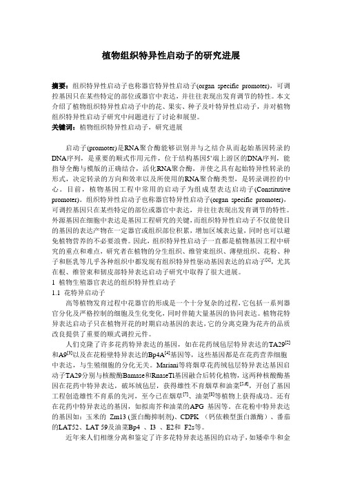
植物组织特异性启动子的研究进展摘要:组织特异性启动子也称器官特异性启动子(organ specific promoter),可调控基因只在某些特定的部位或器官中表达,并往往表现出发育调节的特性。
本文介绍了植物组织特异性启动子中的花、果实、种子及叶特异性启动子,并对植物组织特异性启动子研究中问题进行了讨论和展望。
关键词:植物组织特异性启动子,研究进展启动子(promoter)是RNA聚合酶能够识别并与之结合从而起始基因转录的DNA序列,是重要的顺式作用元件,位于结构基因5’端上游区的DNA序列,能指导全酶与模版的正确结合,活化RNA聚合酶,并使之具有起始特异性转录的形式,决定转录的方向和效率以及所使用的RNA聚合酶类型,是转录调控的中心。
目前,植物基因工程中常用的启动子为组成型表达启动子(Constitutive promoter)。
组织特异性启动子也称器官特异性启动子(organ specific promoter),可调控基因只在某些特定的部位或器官中表达,并往往表现出发育调节的特性。
外源基因在细胞中表达是基因工程研究的关键,而组织特异性启动子不仅能使目的基因的表达产物在一定器官或组织部位积累,增加区域表达量,同时也可以避免植物营养的不必要浪费。
因此,组织特异性启动子一直都是植物基因工程中研究的重点和难点,研究者在植物的分生组织、维管束组织、薄壁组织、花粉、种子和胚乳等几乎各种组织中都发现有组织特异性驱动基因表达的启动子[1],尤其在根、维管束和韧皮部特异表达启动子研究中取得了很大进展。
1 植物生殖器官表达的组织特异性启动子1.1 花特异启动子高等植物发育过程中花器官的形成是一个十分复杂的过程,它包括一系列器官分化及严格控制的细胞及生化变化,同时伴随大量基因的协同表达。
植物花特异表达启动子只在植物开花的时期启动基因的表达,它的分离克隆为花卉的品质改良提供了重要的顺式调控元件。
人们克隆了许多花药特异表达的基因,如在花药绒毡层特异表达的TA29[2]和A9[3]以及在花粉壁特异表达的Bp4A[4]基因等,这些基因都是在花药营养细胞中表达,与生殖细胞的分化无关。
日本 PCP Detection of DOPA 4,5-Dioxygenase (DOD) Activity Using
∗ C orresponding author: E-mail, n ove@cc.tuat.ac.jp ; Fax, +81-42-388-7239.Plant Cell Physiol. 50(5): 1012–1016 (2009) doi:10.1093/pcp/pcp053, available online at © The Author 2009. Published by Oxford University Press on behalf of Japanese Society of Plant Physiologists.All rights reserved. For permissions, please email: journals.permissions@B etalains are synth esized in fl owers, fruits and oth er tissues of the plant order Caryophyllales. Betalamic acid is th e ch romoph ore of betalain pigments synth esized by aring-cleaving enzyme reaction onL -dih ydroxyph enyla lanine (DOPA). Alth ough reverse genetic evidence h as proven that DOPA 4,5-dioxygenase (DOD) is a key enzyme of b etalain b iosynthesis, a ll a ttempts t o d etect r ecombinant plant DOD activity in vitro have failed. Here, we report on the formation of betalamic acid from DOPA under suitable assay conditions using recombinant MjDOD produced by E scherichia coli . This is the fi rst report showing biochemical evidence for DOD activity in vitro. K eywords:B etalain •B etalamic acid •D OPA dioxygenase • M irabilis jalapa .A bbreviations :C CD ,c arotenoid cleavage dioxygenase ; c D OPA5GT ,c yclo -DOPA 5-O -glucosyltransferase ;D AD ,d iode array detector ;D OD ,D OPA 4,5-dioxygenase ;D OPA ,L -dihydroxyphenylalanine ;G US ,β-glucuronidase ;L B ,L uria–Bertani ;M S ,m ass spectrometry ;O RF ,o pen reading frame. T he nucleotide sequence of M jDOD cDNA reported in this paper has been submitted to the DDBJ database with the accession number AB435372.B etalains are water-soluble nitrogenous pigments in plants of the order Caryophyllales, with the exception of Caryophyllaceae and Molluginaceae, but have never been detected with anthocyanins in the same species. Betalains are classifi ed into two groups — r ed betacyanin and yellow betaxanthin — b oth of which contain betalamic acid as the chromophore. Despite extensive study of the biosynthetic pathway of anthocyanins/fl avonoids, only a few enzymes and genes involved in the biosynthesis of betalains have been identifi ed. The betalain biosynthetic pathway comprises several enzymatic reaction steps and some spontaneouschemical reaction steps ( T anaka et al. 2008 ).L -Dihydroxy-phenylalanine (DOPA) is synthesized from tyrosine by tyrosine hydroxylase. DOPA undergoes an extradiol cleavage by DOPA 4,5-dioxygenase (DOD) to produce 4,5- s eco -DOPA following spontaneous chemical recyclization to form beta-lamic acid ( F ig. 1). Betalamic acid conjugates with an amino acid or amine to form betaxanthin, and with c yclo -DOPA glucoside to form betacyanin. Thus, DOD, which catalyzes betalamic acid formation, might be a crucial enzyme in beta-lain biosynthesis. I n 2004, a plant D OD candidate cDNA, designated P gDOD , was isolated from P ortulaca grandifl ora by the cDNA subtraction method. Using a particle bom-bardment method, the DOD candidate was introduced into and expressed in white petals of P . grandifl ora to give red spots on the white, where betalamic acid was synthesized to complement betacyanin synthesis. This reverse in vivo genetic evidence indicated that this candidate should be involved in betalain synthesis ( C hristinet et al. 2004 ). How-ever, attempts to detect DOD enzyme activity in vitro using the protein produced in an E scherichia coli expression systemD etection of DOPA 4,5-Dioxygenase (DOD)Activity Using Recombinant Protein Prepared fromE scherichia coli Cells Harboring cDNA Encoding D OD from M irabilis jalapaN obuhiro S asaki 1 , ∗,Y utaka A be 1,Y ukihiro G oda 2,T aiji A dachi 3,K ichiji K asahara 4and Y oshihiro O zeki 11 D epartment of Biotechnology and Life Science, Faculty of Engineering, Tokyo University of Agriculture and Technology, 2-24-16 Naka-cho, Koganei, Tokyo, 184-8588 Japan2 D ivision of Pharmacognosy, Phytochemistry and Narcotics, National Institute of Health Sciences, 1-18-1 Kamiyoga, Setagaya-ku, Tokyo, 158-8501 Japan3 I nstitute for Plant Biotechnology Research & Development, Ltd., 1-46-901, Hama 5-Chome, Tsurumi-Ku, Osaka, 538-0035 Japan 41128Moro-oka,Kohoku-ku,Yokohama,Kanagawa,222-0002Japan Short Communicationat Fujian Agriculture and Forestry University on December 13, 2012/Downloaded fromfailed ( C hristinet et al. 2004 ), possibly because of instability of the PgDOD protein produced by E . coli or inappropriate assay conditions for DOD enzyme activity.H ere we report the isolation of a cDNA encoding DOD from M irabilis jalapa (M jDOD ) and the formation of beta-lamic acid in vitro under appropriate assay conditions using a crude extract of recombinant MjDOD expressed in E . coli . A candidate cDNA of DOD from M . jalapa petals was isolated by degenerate PCR using the primers designed from the conserved amino acid region of B eta vulgaris DOD (B vDOD , AJ583017) and P . grandifl ora DOD (P gDOD ,AJ580598). The full length of the putative open reading frame (ORF) of the M jDOD cDNA comprised 801 bp encod-ing a protein of 267 amino acids. The nucleotide sequence of M jDOD cDNA showed 67 and 63 % similarity to B vDOD and P gDOD , respectively, and the deduced amino acid sequence of MjDOD had 55 and 54 % identity to BvDOD and PgDOD,respectively . T he M jDOD transcripts were detectable in petals at devel-opmental stage 2, and they were abundant in petals during stages 3–5 (fully opened fl ower), and especially at stage 5, but were below the level of detection in leaves, stems androots ( F ig. 2A, B). These results were similar to those for betanin accumulation ( S asaki et al. 2004 ) and the expression profi le of the cyclo -DOPA 5-O -glucosyltransferase (c D OP-A5GT ) gene involved in betacyanin biosynthesis ( S asaki et al. 2005 ). In M . jalapa , two loci involving fl ower color have been identifi ed: locus C is responsible for fl ower color and locus R for the accumulation of c yclo -DOPA glucoside (see Fig. S3 in T anaka et al. 2008 ). We identifi ed two lines of M . jalapa plants bearing white fl owers. The genotype of one line was presumed to be c cR- containing c yclo -DOPA glucoside in theF ig.2Analysis of M jDOD gene expression. Northern hybridization for poly(A) + RNA (1 µg) from M . jalapa petals, leaves, stems and roots (A),and total RNA (25 µg) from petals at each developmental stage (B) or for various colors of petals (C) was performed using32P -labeled M jDOD cDNA as a probe. A carnation a ctin cDNA probe was used for the loading control (A, B) and ethidium bromide stain of rRNA is shown for the loading control (C).F ig. 1 Scheme of the DOPA 4,5-dioxygenase reaction. It is possible to generate ( S )- and ( R )-betalamic acid (diasteroisomers at the C-6 position) chemically at the spontaneous recyclization. Here, ( S )-betalamic acid is included in the fi gure because the results showed that this was the major product of the DOD reaction (see text).Detection of DOPA 4,5-dioxygenase activityat Fujian Agriculture and Forestry University on December 13, 2012/Downloaded frompetals (line A in F ig. 2C) and that of the other was presumed to be c crr, which lacked c yclo-DOPA glucoside (line B in Fig. 2C). The M jDOD gene was expressed in red, yellow and scarlet petals, but the transcripts were undetectable in white petals in both lines ( F ig. 2C). These results indicated that M jDOD might be a crucial gene for the coloration of M. jalapafl owers, assuming that M jDOD corresponds to the C gene.P revious attempts to detect the enzyme activity of the extradiol dioxygenase using recombinant PgDOD expressed in E. coli had failed ( C hristinet et al. 2004 ). I n plants, the biochemical properties of two types of dioxygenases, i.e. 2-oxo-glutarate-dependent dioxygenase ( B ritsch and Grise-bach 1986 ,B ritsch et al. 1992 ) and carotenoid cleavage diox-ygenase (CCD), have been well characterized ( S chwartz et al. 1997 ,V ogel et al. 2008 ). 2-Oxo-glutarate-dependent dioxy-genase has been characterized as a hydroxylase, e.g. fl avanone 3-hydroxylase is a representative 2-oxo-glutarate-dependent dioxygenase involved in flavonoid biosynthesis. CCD is involved in the biosynthesis of ABA and apocarotenoid in higher plants. Both enzymes require ferrous ion and ascorbic acid to maintain iron in the proper redox state for their activity. The enzymatic reaction mechanism of DOD was thought to be similar to that of CCD because DOD and CCD catalyze cleavage of the conjugated double bond of aromatic rings and that of carotenoid, respectively. It had been sup-posed that ferrous ion and ascorbic acid might be required for the enzyme activity of recombinant DOD.F irst, the recombinant MjDOD protein was expressed using a yeast expression system under various reaction con-ditions. We succeeded in detecting the activity using a crude extract from yeast harboring M jDOD cDNA in the optimized reaction mixture containing ascorbic acid without addition of ferrous ion, which might be contained in the extract car-ried from the yeast cytosol (data not shown). Using this reaction condition in an E. coli expression system, DOD activity of the recombinant MjDOD was detected success-fully. The reaction mixture appeared as a yellow color after incubation for 20 min at 30 °C, but the crude extract from the E. coli harboring the β-glucuronidase(G US) gene as the control did not cause a color change in the reaction mixture (F ig. 3A). The reaction products were separated on a HPLC-photodiode array detector (DAD) system equipped with a reversed-phase column. Two peaks were detected in the reaction mixture containing recombinant MjDOD. The retention time and spectrum of the major peak agreed with those of authentic betalamic acid ( F ig. 3B).HPLC–mass spectrometry (MS) analysis revealed that the [M +H]+ion of this major peak, which was produced by the recombinant MjDOD enzymatic reaction, was observed at m/z212 in the positive ion mode, which agreed with the [M +H]+ ion of authentic betalamic acid. Because betalamic acid was formed from DOPA by the DOD enzymatic reaction via 4,5- s eco-DOPA, the minor broad peak was suspected to be 4,5- s eco-DOPA, whose UV/visible spectrum ( λmax= 385 nm, F ig. 3C arrow) was similar to that in a previous report ( T erradas and Wyler 1991 ).A lthough MjDOD activity could be detected without the addition of extra ferrous ion to the reaction mixture, as shown above, the requirement for ferrous ion in the reaction remained. To refine the effect of ferrous ion in the DOD enzymatic reaction, the crude extract was dialyzed to remove iron ions carried over from the cytosol, and iron ion or ascor-bic acid, or both, was added to the reaction mixture. Com-pared with a relative activity of 100 %in the dialyzed extract without iron ion or ascorbic acid, the activities in the presence of Fe 2+a lone,Fe2+a nd ascorbic acid, and Fe 3+and ascorbic acid in the reaction mixture were 182, 397 and 267 %, respectively. This result indicated that MjDOD requires iron ion and ascorbic acid for its enzymatic activity and that ascorbic acid is required for iron to be in the proper redox state, as in other plant dioxygenases ( S chwartz et al. 1997 ,V ogel et al. 2008 ).W e isolated D OD cDNAs from the bracts of B ougainvillea glabra(B gDOD, AB435373) and roots of B. vulgaris(B vDOD, AJ583017), and both recombinant BgDOD and BvDOD in the crude extracts prepared from E. coli showed DOD activity under the conditions described above (data not shown). Therefore, regardless of the originating species, addition of ascorbic acid to the reaction mixture appears to be important for detecting the recombinant DOD activity.W e reported previously on the c D OPA5GT activity of M. jalapa( S asaki et al. 2004 ,S asaki et al. 2005 ). The forma-tion of c yclo-DOPA5-O-glucoside was confirmed by the detection of betanin and isobetanin, which were derived chemically during the post-enzymatic reaction by the con-densation of c D OPA5GT reaction products with chemically synthesized betalamic acids by alkaline hydrolysis of red beet extract. In the mixture of the condensation reaction, almost equal amounts of betanin derived from ( S)-betalamic acid and isobetanin from ( R)-betalamic acid (diasteroisomers at C-6 position) were detected (see F ig. 2,S asaki et al. 2005 ), suggesting that equal amounts of ( S)- and ( R)-betalamic acid were synthesized chemically during alkaline hydrolysis of red beet extract. To determine whether the product of the recombinant MjDOD reaction was ( S)- or ( R)-betalamic acid, the MjDOD assay mixture was chemically condensed with c yclo-DOPA after the termination of the enzyme reac-tion under acidic conditions. Two peaks were observed on the HPLC chromatogram: a major peak corresponding to the retention time of betanidin and a minor peak corre-sponding to that of isobetanidin (C-15 diasteroisomer of betanidin, F ig. 3C). This result indicated that the main product of the recombinant DOD enzyme reaction was ( S)-betalamic acid (approximately 88 %of total betalamic acid molecules). To confi rm that the proportion of the amount of ( S)-andN. Sasaki et al.at Fujian Agriculture and Forestry University on December 13, 2012/Downloaded from( R )-betalamic acids catalyzed by the recombinant MjDOD enzyme reaction corresponded to that of the ( S )-and ( R )-betalamic acids synthesized in vivo, the ratio of betanin (betanidin 5- O -glucoside) and isobetanin (isobetanidin 5- O -glucoside) in the acidic methanol extract from the petals of M . jalapa was analyzed. The extract contained a large amount of betanin (approximately 94 % of both betacya-nins) in comparison with a trace amount of isobetanin(F ig. 3D ), implying that ( S )-betalamic acid was the main product in vivo similar to that of the enzyme reaction in vitro. We concluded that the DOD enzymatic activities in vitro and in vivo are similar.M aterials and Methods D OPA was purchased from Tokyo Kasei Kogyo (Tokyo, Japan). Betanin, isobetanin, betalamic acid and c yclo -DOPA were prepared as described previously ( S asaki et al. 2005 ). A n M jDOD candidate cDNA was isolated by degenerate PCR. The fi rst-strand cDNA for the template was prepared from red petals of M . jalapa as described previously ( S asaki et al. 2005 ). Two antisense degenerate primers were designed based on the conserved region of the amino acid sequences B vDOD (AJ583017) and P gDOD (AJ580598). The fi rst PCR was performed for 30 cycles (92 ° C for 30 s, 52 ° C for 30 s and 72 ° C for 1 min) using GeneRacer ™5′ primer (I nvitrogen, Carlsbad, CA, USA) and DOD-Rv1 primer (5 ′-TGRCANACN GGDATRTCNGCYTCNGG-3 ′ , corresponding to the amino acid sequence PEADIPVCQ). For the nested PCR, the Not-GR primer ( S asaki et al. 2005 ) and the DOD-Rv2 primer (5 ′ -GCNCCCATNGCNACRTGNARNGG-3 ′ , corresponding to the amino acid sequence PLHVAMGA) were used. After the nested PCR (30 cycles of 92 ° C for 30 s, 54 ° C for 30 s and 72 ° C for 1 min), the products were introduced into pBlue-script SK + followed by determination of the nucleotide sequences.A gene-specifi c sense primer of M jDOD cDNA was syn-thesized (5 ′-A TG A AAGGAACATACTATAT-3′;underlining shows the putative fi rst ATG) to prepare a full-length M jDOD cDNA by PCR (30 cycles of 92 ° C for 30 s, 58 ° C for 30 s and 72 ° C for 2 min) together with the GeneRacer ™3′primer using the same fi rst-strand cDNAs as noted above. The second and third round PCRs using the diluted DNA frag-ments amplifi ed by the previous round of PCR as a templatewere performed sequentially using the primers DODattB1F ig.3Production of betalamic acid by the DOD assay. When the crude extract from E . coli harboring M jDOD cDNA was mixed withDOPA, the color of the reaction mixture turned yellow, whereas nocolor change was observed in the mixture containing the crude extracts from E . coli harboring G US (A). HPLC elution profi les of the MjDOD assay mixture and authentic betalamic acid (B), and the products of the MjDOD assay mixture condensed with c yclo -DOPA and the extracts from petals of M . jalapa (C).Detection of DOPA 4,5-dioxygenase activityat Fujian Agriculture and Forestry University on December 13, 2012/Downloaded from(5 ′-TGTACAAAAAAGCAAAGATGAAAGGAACATAC-3′) and GR-attB2 (5 ′-TGTACAAGAAAGCTGACGCTACGTAA CGGCATGACAGTG-3 ′) in the second round, then using the primers a ttB1-SD, (5 ′-GGGGACAAGTTTGTACAAAAAAGCA GAAGGAGATATAATAATG-3 ′) and attB2R (5 ′-GGGGACC ACTTTGTACAAGAAAGCTG-3 ′) to add attB1 and Shine–Dalgarno sequences just upstream of the putative start codon and the attB2 sequence at the 3 ′end. The fi nal ampli-fi ed product was introduced into pDONR221 (I nvitrogen) using BP clonase (Invitrogen).M jDOD gene expression was analyzed by Northern hybrid-ization as described previously ( S asaki et al. 2005 ) using the M jDOD cDNA fragment prepared from that introduced into pDONR221 as a probe.M jDOD cDNA in pDEST221 was introduced into the E. coli expression vector pDEST15 (I nvitrogen), which was transformed into BL21-AI (I nvitrogen). The transformants were grown in 5 ml of Luria–Bertani (LB) medium contain-ing 50 µg ml −1ampicillin overnight at 30 °C.Pre-cultured cells in 1.25 ml were inoculated into 250 ml of LB medium containing 50 µg ml −1ampicillin and cultured until the OD600 reached 0.6, followed by addition of 5 ml of 20 %arabinose and further culturing for 4 h at 16 °C. The harvested cells were washed with the extraction buffer (100 mM potassium phosphate, pH 7.2), resuspended in 300 µl of the extraction buffer, and then disrupted by sonication. After centrifuga-tion of the lysate at 20,000 ×g for 2 min, the supernatant was used as a crude extract for the enzyme assay. The reaction mixture (100 µl) comprised 0.1 µmol DOPA, 1 µmol ascorbic acid, 9 µmol potassium phosphate, pH 7.2, and 20–200 µg of the protein of the crude extract. The amount of protein was quantifi ed using a Coomassie Plus ™Protein Assay Reagent (Pierce Biotechnology, Rockford, IL, USA) with bovine serum albumin as the standard. A standard enzyme assay was per-formed at 30 °C for 20 min. The crude extract prepared from E. coli harboring the G US gene was used as the control. To examine the effect of iron ion on the MjDOD enzymatic reaction, the crude extract was dialyzed with a 1,000-fold excess volume of 100 mM potassium phosphate, pH 7.2, for 16 h, and the enzymatic activity of the dialyzed extract was measured in a reaction mixture of 100 µl containing 0.1 µmolFeCl2or FeCl3and/or 1 µmol ascorbic acid at 30 °C for 5 min.The enzymatic reaction was terminated by the addition of 1 %phosphoric acid at the fi nal concentration, and the rela-tive activity was calculated based on the area of the HPLC chromatogram monitored at 405 nm.B etalamic acid was separated using HPLC-DAD (LaChrom Elite system, Hitachi Hi-technologies, Tokyo, Japan) equipped with a reversed-phase column (4.6 mm i.d. ×250 mm; Develo-sil ODS-SR-5, Nomura Chemicals, Aichi, Japan) and using a linear elution gradient (1 ml min −1) of 25–70 %methanol in solvent A (1.5 %phosphoric acid) for 20 min, and was moni-tored at 405 nm. Betanidin and isobetanidin ( F ig. 3C)were separated using a linear elution gradient (1 ml min−1) of 10–40 %methanol in solvent A for 20 min and monitored at 535 nm. Formation of betalamic acid by the enzymatic reac-tion was confi rmed by HPLC-MS with a MassLynx system (Waters Corp., Milford, MA, USA). Betanin and isobetanin accumulated in the fully opened red petals of M. jalapa ( F ig. 3D) were extracted with 1.5 %phosphoric acid/80 % methanol containing 10 mM ascorbic acid, and then ana-lyzed by HPLC under the same conditions as described above.R eferencesB ritsch,L.and G risebach,H.(1986)P urification and characterization of ( 2S)-fl avanone 3-hydroxylase from P etunia hybrida.E ur.J.Biochem. 156:569–577.B ritsch,L.,R uhnau-Brich,B.and F orkmann,G.(1992)M olecular cloning, sequence analysis, and in vitro expression of fl avanone 3 β-hydroxylase from P etunia hybrida.J. Biol. Chem.267:5380–5387.C hristinet,L.,B urdet,F.X.,Z aiko,M.,H inz,U.and Z ryd,J.P.(2004) C haracterization and functional identifi cation of a novel plant 4,5-extradiol dioxygenase involved in betalain pigment biosynthesis in P ortulaca grandifl ora.P lant Physiol.134:265–274.S asaki,N.,A dachi,T.,K oda,T.and O zeki,Y.(2004)D etection of UDP-glucose: c yclo-DOPA 5-O-glucosyltransferase activity in four o’clocks ( M irabilis jalapa L.).F EBS Lett.568:159–162.S asaki,N.,W ada,K.,K oda,T.,K asahara,K.,A dachi,T.and O zeki,Y. ( 2005 )I solation and characterization of cDNAs encoding an enzyme with glucosyltransferase activity for c yclo-DOPA from four o’clocks and feather cockscombs.P lant Cell Physiol.46:666–670.S chwartz,S.H.,T an,B.C.,G age,D.A.,Z eevaart,J.A.and M cCarty,D.R. ( 1997 )S pecifi c oxidative cleavage of carotenoids by VP14 of maize . S cience276:1872–1874.T anaka,Y.,S asaki,N.and O hmiya,A.(2008)B iosynthesis of plant pigments: anthocyanins, betalains and carotenoids .P lant J.54 : 733–749.T erradas,F.and W yler,H.(1991)2,3-and4,5-secodopa,the biosynthetic intermediates generated from L-DOPA by an enzyme system extracted from the fl y agaric , Amanita muscaria L., and their spontaneous conversion to muscaflavin and betalamic acid, respectively,and betalains.H elv. Chim. Acta74:124–140.V ogel,J.T.,T an,B.C.,M cCarty,D.R.and K lee,H.J.(2008)T he carotenoid cleavage dioxygenase 1 enzyme has broad substrate specificity, cleaving multiple carotenoids at two different bond positions .J. Biol. Chem.283:11364–11373.(Received March 17, 2009; Accepted April 8, 2009)N. Sasaki et al.at Fujian Agriculture and Forestry University on December 13, 2012/Downloaded from。
Introduction Human microRNA Polyadenylation Signals
Human microRNA Polyadenylation SignalsEric J. Devor, Ph.D.Molecular Genetics and BioinformaticsIntegrated DNA TechnologiesIntroductionMicroRNAs (miRNAs) are small non-coding RNAs that are involved in post-transcriptional gene regulation (cf., Bartel, 2004). First identified in C. elegans just over a decade ago (Lee et al., 1993), miRNAs have been identified in virtually every metazoan and plant species examined. Experimental evidence is rapidly accumulating that shows miRNAs to play key roles in process such as cellular differentiation, cell death, and fat storage. Some miRNAs appear to be ancient (Pasquinelli et al., 2000; Floyd and Bowman, 2003) while others, particularly those found in only a few plant or animal taxa, may be quite young in an evolutionary sense. Allen et al. (2004) offered a model of miRNA evolution based upon an analysis of sequences in A. thaliana but the origin and subsequent development of miRNAs remains an open issue.MicroRNA biogenesis has been mapped to at least a first-order level. An outline of miRNA development is presented in Bartel (2004). In general, an miRNA is composed of a highly conserved core sequence of 21-23 nucleotides, the mature miRNA, contained within a less well conserved precursor sequence ranging in size from 60 nucleotides to more than 120 nucleotides, the pre-miRNA. This sequence is, in turn, part of a larger primary transcript, the pri-miRNA, that may contain a single pre-miRNA or two or more pre-miRNAs arranged as paired or polycistronic transcripts. Following transcription, pre-miRNAs form a characteristic stem-loop structure that is processed by the RNase III enzyme DROSHA (Lee et al., 2003) in concert with accessory proteins such as PASHA and DCRG (Gregory et al., 2004; Denli et al., 2004). The pre-miRNA is thus released to be exported from the nucleus whereupon it is further processed by the DICER/RISC complex releasing the mature miRNA to carry out its regulatory function.Recently, Bracht et al. (2004), Cai et al. (2004) and Lee et al. (2004) presented evidence that at least some pri-miRNAs are both capped and polyadenylated prior to DROSHA processing and can act as mRNAs. While several publications have noted an absence of consistent 5’ or 3’ sequence features outside pre-miRNAs (Berezikov et al., 2005; Ohler et al., 2004), if pri-miRNAs are polyadenylated there should be functional polyadenylation signals 3’ of the pre-miRNA sequence. Here, a survey of the presence or absence of polyadenylation signals downstream of more than 100 human microRNAs is presented. Most of these miRNAs possess at least one polyadenylation signal. Results of this survey indicate that use of the canonical AATAAA in pri-miRNAs is less than that for other RNAs. In addition, paired and poly-cistronic transcripts generally do not displayinter-cistronic signals and the occurrence and type of polyadenylation signal is slightly different for miRNAs transcribed in introns versus those that are in intergenic regions. Overall, however, data presented here do indicate that the great majority of human microRNA loci possess potentially valid polyadenylation signals independent of whether or not they lie in introns and that the occurrence of such signals in paired and polycistronic loci is consistent with single transcript processing.MethodsRelease 6.0 of the MicroRNA Registry (Ambros et al., 2003; Griffiths-Jones, 2002) contains information on 227 human miRNAs. Registry entries include both the mature and the precursor sequence as well as the chromosome coordinates of each miRNA. These data were used to arbitrarily select 72 human miRNA “loci.” The only selection criteria that were employed were to ensure that as many of the human chromosomes as possible were represented and that if a selected miRNA was found to be part of a pair or a group of three or more, all of the miRNAs were included from the grouping and are regarded here as a single “locus.” Fifty of those thus selected are presumed to be single transcript miRNAs as there are no other known miRNAs nearby. A further 16 loci consist of two miRNAs that appear to form a single transcript and six additional miRNAs were chosen that appear to form a polycistronic transcript. The MicroRNA Registry identification and the chromosome coordinates for each of these loci are presented in Table 1. Also shown in Table 1 is the genomic context of each miRNA. For those miRNAs found to reside in an intron, the GenBank identification of the gene is given.Chromosome coordinates were used to retrieve the genomic sequence of each miRNA as well as a 2kb region downstream of the 3’ end of the pre-miRNA. In the case of tandem or polycistronic miRNAs, the sequences selected started at the 5’ end of the upstream member of the transcript and then continued 2kb downstream from the 3’ end of the of the 3’-most member. MiRNA sequences in each download were verified with the microRNA Registry precursor sequences and the genome location and strand orientation was verified by BLAST. Finally, identification of the canonical AATAAA polyadenylation signal hexamer as well as the three most common single nucleotide variant hexamers ATTAAA, AGTAAA, and TATAAA in each sequence was carried out by means of a simple text search.Once the data set was assembled and annotated (see Supplemental Materials), polyadenylation signals were “validated” using DNAFSMiner (Liu et al., 2003, 2005; available on-line at .sg/DNAFSMiner/). Based upon information provided in DNAFSMiner, a cutoff for validation was set at a score of 0.500. This cutoff represents a sensitivity and precision of at least 75% in CDS sequences (Liu et al., 2005). For those miRNA 3’ sequences in which no validated polyadenylation signal could be found using this criterion, the search was extended an additional 2kb. No further extensions were attempted beyond the 4kb range.A second data set composed of mouse ortholog miRNAs was compiled using the same criteria as for the human data set. These sequences are also provided in the Supplemental Materials.Table 1Primary Human miRNA Data Used in This StudyI. Single Transcript miRNAs1miRNA ID2 Chr Chr Coordinates3 Strand Intron Gene34a 1 9145993-9146102 - N186 1 71245335-71245420 - Y ZNF265137 1 98223647-98223748 - Y FLJ35409 197 1 109853557-109853631 + N205 1 205993873-205993982 + N149 2 241115408-241115496 + Y GPC1216 2 56127736-56127845 - N217 2 56121753-56121862 - N198 3 121597205-121597266 - Y FSTL128 3 189889271-189889356 + Y LPP95 4 8125099-8125179 - Y ABLIM2146 5 159844937-159845035 + N340 5 179374909-179375003 - Y RNF130339 7 835810-835903 - Y MGC11257 25 7 99335834-99335917 - Y MCM7124a-1 8 9798308-9798392 - N383 8 14755318-14755390 - N151 8 141811845-141811934 - Y PTK2320 8 22158420-22158501 - N31 9 21502114-21502184 - N32 9 108888064-108888133 - Y C9orf5346 10 88014424-88014509 - Y GRID1107 10 91342484-91342564 - Y PANK1210 11 558089-558198 - N130a 11 57165247-57165335 + N326 11 74723784-74723878 - N331 12 94204664-94204757 + N342 14 99645745-99645843 + Y EVL345 14 99843949-99844046 + N134 14 100590777-100590849 + Y LOC440201 203 14 103653495-103653604 + Y LOC374569 190 15 60903209-60903293 + Y TLN2184 15 77289185-77289268 + N328 16 65793725-65793799 - Y ELMO322 17 1563947-1564031 - N195 17 6861658-6861744 - N144 17 24212677-24212762 - N21 17 55273409-55273480 + N338 17 76714278-76714344 - N187 18 31738779-31738887 - N330 19 50834092-50834185 - Y EML2150 19 54695854-54695937 - NTable 1 (cont.)103-2 20 3846141-3846218 + Y PANK2296 20 56826065-56826144 - N155 21 25868163-25868227 + Y BIC33 22 40621448-40621516 + Y SREBF2188 X 49471145-49471230 + N223 X 65021733-65021842 + N220 X 122421841-122421590 - Y LOC402422 424 X 133406164-133406261 - Y MGC16121 II. Paired miRNAs200a, 200b 1 1142407-1143255 + N181b-1, 213 1 195559659-195559768 - N29c, 29b-2 1 204363592-204364263 - N425, 191 3 49032585-49033146 - Y FLJ10496 15b, 16-2 3 161605078-161605315 + Y SMC4L1 96, 183 7 129008483-129008805 - N29a, 29b-1 7 130018761-130019553 - N192, 194-1 11 64415185-64415487 - N34b, 34c 11 110888873-110889450 + N200c, 141 12 6943123-6943615 + N16-1, 15a 13 49521110-49521338 - Y DLEU2 132, 212 17 1899952-1900424 - Nlet7a-3,let7b 22 44829148-44830167 + N221, 222 X 45361839-45362784 - N98, let7f-2 X 53466224-53467256 - Y UREB1105-1, 105-2 X 151231259-151233532 - Y GABRA3 III. Polycistronic miRNAs4367, 302d, 302a,302c, 302b 4 113926634-113927317 - N23b, 27b, 24-1 9 94927045-94927925 + Y C9orf3 17, 18, 19a,20, 19b-1, 92-1 13 90800860-90801646 + Y C13orf25 24-2, 27a, 23a 19 13808101-13808473 - N371, 372, 373 19 58982741-58983839 + N92-2, 19b-2,106a X 133029088-133029828 - N1Based upon known miRNAs in the miRNA Registry2Unless otherwise indicated, the full miRNA Registry ID for each entry includes the prefix hsa-mir.3Chromosome coordinates as given in the miRNA Registry4Polycistronic is taken here to mean three or more miRNAsResultsPrimary Locus InformationSeventy-two microRNA loci, totaling 105 human microRNAs, were selected for this study. Using the chromosome coordinates of each locus, genomic context, including chromosome location, strand orientation, and neighborhood, was established. For loci in intergenic sequences, neighborhood refers to the distance in both directions to the nearest annotated genes. For intronic loci, neighborhood refers to the size of the intron and the microRNA’s position within it. For 64 of the 72 loci, neighborhoods were found to be unremarkable. That is, microRNA loci in intergenic sequences were located tens to hundreds of kilobases from annotated genes and microRNA loci in introns were well inside large introns, usually more than 10kb in size, and transcribed in the same direction as the host mRNA. Eight loci were found not to fit this picture for one reason or another and these will be discussed below. These are single transcripts hsa-miR-25, hsa-miR-134, hsa-miR-203, hsa-miR-328, hsa-miR-155, and hsa-miR-220 and polycistronic transcripts [hsa-miR-367, hsa-miR-302a, hsa-miR-302b, hsa-miR-302c, hsa-miR-302d] and [hsa-miR-17, hsa-miR-18, hsa-miR-19a, hsa-miR-20, hsa-miR-19b-1, hsa-miR-92-1].Of the 72 loci, 50 are presumed solitary transcripts, 16 are paired transcripts (32 miRNAs), and six are polycistronic transcripts (23 miRNAs). Overall, 46 of the 72 loci (63.9%) are located in intergenic sequence and 26 of the 72 loci (36.1%) are located within introns. This is consistent with the prevailing view that about two-thirds of microRNAs are found in intergenic regions and about one-third are found in introns. Of the 46 intergenic miRNAs 31 (67.4%) are solitary, 11 (23.9%) are paired, and 4 (8.7%) are polycistronic. Of the 26 intronic miRNAs 19 (73.1%) are solitary, 5 (19.2%) are paired, and 2 (7.7%) are polycistronic.Polyadenylation SignalsThe text search of all of the assembled and annotated 3’ sequences revealed more than two hundred instances of one or another of the four polyadenylation signal hexamers. Some could be discounted immediately as they were found to be part of a repetitive sequence including the 3’ regions of several Alu repeats that were identified. This served to eliminate a few hexamers from consideration but, as for the remainder, the mere presence of an appropriate hexamer sequence or sequences is no guarantee that the putative transcript is polyadenylated there, or anywhere for that matter. Numerous reports suggest that valid polyadenylation sites have additional sequence features. Colgan and Manley (1997) cite the presence of a highly variable G/U-rich motif located 20 to 40 bases 3’ of the polyadenylation signal in approximately 70% of mRNAs. It is further noted that these motifs are often characterized by single or multiple stretches of up to five consecutive Us frequently interrupted by a single G (cf., MacDonald et al., 1994, Takagaki and Manley, 1997). Zarudnaya et al. (2003) reiterate this theme by noting that the downstream sequence elements are poorly conserved G/U- or U-rich sequences. They suggest that there is no good consensus among the downstream elements with various sequence features occurring at various distances from each other. Arhin et al. (2002)showed a variety of sequences in which U- and G-rich elements lie downstream from AAUAAA and AUUAAA polyadenylation signals in mammalian genes. A very generalized model is proposed by Liu et al. (2003) for in silico prediction of valid polyadenylation signals. They note a rare and poorly defined upstream element (USE) followed by the polyadenylation signal (PAS) followed, in turn, by a CA dimer poly-A site 10-30 bases away and a G/U-rich downstream element (DSE) a further 20-40 bases away. It should be noted that all of the sequences shown by Arhin et al. (2002) fit this generalized model.Using the Liu et al. (2003) model (available at http://research.i2r.a-.sg/DNAFSMiner/), each of the four potential polyadenylation signals was evaluated in each 3’ sequence. DNAFSMiner identified valid polyadenylation signals in 59 of the 72 microRNA loci (81.9%). This included 38 of 50 single transcript loci (76.0%), 15 of 16 paired miRNAs (93.8%), and all six of the polycistronic loci. Overall, 39 of 46 miRNA loci located in intergenic sequences (84.8%) and 20 of 26 intronic miRNA loci (76.9%) were found to have at least one valid polyadenylation signal. The average DNAFSMiner score among the signals with a score above 0.500 was 0.709 (n = 125; range 0.501 to 0.957). No difference was seen for average valid score among the transcript types but miRNA loci in introns did have a slightly higher average than did miRNA loci in intergenic sequences (0.733, n = 41 versus 0.698, n = 84). Examples of DNAFSMiner validated polyadenylation signals are given in Table 2.There is no way to determine which, if any, of the validated polyadenylation signals is in fact a real polyadenylation site using the data available. Therefore, comparisons of signal types and distances among these miRNAs are based upon choosing the hexamer with the highest score for each locus. Thirty of the 59 loci with a valid signal (50.8%) had only one signal whereas the range was from two to six valid signals among the other 29 loci. Overall, the average number of valid signals per locus is 2.02 with no differences observed by locus type or genomic context.Average distance from the 3’ end of pre-miRNA sequences to the highest scoring polyadenylation signal is 992.5bp. Genomic context does appear to be something of a factor for loci having two or more miRNAs. Single transcript miRNAs display an average distance to the highest scoring signal of 1044.8bp in intergenic space (n = 24) and 1108.4bp in introns (n = 14). On the other hand, multiple miRNA loci in intergenic space display an average distance of 1077.3bp (n = 15) versus those in introns where the average distance is 461.7bp (n = 6). It is important to be mindful of the small sample sizes here, however.Looking at the specific hexamer sequences comprising the validated signals shows that the canonical AATAAA sequence is the most common. This sequence is the highest scoring polyadenylation signal in 30 loci (50.8%). ATTAAA is next with 14 loci(23.7%). The other “minor” signals AGTAAA and TATAAA account for 14 of the remaining 15 loci. One locus, hsa-miR-22, did not display any of the above polyadenylation signal sequences even out to the four kilobase limit imposed on the extended searches. However, another minor sequence, ACTAAA, was found at 355bpwith a DNAFSMiner score of 0.609. The only noteworthy aspect of polyadenylation signal sequence distribution among these microRNAs is that ATTAAA is the only signal with the highest score among the six intronic loci having more than one miRNA in the presumed transcript. Again, the small sample sizes must be kept in mind. Thus, overall, polyadenylation signal sequence distribution by both transcript class and genomic context is unremarkable compared to the “real” 3’ UTRs presented by other investigators.Table 2Validated polyadenylation signals from several human microRNA loci. The poly-A signal is shown in BOLD, intervening CA dimers are underlined and G/U- and U-rich sequences are italicized.Single Transcript miRs:miR-34a AAUAAA AAGGGACAG GUUUUGmiR-197 AAUAAA AAUUCAACAGUACGGUGUAAAUUUAAAAUGAGGCAAAAGAAAGAUAAUGACCAA UUUUUmiR-149 AAUAAA GGCUGCAGAGUGAAGGAUGGUCCCUGGCGAGUGUACCUGUGUCCCAGUCUUCUUGCCUGAGGGCGAGGAGUGGGGAAGGAGmiR-146 AAUAAA UGUAUGUAGGAAUACGUGUGUUGGAAAGAUGUACAUCAAUUUGCUAACAAUGGUUAUCUCUGAC GUGGUGGGAUUUGAGAUGUGUUUUUCUUUUUGGUUGUmiR-25 AAUAAA A GUGUUUGU AGAUUGUCAUCUUCUAGCCUGGGCCUGACUUCCAUUAAAACA GGGUUUUGUGCGUUUUUmiR-383 AAUAAA UUGUU CAACUGAGGTGAAAAUUAAUUCUAGAAUAUUUUCAGGCAA UUGUUGGUAGGGUGUUUGUUmiR-346 AAUAAA ACACCUGACCCGCA GGUGGGCAGCCUGGAUUmiR-107 AUUAAA GAACACAGUAGAUCUGUAAUGGAG UUUUUmiR-331 AUUAAA CUCUCCCCCCAAAUUUAAUGGUUUC UGUUUU CUUAAA UGUUUUUmiR-144 AAUAAA GAUGAGAACCAGAAUGCUUAUAUUUUAUUAGUAUCCAAGAC UGGGGAGAGGGAUGGGGUGGGmiR-330 AAUAAA GCUGGAAGAGGAGAGGGAAUA GGGAUGGGGCUGGUGUmiR-103-2 AUUAAA CA UUUGUUGG AAUAUCAUAACUUC UUUUUPaired miRs:miR-425,191 AUUAAA AGGAA UGGGGGUGGGGGUGGGGGGUG AGUAAGUUmiR-181b-2,213 AUUAAA UUUGAUCACAGCA GUUUUmiR-29c,29b-2 AAUAAA UACUAUGUUCAGUUUAUCAUGUC UGUGUUUUUmiR-15b,16-2 AAUAAA AGCAAAACUA UUUUGUGmiR-34b,34c AAUAAA GUGCCAUAAAGACAUAAA UUGUGGUUAUUGlet7a-3,let7b AAUAAA GCUGUUCUGACCAUGAACUUGGAACUGGCCCCUUUCAUUCUGGAAGCCCAGGGAC UGGGCUCUGGGGGCGCGUCUGCUGCGGGACAGUGUGGAGGPoly-cistronic miRs:miR-367, 302a,302b, 302c,302d AAUAAA UAAAAAAGCUCUCCGUUAAGACA UGGGGCUGUGAAAUGGGUGmiR-17, 18, 19a,19b-1, 20, AUUAAA GAAAA UGUGU AAC UUUUU GUGUAAATACAUCUUGUC UUGUUUU CAUUCAAAAACAUUUCAC 92-1 UUUUGGGGUUGCGUGUmiR-24-2, 23a,27a AUUAAA AGGGAAGCCAGC UGUGU CCUUUCCGGCC UGGAGGmiR-371, 372,373 AAUAAA AAUUGGCAUAUUC UGGGUGGGConservation in Mouse OrthologsAgain using chromosome coordinates presented in the microRNA Registry, a similar set of sequence data was compiled for all mouse orthologs of the miRNAs in the human data set that could be identified. Of the total of 72 miRNA loci in the human data set amouse ortholog was found for all but five. The human miRNAs for which no comparable mouse locus could be found were the single transcripts hsa-miR-197, hsa-miR-198, hsa-miR-220, and hsa-miR-95, and the paired transcript hsa-miR-105-1/hsa-miR-105-2.For the 67 mouse microRNA orthologs (a total of 95 mouse microRNAs) that were identified, chromosome location, strand orientation, and neighborhood was confirmed. Every intronic miRNA was found to map to an intron in the orthologous mouse gene or to a sequence that was highly similar but not yet annotated in the mouse genome. Once each ortholog was thus validated, 2kb 3’ sequences were compiled. Using DNAFSMiner, valid polyadenylation signals were identified in 46 mouse 3’ sequences. These included 32 single transcript loci, 9 paired transcript loci, and 5 polycistronic transcript loci.Comparing valid polyadenylation signals pairwise by miRNA locus showed that few human and mouse microRNA loci have the same signal hexamers and there is very little 3’sequence similarity in general as would be expected for a non-coding region. However, among the 67 mouse microRNAs there are 43 for which both the human microRNA and the corresponding mouse ortholog with a valid polyadenylation signal (64.2%). Similarly, there are five loci for which neither the human or the mouse ortholog has a valid polyadenylation signal (using the validation criterion of a DNAFSMiner score of 0.500 as the cutoff). This is an overall concordance of 71.6%. Discordant loci were represented by 16 human microRNAs that have a valid polyadenylation signal and the mouse ortholog does not and three human microRNAs that do not have a valid polyadenylation signal but the mouse ortholog does. In most of these discordant cases polyadenylation signal hexamers are present but do not meet the minimum criteria established above (see Supplemental Materials).In general, the mouse data, taken together with the human data, indicate that the presence of potentially valid polyadenylation sites is probably the rule for mammalian microRNAs rather than the exception. It would not be surprising to find that this finding extends to all microRNA loci.MicroRNA loci in unusual genomic contextsAs noted at the outset, most of the microRNA loci examined here, whether single or multiple transcript loci, lie in a “conventional” genomic context. That is, loci in intergenic sequences are not at all close to annotated genes and intronic loci are well clear of exons and are transcribed in the same direction as the gene in which they are located. There are a few of the miRNAs selected for this study that were found to present in what can be called an usual genomic context. Hsa-miR-25 is located in a small intron near the 3’ UTR of the gene MCM7. DNAFSMiner chose the same polyadenylation signal for the gene as for the microRNA. The same location and signal circumstance obtains for the mouse ortholog mmu-miR-25.The locus hsa-miR-134 lies in a small intron of LOC440201 just 5’ from two small, closely spaced exons. The polyadenylation signal chosen by DNAFSMiner lies in the intron beyond these two exons. Evidence for LOC440201 consists of computationalresults in annotation and two ESTs. The mouse ortholog, mmu-miR-134, is located in the same position in a sequence identified by a BLAST search of the presumed LOC440201 mRNA sequence. A nearly identical circumstance was observed for hsa-miR-330/mmu-miR-330.The locus hsa-miR-203 also lies in an annotated gene, LOC374569. In this case, however, the microRNA is inside the presumed coding region in an exon and is transcribed in the same direction. The one identified polyadenylation signal (AATAAA) received a DNAFSMiner score of 0.925 and is in a position just 3’ of the termination codon of the locus.The locus hsa-miR-328 lies in the gene ELMO3 on chromosome 16. This miRNA is unusual in that it lies in the small intron 11 but is transcribed in the opposite direction and failed to display any polyadenylation signals. The same genomic context is found for the mouse ortholog, mmu-miR-328 with the exception that the microRNA begins in the 5’ end of an exon and extends in to the preceding intron.An interesting circumstance surrounds hsa-miR-220 on chromosome X. The microRNA sequence lies completely within the coding region of a beta-tubulin processed pseudogene, ΨTUBB5 (LOC402422), and is transcribed in the opposite orientation. There is no rodent ortholog for this microRNA locus but the precursor sequence of hsa-miR-220 is reasonably well conserved in the coding regions of human beta-tubulin genes as well as in numerous mammalian beta-tubulins including mouse, rat, dog, cow, and pig. The question that naturally arises from this situation is how and why the creation of a processed pseudogene led to the activation of a microRNA. This is being addressed in a separate investigation.The last of the solitary transcript microRNA loci that deserves mention is hsa-miR-155 located on chromosome 21. This microRNA lies completely within the transcribed sequence of BIC, an RNA transcript in Burkitt and Hodgkin lymphomas (cf., Metzler et al., 2004; van den Berg et al., 2003). The complete BIC transcript is 12,969bp long with the miR-155 precursor sequence just over 1kb from the 3’ end. The validated AATAAA polyadenylation signal (0.691) observed for hsa-miR-155 is at the 3’end of the BIC transcript. To date, no other portion of the BIC transcript has been associated with any other function. Thus, it is likely that the entire 12,969nt BIC transcript is the primary miRNA transcript (pri-miRNA) for this locus.Finally, two polycistronic miRNA loci merit mention. The locus [hsa-miR-367, hsa-miR-302a, hsa-miR-302b, hsa-miR-302c, hsa-miR-302d] lies just 5’ of the first coding sequence of the chromosome 4 gene HDCMA18P and is transcribed on the opposite strand. Numerous valid polyadenylation signals are present and these are opposite the multiple small sequence fragments that make up the 5’ UTR of HDCMA18P. The annotated sequence of both strands at this locus is very complex. The mouse ortholog of this locus is located in the same opposite orientation at the 5’ end of the gene encoding the HDCMA18P ortholog protein D3Wsu161e.Evolution of the cluster containing [hsa-miR-17, hsa-miR-18, hsa-miR-19a, hsa-miR-20, hsa-miR-19b-1, hsa-miR-92-1] is well documented (Tanzer and Stadler, 2004). The genomic context of this cluster is less well known. The cluster resides completely within the 3’ UTR of the gene currently known as C13orf25. This gene has been implicated as the target for 13q31-q32 amplification in malignant lymphoma (Ota et al., 2004).C13orf25 produces two transcripts and it is Transcript B that contains the microRNA cluster. The mouse ortholog of the cluster lies on chromosome 14. The sequences surrounding the cluster in the mouse genome are more than 90% identical to humanC13orf25 sequences. Both clusters have highly supported ATTAAA polyadenylation signals very near the 3’ end (0.842 in human and 0.833 in mouse). As yet, there is no definitive relationship postulated between these microRNAs and the putative C13orf25 protein.DiscusssionSeveral investigators have observed that microRNA transcripts are both capped and polyadenylated (cf., Bracht et al., 2004; Cai et al., 2004; Lee et al., 2004). As a first step in determining whether polyadenylation of microRNA transcripts is a common occurrence a sample of 72 microRNA loci containing 105 human microRNAs was selected from the MicroRNA Registry. These loci were selected simply by Registry ID number without regard to their location, genomic context, or whether or not they were part of a putative multicistronic group. The sample so selected is a reasonable cross section of human microRNAs as about two-thirds are located in intergenic sequence and one-third in introns and the majority appear to be solitary transcripts. In addition, a comparative sample consisting of mouse orthologs was compiled.A survey of potentially valid polyadenylation signals was carried out on both human and mouse sequences lying 3’ of the microRNAs using DNAFSMiner. Results of this survey are fairly straight-forward. Most microRNA loci, whether composed of single or multiple miRNA transcripts and whether they are located in intergenic sequence or in an intron, can be expected to have at least one valid polyadenylation signal within one or two kilobases of the 3’end of the pre-miRNA sequence. The question of a statistically valid polyadenylation actually being a polyadenylation site is still open.A secondary result of this survey is that the method of confirming the genomic context of each microRNA locus selected revealed that a small portion of microRNAs can be expected to be in a context that is unusual for some reason. This expectation is best conveyed by simply saying that, in a genome the size of the average mammalian genome, if something can happen, it probably has at least once. Clearly, the genomic context of the few unusual microRNAs seen in this survey has not prevented them from being expressed and, presumably, functional. It is important to also note that the microRNAs that were deemed unusual were most often transcribed on the strand opposite the genomic feature that made them unusual.The exercise of identifying potential polyadenylation sites in silico is just that- an in silico exercise. The importance of these results will not be known until the in vivo reality。
群体性孤独——社交媒体使用对孤独感的影响
[18]
这一点ꎬ我们将在下一节详细讨论ꎮ
ꎮ 关于
( 二) 社交媒体使用方式的类型学分析
2021 年ꎬ埃米莉欧戴( Emily B. O’ Day) 和理查德亨贝格( Richard G. Heimberg) 在一项最新的
“群体性孤独” 只不过是人们对新媒介技术产生的道德恐慌而已 [4] ꎮ 在实证研究方面ꎬ不同学者的研究
结论也很不一致ꎮ 有研究发现ꎬ频繁使用社交媒体会加剧孤独感 [20] [21] [22] ꎻ也有研究发现ꎬ使用社交媒
体能缓解孤独感 [5] ꎻ还有研究发现ꎬ社交媒体使用时间与孤独感之间呈先下降后上升的“ U” 形关系 [1] ꎮ
在实证研究方面ꎬ国内学者大多以社交媒体使用频率作为自变量来研究其对孤独感的影响ꎮ 虽然频繁
使用与病态化使用之间存在正相关关系ꎬ但它们是两个不同的概念ꎬ不可混为一谈ꎮ 我们认为ꎬ要想厘
清社交媒体使用与孤独感之间的复杂关系ꎬ就必须对人们使用社交媒体的方式进行深入的类型学分析ꎮ
具体来说ꎬ我们可以根据使用频率和是否上瘾这两个维度将社交媒体的使用者分为以下三种类型ꎮ
piro) 认为ꎬ克劳特等的研究对象主要是大学生和社区精英ꎬ这些群体即便不使用互联网也会变得越来
越孤独ꎬ因此ꎬ不能由此得出互联网增加孤独感的结论 [17] ꎮ 此外ꎬ很多心理学研究也指出ꎬ孤独者更倾
向使用互联网来缓解孤独ꎬ因此是孤独影响了其对互联网的使用ꎬ而不是相反ꎬ克劳特等混淆了互联网
使用与孤独感之间的因果方向ꎬ因而错误地得出了互联网会加剧孤独的结论 [6] ꎮ
关于社交媒体使用和心理健康的研究综述中写道ꎬ要弄清社交媒体如何影响孤独、焦虑等负向心理状
2007_Isolation and characterization of antibacterial substances
1Graduate School of Marine Science and Technology, Tokyo University of Marine Science and Technology, 4-5-7, Konan, Minato-ku, Tokyo 108-8477, Japan
Table 1. Location of sampling stations in Otsuchi Bay
6
Station Latitude (N) Longitude (E) Depth (m)
1
39 22.0780
142 00.6640
100.0
5
2
39 21.9000
141 59.6260
INTRODUCTION
Natural organic compounds produced by microorganisms are an important screening target for a variety of bioactive substances. Compounds of actinomycetal origin, in particular, have been valuable in the field of bioactives. However, the rate of discovery of novel substances from microorganisms, especially from actinomycetes of terrestrial origin, has decreased recently. Most actinomycetes were believed to terrestrial; however, some strains have also been found in marine environments, though no marine actinomycetes with special characteristics have been isolated. In 2002, Mincer et al.1) isolated actinomycetes from a marine environment, requiring seawater for growth, and these strains were designated marine actinomycetes.