ibidi伤口愈合与细胞迁移培养插件Wound Healing culture inserts
细胞迁移实验报告
细胞迁移实验报告一、实验目的本实验旨在研究细胞在不同条件下的迁移能力,深入了解细胞迁移的机制和影响因素,为相关疾病的治疗和药物研发提供理论依据。
二、实验原理细胞迁移是一个复杂的动态过程,涉及细胞骨架的重组、黏附分子的调节以及细胞内外信号传导等多个方面。
在本实验中,我们利用划痕实验和 Transwell 小室实验两种方法来观察和定量分析细胞的迁移情况。
划痕实验是通过在细胞单层上制造线性划痕,模拟细胞损伤,然后在一定时间内观察细胞向划痕区域迁移的过程。
Transwell 小室实验则是利用具有通透性的膜将上下室隔开,细胞在上室接种后,能够通过膜上的小孔向下室迁移,通过对下室细胞的计数来评估细胞的迁移能力。
三、实验材料与方法(一)实验材料1、细胞株:选用了_____细胞株。
2、主要试剂:包括细胞培养基、胎牛血清、胰蛋白酶、PBS 缓冲液、结晶紫染液等。
3、实验器材:培养皿、Transwell 小室、移液器、显微镜等。
(二)实验方法1、细胞培养将_____细胞株接种在含有 10%胎牛血清的培养基中,置于 37°C、5% CO₂的培养箱中培养,待细胞融合度达到 80% 90%时进行传代和实验。
2、划痕实验(1)在 6 孔板中接种细胞,培养至融合度达到 90%以上。
(2)使用无菌的200 μL 移液器枪头在细胞单层上垂直划痕,形成无细胞的划痕区域。
(3)用 PBS 缓冲液轻轻冲洗细胞 2 3 次,去除脱落的细胞。
(4)加入无血清培养基,置于培养箱中培养,在 0 h、12 h、24 h 时分别在显微镜下观察并拍照,测量划痕宽度,计算细胞迁移率。
3、 Transwell 小室实验(1)将 Transwell 小室放入 24 孔板中,在上室加入无血清培养基预处理 30 分钟。
(2)消化收集细胞,用无血清培养基重悬,调整细胞浓度为_____个/mL。
(3)在上室加入200 μL 细胞悬液,下室加入600 μL 含 10%胎牛血清的培养基。
病理损伤的修复
三、影响创伤愈合的因素 (一)全身性因素
年龄 营养
(三)影响骨折愈合的因素 是否及时正确复位 是否牢固固定 全身和局部功能锻炼
(二)局部因素
感染和异物 局部血液循环 神经支配 电离辐射
修复的概念和形式 细胞的再生能力
影响细胞再生的因素
5. 蛋白多糖和透明质酸→水合凝胶 抑制细胞间黏附,促进细胞迁移
连接蛋白 核心蛋白 透明质酸
硫酸软骨素
硫酸角质素 图 5—15 基质分子筛结构示意图
(二)生长因子(Growth factor) 作用:刺激同类或同一胚层发育来的细胞增生,并刺激细胞移动、收缩和分化。
细胞损伤
细胞因子
第二信使
细胞外基质合成
转分化(Trans-differentiation):部分成体干细胞可向与本身组织无关的其他组织 类型的成熟细胞分化。
干细胞生物工程:利用干细胞在一定条件下进行分化,形成任何类型的组织和器官,实现 组织器官等的无排斥移植。
多利,1996年生,2003年卒
人造膀胱
三、各种细胞的再生过程
(一)上皮组织的再生 1. 被覆上皮再生:鳞状上皮、腺上皮
黏附性糖蛋白和整和素 n 纤维连接蛋白(fibronectin)(E、F) n 层粘连蛋白(laminin)(E) n 整和素(integrins) (E)
介导细胞-细胞外基质黏附 改变细胞生长、形态、分化、运动 增强血管内皮细胞对生长因子敏感性
E:上皮细胞 F:成纤维细胞
4. 基质细胞蛋白 一组能影响细胞-基质相互作用的分泌性蛋白 ① 骨连接素(血管生成/抑制) ② 血栓粘合素(血管生成/抑制) ③ 骨桥蛋白(白细胞趋化因子) ④ 细胞粘合素(调控细胞粘附)
干细胞ELECTRIC STIMULATION AT 448 kHZ - 2014 (ENG)v2
干细胞ELECTRIC STIMULATION AT 448 kHZ - 2014 (ENG)v2 448 kHz 的电刺激提升人类间充质干细胞的增殖介绍前体细胞在组织再生中起着至关重要的作用。
增殖后新细胞恢复组织原有功能。
间充质干细胞(MSC) 是参与病变再生的增殖期,存在于几乎所有的成人组织。
传统上,基于电或电磁刺激的物理疗法已在创伤性或退行性组织病变的再生以及在美容医学(1-7)。
在这些疗法中,电容电阻电转移(CRET) 是一种非侵入性的电热疗法策略,基于电流的应用射频范围为400 kHz-450 kHz。
近期体外结果表明,当以热电流密度管理时,CRET 在人类癌细胞中引起细胞毒性,例如热通过在靶向肿瘤组织中注射金属微粒来增强效果(8)。
在细胞水平上,CRET影响不仅限于热效应。
CRET 刺激在亚热(非加热)剂量可诱导抗增殖和培养的人类癌细胞系中的细胞毒性反应,但不是在人外周血单个核细胞的原代培养中(9-13)。
这些实验结果可以解释为支持现有证据表明CRET 医学疗法的效果不仅是由于温度升高,而且还直接细胞对电刺激本身的反应。
关于组织再生,CRET 疗法目前用于物理康复和运动医学,以治疗肌肉、骨骼、韧带和十个唐氏病变(14-16)。
CRET加速损伤恢复包括普遍减少受损区域的扩展,连同抗炎过程、镇痛和恢复肌肉功能(17-20)。
本研究的目的是研究细胞增殖促进是否是在亚热电流密度下参与CRET 诱导的组织再生的现象之一,在脂肪来源的干细胞(ADSC) 中,ADSC 是一种海安会。
材料和方法细胞培养从皮下脂肪中分离出脂肪干细胞来自四名健康捐赠者的样本(两名男性,分别为65 岁和69 岁,以及两名29 岁和35 岁的女性)。
ADSC 从第三段到第八段实验中使用。
CRET曝光细胞暴露于CRET 电流(如图1 所示)包括448 kHz 电流的5 分钟脉冲,亚热密度为50μA/mm2,间隔 4 小时的脉冲间隔,总周期为48 小时。
力生长因子在兔膝关节软骨细胞增殖和迁移中的作用
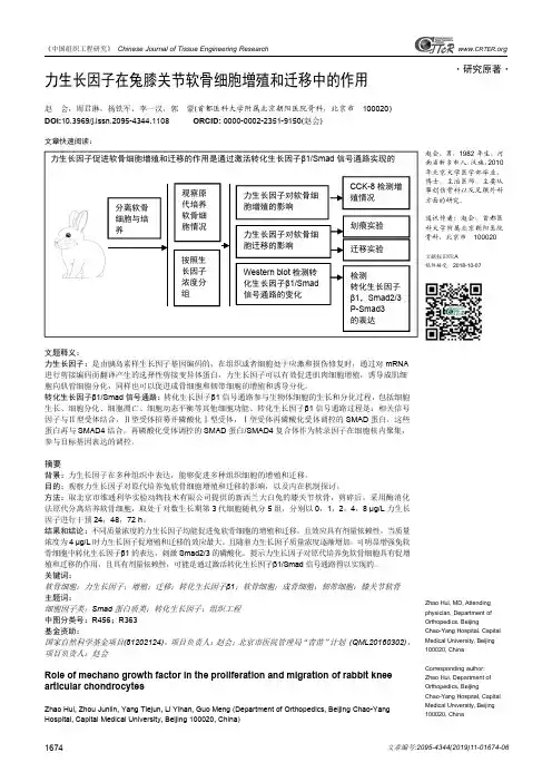
《中国组织工程研究》Chinese Journal of Tissue Engineering Research文章编号:2095-4344(2019)11-01674-061674www.CRTER .org·研究原著·赵会,男,1982年生,河南省新乡市人,汉族,2010年北京大学医学部毕业,博士,主治医师,主要从事创伤骨科以及足踝外科方面的研究。
通讯作者:赵会,首都医科大学附属北京朝阳医院骨科,北京市100020文献标识码:A稿件接受:2018-10-07Zhao Hui,MD,Attending physician,Department of Orthopedics,Beijing Chao-Yang Hospital,Capital Medical University,Beijing 100020,China Corresponding author:Zhao Hui,Department of Orthopedics,Beijing Chao-Yang Hospital,Capital Medical University,Beijing 100020,China力生长因子在兔膝关节软骨细胞增殖和迁移中的作用赵会,周君琳,杨铁军,李一汉,郭蒙(首都医科大学附属北京朝阳医院骨科,北京市100020)DOI:10.3969/j.issn.2095-4344.1108ORCID:0000-0002-2351-9150(赵会)文章快速阅读:文题释义:力生长因子:是由胰岛素样生长因子基因编码的,在组织或者细胞处于应激和损伤修复时,通过对mRNA 进行剪接编码而翻译产生的选择性剪接变异体蛋白,力生长因子可以有效促进肌肉细胞增殖,诱导成肌细胞向肌管细胞分化,同样也可以促进成骨细胞和韧带细胞的增殖和诱导分化。
转化生长因子β1/Smad 信号通路:转化生长因子β1信号通路参与生物体细胞的生长和分化过程,包括细胞生长、细胞分化、细胞凋亡、细胞动态平衡等其他细胞功能。
lecture_22
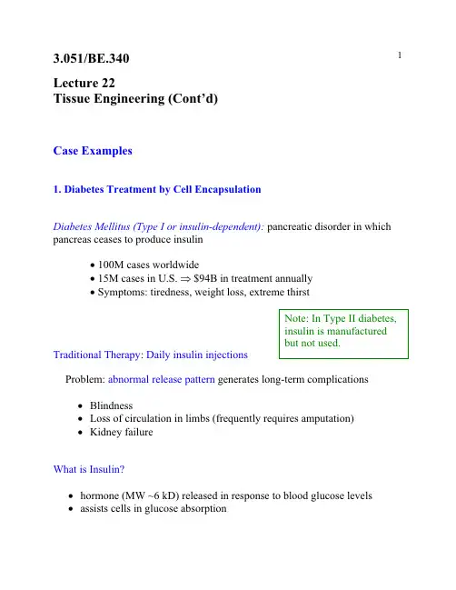
Lecture 22Tissue Engineering (Cont’d)Case Examples1. Diabetes Treatment by Cell EncapsulationDiabetes Mellitus (Type I or insulin-dependent): pancreatic disorder in which pancreas ceases to produce insulinx100M cases worldwidex15M cases in U.S. $94B in treatment annuallyx Symptoms: tiredness, weight loss, extreme thirstTraditional Therapy:Daily insulin injectionsProblem:abnormal release pattern generates long-term complicationsx Blindnessx Loss of circulation in limbs (frequently requires amputation)x Kidney failureWhat is Insulin?x hormone (MW ~6 kD) released in response to blood glucose levels x assists cells in glucose absorptionPancreas : an endocrine gland, incorporating:¾Pancreatic Acini: release digestive enzymes to the duodenum ¾Islets of Langerhans (1-2% of pancreas vol.): release insulin to bloodstream Cell Encapsulation Therapyx 1st reported in mid-1970’s (W.L. Chick, Joslin Res. Lab., Boston)glucose homeostasis achieved in rats x Tubular implants in pancreatectomized dogs, early 1990’s (R.P. Lanza)PVC-PAN membraneacrylic housing-Implanted in peritoneal (abdominal) cavity-Membrane connected to vasculature by PTFE grafts-No exogenous insulin required for > 10 weeks-Little fibrosis up to 30 weeks (biocompatible acrylic)-Mechanisms of failure:a)membrane rupture (80-90% of devices by 5-7 mo.)b)thrombosisc)infectiond)loss of islet function, islet necrosisx Human trials 1994 (Sharp & Lacy)-Implanted subcutaneously-PAN-PVC hollow fiber (1.5 cm length)-Human islets in alginate matrix-90-95% islet viability after 2 weeksLimitation: Insulin quantity—several meters of fiber for therapeutic # of cells Possible solutions:a)“super” cell lines—high insulin output, low nutrient needsb)advances in angiogenesisReferencesR.H. Li, “Materials for cell encapsulation”, Adv. Drug Delivery Rev. 33 (1998) 87-109. R.P. Lanza, R. Langer, W.L. Chick, Principles of Tissue Engineering, R.G. Landes Co.: Austin, TX, 19972.In Vitro Cartilage RegenerationBasics of Cartilage:x incorporates a single cell type (chondrocytes)x low vascularization (limits in vivo regeneration capability)x provides joint lubrication (knee)x basis of soft structural members (nose, ears)Conditions:x torn cartilage (athletic injury) (~0.5M/yr in U.S.)x rheumatoid arthritis (enzymes from phagocytes degrade cartilage)xmenisciIn vitro Chondrogenesisx Materials: nonwoven PGA fiber mesh or salt-leached; ~95% porous x Procedure: scaffold prewet with culture medium & seededx PGA matrix replaced over time by cells, collagen & GAGsSalt-leached scaffoldmade by 3.082 students10% polylactideLimitations:a)Low solids content: poor mass transfer in cultureb)Irregular tissue morphology: lack of flow field in vitro Revised Approach: Bioreactors(R. Langer, MIT)Spinner Flaskmediumtissue constructsmagnetic stirbarRotating Vesselfluidreplacementtissue constructs(free-floating)Cells GAG Collagen H2O Static41015Rotating7301988Natural4384275Bioreactor-Grown Tissues:x tissue dimensions close to original scaffoldx higher solids content than staticx histology mimics natural cartilage~ 30 P m capsuleflat cells & collagen:gives stiffnessuniform mix of cells,GAG, collagen Commercial Status: Genzyme (Cambridge, MA)FDA-approved for autologous knee cartilage repairsRemaining Challenges:a) scale-up of tissue dimensionsb) improved mechanical properties2.In Vivo Nerve RegenerationCentral Nervous System (CNS):Brain & Spinal cordSpinal cord: neurons coated by oligodendrocytes—secretions inhibitregeneration Peripheral Nervous System (PNS):nerve branches that process informationfrom the environment; some regenerative capacity: axon bundles surrounded by connective tissueNerve : several fasicles encased in epinurium- generate myelin (insulator)- secrete neutrophic factorsCondition:Severed nervex loss of support (neurotrophic factors)x scar ingrowthx displacement of resprouting axonsNerve Guidance Channels: use regenerative capacity of PNS HistoryWWI- Rubber tubes used to guide axon growth- low biocompatibility1960’s- Silastic trials (crosslinked silicone rubber)Today- Limited clinical use- Silicone & PGA devices- largest bridgeable gap ~ 1 cmproximal stump (to spine)Case Study: Severed rat sciatic nerve in silicone guideTime elapsed Morphologyhours influx of serum, fibrin, neurotrophicfactorsone week longitudinally oriented fibrin coalescesto bridge stumpscells invade: macrophages, fibroblasts,Schwann, endothelialaxons sprout from proximal stump four weeks axon sprouts reach distal stump(~ 1cm)Regenerated Nervesx less axons & thinner sheathx slower signal conductionx lower amplitude signalChannel Design ConsiderationsResorbable vs. Nonresorbableresorbable—larger inflammatory response nonresorbable—susceptible to compression injuryImpermeable vs. Porousimpermeable—poor nutrient, waste and O2 transport porous—interference by wound healing mechanisms, poor orientation semipermeable—MW cutoff 50-100 kD enables transport & sequesters GF Schwann cell-seeded gels- secrete neurite-promoting basal lamina, NGFs- can be genetically engineered to secrete neurotrophins- evidence of CNS regeneration support (optic nerve regen. demonstrated) Remaining Challenges:a) large-gap repairb) CNS regeneration (spinal cord work at MIT)Important Remaining Challenges in Tissue Engineering1. angiogenesis/mass transport limitations2. delivering appropriate signals to cells3. providing appropriate mechanical stimulus for growth。
细胞培养 英文版
Our Cell Line
• An immortal cell line, but not tumorogenic, will reach contact inhibited state • Originally used to study heat shock response
• These cells maintain a number of differentiated cell functions of hepatocytes. The cells possess inducible and stable cytochrome P450 (CYF) activityultures
• Primary Culture – Cells taken directly from a tissue to a dish • Secondary Culture – Cells taken from a primary culture and passed or divided in vitro. – These cells have a limited number of divisions or passages. After the limit, they will undergo apoptosis.
Growing Cells in Culture
Part 1: Terminology
Cell Culture
• Pros • Use of animals reduced • Cells from one cell line are homogenous and have same growth requirements, optimizing growing patterns. • In vitro models allow for control of the extracellular environment • Able to monitor various elements and secretions without interference from other biological molecules that occurs in vivo
- 1、下载文档前请自行甄别文档内容的完整性,平台不提供额外的编辑、内容补充、找答案等附加服务。
- 2、"仅部分预览"的文档,不可在线预览部分如存在完整性等问题,可反馈申请退款(可完整预览的文档不适用该条件!)。
- 3、如文档侵犯您的权益,请联系客服反馈,我们会尽快为您处理(人工客服工作时间:9:00-18:30)。
ibidi 伤口愈合与细胞迁移实验
细胞划痕实验是一种操作简单,经济实惠的研究细胞迁移/肿瘤侵袭的体外试验方法。
这种方法的原理是,当细胞长到融合成单层状态时,在融合的单层细胞上人为制造一个空白区域,称为“划痕”。
划痕边缘的细胞会逐渐进入空白区域使“划痕”愈合。
基本步骤:“划痕”的制造,细胞迁移期间图像的获得以及后期数据的处理。
特点:
1.在一定程度上模拟了体内细胞迁移的过程。
2.非常适合研究细胞与胞外基质(ECM),细胞与细胞之间相互作用引起的细胞迁移。
3.与包括活细胞成像在内的显微镜系统兼容,可用于分析细胞间的相互作
4.研究细胞迁移的体外实验中最简单的方法。
传统实验方法:细胞融合后,用枪头在细胞层表面制造划痕实验方法缺陷:人工制造的“划痕”很难保证一致性,重复性差。
ibidi Culture-Insert
生物相容性硅胶制成的插件,可产生确定的500μm “划痕”区域。
适合于伤口愈合实验,细胞迁移实验,2D侵袭和共培养。
技术参数:
材料:生物相容硅胶材料
规格:两孔(70μl/孔)
尺寸(长x宽x高):8.4 mm x 8.4 mm x 5 mm 底部:可黏附底部
每孔生长面积:0.22 cm2
产生“划痕”宽度:500 μm
操作步骤:
其他应用:
ibidi Culture Insert与传统划痕实验比较:
Ibidi Culture Insert提供重复性更好的实验结果
实验结果分析:
Data Acquisition/数据采集:
显微镜用于评估伤口愈合的过程。
根据你的关注点和兴趣,不仅可以视频显微镜,还可以进行定点的成像观察。
ibidi加热与孵育系统可以直接在培养的条件下进行活细胞成像,不用将样品反复的从孵育室移至显微镜上。
Wound Healing Image Analysis/伤口成像分析– WimScratch
是一个伤口愈合和细胞迁移实验的成像分析软件
1. 全面解决伤口愈合实验-从样品准备到成像分析只需要几步
2. 客观的和可再生分析
3. 简单并且快速的数据处理-几分钟出结果
4. 不需要额外的硬件或者软件
WimScratch 自动分析实验结果
WimScratch是基于网站的自动化分析平台,无需任何软件和硬件,只要将图片上传到数据分析平台,几分钟之后结果就可以发送到注册邮箱。
分析数据包括:
a) 细胞覆盖面积
b) 终止速度(平均≥5个图像)
c) 加速特征(平均≥5个图像)
d) 概览图
e) 中间接合处的近似值(平均≥5个图像)
ibidi的伤口愈合成像分析解决方案可以评估细胞迁移,仅仅通过4步就可以得到结果。
订购信息:
相关产品:。