Hormonal regulation of the novel adipocytokine visfatin in 3T3-L1_2006
脂联素演示教学

脂联素的发现
吴健雄Байду номын сангаас院 2015.10.21
Maeda 等Q 的类似物, 因其与胶原质Ⅹ、Ⅷ和补体因 子C1q 有高度相似, 并且在脂肪组织中基因转录产物最丰富, 故称为 apM1;
Nakano 等( 1996) 用明胶亲和层析分离人血浆蛋白时发现 了该蛋白质, 故命名为明胶结合蛋白( GBP28);
脂联素结构
吴健雄学院 2015.10.21
脂联素通过3个球形结构域单体连接成三聚体,4-6个三 聚体通过胶原结构域链接形成低聚体或者高级结构,其在 血浆内的浓度为5-30μg/ml,有全长和球形两种循环形式。
图2 脂联素的三聚体和低聚体
脂联素含量在人群中的差异 吴健雄学院 2015.10.21
人类脂联素mRNA在胚胎发育的中晚期开始表达 , 起缘于中 胚层和外胚层。 分娩时脐带血中脂联素水平(11.7-35.7μg/ml) 明显高于年长儿和成年人。 脂联素的表达和分泌总体来说 是女性(11.7 μg/ml)高于男性(7.9 μg/ml)。
脂联素与能量代谢
吴健雄学院 2015.10.21
脂联素具有调节葡萄糖转运的作用 。研究发现通过静脉 注射脂联素可以降低肝脏磷酸烯醇丙酮酸羧化酶和葡糖6-磷酸酶的mRNA 表达 , 抑制肝糖生成酶的表达 , 从而减 少了内源性糖的生成。
脂联素与能量代谢
吴健雄学院 2015.10.21
脂联素能够影响机体处理糖类和脂肪的能力。脂联素 可能通过抑制TNF-α通路增强胰岛素PI-3K通路,增强胰岛素 的敏感性。同时β细胞是脂联素直接作用的靶细胞,血清脂 联素浓度和β细胞上脂联素受体的多少决定了脂联素对β细 胞的作用强弱,这暗示了脂联素对调节糖代谢有着双重作 用,即增强胰岛素敏感性及改善β细胞功能。
黄芪多糖对3T3

黄芪多糖对前脂肪细胞增殖和分化的影响(一)作者:刘毅,王文健,陈伟华,应健【关键词】黄芪多糖;增殖;分化;mRNA;前脂肪细胞近年来许多研究表明,黄芪及其主要成分黄芪多糖(Astragaluspolysaccharides,APS)可以调节糖脂代谢,具有改善胰岛素抵抗的作用,因此广泛运用于2型糖尿病、肥胖和代谢综合征等代谢相关性疾病的防治〔1~4〕。
在以往的实验中,我们发现黄芪多糖可增加脂肪细胞葡萄糖的摄取和利用,促进前脂肪细胞的分化〔3〕,为了进一步探讨其影响前脂肪细胞增殖和分化的机制,本研究从影响脂肪细胞分化的两个主要基因过氧化物体增殖剂活化受体γ(peroxisomeproliferatoractivatedreceptorγ,PPARγ)和CAAT/增强子结合蛋白α(CAAT/enhancerbindingproteinα,C/EBPα)mRNA与蛋白质水平,观察了APS对其的影响。
1材料与方法1.1药物与试剂前脂肪细胞株,AmericanTypeCultureCollection(ATCC)产品;达尔伯克改良伊格尔培养基(Dulbecco'smodifiedeagle'smedium,DMEM)、特级新生小牛血清、胎牛血清,Gibco公司产品;胰蛋白酶、青霉素G钠盐、硫酸链霉素、焦碳酸二乙酯(diethylpyrocarbonate,DEPC)和溴化乙啶,Amresco公司产品;APS由复旦大学内分泌研究所提供;胰岛素、地塞米松、异丁基甲基黄嘌呤、二甲氧唑黄{〔〕}、牛血清白蛋白(bovineserumalbumin,BSA)、酶标二抗,Sigma 公司产品;罗格列酮(rosiglitazone,ROS),浙江天马医药化工有限公司产品;Trizol试剂,Invitrogen公司产品;硝酸纤维素膜,Whatman公司产品;PPARγ和C/EBPα抗体,Adipogen 公司产品;,Fermentas公司产品;BIORADIcycler5色荧光定量PCR仪、紫外凝胶摄像分析仪和图像分析软件,SygeneGeneGenius公司产品。
霍格兰营养液配方
COMMENTS FOR THE AUTHOR:Reviewer #1: The manuscript CEMN-D-12-01363 by Chen and collaborators describes the expression and distribution of the transcription factor FOXO3a and the kinase inhibitor p27kip1 in the retina of the DBA/2J mouse relative o the C57/BL6 mice.The manuscript contains some original observations.SUMMARY:While Foxo3a inhibits cell cycle progression via control of p27kip1 (cyclin kinase inhibitor) during the G1-S phase transition in various cell lines, the authors believe these two genes/gene products are worthy candidates for study in differentiated neurons and glia in a mouse model for glaucoma.They looked for the spatial and temporal distributions in the retinas of the DBA/2J mice (D2) grouped by factors such as age, IOP, slit lamp and ophthalmoscope inspection to delineate non-glaucomatous,pre-glaucomatous and glaucomatous animal groups.The authors used light microscopy, RT-PCR, and immunocytochemistry to look at the spatial and temporal distributions and expression levels of foxo3a and p27kip1 and made use of TUNEL and antibodies to activated caspase-3 as measures of cell death.Their results indicated that - Westerns: foxo3a and p27kip proteins were reduced in time/condition relative to control mice retina; RT-PCR: p27kip mRNA may also be reduced with time/condition.Immuno-histology was used to show that - foxo3a and p27kip are diffusely expressed in retina of D2 and B6 mice and decreased in Muller (GS+) and astrocytes (GFAP+). However, foxo3a increased in RGCs (neuN+) of D2 mice and activated caspase 3 was evident in RGCs (NeuN+) of D2 mice. TUNEL was evident in RGC layer of D2 miceThe authors concluded that - foxo3a and p27kip1 are involved in neural cell loss in D2 miceCRITIQUE:INTRODUCTION:The experimental plan to group the animals into 3 groups is a good idea, although, it has been shown previously that there are changes the expression of some genes that occur prior to the full blown glaucomatous condition. In this case, it appears that FOXO3a and p27kip1 may be somewhat late and perhaps downstream to the primary signaling events leading to loss of vision."ubiquitously" seems to be redundant in first sentence of second paragraph. The listed citations do not give appropriate credit to many important studies. For example:1) The introduction to this paper needs additional references for the second sentence in the 1st paragraph, especially for each of themechanisms suggested as primary causes of RGC cell death. The author should add excitotoxicity as one of the postulated mechanisms here - especially as foxo3a has been implicated in apoptosis.2) Paragraph 4 of the introduction - the authors may want to cite the 2001 Nobel laureates for their discoveries of the role of cyclins and CDKs in cell cycle progression.3) The authors must cite Ophthalmol Eye Dis. 2010 March 11; 1: 23-41, a microarray study where up-regulation of GFAP (Muller cells and astrocytes) and Iba1 (microglia) were shown and where a CDI was also shown to be increased in the DBA/2J mouse retina. At the very least this should be in the last paragraph of the introduction, and probably, also used in the discussion.4) The authors should also cite Invest Ophthalmol Vis Sci 2006;47:977-85 another important gene expression study. This should also go, at least in the last paragraph of the introduction.METHODS:Can the authors state how they determined an illumination level of 50-60 lux for housing the animals? Just curious.I would like to see a table for the 3 DBA/2J animal groups with the various factors used to assign animals to this or that group. This might be helpful to investigators who use these animals.Catalogue numbers should be used to identify the antibodies that were used. RESULTS:In second and fifth lines of the first paragraph, what is meant by the term "control group" here? Are these B6 or are they D2- non glaucomatous mice? You also use the term "control" for no-primary controls, for example, in the figure legends. So always be specific.The last sentence of the first paragraph states that neurodegeneration induced a decrease in foxo3a and p27kip1. The term "induced" may be inappropriate here.The second paragraph speaks of a decrease in immuno-staining. This is difficult to discern, perhaps due to the use of cresyl violet as a counterstain. It might be better to not use a nuclear counterstain. I cannot see the translocation from nucleus to cytoplasm. Again this might have been possible without the dark blue counterstain.I cannot see much overlap between foxo or p27 with the Muller cell glutamine synthase in figure3. While the author has boxed a possible example, there are many instances where the labeling is either red or green. There are hints of foxo and p27 in the middle layer of the INL and in the OPL, but the figure could be improved.There is some evidence in Figure 4 for double labeled cells containing both markers for GFAP and Foxo but all-GFAP or all-Foxo labeled cells are also to be seen. Can the authors explain this?The statement indicating a cause and effect relationship between FOXO andp27 may be misplaced, at least in this part of the results section. There does appear to be a correlation between the relative abundance of FOXO and p27 in the retina as judged from western blots of the whole retina, but you have also made a case for increased abundance of FOXO in RGCs. Thus, it is not clear that your argument is consistent at the cellular level, or at least, it is not made clear to this reviewer.It is not clear that figure 8 adds anything that has not already been reported and the data are not that convincing anyway. It might have been useful to show the presence of cleaved caspase or TUNEL in double labeled cells containing FOXO or p27 because the direct connection between FOXo and p27 with cell death is somewhat tenuous in this manuscript. DISCUSSION:Much is lost in translation and this is particularly true of the discussion section where misleading statements such as "The present data reveal that down-regulation of p27kip1 through FOXO transcriptional inhibition was associated with ----" and "our results demonstrated that Foxo3a as a positive regulator of p27kip1 was associated with ---" can be found. There is little integration with the existing data on the DBA/2J mouse or any other animal model of glaucoma.Reviewer #2: In this study the authors show that the transcription factor FOXO3a and its downstream gene p27kip1 are decreased in glaucomatous DBA/2J retinas. The decreased expression of these proteins was found in the Müller cells and astrocytes. The authors also found increased expression of FOXO3a in RGCs from glaucomatous retinas. The latter finding seems to be correlated with the apoptosis of RGCs, observed with TUNEL and Caspase3 staining in glaucomatous retinas.Major points:1) The authors were able to show that FOXO3a and p27kip1 expression levels are changing in glaucomatous retinas, however the study lacks a direct connection between their findings and the cellular processes in which these proteins are involved. Downregulation of FOXO3a and p27kip1 have been proposed to be involved in cell proliferation, while upregulation of FOXO3a in neurons is associated with apoptosis. It will be interesting if the authors are able to establish the connection between the expression of these proteins and the cellular events mentioned above. A previous study has shown the involvement of the PTEN-Akt-FOXO3a pathway in neuronal apoptosis in brain development after hypoxia-ischemia (Li et al., J Cereb Blood Flow Metab, 2009). In this study, the expression of Bim, an apoptosis related protein that is downstream of FOXO3a, is increased as a response of FOXO3a nuclear translocation. The authors could demonstrate the link between FOXO3a and apoptosis byshowing dephosphorylation in the PTEN-Akt-FOXO3a pathway and the consequent increased levels of Bim in glaucomatous RGCs. In this way the authors might propose a possible mechanism of apoptosis that could betargeted therapeutically in glaucoma.2) It is necessary to include an optic nerve assessment of degeneration for the establishment of glaucoma in these mice. The use of an axonal antibody in the optic nerve could be a good alternative.。
瘦素对肥胖饮食小鼠的脂质合成相关基因表达影响的研究

提取RNA,将RNA通过逆转录HiFiScript cDNA 第一链合成试剂盒合成cDNA,以cDNA为模板,在 荧光定量PCR仪上进行检测,以GAPDH为内参,计 算出各组FAS、ACC-1、PPARγ、SREBP-1的相对 表达量。操作体系: RNase Free dH20 9.5μL、cDNA 1 μL、Forward Primer 1 μL、Reverse Rrimer 1 μL、 2×qPCR Mixture12.5 μL。反应程序,三步法:预变 性:95℃、10min,变性:95℃、10s,退火:58℃、 30s,延伸:72℃、30s,40个循环。融解曲线分析: 95℃、15s,58℃、1min,95℃、15s,58℃、15s, 58℃、15s,95℃、0.5s。引物如表1。 1.7 统计学方法
of mice in high-fat group was significantly decreased (P<0.01). Compared with the high-fat group, the expression of PPARγ mRNA in
liver tissue of mice in low-dose and high-dose leptin group was increased, the expression of ACC-1, FAS and SREBP1 mRNA in liver
固醇调节元件结合蛋白(sterol regulatory element binding protein-1,SREBP-1)作为脂质合成的关键 转录因子,被活化后能够启动下游靶基因脂肪酸合 酶(Fatty acid synthase,FAS)、乙酰辅酶A羧化酶 (Acetyl-Coa carboxylase,ACC-1)等与脂质合成相 关的关键酶合成,进而参与脂质的合成。FAS催化长 链脂肪酸合成,ACC-1催化丙二酰辅酶A合成,因此 在脂肪酸和甘油三酯的代谢过程中发挥重要作用。过 氧化物酶体增殖物激活受体(Peroxisome proliferatoractivated receptor,PPAR)是参与脂质合成、分泌及 积累的转录因子。其中PPARγ促进脂肪代谢限速酶合 成,进而增强脂肪代谢限速酶的抗脂质分解作用,使 本该流向肌肉及肝脏的游离脂肪酸流向脂肪组织,进 而减少糖异生。激活PPARγ后,脂质细胞不仅形态上 发生一些变化,还会出现脂质的积累及胰岛素的敏 感等现象。因此,通过调控PPARγ、FAS、ACC1、 SREBP-1的表达对于肥胖引起的脂质积聚具有重要的 意义。
合文

合文®- 控制血清甘油三酯水平
与标准的大豆油相比 ,合文®组血清甘油三酯浓度显著降低。
Schlotzer E, Kanning U. Elimination and tolerance of a new parenteral lipid emulsion (SMOF)--a double-blind cross-over study in healthy male volunteers. Ann NutrMetab. 2004;48(4):263-8.
2013 PN 合文 0001
14
合文®- 改善炎症平衡
一项针对外科术后患者的前瞻性、随机对照研究中,输注合文®后的促炎 症细胞因子IL-6水平显著低于橄榄油/大豆油组。
2013 PN 合文 0001 15
Schade I, Röhm KD, Schellhaass A, et al. Inflammatory response in patients requiring parenteral nutrition: comparison of a new fish-oil-containing emulsion (SMOF®) versus an olive/soybean oil-based formula. Critical Care. 2008;12(Suppl 2):P144.
Schulzki C, Mertes N, Wenn A et al.Effects of a new type of lipid emulsion based on soybean oil, MCT, olive oil and fish oil (SMOF) in surgical patients. Clinical Nutrition .1999; 18, (Suppl 1):7.
具有胰岛素效应的新脂肪细胞因子-Visfatin
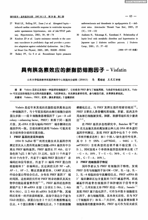
蛋 白多肽— —前 B细 胞集 落增强 因子 (r Bcl pe— e l cl y— nac gf tr B F o n ehni c ,P E )来 源 于 同 一 基 因 o n ao
片段 ,其 c N D A片段与编码 P E 5 B F ’端非翻译区的
基 因序 列一 致 。 目前 的研 究 表 明 Vs t ian可 能具 有 fi 自分 泌和旁 分 泌的双 重功能 。
磷酸化位点 ,与 P E B F发挥生理作用密切相关…。
PE B F主要在人 类 骨 髓 间质 细 胞 、肝 脏 、肌 肉及 外 周血 淋 巴细胞 被发 现 ,肺 脏 、胎膜 等组织 有少 量表 达 ] 。 在 PE B F基 因 多态 性 研 究 中 ,B thr j ot e 等 对 c
19 94年 SJa 等 利 用 克 隆 技术 及 基 因序 列 all l
测定首次从人类外周血淋巴细胞 c N D A基 因库中分
离出PE B F编码 基 因。P E B F基 因长 3 . k ,位 于 74 b 染 色体 7 2 . 和 7 3 . 3之间 ,包 含 1 个 外显 子 q2 1 q 13 1
3 As k wa H , T k ma a K, Ka k miF. R lt n hp o 0 aa ou g wa a e ai s i f o
2 K usnJ 8 n d o D
a. L p n rss n e e tn st h OO 1 e f it c xed o te C /- i e a
t 可促进脂肪细胞分化成熟和脂肪蓄积 ,与肥胖相关 ;有类胰岛素样作用 ,参与糖脂代谢 ,改善胰 岛索抵抗 。 i n 关键词 V s t ;P E ;肥胖 ;胰岛素抵抗 ;2型糖尿病 i an B F fi
IL-1beita的最新成果
Mol. Cells, V ol. 21, No. 2, pp. 174-185The Modulation of Inflammatory Gene Expression by Lipids: Mediation through Toll-like ReceptorsJoo Y. Lee1,* and Daniel H. Hwang21 Department of Life Science, Gwangju Institute of Science and Technology, Gwangju 500-712, Korea;2 USDA, ARS, Western Human Nutrition Research Center, and Department of Nutrition, University of California, Davis, CA 95616, USA.(Received March 29, 2006; Accepted March 31, 2006)Toll-like receptors (TLRs) were evolved to detect in-vading pathogens and to indu ce innate immu ne re-sponses in order to mount host defense mechanisms. It becomes apparent that the activation of certain TLRs is also modu lated by endogenou s molecu les inclu ding lipid components, fatty acids. Results from epidemiol-ogical and animal studies demonstrated that saturated and polyu nsatu rated dietary fatty acids can differen-tially modify the risk of development of many chronic diseases. Inflammation is now recognized as an impor-tant underlying etiologic condition for the pathogene-sis of many chronic diseases. Therefore, if the activa-tion of TLRs and consequ ent inflammatory and im-mu ne responses are differentially modu lated by types of lipids in vivo, this would suggest that the risk of the development of chronic inflammatory diseases and the host defense against microbial infection may be modi-fied by the types of dietary fat consumed. Keywords: Fatty Acids; Inflammation; Lipids; Toll-like Receptors.IntroductionLipids can be de fine d as biological substance s that dis-solve in organic solvents, but not in water. Together with carbohydrate s and prote ins, lipids are the main constitu-e nts of plant and animal ce lls. The y se rve as structural compone nts of ce llular me mbrane s and provide e ne rgy sources. Fatty acids are the major dietary lipid.Fatty ac-ids are hydrocarbon molecules of various lengths and de- grees of unsaturation containing a carboxylic acid moiety * To whom correspondence should be addressed.Tel: 82-62-970-2505; Fax: 82-62-970-2484E-mail: joolee@gist.ac.kr at one e nd. Fatty acids that contain no carbon-carbon double bonds are termed saturated fatty acids while those that contain double bonds are unsaturate d fatty acids. Fatty acids and the ir de rivative s are e sse ntial biological mole cule s as the compone nts of ce llular me mbrane and the major components of stored fat in the form of triacyl-glyce rols. More ove r, fatty acids function as signaling mole cule s that re gulate ge ne e xpre ssion re late d to me ta-bolic pathways or inflammatory diseases. Fatty acids are known to affe ct the ce llular physiological function (pro-liferation, differentiation, apoptosis, metabolism) and also implicated in the modulation of a variety of pathological states including obesity, hyperlipidemia, diabetes, athero-scle rosis, hype rte nsion, inflammation, and cance r. The type s of fatty acids in ce llular lipids are se nsitive ly af-fected by those of dietary fat.Many epidemiological, experimental and clinical studies showe d that polyunsaturate d fatty acids (PUFAs) re duce the risk of cancer and many inflammatory chronic diseases whereas saturated fatty acids are known to increase the risk of cardiovascular dise ase s including coronary he art dis-eases and stroke (Clarke et al., 2002; Hwang, 2000; Serhan and Savill, 2005). The mole cular me chanism as to how different types of fatty acids modify the risk of the devel-opme nt of chronic dise ase s has not be e n cle arly unde r-stood. Evide nce gaine d re ce ntly de monstrate d that satu-rated fatty acids induce the expression of pro-inflammatory ge ne products including COX-2, iNOS and IL-1α in Abbreviations: COX-2, cyclooxygenase-2; DHA, docosahex-aenoic acid; EPA, eicosapentaenoic acid; iNOS, inducible nitric oxide synthase; IFNβ, interferon β; IRF3, interferon-regulatory factor 3; LPS, lipopolysaccharide; MyD88, myeloid differentia-tion factor 88; Ox-PAPC, oxidized-1-palmitoyl-2-arachidonyl-sn-glycero-3-phosphorylcholine; PUFAs, polyunsaturated fatty acids; TLRs, Toll-like receptors; TRIF, TIR domain-containing adapter inducing IFNβ.MoleculesandCells©KSMCB 2006Joo Y. Lee & Daniel H. Hwang 175macrophages (RAW264.7 cells) (Lee et al., 2001). Lauric acid (C12:0) and palmitic acid (C16:0) were the most po-tent ones in inducing COX-2 expression among various size of saturated fatty acids (C8:0-C18:0). The stimulation of macrophages with saturated fatty acid induced the acti-vation of NFκB as determined by the degradation of IκBαand the phosphorylation of p65. Saturated fatty acid also induced the phosphorylation of other signaling components such as ERK, JNK, and Akt in macrophages. In contrast to saturated fatty acids, various unsaturated fatty acids including n-3 PUFAs (EPA, C20:5n-3; DHA, C22:6n-3) and n-6 PUFAs (linoleic acid, C18:2n-6) inhibited the expression of inflammatory genes such as COX-2, iNOS and IL-1α in macrophages (Lee et al., 2001). The activa-tion of NFκB and the phosphorylation of Akt induced by LPS were suppressed by DHA (Lee et al., 2003a). How-ever, the mechanism through which different types of fatty acids differentially modulate inflammatory gene expression has not been fully understood.Toll-like receptors play an important role in inducing innate immunity and inflammatory responses and linking to the activation of adaptive immune responses and host de-fense systemTolls, identified in Drosophila, are first known to be in-volved in the development of dorsal-ventral pattern in em-bryos. L ater, it has been revealed that Tolls participate in the induction of immune responses by producing anti-fungal peptide, drosomycin in adult Drosophila. Toll-like receptors (TLRs) are the mammalian homolog of Toll and Type I transmembrane receptors with extracellular leucine-rich repeat (L RR) motifs and a cytoplasmic Toll/IL-1R (TIR) homology domain (Medzhitov et al., 1997). So far, eleven TLRs have been identified in human while thirteen can be found in the mouse genome. TL Rs recognize con-served pathogen-associated molecular patterns (PAMPs) of invading microorganisms, of which structures include lip-ids, carbohydrates, nucleic acids and various proteins (Aderem and Ulevitch, 2000; Akira et al., 2001; Medzhitov and Janeway, 1997). Each TL R senses different PAMPs although the direct interaction between ligands and recep-tors has not still been demonstrated. The representative TL R agonists include lipo-polysaccaride (L PS) for TL R4, bacterial lipopeptides and peptidoglycan for TLR2, double-stranded RNA for TL R3, flagellin for TL R5, and single-stranded RNA and unmethylated CpG motifs in bacterial DNA for TLR7 and 9, respectively (Aderem and Ulevitch, 2000; Medzhitov and Janeway, 2000). It is now well estab-lished that TLRs play a critical role in triggering and regu-lating innate and adaptive immune responses that ulti-mately lead to the elimination of the invading pathogens in mammals. The innate immune responses triggered by TLR activation accompany with the expression of pro-inflammatory proteins evoking inflammatory responses. Recent evidence now suggests that non-microbial molecules can activate TL Rs. TL R4 can be activated by fibronectin, fibrinogen, heparan sulfate, and taxol (Byrd-Leifer et al., 2001; Okamura et al., 2001). Heat shock proteins also induce the activation of TL R2 and TL R4 (Asea et al., 2002; Ohashi et al.,2000; Vabulas et al., 2001) although it is still controversial if the activation of TL R4 by heat shock protein 60 may be due to the con-tamination with L PS (Gao and Tsan, 2003). Host RNA and DNA derived from damaged cells can activate TLR3 and TLR9 (Brentano et al.,2005). Endogenous TLR ago-nists released from host in the body may cause sterile in-flammatory responses and simultaneously unnecessary immune responses.Therefore, the activation of TL Rs by microbial patho-gens, tissue injury, and stress leads to the expression of mediators for both immune and inflammatory processes. The original purpose of TL R activation is to initiate and to amplify immune responses as host defense system against microbial infection. However, the accompanying inflammatory responses are unavoidable. If the inflamma-tory responses are not appropriately controlled and are prolonged, tissue injury may be progressed and chronic inflammatory diseases can be further developed. It is well known that excessive activation of TLRs can lead to the development of severe systemic inflammation including septic shock with high mortality. Moreover, the close rela-tionship between TLRs and various chronic inflammatory diseases was suggested (Table 1). Many epidemiological studies showed that the polymorphism of TLRs decreases the susceptibility and tends to be protective to the devel-opment of many chronic inflammatory diseases including alzheimer’s disease, atherosclerosis, diabetes, and cancer (Chen et al., 2005; Kiechl et al., 2002). The knockout of TLR4 or MyD88 reduced the progression and severity of atherosclerosis in animals fed with high fat western diet (Bjorkbacka et al., 2004; Michelsen et al., 2004). Myocar-dial infarction injury was less in mice with inactive mutation of TLR4 (C3H/HeJ) (Oyama et al.,2004). Mice deficient of MyD88 did not develop streptococcal cell wall-induced ar-thritis (Joosten et al., 2003). TLR4 expression was increased in case of ischemic heart (Frantz et al., 1999). TLR2 expres-sion was enhanced in fibroblasts and macrophages of syno-vial tissues from patients with rheumatoid arthritis (Seibl et al., 2003). The endogenous TL R4 agonists such as heat shock proteins, fibrinogen, and hyaluronan are found in ar-thritis joints.The downstream signaling pathways of Toll-like receptorsTLRs stimulated with agonist recruit adaptor molecules to176 Modulation of Toll-like Receptor Signaling by LipidsTable 1. Experimental evidences suggesting the involvement of TLRs in chronic inflammatory diseases.Types of study TLRs involvedDiseasesFeaturesGene polymorphismTLR4 Asp299GlyAlzheimer’s disease (Minoretti et al ., 2006)Diabetes (Kolek et al ., 2004) Coronary artery disease (Kolek et al ., 2004)Atherosclerosis (Kiechl et al ., 2002)Gene polymorphism is associ-ated with protection from the development of the diseases TL R 4 sequence variant Multiple SNPs at the TLR6- TLR1-TLR10 gene clusterProstate cancer (Chen et al ., 2005; Zheng et al ., 2004)Prostate cancer (Sun et al ., 2005)TLR5 stop codon Systemic lupus erythematosus (Hawn et al ., 2005)TL R 4 Asp299Gly TLR4 Thr399Ile Ulcerative colitis/ Crohn’s disease (Franchimont et al ., 2004) Ulcerative colitis (Torok et al ., 2004)The mutation was increased inpatients TL R 2 Arg753Gln Atherosclerosis (Hamann et al ., 2005)Knockout mice TLR4 Atherosclerosis (Michelsen et al ., 2004) TL R 2 Atherosclerosis (Mullick et al ., 2005)Renal Ischemia/reperfusion injury (Leemans et al ., 2005)The severity and progress ofdisease was reduced in knockoutmice Experimental disease model TL R 3 Diabetes (Wen et al ., 2004) TL R 2/TL R 3/TL R 9 Arthritis (Deng et al ., 1999; Liu et al ., 2001; Zare et al ., 2004)The treatment with TLR agonist to animals induced experimental disease modelInfection TL R 2/TL R 4 Atherosclerosis (Chlamydia Pneumonia ) (Netea et al ., 2002) Gastric cancer (Helicobacter Pylori ) (Gobert et al ., 2004; Mandell et al ., 2004) C. Pneumonia is TLR2 agonistH. Pylori is TLR 2 and TLR 4 agonistTLR expression TLR2 Rheumatoid arthritis (Seibl et al ., 2003) TLR4 Ischemic heart (Frantz et al ., 1999) Inflammatory bowel diseases (Cario and Podolsky, 2000)TL R 1/TL R 2/TL R 4 Atherosclerosis (Edfeldt et al ., 2002)TLR expression was upregulatedin tissues from patients with di-seasesactivate downstream signaling pathways. Four major adap-tors have been discovered up to now: MyD88, Mal (MyD88 adaptor-like)/TIRAP (TIR domain-containing adaptor protein), TRIF (TIR-domain-containing adaptor inducing interferon-beta) and TRAM (TRIF-related adaptormolecule). Each TLR uses different combinations of adap-tor molecules to provide the signaling specificity for differ-ent TLRs. In general, the activation of TLR4 triggers the activation of both MyD88-dependent and -independent (TRIF-dependent) signaling pathways. TLR2 and TLR9Joo Y. Lee & Daniel H. Hwang 177Fig. 1. Toll-like receptor-media ted downstrea m signa ling pa th-wa ys. Different Toll-like receptors (TLRs) trigger signa ls via different combina tions of a da ptors. TLR4 a ctiva tion recruits four major adaptors, MyD88, TIRAP, TRIF, and TRAM. TLR3 signaling is mostly dependent on TRIF whereas TLR2 requires MyD88 a nd TIRAP. The a ctiva tion of downstrea m signa ling pathways of TLR5, TLR7, and TLR9 is dependent on MyD88. MyD88-dependent pa thwa y includes the a ssocia tion a nd phos-phoryla tion of IRAK4 a nd IRAK1. TRAF6 is recruited to the receptor complex resulting in the a ctiva tion of IKK complex, which consists of IKKα, IKKβ, a nd IKKγ. The IKK complex phosphoryla tes IκB lea ding to the degra da tion of IκB a nd the tra nsloca tion of NFκB into the nucleus, which induces the ex-pression of ta rget genes. TRIF-dependent pa thwa y is essentia l for the MyD88-independent pa thwa y. IKKε a nd TBK1 a re the downstrea m kina ses of TRIF lea ding to the a ctiva tion of IRF3 a nd the expression of Type I Interferons (IFNs). RIP1 is a cti-vated through TRIF pathway resulting in the activation of NFκB. TLR7 and TLR9 induce the expression of Type I IFNs through MyD88-IRF7 pathway.activation primarily induce the activation of MyD88-dependent pathway whereas TLR3 activation primarilyleads to the activation of MyD88-independent (TRIF-de-endent) pathway (Fig. 1).MyD88, the common downstream adaptor molecule formost TLRs, interacts directly with TIR domain of TLRs(Burns et al., 1998; Medzhitov et al., 1998). MyD88 re-cruits IL-1 receptor-associated kinase-4 (IRAK-4) throughthe interaction with death domain and induces IRAK-4phosphorylation. The phosphorylated IRAK-4 then inducesthe phosphorylation and activation of IRAK-1 (Suzuki et al., 2002). IRAK-1 associates with tumor necrosis factor (TN F) receptor-associated factor 6 (TRAF6), which inturn recruits TAB-1 (transforming growth factor-β-ac-tivated kinase-1 (TAK-1)-binding protein-1) and TAB-2leading to the activation of TAK-1 kinase (Ninomiya-Tsuji et al., 1999). TAK-1 activates the canonical IKKα/β/γcomplex resulting in the activation of NF-κB transcription factor. TAK-1 also activates the upstream kinases of p38 and JNK. As a result, inflammatory target genes including COX-2 and cytokines are expressed (Muzio et al., 1998). Synthetic bacterial lipopetides (TLR2 agonist) and CpG DN A (TLR9 agonist), can neither activate N FκB nor in-duce the expression of inflammatory cytokines in macro-phages derived from MyD88-deficient mice demonstrat-ing that TLR2 and TLR9 require primarily MyD88-dependent signaling pathway (Hacker et al., 2000; Schnare et al., 2000).Toll-IL-1 receptor domain-containing adapter protein (TIRAP)/myeloid differentiation protein 88 adapter-like (Mal) was initially considered as an MyD88-independent adaptor (Fitzgerald et al., 2001; Horng et al., 2001). However, the study using TIRAP/Mal knockout mice demonstrated that TIRAP/Mal is not involved in the MyD88-independent pathway but rather cooperates with MyD88 in the signaling pathways of TLR2 and TLR4, but not TLR3, TLR5, TLR7, or TLR9 (Horng et al.,2002). Unlike TLR2 and TLR9 agonists, LPS (TLR4 agonist) and dsRNA (TLR3 agonist) can still induce the activation of N FκB and MAPKs, the production of IL-18, and the maturation of dendritic cells in a delayed fashion in MyD88-deficient cells (Kaisho et al., 2001; Kawai et al., 1999; Seki et al., 2001). These results suggested the pres-ence of an additional adaptor that is independent of MyD88 in the signaling pathways of certain TLRs. TIR domain-containing adapter inducing IFN-β (TRIF)/Toll-interleukin 1 receptor domain (TIR)-containing adaptor molecule (TICAM)-1 has been reported as an adaptor molecule of TLR3 and TLR4 responsible for MyD88-independent signaling pathway (Oshiumi et al., 2003a; Yamamoto et al., 2002). TLR4 signaling pathways require both MyD88 and TRIF as the major adaptor molecules since the activation of N FκB and JNK1 mediated through TLR4 was completely abolished in MyD88/TRIF double-knock-out cells (Yamamoto et al., 2003a). For TLR3 sig-naling, TRIF is believed to be the main adaptor for the ac-tivation of NF-κB and IRF3. TRIF induces the activation of the transcriptional regulator, IRF3 and the expression of IFNβ and IFN-inducible genes through the activation of TBK1 and IKKε as IRF3 kinases (Fitzgerald et al., 2003a; Toshchakov et al., 2002). TRIF also induces the activation of N FκB with delayed fashion while MyD88-dependent pathways mediate the quick activation of NFκB (Sato et al., 2003). RIP1 interacts with TRIF leading to the activation of NFκB although the detail downstream signaling pathways through which RIP1 activates NFκB are not clearly identi-fied (Meylan et al., 2004). It was also suggested that IκBαdegradation induced by TRIF pathway is mediated through the interaction between TRIF and TRAF6 thereby leading to the activation of IKKβ since TRAF6 was shown to asso-ciate with the N-terminal part of TRIF (Jiang et al., 2004; Sato et al., 2003). In addition, polyI:C-induced NFκB acti-vation was completely abolished in TRAF6-deficient mouse embryonic fibroblasts (Jiang et al., 2004). However, Gohda et al. showed that polyI:C-induced IκBα degrada-178 Modulation of Toll-like Receptor Signaling by Lipidstion and cytokine production was not affected in macro-phages derived from TRAF6-deficient mice (Gohda et al., 2004). Therefore, it is still unclear whether TRAF6 is in-volved in N FκB activation mediated through the TLR3-TRIF pathway. Recently, it was reported that the delayed NFκB activation by TLR4 agonist (LPS) shown in MyD88-independent pathway is mediated through IRF3-dependent expression of TN F-α (Covert et al.,2005). TN F-α pro-duced by LPS challenge mediates a positive feedback loop to further stimulate IKK activation (Werner et al.,2005). TRIF needs another adaptor to confer the activation of TLR4 by agonist. TRIF-related adaptor molecule (TRAM), also called TIR domain-containing protein (TIRP) (Bin et al., 2003) and TIR-containing adaptor molecule-2 (TICAM-2), was discovered as a novel adaptor molecule in TLR sig-naling. TRAM associates with TLR4, but not TLR2, TLR5, TLR6, TLR7, TLR8 and TLR9 (Fitzgerald et al., 2003b; Oshiumi et al., 2003b). TRAM interacts with both TLR4 and TRIF as a bridging protein and is involved in the activation of NF-κB and IRF3 as an upstream compo-nent of TRIF in TLR4 signaling (Fitzgerald et al.,2003b; Yamamoto et al., 2003b).Fatty acids acylated in certain TLR agonists are important in agonistic activityCertain types of TLR agonists such as lipid A moiety of LPS (TLR4 agonist) and lipopeptides (TLR2 agonist) contain saturated fatty acid moiety such as lauric acid, myristic acid and palmitic acid (Munford and Hall, 1986). Lipid A of Escherichia coli and Salmonella typhimurium is a β,1-6 linked disaccharide of glucosamine, acylated with R-3-hydroxylaurate or myristate and phosphorylated at positions 1 and 4′. The 3-hydroxyl groups of these satu-rated fatty acids are further 3-O-acylated by lauric acid, myristic acid or palmitic acid. Bacterial lipopeptides such as S-(2,3-bis(palmitoyloxy)-(2-RS)-propyl)-N-palmitoyl-(R)-Cys-(S)-Ser-(S)-Lys(4)-OH trihydrochloride (Pam3Cys), are triacylated at N-terminal cysteine residue with satu-rated fatty acids. Mycoplasmal lipopeptides are diacylated with saturated fatty acids (Takeuchi et al., 2001; 2002). Furthermore, the number of acylated fatty acids in the molecule discriminates whether TLR2 requires TLR1 or TLR6 as a partner to recognize each type of agonist. Tri-acylated bacterial lipopeptides activate TLR2 dimerized with TLR1 while diacylated lipopeptides like macrophage activating lipopeptide-2-kDa (MALP-2), are recognized by TLR2 and TLR6 dimer (Takeuchi et al., 2001; 2002). If the acylated saturated fatty acids on TLR2 and TLR4 agonist are deacylated or replaced with unsaturated fatty acids, the agonists lose their activity or act as antagonists (Kitchens et al., 1992; Krauss et al., 1989).Deacylation of LPS by host lipase such as acyloxyacyl hydrolase (AOAH) is one of the mechanisms to inactivate LPS in vivo and to regulate immune responses against bacterial infection (Lu et al.,2005). Together, these imply that fatty acid moiety acylated in TLR agonist molecules plays an important role in the recognition of agonists by the receptor and the activation of the receptor. Saturated fatty acid induces the activation of TLR2 and TLR4The expression of inflammatory genes such as COX-2 or iN OS induced by saturated fatty acid was blocked by a dominant-negative mutant of TLR4, TLR2, or TLR6. Ec-topical expression of TLR4 with MD2 and CD14 in 293T cells confers the responsiveness to saturated fatty acid (Lee et al., 2003a). Saturated fatty acid also activated TLR2 dimerized TLR1 or TLR6 leading to the activation of N FκB and the expression of COX-2 and IL-8 in macrophages and human colon epithelial cells (SW620) (Lee et al., 2004). However, saturated fatty acid failed to induce N FκB activation when individual TLR3, 5, or 9 was ectopically expressed in 293T cells. Saturated fatty acid activates TLR2 dimers and TLR4 for which cognate ligands from bacterial origin require acylated fatty acids in their molecules.The activation of TLR4 by saturated fatty acid required MD2 and CD14 since the expression of TLR4 alone in 293T cells was not sufficient to confer the responsiveness to saturated fatty acid (Lee et al., 2003a). The activation of TLR4 by LPS includes various proteins. LPS forms a complex with first, LPS-binding protein (LBP) in blood and then with CD14, TLR4, and MD2. The physical prox-imity between TLR4, CD14, and MD2 was induced by LPS as demonstrated by fluorescence resonance energy transfer (FRET) techniques (da Silva Correia et al., 2001; Jiang et al., 2000). CD14 is a cell surface 55-kDa glyco-protein with a glycosylphosphatidylinositol (GPI) mem-brane anchor (Haziot et al., 1988; Wright et al., 1990). MD2, a 20- to 30-kDa glycoprotein, interacts with the extracellular domain of TLR4 and provides greater speci-ficity for recognition of ligands and more efficiency for responsiveness to LPS (Shimazu et al.,1999). The bio-chemical interaction between saturated fatty acids and TLR has not been entirely unveiled yet.Saturated fatty acid induced the activation of both MyD88-dependent and TRIF-dependent signaling path-ways of TLRs. A dominant-negative MyD88 inhibited saturated fatty acid-induced N FκB activation in macro-phages. In addition, saturated fatty acid-induced N FκB activation was inhibited by a dominant-negative mutant of MyD88-dependent downstream signaling components such as TIRAP, IRAK-1, TRAF-6, IKKβ, and IκBα in 293T cells reconstituted with TLR4/MD-2 or TLR2/6. These results demonstrate that saturated fatty acid induced the activation of MyD88-dependent signaling pathways me-Joo Y. Lee & Daniel H. Hwang 179diated through the activation of TLR4 or TLR2. Saturated fatty acid increased ISRE-luciferase activity which is dependent on TRIF/IRF3 activation in RAW 264.7 cells. The activation of ISRE induced by saturated fatty acid was suppressed by a dominant mutant of TLR4, TRIF and IRF-3, but not by TLR2 dominant mutant in RAW264.7 cells. Therefore, saturated fatty acid activates TRIF-dependent signaling pathway mediated through the activation of TLR4, but not TLR2 in macrophages.PI3K/AKT pathway is one of the downstream pathways activated by saturated fatty acid. A dominant-negative mutant of AKT inhibited NFκB activation induced by saturated fatty acid in 293T cells transfected with TLR2/ TLR6 or TLR4/MD2. Saturated fatty acid-induced NFκB activation and CO X-2 expression was inhibited by a pharmacological PI3K inhibitor (LY294002), and a domi-nant-negative mutant of PI3K or AKT in macrophages (RAW264.7). Saturated fatty acid induced the rapid and transient phosphorylation of AKT.Unsaturated fatty acids inhibit TLR-mediated signaling pathways and target gene expression In contrast to the stimulatory effect of saturated fatty ac-ids on TLR activation, various unsaturated fatty acids suppressed NFκB activation induced by TLR4 agonist, LPS. Human diet study consistently showed the inhibitory effect of unsaturated fatty acids on inflammatory gene expression by TLR4 agonist (LPS). LPS-induced COX-2 expression was decreased in blood peripheral monocytes by the consumption of meals containing fish oil, a major source of n-3 PUFAs (DHA and EPA) (Lee et al., 2003b). Cytokine production (IL-2, IL-1, and TNFα) was also reduced in LPS-stimulated human mononuclear cells by fish oil intake at the dose of 18 g/day for six weeks (Endres et al.,1989; 1993).DHA appears to be a pan-inhibitor for various TLRs. DHA suppresses NFκB activation and CO X-2 expression induced by various TLR agonists such as lipopeptides (TLR2), double-stranded RNA (TLR3), LPS (TLR4), flag-ellin (TLR5), and CpG (TLR9) in macrophages (Lee et al., 2004). DHA also inhibits NFκB activation and COX-2 ex-pression induced by a constitutively active TLR4 in RAW264.7 and 293T cells. Therefore, DHA inhibits both ligand-dependent and ligand-independent activation of TLRs. However, DHA is unable to inhibit NFκB activation and COX-2 expression induced by the downstream compo-nents of TLRs such as MyD88, NIK, and AKT. Thus, the potential target of unsaturated fatty acid may be the TLR itself or the proximal events leading to TLR activation, but not the downstream signaling components.N-3 PUFAs (DHA and EPA) are much more potent in-hibitors of TLR4 and TLR2 activation as compared with n-6 PUFAs (arachidonic acid and linoleic acid) or n-9 UFA (Oleic acid). This may account for the reason why n-3 PUFAs are considered as more effective anti-inflamma-tory compounds than n-6 PUFAs. Phosphatidylglycerol(PG) containing an unsaturated fatty acid inhibited LPS-induced NFκB activation and the inhibitory activity wasdependent on the types of fatty acid moiety (Hashimoto et al., 2003). However, triglyceride with unsaturated fatty acid (DHA or linoleic acid) was much less potent to in-hibit LPS-induced CO X-2 expression. Unsaturated fattyacids including linoleic acid, arachidonic acid, EPA, andDHA enhanced the expression of CO X-2 in mammaryepithelial cell line (184B5) (Meade et al.,1999). DHA didnot suppress CO X-2 expression induced by TNF-α incolon epithelial tumor cells (HT-29) (Lee et al., 2001).These suggest that the effect of unsaturated fatty acids onCO X-2 expression seems to be cell type- or stimuli-specific.Inhibitory effect of unsaturated fatty acids: metabolites, oxidation products, or not?Unsaturated fatty acids are enzymatically metabolized by lipoxygenases, CO X, or cytochrome P450. Unsaturated fatty acids can be also non-enzymatically oxidized. 17R-hydroxy series of docosanoids produced from DHA by aspirin-modified COX-2 possess potent anti-inflammatory effects (Serhan et al., 2002). Oxidized phospholipid prod-ucts exerted anti-inflammatory activities by inhibiting neutrophil binding, E-selectin expression, and NFκB acti-vation induced by LPS in endothelial cells (Bochkov et al., 2002; Leitinger et al., 1999). In contrast, unoxidized phospholipids do not show such inhibitory effect. In other studies, oxidation products of phospholipids stimulated endothelial cells to produce inflammatory gene products such as MCP-1 and IL-8 and increased monocyte binding to endothelial cells (Leitinger et al., 1999; Subbana-gounder et al., 2002; Walton et al.,2003) suggesting the pro-inflammatory role of oxidized phospholipids. Never-theless, a report that phosphatidylglycerol (PG) contain-ing an unsaturated fatty acid, but not a saturated fatty acid moiety, inhibited LPS-induced NFκB activation (Hashimoto et al., 2003) suggests that the inhibitory ef-fect of PG depends on the types of acylated fatty acid, but not on the phospholipid backbone.Other lipid mediators that regulate TLR acti-vationMinimally oxidaized LDL (mmLDL) triggers cytoskeletal rearrangement in macrophages through TLR4. LDL oxi-dized by 15-lipoxygenase stimulates the activation of PI-3K and AKT and the phosphorylation of ERK1/2. MmLDL induced NFκB p65 translocation to the nucleus and the。
黄芪多糖对脂肪细胞释放肿瘤坏死因子-α及白细胞介素-6的影响
黄芪多糖对脂肪细胞释放肿瘤坏死因子-α及白细胞介素-6的影响刘琼;翁孝刚【期刊名称】《河南师范大学学报:自然科学版》【年(卷),期】2015(0)3【摘要】目的:探讨黄芪多糖对脂肪细胞释放肿瘤坏死因子-α(TNF-α)、白细胞介素-6(IL-6)的抑制作用及机制.方法由3T3-L1细胞诱导分化成熟的脂肪细胞随机分为实验组和对照组.实验组脂肪细胞以0.1g·L-1黄芪多糖培养基干预,对照组脂肪细胞普通培养基培养.ELISA法测两组脂肪细胞及培养基中TNF-α、IL-6蛋白的含量,及逆转录-聚合酶链反应检测两组脂肪细胞TNF-α蛋白和IL-6蛋白mRNA水平.结果实验组脂肪细胞及培养基中TNF-α、IL-6蛋白的含量低于对照组(P=0.000),而且实验组脂肪细胞TNF-α蛋白和IL-6蛋白mRNA水平低于对照组(P=0.000).结论:黄芪多糖可抑制脂肪细胞释放炎症细胞因子,控制脂肪组织的慢性炎症反应.【总页数】4页(P121-124)【关键词】黄芪多糖;脂肪细胞;肿瘤坏死因子-α;白介素-6【作者】刘琼;翁孝刚【作者单位】新乡医学院第一临床学院内分泌科【正文语种】中文【中图分类】Q455;R965【相关文献】1.双异丙酚预处理对内毒素诱导的人中性粒细胞释放白细胞介素-8和肿瘤坏死因子-α及相应mRNA表达的影响 [J], 池信锦;程楠;黑子清;罗刚健;罗晨芳2.沙利度胺对间质性肺疾病肺泡巨噬细胞释放白细胞介素和肿瘤坏死因子-α的影响 [J], 叶俏;陈宝敏;童朝晖;Nakamura S;Sarria R;Costabel U;Guzman J3.肿瘤坏死因子α和白细胞介素6致脂肪细胞胰岛素抵抗的不同机制 [J], 曾天舒;陈璐璐;潘世秀4.地塞米松对肺损伤大鼠肺泡巨噬细胞释放肿瘤坏死因子-α和白细胞介素-6的影响 [J], 袁志明;陈光瑾;杜文彬5.体外动态力学刺激对SD幼鼠前脂肪细胞形态及肿瘤坏死因子表达释放影响的研究 [J], 谢西梅;陈波;崔瑾;杨运宽因版权原因,仅展示原文概要,查看原文内容请购买。
黄芪多糖调控TXNIP
生命科学仪器 2023年第21卷/第6期研究报告65项目来源:河北省中医药管理局中医药类科研指令性计划课题(2021444)黄芪多糖调控T X N I P /N L R P 3炎症小体对高糖诱导的人视网膜内皮细胞凋亡的作用机制陶雯璇 李君卿 张元坤 张京红(张家口市第四医院,河北张家口075000)摘要 目的探究黄芪多糖调控T X N I P /N L R P 3炎症小体对高糖诱导的人视网膜内皮细胞凋亡的作用机制㊂方法将h R E C s 细胞随机分为C o n t r o l 组㊁H G 组㊁A P S 组和A P S +p c D N A 3.1-T X N I P 组㊂采用C C K-8和流式细胞仪检测细胞存活率和凋亡率;D C F H-D A 荧光探针检测活性氧(R O S)水平;检测细胞中氧化应激和炎症因子水平;蛋白质印迹法检测细胞中T X N I P ㊁N L R P 3㊁I L -1β和ca s p a s e -1蛋白表达㊂结果和C o n t r o l 组相比,H G 组细胞存活率㊁S O D 活性和G S H-P x 含量明显降低,细胞凋亡率㊁R O S 相对荧光强度㊁M D A 含量㊁I L -1β和IL -18含量及细胞中T X -N I P ㊁N L R P 3㊁I L -1β㊁c a s p a s e -1蛋白表达明显增加(P <0.05);和H G 组相比,A P S 组细胞存活率㊁S O D 活性和G S H -P x 含量明显增加,细胞凋亡率㊁R O S 相对荧光强度㊁M D A 含量㊁I L -1β和IL -18含量及细胞中T X N I P ㊁N L R P 3㊁I L -1β㊁c a s p a s e -1蛋白表达明显降低(P <0.05);和A P S 组相比,A P S +p c D N A 3.1-T X N I P 组细胞存活率㊁S O D 活性和G S H-P x 含量明显降低,细胞凋亡率㊁R O S 相对荧光强度㊁M D A 含量㊁I L -1β和IL -18含量及细胞中T X N I P ㊁N L R P 3㊁I L -1β㊁c a s p a s e -1蛋白表达明显增加(P <0.05)㊂结论黄芪多糖可抑制高糖诱导的视网膜内皮细胞凋亡㊁炎症反应和氧化应激损伤,其作用机制可能和抑制T X N I P /N L R P 3通路激活有关㊂关键词 黄芪多糖;高糖诱导的视网膜内皮细胞;凋亡;T X N I P /N L R P 3通路M e c h a n i s m o f a s t r a g a l u s p o l y s a c c h a r i d e m o d u l a t i o n o f T X N I P /N L R P 3i n f l a m m a t o r y ve s i c l e s o n h i g h g l u c o s e -i n d u c e d a p o pt o s i s i n h u m a n r e t i n a l e n d o t h e l i a l c e l l s T a o W e n x u a n L i J u n q i n g Z h a n g Y u a n k u n Z h a n g J i n g h o n g (Z h a n g j i a k o u F o u r t h H o s p i t a l ,Z h a n g ji a k o u 075000,C h i n a )ʌA b s t r a c t ɔO b je c t i v e :T o i n v e s t i g a t e t h e m e c h a n i s m of A s t r ag a l u s p o l y s a c ch a ri d e m o d u l a t i n g T X N I P /N L R P 3i n f l a m m a t o r y v e s i c l e s o n h i g h g l u c o s e -i n d u c e d a p o p t o s i s i n h u m a n r e t i n a l e n d o t h e l i a l c e l l s .M e t h o d s :T h e h R E C s c e l l s w e r e r a n d o m l yd i v i de d i n t o C o n t r o l g r o u p ,H G g r o u p ,A P S g r o u p ,a n d A P S +p c D N A 3.1-T X N I P g r o u p s .C e l l s u r v i v a l a n d a p o pt o s i s w e r e d e t e c t e d b y C C K -8a n d f l o w c y t o m e t r y ,r e s p e c t i v e l y ;r e a c t i v e o x y g e n s p e c i e s (R O S )l e v e l s w e r e d e t e c t e d b y DC F H -D A f l u o r e s c e n t p r o b e ;o x i d a t i v e s t r e s s a n d i n f l a m m a t o r yf a c t o r l e v e l s w e r e d e t e c t e d i n t h e c e l l s ;a n d T X N I P ,N L R P 3,I L -1β,a n d c a s p a s e -1p r o t e i n e x p r e s s i o n w e r e d e t e c t e d i n t h e c e l l s b y p r o t e i n b l o t t i n g .R e s u l t s :C o m p a r e d w i t h t h e C o n -t r o l g r o u p ,t h e c e l l s u r v i v a l r a t e ,S O D a c t i v i t y a n d G S H-P x c o n t e n t w e r e s i g n i f i c a n t l y l o w e r i n t h e H G g r o u p,a n d t h e a p o p t o s i s r a t e ,r e l a t i v e f l u o r e s c e n c e i n t e n s i t y o f R O S ,M D A c o n t e n t ,I L -1βa n d I L -18c o n t e n t ,a n d t h e e x p r e s s i o n o f T X N I P ,N L R P 3,I L -1β,a n d c a s p a s e -1p r o t e i n s i n t h e c e l l s w e r e s i g n i f i c a n t l y i n c r e a s e d (P <0.05);C o m p a r e d w i t h t h e H G g r o u p ,t h e c e l l s u r v i v a l r a t e ,S O D a c t i v i t y a n d G S H-P x c o n t e n t i n t h e A P S g r o u p w e r e s i g n i f i c a n t l yi n c r e a s e d ,a n d t h e a p o p t o s i s r a t e ,r e l a t i v e f l u o r e s c e n c e i n t e n s i t y o f R O S ,M D A c o n t e n t ,I L -1βa n d I L -18c o n t e n t ,a n d c e l l u l a r e x p r e s -s i o n o f T X N I P ,N L R P 3,I L -1β,a n d c a s p a s e -1p r o t e i n s w e r e s i g n i f i c a n t l y d e c r e a s e d (P <0.05);C o m p a r e d w i t h t h e A P S g r o u p ,c e l l s u r v i v a l ,S O D a c t i v i t y a n d G S H-P x c o n t e n t w e r e s i g n i f i c a n t l y l o w e r i n t h e A P S +p c D N A 3.1-T X N I P g r o u p ,a n d a p o p t o s i s r a t e ,r e l a t i v e f l u o r e s c e n c e i n t e n s i t y o f R O S ,M D A c o n t e n t ,I L -1βa n d I L -18c o n t e n t ,a n d c e l l u l a r e x -p r e s s i o n o f T X N I P ,N L R P 3,I L -1β,a n d c a s p a s e -1p r o t e i n s w e r e s i g n i f i c a n t l y i n c r e a s e d (P <0.05).C o n c l u s i o n :A s t r a g a -l u s p o l y s a c c h a r i d e i n h i b i t s h i g h g l u c o s e -i n d u c e d a p o p t o s i s ,i n f l a m m a t o r y r e s p o n s e a n d o x i d a t i v e s t r e s s d a m a ge i n r e t i n a l e n -d o t h e l i a l c e l l s ,a n d i t s m e c h a n i s m of a c t i o n m a y b e r e l a t e d t o t h e i n h i b i t i o n o f T X N I P /N L R P 3p a t h w a y ac t i v a t i o n .ʌK e y wo r d s ɔA s t r a g a l u s p o l y s a c c h a r i d e ;h i g h g l u c o s e -i n d u c e d r e t i n a l e n d o t h e l i a l c e l l s ;a p o p t o s i s ;T X N I P /N L R P 3p a t h -w a y中图分类号:R 45 文献标识码:A D O I :10.11967/2023211213糖尿病视网膜病变(d i a b e t i c r e t i n o p a t h y,D R )是糖尿病常见的一种微血管病变,主要病变为人视网膜内皮细胞(h u m a n r e t i n a l e n d o t h e l i a l c e l l s ,h R E C s)功能障碍,其也是视力丧失的主要原因,影响患者的生活质量[1]㊂D R 发病机制复杂,近年来研究发现[2],慢性视网膜炎症是导致D R 发病的主要因素,当白细胞浸润到视网膜释放炎性递质,通过破坏血-视网膜屏障(b l o o d r e t i n a l b a r r i e r ,B R B )使血管渗漏和新血管形成㊂核苷酸结合寡聚化结构域样受体蛋白3(n u c l e o t i d e -b i n d i n g o l i go m e r i z a t i o n d o -m a i n -l i k e r e c e pt o r p r o t e i n 3,N L R P 3)炎症小体会破坏B R B 参与D R 进程[3];N L R P 3还能通过结合研究报告生命科学仪器 2023年第21卷/第6期66其介质硫氧还蛋白相互作用蛋白(t h i o r e d o x i n -i n -t e r a c t i n g pr o t e i n ,T X N I P )激活N L R P 3炎症小体[4]㊂由此可见,抑制T X N I P /N L R P 3通路激活可能是治疗D R 的新靶点㊂黄芪多糖(a s t r a g a l u s p o l y s a c h a r -i n ,A P S)是黄芪中一种活性最强的有效成分,现代研究证实[5],A P S 具有多种生物学功能,如抗氧化㊁抗炎㊁抗凋亡㊁改善微循环等,但其对D R 的作用及机制报道较少㊂因此,本研究通过D R 体外实验模型,旨探究A P S 能否调控T X N I P /N L R P 3炎症小体影响D R 发生发展㊂1 材料与方法1.1 试剂与仪器 人视网膜内皮细胞(h R E C s)购自中国科学院上海细胞库㊂黄芪多糖(国药准字Z 20040085,天津赛诺制药有限公司,规格250m g/瓶);T X N I P ㊁N L R P 3㊁I L -1β和ca s p a s e -1抗体(英国A n c a m 公司);T X N I P 过表达载体(pc D N A 3.1-T X N I P )(广州瑞博生物公司);细胞活力/毒性(C E L L C O U N T I N G K I T-8,C C K-8)试剂盒㊁氧化应激指标检测试剂盒及炎症因子E L I S A 检测试剂盒(南京健成生物研究所);流式细胞仪检测试剂盒(美国B D 公司);低温高速离心机(型号:M i -c r o 17R ,德国T h e r m o 公司);酶标仪(型号C M a x -P l u s ,美国M o l e c u l a r D e v i c e s 公司);显微镜(型号A E 2000型,美国M o t i c 公司)㊂1.2 细胞培养及分组 将h R E C s 培养于R P M I -1640培养基中,含10%胎牛血清㊁100U /m L 青霉素和链霉素,将培养基置于37ħ㊁5%C O 2的培养箱24h ,收集处于对数期的细胞㊂将h R E C s 细胞随机分为C o n t r o l 组(h R E C s 细胞培养于葡萄糖含量为5m m o l /L 的正常培养基)㊁H G 组(h R E C s 细胞培养于含25m m o l /L 葡糖糖培养基)㊁A P S 组(h R E C s 细胞培养于含25m m o l /L 葡糖糖和300g/L 黄芪多糖培养基[8])和A P S+p c D N A 3.1-T X N I P 组(将h R E C s 细胞转染4μg p c D N A 3.1-T X N I P 质粒,后培养于含25m m o l /L 葡糖糖和300g/L 黄芪多糖培养基),各组细胞均培养48h 后进行后续研究㊂1.3 C C K -8检测细胞增殖能力 将h R E C s 细胞接种于96孔板中,每孔细胞密度为1.5ˑ104个/m L ,培养细胞于37ħ环境及质量分数为5%C O 2的培养箱,分别在培养24h ㊁48h ㊁72h 时将C C K -8溶液(100μL )加入到孔板中,后培养细胞于37ħ环境及质量分数为5%C O 2的培养箱,3.5h 后于酶标仪450n m 处测定酶标仪D O 值㊂该实验重复3次取均值㊂1.4 流式细胞仪检测细胞凋亡 将h R E C s 细胞接种于96孔板中,每孔细胞密度为1.5ˑ104个/m L ,培养细胞于37ħ环境及质量分数为5%C O 2的培养箱24h ,分别将1ˑb i n d i n g bu f f e r ㊁10μL a n n e x i n V 和20μL P I 加入到孔板中培养,后将原有的培养基弃掉,将提前配制好的A n n e x i n V /P I 染液加入到孔板中,常规孵育,将胰蛋白酶裂解液分别加入到孔板的每孔中,裂解细胞呈悬液于流式细胞仪上检测㊂该实验重复3次取均值㊂1.5 2,7-二氯荧光素二乙酸酯(2',7'-D I C H L O -R O F L U O R E S C I N D I A C E T A T E ,D C F H-D A )荧光探针检测活性氧(R E A C T I V E O X Y G E N S P E C I E S,R O S)水平将h R E C s 细胞接种于6孔板中孵育1.5h ,P B S 洗涤细胞后,加入提前用P B S 稀释为10μm o l/L 的D C F H-D A 荧光探针,孵育于37ħ避光的环境中,30m i n 后用P B S 再次洗涤细胞,P B S 重悬收集细胞,通过荧光显微镜观察细胞中R O S 水平㊂该实验重复3次取均值㊂1.6 检测细胞中氧化应激和炎症因子水平 将各组h R E C s 中加入R I P A 裂解细胞,根据试剂盒说明书,分别检测细胞裂解液中S O D ㊁M D A ㊁G S H-P x㊁I L -1β㊁I L -18水平㊂1.7 蛋白质印迹法检测细胞中T X N I P ㊁N L R P 3㊁I L-1β和ca s p a s e -1蛋白表达 用B C A 法测定细胞中总蛋白浓度㊂热水煮沸蛋白样品(终浓度为2μg㊃m L -1)10m i n ,保存于-20ħ环境㊂取蛋白上样50μg ,加入将上样缓冲液后煮沸,将变性的蛋白样品用S D S -P A G E 分离,后转移蛋白样品于P V D F 膜上,加入质量分数为5%的脱脂牛奶(5%)封闭,1h 后用T B S T 溶液清洗P V D F 膜,后加入一抗(1:1000),在4ħ避光环境下孵育一抗过夜;后将H R P标记的二抗(1:5000)加入细胞中,常温孵育2h㊂将E C L 发光液加入细胞显影,曝光后对各组细胞蛋白条带用I m a g e L a b 软件分析㊂1.8 统计学分析 实验数据采用G r a p h p a d p r i a m 8.0软件进行统计学分析㊂数据以均值ʃ标准差(M e a n ʃS D )表示㊂不同组间数据采用重复测量方差和单因素方差(o n e -w a y A N O V A )分析,各组间两两比较采用L S D-t 检验,实验结果以P <0.05表示差异有统计学意义㊂2 结果2.1 各组细胞增殖和凋亡率比较 和C o n t r o l 组相比,H G 组细胞存活率降低,细胞凋亡率增加(t 存活率=10.80,t 凋亡率=13.04,P<0.0001);和H G 组相比,A P S 组细胞存活率增加,细胞凋亡率降低(t 存活率=9.408,t 凋亡率=8.846,P <0.0001);和A P S 组相比,A P S +p c D N A 3.1-T X N I P 组细胞存活率降低,细胞凋亡率增加(t 存活率=8.318,t 凋亡率=8.126,P <0.0001)(图1)㊂2.2 各组细胞R O S 水平比较 H G 组细胞中R O S相对荧光强度高于C o n t r o l 组(t=16.91,P<0.0001);A P S 组细胞中R O S 荧光相对强度低于H G 组(t =12.93,P <0.0001);A P S +p c D N A 3.1-T X N I P 组细胞中R O S 相对荧光强度高于A P S 组(t生命科学仪器 2023年第21卷/第6期研究报告67=12.50,P <0.0001)(图2)㊂A :各组细胞存活率比较;B:流式细胞仪检测细胞凋亡;C :各组细胞凋亡率比较㊂v s C o n t r o l 组*P <0.05;v s H G 组#P <0.05;v s A P S 组әP <0.05㊂图1 各组细胞增殖和凋亡率比较n =6A :D C F H-D A 荧光探针检测细胞中R O S 水平;B :各组细胞中R O S 水平比较㊂和C o n t r o l 组相比*P <0.05;和H G 组相比#P <0.05;和A P S 组相比әP <0.05㊂图2 各组细胞R O S 水平比较n =62.3 各组细胞中氧化应激指标比较 和C o n t r o l 组相比,H G 组细胞中S O D 活性和G S H-P x 含量降低,M D A 含量增加(t S O D =10.78,t G S H-P x=10.87,t M D A =12.38,P <0.0001);和H G 组相比,A P S 组细胞中S O D 活性和G S H-P x 含量增加,M D A 含量降低(t S O D =10.63,t G S H-P x =7.801,t M D A =7.757,P<0.0001);和H G 组相比,A P S+p c D -N A 3.1-T X N I P 组细胞中S O D 活性和G S H-P x含量降低,M D A 含量增加(t S O D =9.362,t G S H-P x =7.549,t M D A =8.571,P <0.0001)(图3)㊂A :细胞中S O D 活性;B :细胞中M D A 含量比较;C :细胞中G S H-P x 含量比较㊂和C o n t r o l 组相比*P <0.05;和H G 组相比#P <0.05;和A P S 组相比әP <0.05㊂图3 各组细胞S O D 活性和M D A ㊁G S H-P x 含量比较n =62.4 各组细胞中炎症因子含量比较 和C o n t r o l 组相比,H G 组细胞中I L -1β和IL -18含量增加(t I L -1β=11.78,t I L -18=13.49,P<0.0001);和H G 组相比A P S 组细胞中I L -1β和IL -18含量均降低(t I L -1β=8.791,t I L -18=9.094,P<0.0001);和A P S 组相比,A P S +p c D N A 3.1-T X N I P 组细胞中I L -1β和IL -18含量增加(t I L -1β=7.832,t I L -18=8.585,P <0.0001)(图4)㊂A :细胞中I L -1β含量比较;B :细胞中I L -18含量比较㊂和C o n t r o l 组相比*P <0.05;和H G 组相比#P <0.05;和A P S 组相比әP <0.05㊂图4 各组细胞中I L -1β和IL -18含量比较n =62.5 各组细胞中T X N I P ㊁N L R P 3㊁I L-1β㊁c a s p a s e -1蛋白表达比较 和C o n t r o l 组相比,H G 组细胞中T X N I P ㊁N L R P 3㊁I L -1β㊁c a s p a s e -1蛋白表达均增加(t T X N I P =11.56,t N L R P 3=9.311,t I L -1β=9.640,t c a s p a s e -1=12.93,P<0.0001);和H G 组相比,A P S 组细胞中TX N I P ㊁N L R P 3㊁I L -1β㊁c a s p a s e -1蛋白均降低(t T X N I P =8.993,t N L R P 3=6.114,t I L -1β=6.020,t c a s p a s e -1=10.47,P<0.0001);和A P S 组相比,A P S +p c D N A 3.1-T X N I P 组细胞中T X N I P ㊁N L R P 3㊁IL -1β㊁c a s p a s e -1蛋白均增加(t T X N I P =9.242,t N L R P 3=6.123,t I L -1β=6.015,t c a s p a s e -1=10.65,P <0.0001)(图5)㊂A :细胞中T X N I P ㊁N L R P 3㊁I L -1β㊁c a s p a s e -1蛋白条带图;B :细胞中T X N I P 蛋白表达比较;C :细胞中N L R P 3蛋白表达比较;D :细胞中I L-1β蛋白表达比较;E :细胞中c a s pa s e -1蛋白表达比较㊂和C o n t r o l 组相比*P <0.05;和H G 组相比#P <0.05;和A P S 组相比әP <0.05㊂图5 各组细胞中T X N I P ㊁N L R P 3㊁I L -1β㊁c a s pa s e -1蛋白表达比较n =63 讨论目前,临床目前常采用激光凝术㊁玻璃体手术等方法治疗D R ,虽能改善患者症状,但由于会损伤视力及使产生不适使患者不易接受㊂因此,探究有效治疗D R 药物对改善患者生活质量尤为重要㊂S u n 等[6]学者研究发现,A P S 治疗糖尿病大鼠,可有效改善心肌组织损伤㊂彭涛等[7]研究证实,A P S 对急性高眼压大鼠视网膜可发挥保护作用㊂赖莉研究报告生命科学仪器 2023年第21卷/第6期68等[8]研究证实,A P S 可通过介导T r a f 6/T A K 1信号通路保护视网膜㊂还有研究发现[9],A P S 可通过抑制多种因素诱导的血管内皮细胞凋亡㊂本研究结果显示,H G 组h R E C s 存活率明显降低,凋亡率明显增加,A P S 干预后,h R E C s 存活率明显增加,凋亡率明显降低,提示A P S 可保护视网膜内皮细胞损伤,对D R 发挥治疗作用㊂闫丰华等[10]研究证实,黄芪多糖可减轻大鼠糖尿病视网膜损伤,从而对视网膜发挥保护作用㊂研究发现[11],高糖环境下视网膜内皮细胞中的氧化应激和D R 发生发展密切相关,当细胞内的氧化平衡状态被破坏,导致细胞过度凋亡,进而影响内皮细胞的功能㊂R O S ㊁S O D ㊁M D A 和G S H-P x指标是检测氧化应激反应的常用标志物㊂本研究结果显示,A P S 可抑制高糖诱导h R E C s 中M D A 含量和R O S 含量,增加S O D 活性和G S H-P x 含量,提示A P S 可提高高糖诱导h R E C s 抗氧化能力,减少氧化损伤㊂汪洋等[12]研究发现,达格列净对人视网膜血管内皮细胞氧化应激和凋亡发挥抑制作用,从而通过减轻细胞损伤保护D R ㊂炎性细胞因子可介导D R 进程,当视网膜炎症和白细胞黏附于D R 微血管时,可对血-视网膜屏障的破坏发挥促进作用㊂研究报道[13],I L-1β在D R 发展中发挥重要作用,而I L -1β的成熟及释放都受到N L R P 3炎症小体的调控㊂N L R P 3炎症小体异常激活可通过活化c a s p a s e -1和I L -1β等效应分子介导机体的炎症和免疫过程㊂Z h e n g 等[14]研究证实,阻断糖尿病视网膜中N L R P 3炎症小体的形成可抑制其损伤㊂由此,N L R P 3炎症小体可能是治疗D R 的重要靶点㊂T X N I P 是硫氧还蛋白的内源性抑制蛋白,当其与硫氧还原蛋白的活性部分结合时,会导致细胞内活性氧化物积累,进而导致机体损伤;T X N I P 还能通过激活N L R P 3炎症小体对炎症发挥促进作用㊂冯梅等[15]通过体内实验发现,富氢水可通过抑制T X N I P /N L R P 3通路的激活降低D R 大鼠视网膜血管通透性,减轻视网膜神经元损伤㊂本研究结果显示,A P S 可抑制高糖诱导h R E C s 中T X N I P ㊁N L R P 3㊁I L -1β㊁c a s p a s e -1蛋白表达,由此猜测,A P S 可能通过抑制T X N I P /N L -R P 3通路的激活减轻高糖诱导h R E C s 凋亡㊁炎症和氧化应激损伤,对视网膜发挥保护作用㊂为了验证这一猜想,本研究采用过表达T X N I P 和A P S 共同干预高糖诱导h R E C s ,结果显示,pc D N A 3.1-T X -N I P 可减弱A P S 对高糖诱导h R E C s 凋亡㊁炎症和氧化应激损伤的抑制作用㊂综上所述,黄芪多糖可抑制高糖诱导的视网膜内皮细胞凋亡㊁炎症反应和氧化应激损伤,其作用机制可能和抑制T X N I P /N L R P 3通路激活有关㊂由于时间和成本等因素,本研究未设置单独的过表达T X N I P 分组或抑制T X N I P 组,可能使本研究结果存在一定局限性,在今后的研究中会增加单独过表达T X N I P 组或敲减T X N I P 组进行对照,进一步证实黄芪多糖能通过调控T X N I P /N L R P 3通路对高糖诱导的视网膜内皮细胞发挥作用,进而为临床治疗糖尿病视网膜病变提供更多的实验依据㊂参考文献[1]Y a o X ,Z h a o Z ,Z h a n g W ,e t a l .S pe c i a l i z e d R e t i n a l E n d o t h e l i a l C e l l s M o d u l a t e B l o o d -R e t i n a B a r r i e r i n D i a b e t i c R e t i n o p a t h y[J ].D i a b e t e s ,2024,73(2):225-236.[2]D h a r m a r a ja n S ,C a r r i l l o C ,Q i Z ,e t a l .R e t i n a l i n f l a m m a t i o n i n m u r i n e m o d e l s o f t y p e 1a n d t y p e 2d i ab e t e s w i t h d i a b e t ic r e t i n o pa -t h y [J ].D i ab e t o l o g i a ,2023,66(11):2170-2185.[3]S i n g h L P ,Y u m n a mc h a T ,S w o r n a l a t a D e v i T .M i t o p h a gi c F l u x D e r e g u l a t i o n ,L ys o s o m a l D e s t a b i l i z a t i o n a n d N L R P 3I n f l a m m a -s o m e A c t i v a t i o n i n D i a b e t i c R e t i n o p a t h y:P o t e n t i a l s o f G e n e T h e r -a p y T a r g e t i n g T X N I P a n d T h e R e d o x S y s t e m [J ].O ph t h a l m o l R e s R e p,2018,3(1):O R R T -126.[4]L i a n L ,L e Z ,W a n g Z ,e t a l .S I R T 1I n h i b i t s H i gh G l u c o s e -I n -d u c e d T X N I P /N L R P 3I n f l a m m a s o m e A c t i v a t i o n a n d C a t a r a c tF o r m a t i o n [J ].I n v e s t O p h t h a l m o l V i s S c i ,2023,64(3):16.[5]J i a n g X ,L i Y ,F u D ,e t a l .C a v e o l i n -1a m e l i o r a t e s a c e t a m i n o -p h e n -a g g r a v a t e d i n f l a m m a t o r y d a m a g e a n d l i p i d d e po s i t i o n i n n o n -a l c o h o l i c f a t t yl i v e r d i s e a s e v i a t h e R O S /T X N I P /N L R P 3p a t h -w a y [J ].I n t I m m u n o ph a r m a c o l ,2023,114:109558.[6]S u n S ,Y a n g S ,Z h a n g N ,e t a l .A s t r a g a l u s p o l ys a c c h a r i d e s a l l e -v i a t e s c a r d i a c h y p e r t r o p h y i n d i a b e t i c c a r d i o m y o p a t h y v i a i n h i b i t i n gt h e B M P 10-m e d i a t e d s i g n a l i n g p a t h w a y [J ].P h yt o m e d i c i n e ,2023,109:154543.[7]彭涛,于丹丹,谢美娜,等.黄芪多糖对高眼压大鼠视网膜神经节细胞凋亡的影响[J ].中国临床药理学杂志,2020,36(10):1344-1346.[8]赖莉,覃晖.黄芪多糖通过T R A F 6/T A K 1信号通路调节视网膜神经节细胞的炎症反应[J ].中国中医眼科杂志,2019,29(6):434-437.[9]王忠庆,蔡帆,诸波,等.黄芪多糖对大鼠缺氧/复氧诱导的心肌细胞自噬及凋亡抑制作用的机制探讨[J ].中国循环杂志,2022,37(2):185-192.[10]闫丰华,焦禄安,郑加军,等.黄芪多糖对糖尿病模型大鼠视网膜病变及血清胱抑素C 的影响[J ].热带医学杂志,2019,19(7):813-816,804.[11]H a y d i n ge r C D ,O l i v e r G F ,A s h a n d e r L M ,e t a l .O x i d a t i v e S t r e s s a n d I t s R e g u l a t i o n i n D i a b e t i c R e t i n o p a t h y[J ].A n t i o x i d a -n t s (B a s e l ),2023,12(8):1649.[12]汪洋,王可,刘宝兰.达格列净对高糖诱导人视网膜血管内皮细胞凋亡及氧化应激的影响[J ].国际眼科杂志,2022,22(3):378-382.[13]R a m a n K S ,M a t s u b a r a J A.D y s r e gu l a t i o n o f t h e N L R P 3I n f l a m -m a s o m e i n D i a b e t i c R e t i n o p a t h y a n d P o t e n t i a l T h e r a p e u t i c T a r -ge t s [J ].O c u l I m m u n o l I nf l a m m ,2022,30(2):470-478.[14]Z h e ng X ,Wa n J ,T a n G .T h e m e c h a n i s m s o f N L R P 3i n f l a m m a -s o m e /p y r o p t o s i s a c t i v a t i o n a n d t h e i r r o l e i n d i ab e t ic r e t i n o p a t h y.F r o n t I m m u n o l .2023A pr 25;14:1151185.[15]冯梅,江霞,王艳丽,等.富氢水对糖尿病视网膜病变大鼠硫氧还蛋白相互作用蛋白/核苷酸结合寡聚化结构域样受体蛋白3通路及视网膜血管通透性的影响[J ].安徽医药,2022,26(7):1367-1373.。
去垢剂
在生物学或生物化学实验室使用的去污剂都是作用比较温和的表面活性剂(=表面活性成分),是用来破坏细胞膜(裂解细胞)以释放细胞内的可溶性物质。
它们可以破坏蛋白质-蛋白质、蛋白质-脂质、脂质-脂质之间的连接,使蛋白质发生结构上的变性,防止蛋白质结晶,另外在免疫学实验中还可避免非特异性吸附。
去污剂根据其特性可以分为好几类,因此科学研究中去污剂的选择很关键,取决于后续研究的具体内容。
实际应用中有众多不同的去污剂可以选择。
为了某些特殊的应用,新的去污剂被不断开发出来[]。
在这篇综述中,对一些最常用的去污剂的特点和应用进行了论述。
去污剂是由一个疏水尾端基团和一个极性亲水头端基团组成的有机化合物(图一A)。
在一定的温度条件下,以特定浓度溶解于水时,去污剂分子会形成胶束,疏水基团部分位于胶束内部,而极性亲水基团则在其外部(图一B)。
因此,胶束的疏水中心会结合到蛋白的疏水区域。
一个胶束中,去污剂分子的聚集数目,是用来评价膜蛋白溶解度的一个重要参数[]。
去污剂分子疏水区域的长度和其疏水性成正比,且去污剂的疏水区域非常恒定,而极性头端亲水基团是可变的,可据其特点,把去污剂分为三类:离子型(阴离子或阳离子型),两性离子型和非离子型(见表一)。
在特定的温度下,表面活性剂分子缔合形成胶束的最低浓度,称之为临界胶束浓度(CMC)。
当去污剂低于临界胶束浓度时,只有单体存在;当高于临界胶束浓度时,胶束、单体以及其余不溶于水的非胶束相共存。
同样,胶束形成的最低温度称为临界胶束温度(CMT)。
因此,温度和浓度是去污剂两相分离和溶解性的重要参数。
一般来说,低亲脂或憎油的去污剂的临界胶束浓度会较高。
种类化合物离子去污剂十二烷基硫酸钠(SDS),脱氧胆酸钠,胆酸钠,肌氨酸非离子去污剂tritonX-100,十二烷基麦芽糖苷,洋地黄皂苷,tween20,tween80两性离子去污剂CHAPS离液剂尿素表一:去污剂的分类。
离子去污剂离子去污剂是由一个亲水链和一个阳离子或阴离子的极性头端基团组成。