A Long Noncoding RNA Mediates Both Activation and Repression of Immune Response Genes
血清lncRNA_TGFB2-AS1水平与冠状动脉钙化及其严重程度的关系

血清lncRNA TGFB2-AS1水平与冠状动脉钙化及其严重程度的关系回振宇,张文娟,李 阳摘要 目的:探究血清长链非编码RNA (lncRNA )TGFB2-反义RNA 1(TGFB2-AS1)水平与冠状动脉钙化(CAC )及其严重程度的关系㊂方法:选取2018年5月 2021年7月于丹东市第一医院行CT ㊁冠状动脉造影检查的185例病人为研究对象,根据病人是否发生CAC 将其分为非CAC 组(47例)与CAC 组(138例)㊂比较CAC 组㊁非CAC 组一般资料及血清lncRNA TGFB2-AS1㊁转化生长因子-β1(TGF -β1)水平;比较不同CAC 严重程度病人的血清lncRNA TGFB2-AS1㊁TGF -β1水平;分析CAC 病人血清lncRNA TGFB2-AS1表达水平与TGF -β1的相关性;分析血清lncRNA TGFB2-AS1表达水平诊断CAC 的价值㊂结果:CAC 组病人高血压占比㊁低密度脂蛋白胆固醇(LDL -C )水平㊁糖尿病占比㊁冠心病占比及TGF -β1水平高于非CAC 组(P <0.05),血清lncRNA TGFB2-AS1水平低于非CAC 组(P <0.05);中度㊁重度CAC 组病人血清lncRNA TGFB2-AS1表达水平低于轻度CAC 组(P <0.05),TGF -β1水平高于轻度CAC 组(P <0.05);重度CAC 组病人血清lncRNA TGFB2-AS1表达水平低于中度CAC 组(P <0.05),TGF -β1水平高于中度CAC 组(P <0.05);CAC 病人血清lncRNA TGFB2-AS1表达水平与TGF -β1呈负相关(r =-0.589,P <0.05);血清lncRNA TGFB2-AS1诊断CAC 的受试者工作特征(ROC )曲线下面积(AUC )为0.905,截断值为0.78,此时其敏感度为83.0%,特异度为87.7%㊂结论:CAC 病人血清lncRNA TGFB2-AS1表达水平较低,与TGF -β1相关,其有望成为临床诊断CAC 并评估其严重程度的有效指标㊂关键词 冠状动脉钙化;长链非编码RNA TGFB2-反义RNA 1;转化生长因子-β1;严重程度d o i :10.12102/j.i s s n .1672-1349.2023.18.028 冠状动脉钙化(coronary artery calcification ,CAC )是动脉粥样硬化(atherosclerosis ,AS )的一种病理现象,其可降低血管壁的顺应性㊁弹性,可引发心血管事件[1-2]㊂CAC 不仅与糖尿病㊁年龄等相关,还与长链非编码RNA (long non -coding RNA ,lncRNA )异常表达㊁血管平滑肌细胞(vascular smooth musclecell ,VSMC )表型转化等有关[3-4]㊂研究发现,转化生长因子-β1(transforming growth factor -β1,TGF -β1)是TGF -β家族的一员,其在血管钙化中水平较高,TGF -β1可能参与并影响血管钙化过程[5];而lncRNA TGFB2-反义RNA 1(lncRNA TGFB2-antisense RNA 1,lncRNA TGFB2-AS1)可负性调节TGF -β信号传导[6]㊂但lncRNA TGFB2-AS1在CAC 中的表达水平及其与CAC 严重程度的关系尚不明确㊂基于此,本研究探讨lncRNA TGFB2-AS1在CAC 病人中的表达情况,分析lncRNA TGFB2-AS1与CAC 严重程度的关系,以期为诊断CAC 并评估CAC 病人病情程度提供依据㊂1 资料与方法1.1 一般资料选取2018年5月 2021年7月于丹东市第一医院行CT ㊁冠状动脉造影检查的病人185例为研究对作者单位 丹东市第一医院(辽宁丹东118000),E -mail :hzyuu123@引用信息 回振宇,张文娟,李阳.血清lncRNA TGFB2-AS1水平与冠状动脉钙化及其严重程度的关系[J ].中西医结合心脑血管病杂志,2023,21(18):3431-3434.象,男115例,女70例;年龄40~72(57.14ʃ12.78)岁㊂纳入标准:1)受试者对本研究知情同意;2)临床检查资料齐全;3)受试者行CT ㊁冠状动脉造影检查㊂排除标准:1)既往接受过冠状动脉旁路移植术者;2)合并甲状腺功能亢进㊁急慢性感染者;3)合并结缔组织病㊁骨代谢疾病及肝㊁肾功能不全者;4)合并心律失常㊁肿瘤者;5)合并贫血性疾病或严重血液病者;6)伴有严重心力衰竭,左室射血分数(LVEF )<35%者㊂本研究获得了本院伦理委员会批准㊂1.2 方法1.2.1 样本收集收集病人入院24h 内㊁健康者体检当日4~5mL 外周血,静置28~31min ,分离血清(4500r/min 离心6min ),分装,置于-70ħ冰箱待用㊂1.2.2 反转录聚合酶链式反应(qRT -PCR )法检测血清lncRNA TGFB2-AS1表达水平 解冻适量血清,以TRIzol LS Reagent (美国Invitrogen 公司,10296010)抽提总RNA ;之后,以FirstStrand cDNA Synthesis Kit (上海康朗生物科技有限公司,KL411A )制备cDNA ;最终,以QuantiNova SYBR Green RT -PCR Kit (德国Qiagen 公司,208152)配制扩增体系,并以qRT -PCR 仪(美国ABI 公司,StepOne TM )扩增cDNA ,获取循环阈值CT ㊂qRT -PCR 仪参数:97.2ħ7min ;95ħ30s ,64ħ25s ,62.5ħ15s ,40个循环㊂lncRNA TGFB2-AS1引物序列:正向5'-AGGGAGTGTGGAAATGAGG -3',反向5'-GGGTTTGGGAGTACATTCAAC -3';GAPDH 引物序列:正向5'-ACGGATTTGGTCGTATTGGGCG -3',反向5'-GCTCCTGGAAGATGGTGATGGG-3'㊂血清lncRNA TGFB2-AS1相对表达量以2-әәCT法计算㊂1.2.3酶联免疫吸附法(ELISA)测定血清TGF-β1水平采用人TGF-β1ELISA试剂盒(武汉益普生物科技有限公司,YX-E10113)配制TGF-β1的标准品溶液,解冻血清样本,酶标仪(瑞士T ecan公司,Infinite200Pro)测定TGF-β1血清样本㊁标准品溶液在波长450nm处的吸光度,绘制TGF-β1回归曲线,依据TGF-β1标准回归方程,计算血清TGF-β1水平㊂1.2.4CAC积分利用CT机(美国通用电气公司,GE Lightspeed)进行冠状动脉CT检查扫描㊂扫描参数:螺距为0.516ʒ1,转速为0.35s/r,电流为400~500mA,层厚为0.65mm,电压为120kV㊂扫描范围:从气管隆突处到心脏膈面向下2cm处㊂由2位专业医师对右冠状动脉㊁左前降支㊁左主干㊁左回旋支及其分支进行钙化程度评估㊂以Agaston积分方法计算钙化积分[7-8],钙化积分=钙化面积ˑ钙化灶最大CT值(要求:钙化面积>1mm2,CT 值>130Hu),且若钙化灶最大CT值为130~199Hu㊁> 199~299Hu㊁>299~399Hu㊁>399Hu则分别定义为1分㊁2分㊁3分㊁4分㊂按病人是否发生CAC分为非CAC组(47例)与CAC组(138例),收集两组高血压㊁体质指数(body mass index,BMI)㊁总胆固醇(total cholesterin,TC)㊁糖尿病㊁高密度脂蛋白胆固醇(high-density lipoprotein cholesterol,HDL-C)㊁冠心病㊁三酰甘油(triacylglycerol, TG)㊁低密度脂蛋白胆固醇(low-density lipoprotein cholesterol,LDL-C)㊁吸烟㊁空腹血糖等资料㊂依据钙化积分将CAC组病人分为轻度CAC组(钙化积分< 100分,47例)㊁中度CAC组(100分ɤ钙化积分<400分,48例)㊁重度CAC组(钙化积分ȡ400分,43例)㊂1.3统计学处理应用SPSS25.0软件分析数据㊂符合正态分布的定量资料以均数ʃ标准差(xʃs)表示,两组间比较行独立样本t检验,多组间比较行单因素方差分析,进一步两两比较行SNK-q检验;定性资料以例数㊁百分比(%)表示,采用χ2检验;CAC病人血清lncRNA TGFB2-AS1表达水平与TGF-β1的相关性采用Pearson相关分析;血清lncRNA TGFB2-AS1表达水平诊断CAC的价值采用受试者工作特征(receiver operating characteristic,ROC)曲线评估㊂以P<0.05为差异有统计学意义㊂2结果2.1CAC组㊁非CAC组临床资料比较与非CAC组比较,CAC组病人高血压㊁LDL-C水平㊁糖尿病㊁冠心病差异均有统计学意义(P<0.05),性别㊁TC㊁吸烟㊁BMI㊁TG㊁年龄㊁HDL-C㊁空腹血糖比较差异均无统计学意义(P>0.05)㊂详见表1㊂表1CAC组、非CAC组临床资料比较项目非CAC组(n=47)CAC组(n=138)统计值P 性别[例(%)]男28(59.57)87(63.04)女19(40.43)51(36.96)χ2=0.1790.672年龄(岁)56.05ʃ12.7157.96ʃ12.85t=-0.8830.379 BMI(kg/m2)23.16ʃ3.1123.52ʃ3.20t=-0.6710.503糖尿病[例(%)]是6(12.77)50(36.23)χ2=9.1460.002否41(87.23)88(63.77)冠心病[例(%)]是10(21.28)111(80.43)χ2=54.227<0.001否37(78.72)27(19.57)高血压[例(%)]是17(36.17)90(65.22)χ2=12.130<0.001否30(63.83)48(34.78)吸烟[例(%)]是19(40.43)68(49.28)χ2=1.1020.294否28(59.57)70(50.72)TC(mmol/L) 4.21ʃ1.40 4.53ʃ1.51t=-1.2780.203 LDL-C(mmol/L) 2.21ʃ0.55 2.73ʃ0.68t=-4.739<0.001 TG(mmol/L) 1.74ʃ0.58 1.91ʃ0.64t=-1.6090.109 HDL-C(mmol/L) 1.24ʃ0.32 1.16ʃ0.39t=1.2680.206空腹血糖(mmol/L) 4.87ʃ1.62 5.04ʃ1.68t=-0.6050.5462.2两组血清lncRNA TGFB2-AS1㊁TGF-β1水平比较与非CAC组比较,CAC组血清lncRNA TGFB2-AS1表达水平降低(P<0.05),TGF-β1水平升高(P< 0.05)㊂详见表2㊂表2 两组血清lncRNA TGFB2-AS1㊁TGF -β1水平比较(x ʃs )组别例数lncRNA TGFB2-AS1TGF -β1(pg/mL )非CAC 组47 1.03ʃ0.3430.47ʃ10.88CAC 组1380.52ʃ0.1762.85ʃ22.45t 值13.412-9.503P<0.001<0.0012.3 不同CAC 严重程度病人血清lncRNA TGFB2-AS1㊁TGF -β1水平比较 与轻度CAC 组比较,中度㊁重度CAC 组病人血清lncRNA TGFB2-AS1表达水平降低(P <0.05),TGF -β1水平升高(P <0.05);与中度CAC 组比较,重度CAC组病人血清lncRNA TGFB2-AS1表达水平降低(P <0.05),TGF -β1水平升高(P <0.05)㊂详见表3㊂表3 不同CAC 严重程度病人血清lncRNA TGFB2-AS1㊁mRNA 及TGF -β1水平比较(x ʃs ) 组别例数lncRNA TGFB2-AS1TGF -β1(pg/mL )轻度CAC 组470.67ʃ0.2248.18ʃ17.21中度CAC 组480.50ʃ0.17①61.30ʃ21.89①重度CAC 组430.38ʃ0.12①②80.61ʃ28.80①②F 值30.92622.626P<0.001<0.001注:与轻度CAC 组比较,①P <0.05;与中度CAC 组比较,②P <0.05㊂2.4 CAC 病人血清lncRNA TGFB2-AS1表达水平与TGF -β1的相关性 Pearson 法分析显示,CAC 病人血清lncRNA TGFB2-AS1表达水平与TGF -β1呈负相关(r =-0.589,P <0.001)㊂详见图1㊂图1 CAC 病人血清lncRNA TGFB2-AS1水平与TGF -β1的相关性2.5 血清lncRNA TGFB2-AS1表达水平对CAC 的诊断价值ROC 曲线分析显示,血清lncRNA TGFB2-AS1表达水平诊断CAC 的曲线下面积(area under curve ,AUC )为0.905[95%CI (0.844,0.966)],截断值为0.78,约登指数为0.707,其敏感度为83.0%,特异度为87.7%㊂详见图2㊂图2 血清lncRNA TGFB2-AS1表达水平诊断CAC 的ROC 曲线3 讨 论CAC 是钙盐在冠状动脉壁上沉积的一种病理变化,是动脉粥样硬化的重要特征,与心血管事件发生率相关[9]㊂此外,卿平等[7]研究显示,高血压㊁糖尿病等是CAC 发生的危险因素㊂本研究显示CAC 组病人高血压㊁LDL -C ㊁糖尿病㊁冠心病高于非CAC 组,与卿平等[7]研究结果部分一致,表明了解病人基础病并监控LDL -C 水平有利于临床及时防治CAC ㊂因此,积极寻找与CAC 发生相关的机制,对防治CAC ㊁降低心血管事件发生率甚为重要㊂TGF -β1是一种细胞因子,其可调节VSMC 增殖㊁迁移,影响细胞外基质降解㊁合成,参与免疫炎症反应,与动脉粥样硬化㊁高血压㊁CAC 等关系密切[10-11]㊂研究发现,TGF -β1不仅可促使VSMC 向成骨样细胞分化,还可促进VSMC 钙化[12]㊂另外,TGF -β1在慢性肾脏病中呈高水平,而血管舒缩素可通过抑制TGF -β1表达进而抑制VSMC 钙化,TGF -β1可能是治疗慢性肾脏病病人血管钙化的潜在靶标[13]㊂本研究中CAC 组病人血清TGF -β1水平较高,与Zhao 等[5]研究结果相似,且CAC 越严重,病人血清TGF -β1水平越高,提示TGF -β1可能与CAC 进程密切相关,TGF -β1具有评估CAC 严重程度的潜在价值,推测高水平TGF -β1可能通过影响VSMC 增殖㊁迁移及其向成骨细胞转型,进而影响CAC 病变过程㊂lncRNA 是一类可调节信号传导㊁影响VSMC 表型转换的非编码RNA分子,与冠心病㊁CAC㊁脑梗死等有关[14-15]㊂lncRNA TGFB2-AS1是lncRNA家族的一员,其在肺腺癌中表达失调,可能是治疗肺腺癌的潜在靶标[16];另外,Liu等[17]研究发现,lncRNA TGFB2-AS1可通过调控肝癌细胞增殖㊁凋亡,进而影响肝细胞癌发生发展,其为治疗肝细胞癌提供新思路㊂Papoutsoglou等[6]认为lncRNA TGFB2-AS1与TGF-β有关信号传导过程有关,而lncRNA TGFB2-AS1与CAC的关系尚不清楚,故本研究探讨了lncRNA TGFB2-AS1与CAC的相关性,结果显示,lncRNA TGFB2-AS1在CAC病人血清中呈低表达,且血清lncRNA TGFB2-AS1表达水平随CAC严重程度加重而降低,提示lncRNA TGFB2-AS1可能与CAC病变进程有关,lncRNA TGFB2-AS1有望成为评估CAC严重程度的标志物㊂分析可能原因,lncRNA TGFB2-AS1作为lncRNA家族的成员,其可能通过调控VSMC表型转换㊁影响相关信号转导过程,进而影响CAC的病理过程㊂多层螺旋CT是诊断血管钙化的重要手段,但其价格昂贵,临床应用受限㊂故本研究分析了血清lncRNA TGFB2-AS1诊断CAC的价值,结果显示,血清lncRNA TGFB2-AS1诊断CAC的AUC为0.905,当lncRNA TGFB2-AS1相对表达量<0.78时,CAC发生概率较高,提示lncRNA TGFB2-AS1对CAC有一定诊断价值,测定血清lncRNA TGFB2-AS1表达水平有助于临床诊断CAC,lncRNA TGFB2-AS1有望成为诊断CAC的辅助指标㊂本研究分析CAC病人血清lncRNA TGFB2-AS1表达水平与TGF-β1的相关性,结果显示,血清lncRNA TGFB2-AS1表达水平与TGF-β1呈负相关,提示lncRNA TGFB2-AS1可能与TGF-β1共同参与CAC的发生发展过程,推测lncRNA TGFB2-AS1通过调节TGF-β1相关信号通路,进而影响CAC过程,其机制有待深入探究㊂CAC病人血清lncRNA TGFB2-AS1表达水平较低,与TGF-β1呈负相关,且血清lncRNA TGFB2-AS1㊁TGF-β1水平与CAC严重程度相关,lncRNA TGFB2-AS1可作为临床诊断CAC并评估其严重程度的参考指标㊂但由于本项研究经费和时间限制,未深入探讨lncRNA TGFB2-AS1在CAC中的机制,且样本较少,有待后续扩大样本量进行深入探究㊂参考文献:[1]HAN D,KLEIN E,FRIEDMAN J,et al.Prognostic significance ofsubtle coronary calcification in patients with zero coronary arterycalcium score:from the CONFIRM registry[J].Am J Kidney Dis,2019,73(3):432-434.[2]ERBEL R.Prevention of sudden death:the role of coronary arterycalcification imaging in preventive cardiology[J].Atherosclerosis,2020,309(9):33-38.[3]JALES NETO Z,WICIK L H,GUZMAN L E F,et al.The crosstalkbetween bone metabolism,lncRNAs,microRNAs and mRNAs incoronary artery calcification[J].Genomics,2021,113(1):503-513.[4]PAN W,LIANG J,TANG H,et al.Differentially expressedmicroRNA profiles in exosomes from vascular smooth musclecells associated with coronary artery calcification[J].Int JBiochem Cell Biol,2020,118(1):645-651.[5]ZHAO F,WU Y,YANG W,et al.Inhibition of vascular calcificationby microRNA-155-5p is accompanied by the inactivation of TGF-beta1/Smad2/3signaling pathway[J].Acta Histochem,2020,122(4):551-558.[6]PAPOUTSOGLOU P,TSUBAKIHARA Y,CAJA L,et al.TheTGFB2-AS1lncRNA Regulates TGF-beta signaling by modulatingcorepressor activity[J].Cell Rep,2019,28(12):3182-3198.[7]卿平,李怡霖,贾燕珺,等.内皮素1与冠状动脉钙化的相关性研究[J].中华老年心脑血管病杂志,2013,15(9):916-919.[8]孙玲,郭大璘,李保,等.血浆骨桥蛋白水平与冠状动脉钙化及其严重程度的关系[J].中国药物与临床,2012,12(6):814-816. [9]KHAN M M H,FUJIYOSHI A,SHIINO A,et al.The associationbetween coronary artery calcification and subclinical cerebrovasculardiseases in men:an observational study[J].J AtherosclerThromb,2020,27(9):995-1009.[10]YU H,MA S,SUN L,et al.TGF-beta1upregulates the expressionof lncRNA-ATB to promote atherosclerosis[J].Mol Med Rep,2019,19(5):4222-4228.[11]LI L,XIANG S,WANG B,et al.Dead muscle tissue promotesdystrophic calcification by lowering circulating TGF-beta1level[J].Bone Joint Res,2020,9(11):742-750.[12]王亚萍,何胜虎.过氧化体增殖物激活型受体γ抑制转化生长因子β1诱导的血管平滑肌细胞钙化[J].中国动脉硬化杂志,2018,26(12):1206-1211.[13]LUONG T T D,ESTEPA M,BOEHME B,et al.Inhibition of vascularsmooth muscle cell calcification by vasorin through interferencewith TGFbeta1signaling[J].Cell Signal,2019,64(9):9414-9420.[14]侯攀,郭显,李攀,等.长链非编码RNA与心血管疾病[J].国际心血管病杂志,2019,46(5):285-288.[15]梁晓雪,朱雨岚.急性脑梗死与长链非编码RNA的相关研究[J].脑与神经疾病杂志,2019,27(5):321-324.[16]LING Z G,WEN Z W,TANG Z M,et al.LncRNA TGFB2-AS1regulates lung adenocarcinoma progression via act as a spongefor miR-340-5p to target EDNRB expression[J].AmericanJournal of Translational Research,2020,12(7):3813-3821. [17]LIU W,HUAI R,ZHANG Y,et al.Down-regulation expression ofTGFB2-AS1inhibits the proliferation,migration,invasion andinduces apoptosis in HepG2cells[J].Genes Genomics,2019,41(8):951-959.(收稿日期:2022-01-10)(本文编辑王雅洁)。
Long noncoding RNA CCAT2 promotes breast tumor growth by regulating the Wnt signaling pathway

© 2015 Cai et al. This work is published by Dove Medical Press Limited, and licensed under Creative Commons Attribution – Non Commercial (unported, v3.0)License. The full terms of the License are available at /licenses/by-nc/3.0/. Non-commercial uses of the work are permitted without any furtherpermission from Dove Medical Press Limited, provided the work is properly attributed. Permissions beyond the scope of the License are administered by Dove Medical Press Limited. Information on how to request permission may be found at: /permissions.phpOncoT argets and Therapy 2015:8 2657–2664OncoTargets and TherapyDove presssubmit your manuscript | Dove press2657O r i g i n a l r e s e a r c hopen access to scientific and medical researchOpen access Full Text article/10.2147/OTT .S90485long noncoding rna CCAT2 promotes breast tumor growth by regulating the Wnt signaling pathwayYi cai 1,*Jing he 2,*Dong Zhang 11Department of geriatric Oncology, 2Department of geriatric integrated surgery, The general hospital of chinese People’s liberation army, Beijing city, People’s republic of china*These authors contributed equally to this workAbstract: In addition to protein-coding genes, the human genome makes a large amount of noncoding RNAs, including microRNAs and long noncoding RNAs (lncRNAs). Emerging evidence indicates that lncRNAs could have a critical role in the regulation of cellular pro-cesses such as cell growth and apoptosis as well as cancer progression and metastasis. The lncRNA CCAT2 is dysregulated in several cancers such as colon cancer, non-small cell lung cancer, esophageal squamous cell carcinoma, gastric cancer, and breast cancer; however, the contributions of CCAT2 to breast cancer remain largely unknown. In the current paper, we first confirmed the high expression level of CCAT2 in breast cancer tissues and breast cancer cell lines by reverse transcription quantitative polymerase chain reaction (RT-qPCR) assay, and we further analyzed the relationship between CCAT2 expression and clinical prognostic factors. Also, the biological function of CCAT2 was explored and the results showed silencing of CCAT2 could suppress cell growth in vitro and tumor formation in vivo. Finally, our results revealed that the abnormal expression of CCAT2 could influence the Wnt signaling pathway. In conclusion, lncRNA CCAT2 might be considered as a novel molecule involved in breast cancer development, which provides a potential therapeutic target for breast cancer.Keywords: long noncoding RNAs, CCAT2, breast cancer, Wnt signaling pathwayIntroductionGlobally, breast cancer is the most commonly diagnosed cancer in women, while metastatic disease is the leading cause of cancer-related deaths in this group.1 Therapeu-tic failure and distant metastasis have been a major challenge in the treatment of breast cancer. Thus, exploring more markers to predict responsiveness of treatment, tumor progression, and potential target therapies is becoming more and more important.2,3As we all know, cancer progression involves various changes in gene expression. As a newly discovered class of noncoding genes, emerging evidence suggests that long noncoding RNAs (lncRNAs) may serve as master gene regulators capable of controlling protein-coding and noncoding genes.4 The altered expression of lncRNAs is frequently observed in human cancers, including breast cancer. Alterations in the expression, the primary structure, and secondary structure of lncRNAs as well as changes in their binding proteins are often associated with metastasis, invasion, and patient survival.5 These findings suggested that the potential role of lncRNAs should be further investigated.Ling et al first discovered CCAT2, a novel noncoding RNA mapping to 8q24, in 2013. They found that CCAT2 is highly overexpressed in microsatellite-stable col-orectal cancer and promotes tumor growth, metastasis, and chromosomal instability.6correspondence: Dong ZhangDepartment of geriatric Oncology, The general hospital of chinese People’s liberation army, 28 Fuxing road, haidian District, Beijing city, 100853, People’s republic of china Tel +86 10 6687 6324email zhangdzhangd301@Number of times this article has been viewedThis article was published in the following Dove Press journal: OncoT argets and Therapy 21 September 2015OncoT argets and Therapy 2015:8submit your manuscript | Dove pressDove press2658cai et al Redis et al showed that CCAT2 may represent a valuable predictive marker of clinical outcomes (shorter metastases-free survival and overall survival) for a specific subgroup of breast cancer patients.7 Qiu et al reported that CCAT2 is a lung adenocarcinoma-specific lncRNA and promotes invasion of non-small cell lung cancer, and CCAT2 could be used as a potential biomarker for lymph node metastasis.8 Wang et al reported that CCAT2 correlates with smoking in esophageal squamous cell carcinoma, and that CCAT2 showed higher diagnostic performance than conventional serum biomark-ers.9 In addition, Wang et al reported that upregulation of CCAT2 was correlated with gastric cancer progression, and that CCAT2 might be a potential molecular biomarker for predicting the prognosis of patients.10 These authors sug-gest that CCAT2 might exert its biological function as an oncogene in several kinds of cancers; however, the detailed function of CCAT2 in breast cancer was still uncertain. In the current paper, we investigated the expression of CCAT2 in breast cancer tissues and cell lines. We then exam-ined the relationships between CCAT2 expression levels in tumor tissues and the clinical prognostic features of breast cancer patients. Furthermore, we conducted in vitro assays to demonstrate the biological functions of CCAT2 in the proliferation of breast cancer cells. Finally, the in vivo assay of the tumor xenograft model was carried out to verify the ability of tumor formation.Materials and methods Patients’ samplesThe Ethical Committee of The General Hospital of Chinese People’s Liberation Army for Clinical Research approved this study. Paired breast cancer tissue and adjacent normal breast tissue were obtained from 67 patients who had under-gone surgical breast cancer resection between March 2009 and February 2010 at the Department of Geriatric Oncology and the Department of Geriatric Comprehensive Surgery, in the General Hospital of Chinese People’s Liberation Army. All the participators are female. Average age of breast cancer patients was 51.5±12.5 years (range: 29–71), and the body mass index, considered as a risk factor for breast cancer, was 22.4±2.5 kg/m 2 (range: 19–34). No smoking subjects were included in our study. All the patients were diagnosed by two experienced pathologists according to American Society of Clinical Oncology breast cancer guidelines. The non-tumorous tissue samples were at least 2 cm from the edge of the tumor, contained no obvious tumor cells, and were also evaluated by the pathologists. The tissue samples were obtained from fresh surgical specimens, then frozen in liquidnitrogen immediately, and stored at -80°C. All the samples were obtained with the patients’ informed consent.cell lines and culture conditionsThe human breast cancer cell lines MDA-MB-231 and MCF-7 were purchased from Beijing Zhongyuan Ltd. (Beijing, Peo-ple’s Republic of China). Normal mammary fibroblast cell line Hs578Bst was purchased from Shanghai Kexing Biotech Ltd. (Shanghai, People’s Republic of China). MDA-MB-231 cells were cultured in L-15 medium, and MCF-7 cells and Hs578Bst cells were cultured in Dulbecco’s Modified Eagle’s Medium. All media included 10% fetal bovine serum, 100 U/mL of penicillin, and 100 μg/mL of streptomycin. Both cell lines were cultured at 37°C and 5% CO 2.cell transfectionThe siRNA specifically targeting CCAT2 (si-CCAT2) was commercially constructed by Shanghai GenePharma Co. Ltd (Shanghai, People’s Republic of China), the sequences were S: 5′-GUGCAACUCUGCAAUUUAAUU-3′, S: 5′-UUAAAUUGCAGAGUUGCACUU-3′, and the scram-bled nucleotide was used as the negative control (si-NS). Cells were transfected with siRNAs using Lipofectamine RNAiMAX reagent (Thermo Fisher Scientific, Waltham, MA, USA) according to the manufacturer’s protocol.rna extraction and quantitative reverse transcription polymerase chain reactionRNA was isolated from tissues or cells using TRIzol reagent (Thermo Fisher Scientific), and then the RNA was reverse transcribed using SuperScript First Strand cDNA System (Thermo Fisher Scientific) according to the manufacturer’s instructions. RT-qPCR was performed to detect lncRNA expression, and delta Ct values were used to determine their absolute expression, delta-delta Ct values were used to determine their relative expres-sion as fold changes occur. The PCR amplification was performed for 40 cycles at 94°C for 30 seconds, 60°C for 30 seconds, and 72°C for 30 seconds, on a Thermo Fisher Scientific 7300 Real-Time PCR Sys-tem with 1.0 μL of cDNA and SYBR Green Real-time PCR Master Mix (TransGen Biotech, Beijing, People’s Republic of China). The primers used are listed below: CCAT2: S: 5′-AGACAGTGCCAGCCAACC-3′, AS: 5′-TGCCAAACCCTTCCCTTA-3′; glyceraldehyde-3-phosphate dehydrogenase (GAPDH) was used as an inter-nal control, S: 5′-AATGGACAACTGGTCGTGGAC-3′, and AS: 5′-CCCTCCAGGGGATCTGTTTG-3′.OncoT argets and Therapy 2015:8submit your manuscript | Dove pressDove press 2659CCAT2 promotes breast tumor growthWestern blot analysisThe protein samples were extracted from cultured cells and both the cytoplasmic and nuclear protein were separated and extracted using Nucleoprotein Extraction Kit (purchased from Sangon Biotech, Shanghai, People’s Republic of China) according to the manufacturer’s instructions. The protein content of each tube was calculated via bicinchoninic acid protein quantification method. The protein samples (40 μg) were fractionated by sodium dodecyl sulfate polyacrylam-ide gel electrophoresis (10% polyacrylamide gel). Mouse monoclonal primary antibodies against β-catenin and Actin (C-2) (Santa Cruz Biotechnology Inc., Dallas, TX, USA) and mouse monoclonal primary antibodies against histone 3 (Zhongshanjinqiao Biotech, Beijing, People’s Republic of China) were used for the Western blot.luciferase assayThe TOP/FOP Flash luciferase reporter plasmid system was purchased from Biovector NTCC Ltd (Beijing, People’s Republic of China). Luciferase assays were performed using a luciferase assay kit from Vigorous Biotech (Beijing, People’s Republic of China) accord-ing to the manufacturer’s protocol. First, the MCF-7 and MDA-MB-231 cells were transfected with the appropriate plasmids in 24-well plates. Then, the cells were harvested and lysed for a luciferase assay 24 hours after transfec-tion. Firefly luciferase was used as a base line, and Renilla luciferase was used as the internal control.MTT assayThe MCF-7 and MDA-MB-231 cells transfected with either siCCAT or siNC for 48 hours were suspended and seeded into 96-well plates with a cell density of 5×10³/well, and the final volume was 150 μL/well. MTT solution (20 μL) was added to the plates 12, 24, 48, and 72 hours later. The cells were cultured for another 4 hours at 37°C. Then, the medium was discarded and 150 μL of dimethyl sulfoxide was added and the cells were oscillated for 15 minutes. The absorbance was measured at 490 nm using an enzyme-labeled analyzer.T umor xenograft modelFemale nude mice (4–5 weeks old) were purchased from Vital River Laboratory Animal Technology Ltd. (Beijing, People’s Republic of China). MCF-7 cells were transfected with si-CCAT (100 nM) or siNC (100 nM) respectively. MCF-7 (2×10⁶ cells) in 50% matrigel (BD Biosciences, San Jose, CA, USA) were subcutaneously injected into the right flankof nude mice to develop the xenograft mouse models bearing tumors. The tumor-cell injection was performed as described previously.11 For the tumor growth assay, tumor volume was calculated as follows: tumor volume = width 2 × length/2. The mice were sacrificed at day 24 and the tumors were separated for weighing. All experimental protocols were performed in accordance with ARRIVE (Animal Research: Reporting In Vivo Experiments) guidelines of the UK and were approved by the Institutional Animal Care and Use Committee of the General Hospital of Chinese People’s Liberation Army.statistical analysisAll statistical analyses were carried out using SPSS 17.0 (SPSS Inc., Chicago, IL, USA). Most of the data were analyzed using independent two-tailed Student’s t -test, and overall survival was estimated by using the Kaplan–Meier method. Values of P ,0.05 were considered statistically significant.ResultsCCAT2 is upregulated in breast tumorsFirst, we measured the CCAT2 expression levels by RT-qPCR in a set of 67 matched samples. The result showed that CCAT2 appeared to have higher expression in breast cancer tissues than in adjacent non-tumor tissues (P ,0.05) (Figure 1A). The CCAT2 expression was also examined by RT-qPCR in human breast cancer cell lines MDA-MB-231 and MCF-7. The normal mammary fibroblast cell line Hs578Bst was used as the control. This experiment showed that CCAT2 expression was higher in breast cancer cell lines than in normal mammary fibroblasts (P ,0.001) (Figure 1B).CCAT2 expression correlated with clinical prognostic factorsTo assess the correlation between CCAT2 expression and prognostic factors, we divided the breast cancer patients into two groups according to the CCAT2 expression level in the tumor tissues: low CCAT2 expression group (the change of relative expression , eightfold) and high CCAT2 expression group (the change of relative expression .eightfold) (Figure 2A). As shown in Figure 2B, patients with high CCAT2 expression had a significantly poorer prognosis than those with low expression, and the relative level of CCAT2 expres-sion was correlated with overall survival rate of patients with breast cancer (Figure 2B). Overall, these observations indicated that increased CCAT2 expression is associated with the progression and development of breast cancer.OncoT argets and Therapy 2015:8submit your manuscript | Dove pressDove press2660cai et al suppressing CCAT2 expression decreases cell proliferation and invasion in vitroTo further investigate the role of CCAT2 in human breast cancer cells, si-CCAT2 was designed and transfected into MCF-7 and MDA-MB-231 breast cancer cells. Nonspecific siRNA (si-NS) was used as the negative control. As shown in Figure 3A, cells transfected with si-CCAT2 presented a significantly decreased mRNA expression level of CCAT2 compared with the si-NS group in both cells (P ,0.05) (Figure 3A). The MTT assay showed that the proliferation rate was downregulated in the si-CCAT2-transfected breast cancer cells after 48 hours compared with the si-NS group (P ,0.05) (Figure 3B). Furthermore, to analyze the role of CCAT2 in cell invasion, transwell assays were performed in MCF-7 and MDA-MB-231 cells, and the result indicated that the invasiveness of both the MCF-7 and MDA-MB-231 cellstransfected with si-CCAT2 were greatly decreased compared with the si-NS groups, respectively (Figure 3C). All together, the data demonstrated that suppression of CCAT2 expression could inhibit cell proliferation and invasion of breast cancer cells in vitro.suppressing CCAT2 expression inhibits tumorigenesis in vivoTo further determine the role of CCAT2 on tumorigenesis, MCF-7 cells transfected with either si-NS or si-CCAT2 mixed with matrigel were injected into nude mice. Two weeks after injection, a palpable tumor could be observed, and the results were consistent with the in vitro study; si-CCAT2 sig-nificantly reduced tumor growth at the indicated time (Figure 4A). In addition, tumors derived from the si-CCAT2 group grew at a slower rate than the si-NS group (Figure 4B), andFigure 1 CCAT2 is upregulated in breast tumors.Notes: (A ) The CCAT2 expression levels in breast cancer samples were significantly higher than those in adjacent non-tumor tissues by RT-qPCR assay. GAPDH was used as an internal control. (B ) higher expression levels of CCAT2 were detected in MCF-7 and MDA-MB-231 cells than in normal Hs578Bst cells by RT-qPCR assay. GAPDH was used as an internal control. The experiments were all repeated at least three times. *P ,0.05 vs the control, #P ,0.01.Abbreviations: RT-qPCR, reverse transcription quantitative polymerase chain reaction; GAPDH, glyceraldehyde-3-phosphate dehydrogenase.1RQ WXPRU WLVVXHV'H O W D &W5H O D W L Y H &&$7 H [S U H V V L R Q O H Y H O%UHDVW FDQFHUWLVVXHV+V %VW 0&) 0'$ 0%$%Figure 2 CCAT2 expression correlated with clinical prognostic factors.Notes: (A ) according to the CCAT2 expression level in the tumor tissues, the involved breast cancer patients were divided into a low expression group and a high expression group, with an eightfold change (the average increase fold) as the demarcation point. (B ) Breast cancer patients with high CCAT2 expression showed a significantly poorer prognosis than those with low CCAT2expression according to the Kaplan–Meier overall survival curves. con, tissue from normal person which was normalized to 1.5H O D W L Y H &&$7 H [S U H V V L R Q O H Y H O2Y H U D O O V X U Y L Y D O U D W H/RZ H[SUHVVLRQ +LJK H[SUHVVLRQ0RQWKV&R Q 1R 1R 1R 1R 1R 1R 1R 1R 1R 1R 1R 1R 1R 1R 1R 1R/RZ Q +LJK Q$%OncoT argets and Therapy 2015:8submit your manuscript | Dove pressDove press 2661CCAT2 promotes breast tumor growththe tumor weight in the si-CCAT2 group was significantly less than the si-NS group (Figure 4C). The expression level of CCAT2 was also detected, and the result showed that CCAT2 was knocked down effectively (Figure 4D).suppressing CCAT2 expression affects the Wnt/β-catenin signaling pathwayTo better understand the detailed regulation mechanism of CCAT2 in breast cancer, we first examined whether sup-pressing CCAT2 affects the Wnt signaling pathway, whose activation plays an important role in breast cancer develop-ment. Using the si-CCAT2 group and the si-NS group of the MCF-7 and MDA-MB-231 cells, as well as the Wnt signaling inhibitor FH 535, we found that suppressing the expression of CCAT2 decreased the levels of β-catenin both in the cytoplasm and nucleus via Western blot (Figure 5A), but FH 535 only affected the β-catenin recruitment, and the combination of si-CCAT2 and FH 535 could induce a syn-ergetic effect on Wnt signaling activity. Moreover, CCAT2 knockdown reduced the expression of CCND1 and c-myc (classic downstream genes of the Wnt/β-catenin signalingpathway) via RT-qPCR in both MCF-7 and MDA-MB-231 cells (Figure 5B). In addition, the TOP/FOP Flash luciferase reporter system was employed in MCF-7 and MDA-MB-231 cells; the result showed that suppressing CCAT2 expression inhibited the Wnt/β-catenin signaling pathway transcriptional activity, and combination of siCCAT2 and FH 535 could synergistically inhibit the Wnt signaling (Figure 5C). Thus, these results suggest that suppressing CCAT2 expression affects the Wnt/β-catenin signaling pathway.DiscussionAccumulating evidence shows that lncRNAs play more and more important roles in a wide range of biological processes. Aberrant lncRNA expression is involved in many cancers, and lncRNAs have also been found to function as new regulators in cancer development.12,13 For example, Huang et al reported that lncRNA ANRIL expression was elevated in hepatocellular carcinoma tissues, and the expression of ANRIL was significantly associated with tumor size and liver cancer stage. In addition, knockdown of ANRIL expression could impair cell proliferation and invasion and induce cellFigure 3 suppressing CCAT2 expression decreases cell proliferation and invasion in vitro.Notes: (A ) The transfection efficiency of si-CCAT2 in MCF-7 and MDA-MB-231 cells were indicated by RT-qPCR compared with cells transfected with si-NS. (B ) an MTT assay was performed to investigate the effects of si-CCAT2 on cell proliferation in MCF-7 and MDA-MB-231 cells. The data showed that si-CCAT2 inhibited the cell growth of breast cancer cells. (C ) A transwell assay was performed to investigate the effects of si-CCAT2 on cell invasion in MCF-7 and MDA-MB-231 cells. The data showed si-CCAT2 inhibited the invasion of breast cancer cells. The experiments were all repeated at least three times. *P ,0.05 vs the control.Abbreviations: RT-qPCR, reverse transcription quantitative polymerase chain reaction; h, hour(s); si-CCAT2, siRNA specifically targeting CCAT2; si-NS, nonspecific siRNA; si-NS, scram bled nucleotide used as the negative control.0&)0'$ 0%VL &&$7VL 16&0&)0'$ 0%(PSW\5H O D W L Y H &&$7 H [S U H V V L R Q O H Y H O&H O O S U R O L I H U D W L R Q U D W H I R O G&H O O S U R O L I H U D W L R Q U D W H I R O GVL 16VL &&$7K K K K K KK K K K$%VL &&$7VL 160&3 0'$OncoT argets and Therapy 2015:8submit your manuscript | Dove pressDove press2662cai et al apoptosis both in vitro and in vivo.14 Wang et al presented that lncRNA MEG3 was greatly downregulated in papillary thy-roid carcinoma tissues with lymph node metastasis compared with primary thyroid cancer, and that the downregulation of MEG3 correlated with lymph node metastasis. Biologically, overexpression of MEG3 could inhibit the cell migration and invasion in thyroid cancer cell lines.15 These authors have shown that lncRNAs can function as oncogenes or tumor suppressors during cancer progression.Recently, identification of lncRNAs as biomarkers has helped us to diagnose cancer earlier. For example, Isin et al investigated the exosomal lncRNA GAS5 and lincRNA-p21 levels in urine samples from 30 patients with prostate cancer and 49 patients with benign prostatic hyperplasia. They observed a significant difference in the exosomal lncRNA-p21 levels between prostate cancer and benign prostatic hyperpla-sia patients.16 Xu et al evaluated the feasibility and clinical significance of circulating serum lncRNA RP11-445H22.4 as a biomarker for the detection of breast cancer, and they found a correlation between the levels of serum lncRNA RP11-445H22.4 in breast cancer patients and the clinicopathological factors of these patients.17 Identification of special lncRNAs as biomarkers may help us improve the diagnostic prediction of the malignant state of patients with cancer. Some investigators have begun to identify lncRNAs whose expression is associated with aberrant signaling or unregulated survival of breast cancer cells.18 The overexpression of lncRNA HOTAIR has been associated with enhanced metastasis and invasion of breast cancer cells and can be used as a predictor of overall survival and progression-free survival.19 The expression of lncRNA GAS5 is downregulated in breast cancer samples relative to adjacent unaffected normal breast tissue, and has a distinct tumor suppressive role in breast cancer by inducing apoptosis and suppressing cell proliferation.20 The expression of CCAT2 in normal breast tissue and breast cancer tissue was evaluated, and the results showed that CCAT2 may represent a valuable predictive marker of clinical outcomes (shorter metastases-free survival and overall survival) for a specific subgroup of breast cancer patients, in whom high levels of CCAT2 will indicate that these patients will not benefit from CMF (cyclophosphamide, methotrexate, and 5-fluorouracil) adjuvant chemotherapy.7 In our research, we presented that the expression of CCAT2 was significantly upregulated in breast cancer tissues and breast cancer cell lines, and the expression of CCAT2 correlated with clinical prognostic factors. Biologically, suppressing the expression of CCAT2 could inhibit cell proliferation and invasion in vitro and tumor formation in vivo. With the use of innovative technologies,Figure 4 suppressing CCAT2 expression inhibits tumorigenesis in vivo.Notes: The tumor xenograft model was performed to investigate the effects of si-CCAT2 on tumor formation. (A ) Tumor size was calculated every 3 days after 7 days of injection. The data showed si-CCAT2 inhibited the tumor growth of MCF-7 cells in vivo. *P ,0.05 vs the control. (B ) Tumors were harvested at day 24, and the actual tumor size after harvest was shown. The data showed the xenograft tumors grew slower in the si-CCAT2 group. (C ) Tumors were harvested and weighed at day 24. The data showed the tumor weight was less in the si-CCAT2 group. *P ,0.05 vs the control (D ) The relative expression level of CCAT2 was detected via RT-qPCR. The data showed the expression was decreased in the si-CCAT2 group. *P ,0.05 versus the control.Abbreviations: si-CCAT2, siRNA specifically targeting CCAT2; d, day(s); si-NS, nonspecific siRNA.VL 16VL &&$7 VL 16VL &&$7VL 16VL &&$7&&$77X P R U V L ]H P P7X P R U Z H L J K W P J5H O D W L Y H H [S U H V V L R QG G G G G G G GVL 16VL &&$7$%&'OncoT argets and Therapy 2015:8submit your manuscript | Dove pressDove press 2663CCAT2 promotes breast tumor growthit is now possible to find more ncRNAs used for diagnosis and prognosis of breast cancer. The biological functions and molecular mechanisms for these lncRNAs should be fully explored in the future.Accumulating evidence indicates that the Wnt/β-catenin signaling pathway contributes to the neoplastic process, of which β-catenin is one of the key downstream effectors.21 An inappropriate activation of the Wnt/β-catenin signaling path-way leads to the development of human cancers, including breast cancer.22 The upstream factors of the Wnt/β-catenin signaling pathway and their regulation of carcinogenesis and metastasis in breast cancers is still not well understood.23 In the current paper, we found that the abnormal expres-sion of CCAT2 could influence the Wnt signaling pathway by suppressing β-catenin activity. Using cytoplasmic and nuclear protein isolation, we found that knockdown of CCAT2suppressed both the cytoplasmic and nuclear β-catenin level, suggesting that CCAT2 regulates β-catenin level at the tran-scriptional level; however, the Wnt signaling inhibitor FH 535, which only affects the β-catenin recruitment to target gene promoter, did not affect the cytoplasm β-catenin but only affected the nuclear accumulation of β-catenin. Nevertheless, combination of si-CCAT2 and FH 535 in breast cancer cells indeed had a synergetic effect on Wnt signaling activity. Given the fact that CCAT2 regulates Wnt signaling, it would be helpful for personalized therapy in breast cancer. For example, for patients with high CCAT2 expression, targeting Wnt signaling could be a therapeutic strategy.ConclusionOverall, our findings broaden the scope of known lncRNA CCAT2 functions and aids in the discovery and design ofFigure 5 suppressing CCAT2 expression affects the Wnt/β-catenin signaling pathway.Notes: (A ) a Western blot assay was performed to detect the β-catenin content in the nucleus and cytoplasm of MCF-7 and MDA-MB-231 cells treated with CCAT2 sirna or Wnt signaling inhibitor Fh 535 (10 μM) for 48 hours. The data showed that si-CCAT2 inhibited the β-catenin expression at transcriptional level. Histone 3 was used as an internal control for nuclear protein and actin was used as the control for cytoplasm protein. (B ) The CCND1 and c-myc expression levels were detected by a RT-qPCR assay. The MCF-7 and MDA-MB-231 cells were treated with si-CCAT2 or 10 μM Fh 535, alone or combined, for 48 hours. gaPDh was used as an internal control. (C ) TOP/FOP Flash detection system was used to investigate the effects of si-CCAT2 or FH 535, alone or combined, on Wnt/β-catenin in MCF-7 and MD-MB-231 cells. The data showed si-CCAT2 inhibited the β-catenin transcriptional activity. The experiments were all repeated at least three times. *P ,0.05, and #P ,0.001 vs the control.Abbreviations: RT-qPCR, reverse transcription quantitative polymerase chain reaction; si-CCAT2, siRNA specifically targeting CCAT2; GAPDH, glyceraldehyde-3-phosphate dehydrogenase; si-NS, nonspecific siRNA.VL 16 ±±±±±±± ±±±±±±±VL &&$7 5H O D W L Y H H [S U H V V L R Q O H Y H O5D W L R R I 723 )23 Y D O X H V)+VL 16 ±±±±±±±VL &&$7 )+VL 16 0&)±±±±1XFOHDU β FDWHQLQ +LVWRQH &\WRSODVPLF β FDWHQLQ $FWLQ1XFOHDU β FDWHQLQ +LVWRQH &\WRSODVPLF β FDWHQLQ $FWLQ±±±VL &&$7 )+VL 16 0'$ 0%±±±±±±±VL &&$7)+&&1'0<&0&) 0'$ 0%$%&。
Long Non-Coding RNA SNHG6 as a Potential Biomarker for Hepatocellular Carcinoma
Long Non-Coding RNA SNHG6as a Potential Biomarker for Hepatocellular CarcinomaMaryam Tahmasebi Birgani1&Mohammadreza Hajjari2&Arman Shahrisa3&Atefeh Khoshnevisan2&Zahra Shoja1&Paria Motahari4&Baharak Farhangi5Received:27August2016/Accepted:26April2017#Arányi Lajos Foundation2017Abstract Long Non-coding RNAs(lncRNAs)refer to all non-protein coding transcripts longer than200nucle-otides.Their critical roles in different biological path-ways have been already well established.Altered ex-pression of lncRNAs can be involved in the cancer ini-tiation and/or progression.Since patients with hepatocel-lular carcinoma(HCC)are usually diagnosed in late stages,developing diagnostic methods seems to be es-sential.In this study,the expression levels of different lncRNAs were systematically analysed in different ge-nomic and transcriptome datasets.The analyses showed that SNHG6is among the lncRNAs with distinctive dys-regulation of expression and copy number variation in HCC tumors compared with normal tissues.The results also suggest that the dysregulation of SNHG6is highly cancer type specific.Through co-occurrence analyses,we found that SNHG6and its related co-expressed genes on8q are involved in the structural integrity of ribosome and translation.This comprehensive in silico analysis,provides a resource for investigating SNHG6in hepatocellular carcinoma and lays the groundwork for design of next researches.Keywords Hepatocellular carcinoma.Biomarker.Long noncoding RNA.SNHG6.Systematic analysis IntroductionHepatocellular carcinoma(HCC)is one of the most common cancers worldwide[1].The viral infection(HBV and HCV), alcoholism,non-alcoholic fatty liver,and some hereditary metabolic diseases are the main recognized risk factors for HCC[2–5].Since most of the HCC patients are diagnosed at late stages,when medication is no longer effective,the discovery of sensitive and specific biomarkers for early diag-nosis and treatment is of great attention[6].Long non-coding RNAs(lncRNAs)refer to all lengthy functional transcripts which are actively in-volved in numerous biological processes such as regu-lation of transcription,translation,protein localization, and function,as well as orchestration of cellular scaf-fold.Furthermore,lncRNAs can control the cell cycle, differentiation,apoptosis,and DNA repair through the modulation of epigenome[7,8].Owing to these func-tions,it is not surprising if the aberrant expressions of lncRNAs contribute to disease pathogenesis.The altered expression of lncRNAs in different malignancies along with their tissue-specific expression suggests that these long transcripts may be considered as great biomarkers in cancer diagnosis[9–12].The potential role ofMaryam Tahmasebi Birgani,Mohammadreza Hajjari and Arman Shahrisa contributed equally to this work.*Maryam Tahmasebi Birganitahmasebi-ma@ajums.ac.ir*Mohammadreza Hajjarim-hajari@scu.ac.ir1Department of Medical Genetics,School of Medicine,Ahvaz Jundishapur University of Medical Sciences,Ahvaz,Iran2Department of Genetics,Faculty of Sciences,Shahid Chamran University of Ahvaz,Ahvaz,Iran3Department of Molecular Genetics,Faculty of Biosciences,Tarbiat Modares University,Tehran,Iran4Department of Biotechnology,Iranian Research Organization Science&Technology,Tehran,Iran5Cancer Research Center,Tehran University of Medical Sciences, Tehran,IranlncRNAs in liver malignancies has been recently led into promising novel insights in HCC therapeutic strat-egies [13,14].Therefore,more studies are needed to elucidate the role of lncRNAs in HCC.In recent decades,systematic analyses of the genomic,transcriptomic,and proteomic datasets have become powerful tools in the discovery and the validation of tumor markers [15].Due to the lack of enough supporting evidence to asso-ciate the lncRNAs with HCC,the present study was aimed to perform a systematic genotranscriptomic meta-analysis of the issue.We tried to screen different genomic and transcriptomic datasets in order to find the potential of lncRNAs as prognos-tic and diagnostic tools for HCC.The systematic results can help the researchers with further studies on specific lncRNAs in order to develop predictive biomarkers or therapeutic tar-gets.In the current study,we found that SNHG6,PVT1,and GAS5are potential lncRNAs with a significant role in HCC initiation and progression.Materials and MethodsThe Selection of lncRNAs with High Alteration Frequency in HCC SamplesTo investigate the significance of lncRNAs in liver ma-lignancy,we retrieved 189approved lncRNAs from HGNC ( ).All of the genes wereinterrogated into the cBioPortal database ( )[16,17]for geno/transcriptomic analyses.We queried all the samples from TCGA liver hepatocel-lular carcinoma (TCGA,provisional)with RNA-seq v2data (n =373)in our study and considered RNA dys-regulation with Z-score threshold:±2.TCGA data,as one of the major national and international efforts,in-clude the valid comprehensive data derived from large cohorts.Among different HCC data indexed in TCGA,TCGA liver hepatocellular carcinoma constitutes more number of tissues.The lncRNAs which were altered in more than 10%of the patients were considered as B significant lncRNAs ^for further analyses.It should not be forgot-ten that it was important for us to consider the lncRNAs which were altered at both genomic and transcriptomic levels even if the percent of alteration in patients was near to the threshold of 10%.Regarding to these criteria,these lncRNAs included SNHG6,PVT1,and GAS5due to high levels of alter-ation in both genomic and transcriptomic levels.By means of the R package,the frequency of each genetic alteration was calculated among the cases carrying at least one alteration for the desired genes.Additionally,the co-occurrence between genetic alterations was con-sidered for all of the three genes.The Differential Expression of Significant lncRNAs between Tumor and Normal TissuesTCGA RNA-Seq raw data was extracted in R using the cgdsr extension package (/web/packages/cgdsr /)with a threshold of ±2.The data was then presented as Heatmap plot.The selected lncRNAs were examined in several transcriptomic datasets to explore if any significant difference of expression may exist between normal and tumor tissues.The included datasets were Oncomine ( ),Gene Expression Atlas [18–20],Gene Expression Omnibus:GEO (/geo ),Array express (https:///arrayexpress ),and UCSC cancer genome browser (https:// )[21–26]databases.The results with Fold change > 1.5and P -value <0.01between tumor and normal tissues were considered as significant.In the next step,we also considered any correlation for any pair of genes of interest using Pearson ’s method.In strong positive correlation,the linear correlation coefficient (r)is close to +1;while in strong negative,the correlation is close to −1.Fig.1Genomic alterations (Copy number Variations)of lncRNAs among 373patients with hepatocellular carcinoma.The chart was drawn for the genes which were altered in more than 10%of the patients with hepatocellular carcinoma.The data obtained from cBioPortal ( )Birgani M.T.et al.The Association between lncRNAsand the Clinicopathologic Parameters of Hepatocellular CarcinomaUsing the UCSC Cancer Genome Browser,the asso-ciation of different lncRNAs with clinicopathologic parameters (the histological type,pathologically TNM staging,and grade)was evaluated using Student ’s t -test.Besides,the effect of gene expression dysregula-tion on the patient ’s survival was evaluated using the Kaplan-Meier analysis in cBioPortal.The Log-Rank Test P -Value <0.05was considered as statistically significant.The Comparison of the HCC Profile of lncRNAs with the Profiles of Other CancersIn order to evaluate whether significant lncRNAs fol-low an HCC-specific manner,we examined the geno/transcriptomic alteration of these genes in all of the tumor collections of cBioPortal.Thirty cancers with available RNA-seq data were included at this stage,and the expression data corresponding to genes were extracted.The raw data was filtered based on the z-score >+2and <−2.The mean of the expression levels was calculated using R and the data was presented asheatmaps.Fig.2Altered expression of different lncRNAs in hepatocellular carcinoma with Z score >±2.Sixty out of 189genes were altered among 373patients with HCC.The analysis is done by the R statistical software.The raw data was extracted from cBioPortal ( )Long Non-Coding RNA SNHG6as a potential BiomarkerThe Functional Analysis of Selected lncRNAsAmong 373patients with hepatocellular carcinoma,130cases showed alterations for SNHG6,PVT1,and GAS5.Among these 130cases,we evaluated the genes which were co-expressed with significant lncRNAs using Pearson ’s correla-tion analysis.Then,the genes with correlation value >0.70were uploaded into the GSEA dataset ( )to compute the gene set overlaps matrix based on the GO molecular ing the cBioPortal database,we also drew a network to find any potential contribution of the genes which were co-expressed with sig-nificant lncRNAs.Statistical AnalysisAll of the analyses including the t-test,heatmap,and correla-tion analysis were done by the R statistical software and SPSS.In our analysis,the P -values less than 0.05were considered significant.ResultsSignificant lncRNAs with a Potential Role in Liver MalignanciesAlthough we could not find any mutations in lncRNAs of interest,67out of the 189lncRNAs were found to be altered in their copy numbers at least in one patient.LncRNAs,in-cluding CASC8,PCAT1,PVT1,CCAT1,F ALEC,and GAS5,were the genes whose copy numbers were altered in more than 10%of the patients (Fig.1).We also found 60lncRNAs with altered expression patterns at least in 1%of patients (Fig.2).However,SNHG6,PVT1,and GAS5were the lncRNAs with the highest RNA dysregulation among the 373samples of HCC (Fig.3).For further analysis,we focused on SNHG6,PVT1,and GAS5in which both CNVs (Copy number varia-tions)and RNA dysregulation were higher than other lncRNAs.It is necessary to mentioned that we granted an exception for selection of SNHG6due to some reasons;1)among 189lncRNAs,SNHG6allocated the highest score in total alteration regardless of whether alterations occur atge-Fig.3The expression levels of SNHG6,PVT1,and GAS5among 373patients the patients with hepatocellular carcinoma. a.The chart shows the genes which were altered in more than 10%of the patients with hepatocellular carcinoma. b.The expression levels of SNHG6,PVT1,and GAS5among 130patients with Z score >±2was presented as heatmap.These patients have at least on alteration in SNHG6,PVT1,and GAS5lncRNAs.The analysis is done by the R statistical software.The raw data was extracted from cBioPortal ( )Table 1The frequency of genetic alterations (CNV)of SNHG6,PVT1and GAS5among 373cases of hepatocellular carcinoma using R analysisGAS5SNHG6PVT1Not altered Duplication Not altered Duplication Not altered Duplication Homodeletion 3373634033307651Birgani M.T.et al.nomic or transcriptomic level.2)among 189lncRNAs,SNHG6allocated the highest score at transcriptomic level so although SNHG6apportioned the 9%alteration among the patients at genomic level but due to the two previous reasons,we decided to continue our analysis on it as well as PVT1and GAS5.On the other hand,when we compared the data at both level of genomic and transcriptomic,SNHG6,PVT1and GAS5were common in data of two groups.Regarding to your true comments,we explain clearly the reason of SNHG6selection (in spite of 9%alteration)in the manuscript.In our results,around 35%of the cases (n =130)had an RNA dysregulation in at least one gene,and SNHG6was altered in more cases than PVT1and GAS5.We called these patients B target samples ^in the next steps.The co-occurrence of different genetic alterations for each paired loci was calcu-lated among this group (Table 1).SNHG6,PVT1,and GAS5are Differentially Expressed between Tumor and Normal TissuesAccording to the Cancer Genome Browser,SNHG6,PVT1,and GAS5are significantly upregulated in hepatocellular car-cinoma in comparison with the normal counterparts (P -val-ue =0.001)(Fig.4).Furthermore,the oncomine datasets con-firmed the differential expression of these lncRNAs between cancerous and normal tissues (Table 2).The Overexpression of SNHG6is a Novel Indicator of Reduced Survival in Patients with Hepatocellular CarcinomaWe found that the over-expression of SNHG6in hepato-cellular carcinoma was nearly associated with reduced survival to a median of 19.74months in SNHG6over-Fig.4TCGA hepatocellular carcinoma (LIHC)gene expression by RNA seq (IlluminaHiseq N =423)and the differential expression of SNHG6,PVT1,and GAS5betweenthe primary tumor and the solid normal tissue.The statistical track displayed under the genomic heatmap shows the logarithmic plot of p -values for each genomic position,where the center line indicates a p -value of 1.The primary tumor tissue and the solid normal tissue subgroups were illustrated in red and greenrespectively.The student t -Test was performed to analyze the differential-ly expressed genes between the tumor cells and the normal ones,and P <0.05was considered statistically significant.The red bar indicates that the expression levels of genes are significantly higher in cancerous cells than normal ones.The data was extracted from Cancer Genome BrowserTable 2The differential expression of lncRNAs SNHG6,PVT1and GAS5between normal and tumour samples (Oncomine,the gene expression atlas,and GEO)lncRNA Fold change P value Down-regulatedUp-regulated Experiment typeRef SNHG6 1.983 3.43E-5Non-Tumour Tissue (n =10)HCC (n =35)Human Genome U133Plus 2.0Array [27]PVT1 4.258 1.88E-11Non-Tumour Tissue (n =10)HCC (n =35)Human Genome U133Plus 2.0Array [27]GAS52.692.3E-8Non-Tumour Tissue (n =10)HCC (n =35)Human Genome U133Plus 2.0Array[27]Long Non-Coding RNA SNHG6as a potential Biomarkerexpressing cases,compared with the period of over 48.95months for the remaining cases (Logrank Test P -Value:0.0558)(Fig.5a ).Although the value was not sig-nificant for PVT1and GAS5,studying the clinical associ-ations of these lncRNAs with clinicopathologic parame-ters of HCC represented that all the three genes (SNHG6,PVT1and GAS5)were significantly expressed in high-grade HCC samples compared with low-grade tissues (P -value =0.001)(Fig.5b ).The Association of SNHG6,PVT1,and GAS5with the Progression of Other Human CancersWe evaluated the transcriptomic alterations of these genes among different human tumors.We considered the genes at two levels;first,their frequency among the patients and sec-ond,their related expression among them.In comparison with PVT1and GAS5,SNHG6allocated a good value to itself at both levels.Therefore,SNHG6seems to be astrongerFig.5Study of the clinicalassociation of SNHG6,PVT1,and GAS5with the clinicopathologic parameters of hepatocellular carcinoma.a Kaplan-Meier plots comparing the overall survival in cases with or without SNHG6over-expression.The data was recruited from cBioPortal.b The association of expression of lncRNAs with the histological grade of HCC;we divided tissues based on their grade (G1–2and G3–4as green and red bars re-spectively),which shown in right column.The statistical track which is displayed under the ge-nomic heatmap shows the loga-rithmic plot of p -values for each lncRNA,where the centreline in-dicates a p -value of 1.The bar above the line indicates that the red subgroup (G3–4)is greater than the green subgroup (P -val-ue <0.05).The patients with no pathological data (NA)were omitted from the analysis.The data was extracted from Cancer Genome Browser,TCGA LIHC gene expression by RNA seq (IlluminaHiseq n =423)Birgani M.T.et al.potential biomarker for liver hepatocellular carcinoma in com-parison with PVT1and GAS5.However,PVT1and GAS5can be useful for other cancers such as Uterine Carcinosarcoma (Fig.6).SNHG6is Possibly Involved in the Structural Integrity of Ribosome and TranslationThrough co-occurrence analyses,we found that SNHG6and GAS5have a tendency toward co-occurrence among target samples (n =130),although it was not significant (Table 3).We considered all the genes which had been co-expressed with SNHG6and GAS5.Pearson value >0.7was taken as the puting the gene set overlaps matrix based on the GO molecular function showed that 13of these genes act as molecules contrib-uting to the structural integrity of the ribosome (Table 4).Interestingly,drawn from cBioPortal data,we found that 35%(n =130)of the patients among the target samples had the alteration at least for one of these genes.Among the genes,RPL8,TOP1MT,and RPL30were the top ones which were altered among the patients.We then visualized SNHG6,its co-expressed genes,and the most frequently altered neigh-bour genes network to investigate any probable mode of interaction.Although we did not observe any interaction between SNHG6and these genes,most of the high fre-quently altered genes were located on 8q,near the SNHG6locus (data not shown).DiscussionAlthough the dysregulation of lncRNAs has been report-ed in some studies,their functional mechanism remains to be challenging.Here is a report consideringtheFig.6The genetic alterations of SNHG6,PVT1,and GAS5amongdifferent human cancers.a The frequency of SNHG6,PVT1and GAS5genetic alterations among the patients of 30cancers with available RNA-seq on the portal.b The heatmap of the mean expression levels of SNHG6,PVT1and GAS5among 30cancers with RNA-seq.The heatmaps were drawn using the R software and the raw data of cBioPortal.The grey column represents cancer with no data in case of the gene of interest in the portalTable 3The mutual exclusivity analysis of SNHG6,PVT1and GAS5among 130tumor samples with alterations at least in one gene.The data was recruited from cBioPortal Gene Pairp -V alue Log Odds Ratio AssociationSNHG6PVT1<0.001−1.821Tendency towards mutual exclusivity (significant)SNHG6GAS5<0.0010.344Tendency towards co-occurrencePVT1GAS5<0.001−0.189Tendency towards mutual exclusivityLong Non-Coding RNA SNHG6as a potential Biomarkercontribution of lncRNAs to hepatocellular carcinoma using available bioportals.We queried189approved lncRNAs in cancer gene expression datasets.It was ob-served that the expression levels of68lncRNAs were altered at least in one case.We chose SNHG6,PVT1, and GAS5as lncRNAs with high levels of alteration. We also observed that SNHG6,PVT1,and GAS5were differentially expressed between the HCC tumors tissues and normal tissues.It was also found that SNHG6allo-cated the most RNA dysregulation in cancerous tissues. Although all the three genes were associated with high grades of HCC,the SNHG6up-regulation was more correlated with the shorter survival of patients. However,this value was on the borderline of the statis-tical significance.In a report by Liu et al.,the potential involvement of of SNHG6in portal vein tumor throm-bus,tumor stage,metastasis,and the shorter overall sur-vival of HCC patients was experimentally confirmed [28].The expression and mutation analyses of desired lncRNAs in other human tumours showed the SNHG6 as a potential biomarker.To study the possible function of SNHG6,we recruited all the genes that were co-expressed with it.We classified the co-expressed genes based on the GO molecular function.Interestingly,we found that some of these genes were involved in ribo-some structure/translation and altered in around35%of our analysed HCC samples.Additionally,we found that most of the high frequently altered genes were located on8q21–24,near the SNHG6locus.This data showed that8q may be associated with HCC.There are merely a few studies showing the role of some ribosomal genes including RP36A,RP44in HCC progression [29].It seems that ribosomal proteins are capable to control the gene expression by preparing a selectivity for translating ribosomes[30].The role of the chromosomal alteration,espe-cially8q24in HCC samples,has been the subject of several studies[31].It has been confirmed that this region encodes several lncRNAs involved in tumorogenesis[32].Altogether, this data introduced SNHG6as a good candidate for experi-mental works in HCC researches.However,clinical experi-ments are urgent to evaluate the molecular role of SNHG6in HCC progression as well as its specificity and sensitivity as a biomarker of HCC.Acknowledgments This study was affiliated to Ahvaz Jundishapur University of Medical Sciences and Shahid Chamran University of Ahvaz,Iran.Compliance with Ethical StandardsConflict of Interest The authors declare no conflict of interest. References1.Wang C-H,Wey K-C,Mo L-R,Chang K-K,Lin R-C,Kuo J-J(2014)Current trends and recent advances in diagnosis,therapy, and prevention of hepatocellular n Pac J Cancer Prev16:3595–36042.Mazzocca A,Tahmasebi Birgani M,SabbàC,Carloni V(2014)Tetraspanin-enriched Microdomains and hepatocellular carcinoma progression.Cancer Lett351(1):23–293.Su C-H,Lin Y,Cai L(2013)Genetic factors,viral infection,otherfactors and liver cancer:an update on current n Pac J Cancer Prev14:4953–49604.de Oliveria Andrade LJ,D'Oliveira A,Melo RC,De Souza EC,Costa Silva CA,Parana R(2009)Association between hepatitis C and hepatocellular carcinoma.J Glob Infect Dis1:33–375.El-Serag HB(2012)Epidemiology of viral hepatitis and hepatocel-lular carcinoma.Gastroenterology142(1264–1273):e12616.Zhu K,Dai Z,Zhou J(2013)Biomarkers for hepatocellular carci-noma:progression in early diagnosis,prognosis,and personalized therapy.Biomarker research1:107.Hajjari M,Khoshnevisan A,Shin YK(2014)Molecular functionand regulation of long non-coding RNAs:paradigms with potential roles in cancer.Tumor Biol35:10645–10663Table4TheGeneSet analysis of the genes that are commonly co-expressed with SNHG6and GAS5,based on the GO molecular function.The data was extracted from GSEAGene Set Name Genes in GeneSet(K)Description Genes inOverlap(k)k/K p-value FDR q-valueStructural constituent of ribosome 80Genes annotated by the GO term GO:0003735The action of a molecule thatcontributes to the structural integrityof the ribosome130.1625 3.37E-26 1.33E-2Structural molecule activity244Genes annotated by the GO term GO:0005198The action of a molecule thatcontributes to the structural integrityof a complex or assembly within oroutside a cell130.0533 1.21E-19 2.40E-17RNA Binding259Genes annotated by the GO term GO:0003723Interacting selectively with an RNAmolecule or a portion thereof 60.0309 1.64E-10 2.17E-08Birgani M.T.et al.8.Yarmishyn AA,Kurochkin IV(2015)Long noncoding RNAs:apotential novel class of cancer biomarkers.Front Genet6:1–10 9.Ayers D(2013)Long non-coding RNAs:novel emergent bio-markers for cancer diagnostics.Nature1:31–3510.Hajjari M,Behmanesh M,Sadeghizadeh M,Zeinoddini M(2013)Up-regulation of HOTAIR long non-coding RNA in human gastric adenocarcinoma tissues.Med Oncol30:1–411.Hajjari M,Khoshnevisan A(2013)Potential long non-codingRNAs to be considered as biomarkers or therapeutic targets in gas-tric cancer.Front Genet4:1–312.Hajjari M,Khoshnevisan A,Shin YK(2013)Long non-codingRNAs in hematologic malignancies:road to translational research.Front Genet4:1–213.Shibata C,Otsuka M,Kishikawa T,Ohno M,Yoshikawa T,TakataA,Koike K(2015)Diagnostic and therapeutic application of non-coding RNAs for hepatocellular carcinoma.World J Hepatol7:1–6 14.Fang T-T,Sun X-J,Chen J,Zhao Y,Sun R-X,Ren N,Liu B-B(2014)Long non-coding RNAs are differentially expressed in he-patocellular carcinoma cell lines with differing metastatic potential.Asian Pac J Cancer Prev15:10513–1052415.Goossens N,Nakagawa S,Sun X and Hoshida Y(2015)Cancerbiomarker discovery and validation.Transl Cancer Res4(3):256–26916.Cerami E,Gao J,Dogrusoz U,Gross BE,Sumer SO,Aksoy BA,Jacobsen A,Byrne CJ,Heuer ML,Larsson E,Antipin Y,Reva B, Goldberg AP,Sander C,Schultz N(2012)The cBio cancer geno-mics portal:an open platform for exploring multidimensional can-cer genomics data.Cancer Discov2:401–40417.Gao J,Aksoy BA,Dogrusoz U,Dresdner G,Gross B,Sumer SO,Sun Y,Jacobsen A,Sinha R,Larsson E(2013)Integrative analysis of complex cancer genomics and clinical profiles using the cBioPortal.Sci Signal6:l118.Petryszak R,Keays M,Tang YA,Fonseca NA,Barrera E,BurdettT,Füllgrabe A,Fuentes AM-P,Jupp S,Koskinen S(2015) Expression Atlas update—an integrated database of gene and pro-tein expression in humans,animals and plants.Nucleic Acids Res 44(D1):D746–D75219.Petryszak R,Burdett T,Fiorelli B,Fonseca NA,Gonzalez-Porta M,Hastings E,Huber W,Jupp S,Keays M,Kryvych N,McMurry J, Marioni JC,Malone J,Megy K,Rustici G,Tang AY,Taubert J, Williams E,Mannion O,Parkinson HE,Brazma A(2014) Expression atlas update–a database of gene and transcript expres-sion from microarray-and sequencing-based functional genomics experiments.Nucleic Acids Res42:D926–D93220.Kapushesky M,Adamusiak T,Burdett T,Culhane A,Farne A,Filippov A,Holloway E,Klebanov A,Kryvych N,Kurbatova N, Kurnosov P,Malone J,Melnichuk O,Petryszak R,Pultsin N, Rustici G,Tikhonov A,Travillian RS,Williams E,Zorin A, Parkinson H,Brazma A(2012)Gene expression atlas update–avalue-added database of microarray and sequencing-based func-tional genomics experiments.Nucleic Acids Res40:D1077–D1081 21.Cline MS,Craft B,Swatloski T,Goldman M,Ma S,Haussler D,Zhu J(2013)Exploring TCGA pan-cancer data at the UCSC cancer genomics browser.Sci Rep3:265222.Lopez B,Cline M,Broom B,Margolin A,Omberg L,Weinstein J,Axton M(2013)Thread4:data discovery,transparency and visu-alization.Nat Genet45.doi:10.1038/ng.278923.Goldman M,Craft B,Swatloski T,Ellrott K,Cline M,Diekhans M,Ma S,Wilks C,Stuart J,Haussler D,Zhu J(2013)The UCSC cancer genomics browser:update2013.Nucleic Acids Res41: D949–D95424.Sanborn JZ,Benz SC,Craft B,Szeto C,Kober KM,Meyer L,Vaske CJ,Goldman M,Smith KE,Kuhn RM,Karolchik D,Kent WJ,Stuart JM,Haussler D,Zhu J(2011)The UCSC cancer geno-mics browser:update2011.Nucleic Acids Res39:D951–D959 25.Vaske CJ,Benz SC,Sanborn JZ,Earl D,Szeto C,Zhu J,HausslerD,Stuart JM(2010)Inference of patient-specific pathway activities from multi-dimensional cancer genomics data using PARADIGM.Bioinformatics26:i237–i24526.Zhu J,Sanborn JZ,Benz S,Szeto C,Hsu F,Kuhn RM,KarolchikD,Archie J,Lenburg ME,Esserman LJ,Kent WJ,Haussler D, Wang T(2009)The UCSC cancer genomics browser.Nat Methods6:239–24027.Wurmbach E,Yb C,Khitrov G,Zhang W,Roayaie S,Schwartz M,Fiel I,Thung S,Mazzaferro V,Bruix J(2007)Genome-wide mo-lecular profiles of HCV-induced dysplasia and hepatocellular carci-noma.Hepatology45:938–94728.Cao C,Zhang T,Zhang D,Xie L,Zou X,Lei L,Wu D,Liu L(2016)The long non-coding RNA,SNHG6–003,functions as a competing endogenous RNA to promote the progression of hepatocellular car-cinoma.Oncogene36:1112–112229.Kim JH,You KR,Kim IH,Cho BH,Kim CY,Kim DG(2004)Over-expression of the ribosomal protein L36a gene is associated with cellular proliferation in hepatocellular carcinoma.Hepatology 39:129–13830.Wong QW-L,Li J,Ng SR,Lim SG,Yang H,Vardy LA(2014)RPL39L is an example of a recently evolved ribosomal protein paralog that shows highly specific tissue expression patterns and is upregulated in ESCs and HCC tumors.RNA Biol11:33–41 31.Weber RG,Pietsch T,von Schweinitz D,Lichter P(2000)Characterization of genomic alterations in hepatoblastomas:a role for gains on chromosomes8q and20as predictors of poor outcome.Am J Pathol157:571–57832.Xiang J-F,Yin Q-F,Chen T,Zhang Y,Zhang X-O,Wu Z,Zhang S,Wang H-B,Ge J,Lu X(2014)Human colorectal cancer-specific CCAT1-L lncRNA regulates long-range chromatin interactions at the MYC locus.Cell Res24:513–531Long Non-Coding RNA SNHG6as a potential Biomarker。
lnc-RNA简介

LncRNA长链非编码RNA(Long non-coding RNA, lncRNA)是长度大于200个核苷酸tioneffect)、表观遗传调控、细胞周期调控和细胞分化调控等众多生命活动中发挥重要作用,成为遗传学研究热点。
非编码RNA的提出表观遗传学是研究基因表达发生了可遗传的改变,而DNA序列不发生改变的一门生物学分支,对细胞的生长分化及肿瘤的发生发展至关重要。
表观遗传学的主要机制包括DNA甲基化、组蛋白修饰及新近发现的非编码RNA。
非编码RNA是指不能翻译为蛋白的功能性RNA分子,其中常见的具调控作用的非编码RNA包括小干涉RNA、miRNA、piRNA以及长链非编码RNA。
研究者的大量研究表明非编码RNA在表观遗传学的调控中扮演了越来越重要的角色。
长链非编码RNA◆长度在200-100000 nt(nucleotide,核苷酸)之间的RNA分子◆不编码蛋白◆ lncRNA参与细胞内多种过程调控◆种类、数量、功能都不明确分类◆Antisense lncRNA (反义长非编码RNA)◆Intronic transcript (内含子非编码RNA)◆Large intergenic noncoding RNA (lincRNA)◆Promoter-associated lncRNA(启动子相关lncRNA)◆UTR(untranslated region) associated lncRNA (非翻译区lncRNA)研究背景在近十余年的生命科学研究中非编码调控RNA可谓是研究最火的领域之一,从06年诺奖的siRNA,到这几年异常火爆的microRNA,到即将登场并定能风靡的lncRNA,可谓如火如荼。
RNA不仅仅只承担遗传信息中间载体的辅助性角色,而是更多地承担了各种调控功能。
lncRNA在发育和基因表达中发挥的复杂精确的调控功能极大地解释了基因组复杂性之难题,同时也为人们从基因表达调控网络的维度来认识生命体的复杂性开启新的天地,研究者大部分研究集中于短RNA如 microRNA,piRNA 等一些 ncRNA(非编码RNA)生物生成机制和调控通路,甚至在一些人类复杂疾病中的功能,但是这都只是冰山一角。
lncRNA作用机制
lncRNA 在肿瘤中的研究现状长链非编码RNA(long non-coding RNA, lncRNA)是一类转录本长度超过200nt、不编码蛋白的RNA,这类RNA起初被认为是基因组转录的“噪音”,随着2007年Hotair功能的被发掘,lncRNA的功能渐渐明晰。
据计算,约有93%的转录本为lncRNA1,lncRNA通常位于细胞核和细胞质。
但是lncRNA的基因转录水平一般低于蛋白质编码基因,序列保守性差,承受的进化压力小,但promoter序列通常比较保守。
lncRNA与小分子RNA相比,序列更长、空间结构也较为复杂,参与表达调控的机制也更具有多样性和复杂性。
尽管目前只有一小部分lncRNA的功能有相关报道,但可以明确的是lncRNA参与发育、分化、代谢等多方面的调控。
lncRNA能在表观遗传2、转录3及转录后4水平调控基因表达,参与X染色体沉默、基因组印记以及染色质修饰、转录激活与抑制、核内运输等多种重要的调控过程,与人类疾病的发生、发展和防治都有着密切联系。
研究表明,lncRNA的异常表达与肿瘤的诊断、复发及转移相关5。
lncRNA 以外泌体的形式分泌到细胞外,体液中的lncRNA具有作为生物标志物的潜能,指示肿瘤的进展与恶性程度,指导个性化治疗。
PCA3是一个前列腺癌特异表达lncRNA,在前列腺癌患者尿液中异常升高,已经用于临床前列腺癌诊断6。
血浆中稳定存在的lncRNA也有作为生物标志物的潜能,比如胃癌患者血浆中lncRNA H19显著升高7。
在临床,相同癌症患者接受相同的治疗方式,但往往会表现出不同的临床后果,lncRNA的差异表达是造成这一现象的原因之一8。
癌组织中lncRNA的异常表达通常与转移及预后较差相关。
在胰腺癌中lncRNA HULC表达异常升高,其异常高表达与肿瘤体积、高级别的淋巴结转移与血管浸润显著相关,HULC水平与患者的总体生存率相关9。
Hotair在乳腺、结直肠癌、宫颈癌等多种癌症中表达升高;在宫颈癌中,Hotair的高表达与淋巴结转移相关,且总体生存率较低;通过相应的细胞生物学实验表明,敲减Hotair能够显著抑制宫颈癌细胞的增殖、迁移与侵袭,过表达能引起EMT相关表型10。
Hox基因在马蹄内翻足病因中的研究进展

Tianjin Med J,June2015,Vol.43No.6tensin Aldosterone Syst,2013,14(2):97-102.doi:10.1177/ 1470320312460071.[32]Fang L,Zhou Y,Cao H,et al.Autophagy attenuates diabetic glomerular damage through protection of hyperglycemia-induced podocyte injury [J].PLoS One,2013,8:e60546.doi:10.1371/journal.pone.0060546.[33]Tekirdag KA,Korkmaz G,Ozturk DG,et al.MIR181A regulates starvation-and rapamycin-induced autophagy through targeting ofATG5[J].Autophagy,2013,9(3):374-385.doi:10.4161/au⁃to.23117.[34]Xu Q,Meng S,Liu B,et al.MicroRNA-130a regulates autophagy of endothelial progenitor cells through Runx3[J].Clin Exp Pharmacol Physiol,2014,41(5):351-357.doi:10.1111/1440-1681.12227.(2014-07-22收稿2014-12-08修回)(本文编辑魏杰)Hox基因在马蹄内翻足病因中的研究进展刘凤松,胡钦亮,杜世新△摘要:先天性马蹄内翻足(CCF)是危害儿童健康的常见先天畸形,病因不明,现阶段认为是多基因遗传因素与环境因素共同作用的结果。
关于遗传因素在CCF病因中的研究众多,结合目前研究成果,多个基因可能在CCF发病中起重要作用,如同源异型盒基因(Hox)、PITX1、NAT2、P63、DTDS、COL9A等,这为探讨CCF的预防和治疗提供了更多设想,但是还未确定哪个基因在发病因素中起决定性作用。
双硫死亡在胃肠道肿瘤中研究进展

[28] Ferchaud-Roucher V, Zair Y, Aguesse A, et al. Omega 3improves both apob100-containing lipoprotein turnover and their sphingolipid profile in hypertriglyceridemia[J]. The Journal of Clinical Endocrinology & Metabolism, 2020, 105(10): 3152-3164.[29] Arca M, Borghi C, Pontremoli R, et al. Hypertriglyceridemiaand omega-3 fatty acids: Their often overlooked role in cardiovascular disease prevention[J]. Nutrition, Metabolism and Cardiovascular Diseases, 2018, 28(3): 197-205.[2023-10-01收稿]胃肠道肿瘤包括胃腺癌、结肠腺癌、直肠腺癌等。
其中,结直肠癌是全球第三大常见癌症,也是癌症相关死亡的第四大原因[1-2],而胃癌是全球第五大常见癌症,同时也是癌症死亡的第三大原因[3]。
近年来,我国胃肠道肿瘤的发病率逐年升高[4]。
胃肠道肿瘤的发病大多与遗传、不良的饮食习惯以及幽门螺杆菌感染有关[5]。
最新研究表明,吸烟也是胃肠道肿瘤发生的危险因素之一[6]。
目前,手术仍是胃肠道肿瘤的首选治疗方法[7]。
然而,高昂的费用和术后疼痛给胃肠道肿瘤患者带来了巨大的负担。
此外,胃肠道肿瘤患者复发也较常见,因此,探索其他潜在的治疗方案尤为必要。
半胱氨酸是人体20种氨基酸中含硫氨基酸之一,是蛋白质功能(调节、催化或结合)位点内高度保守的残基,因其独特的化学性质赋予其特殊功能,如与高亲和力金属结合、形成二硫键的能力[8]。
台盼蓝染色细胞存活率检测试剂盒说明书
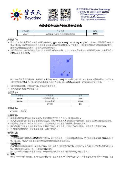
台盼蓝染色细胞存活率检测试剂盒产品编号 产品名称包装 C0011台盼蓝染色细胞存活率检测试剂盒100次产品简介:碧云天生产的台盼蓝染色细胞存活率检测试剂盒(Trypan Blue Staining Cell Viability Assay Kit ),是利用正常的健康细胞能够排斥台盼蓝,而丧失细胞膜完整性的细胞可以被台盼蓝染色研制而成。
严格来说,台盼蓝染色检测的是细胞膜的完整性,通常认为细胞膜丧失完整性,即可认为细胞已经死亡。
台盼蓝染色后,通过显微镜下直接计数或显微镜下拍照后计数,就可以对细胞存活率进行比较精确的定量。
台盼蓝染色后的HeLa 细胞请参考图1。
图1. 细胞台盼蓝染色效果图。
胰酶消化下来的HeLa 细胞,1000g 离心1分钟,弃上清,用适量细胞重悬液重悬后,用等体积台盼蓝染色液(2X)染色。
箭头所示为台盼蓝染色的死亡细胞。
注:本HeLa 细胞经过一定的细胞坏死诱导处理。
台盼蓝染色只需3-5分钟即可完成,并且操作非常简单。
本试剂盒足够检测100个细胞样品。
包装清单:产品编号 产品名称 包装 C0011-1 台盼蓝染色液(2X) 10ml C0011-2细胞重悬液 100ml —说明书1份保存条件:4ºC 保存,一年有效。
注意事项:本试剂盒提供的两种溶液都是无菌的,使用时最好在超净台内进行,避免细菌污染。
本产品长时间存放可能会出现少量颗粒状沉淀,可在37ºC 水浴约10分钟以充分溶解沉淀。
沉淀完全溶解后即可正常使用。
台盼蓝对人体有毒,操作时请特别小心,并注意有效防护以避免直接接触人体或吸入体内。
本产品仅限于专业人员的科学研究用,不得用于临床诊断或治疗,不得用于食品或药品,不得存放于普通住宅内。
为了您的安全和健康,请穿实验服并戴一次性手套操作。
使用说明:1. 收集细胞:对于贴壁细胞先用胰酶和/或EDTA 消化下细胞。
对于悬浮细胞,则可以直接收集细胞。
把收集的细胞在1000-2000g 离心1分钟,弃上清,用1毫升或根据细胞的量用适当细胞重悬液重新悬起细胞。
- 1、下载文档前请自行甄别文档内容的完整性,平台不提供额外的编辑、内容补充、找答案等附加服务。
- 2、"仅部分预览"的文档,不可在线预览部分如存在完整性等问题,可反馈申请退款(可完整预览的文档不适用该条件!)。
- 3、如文档侵犯您的权益,请联系客服反馈,我们会尽快为您处理(人工客服工作时间:9:00-18:30)。
DOI: 10.1126/science.1240925, 789 (2013);341 Science et al.Susan Carpenter Immune Response Genes A Long Noncoding RNA Mediates Both Activation and Repression ofThis copy is for your personal, non-commercial use only.clicking here.colleagues, clients, or customers by , you can order high-quality copies for your If you wish to distribute this article to othershere.following the guidelines can be obtained by Permission to republish or repurpose articles or portions of articles): November 5, 2013 (this information is current as of The following resources related to this article are available online at/content/341/6147/789.full.html version of this article at:including high-resolution figures, can be found in the online Updated information and services, /content/suppl/2013/07/31/science.1240925.DC1.htmlcan be found at:Supporting Online Material /content/341/6147/789.full.html#related found at:can be related to this article A list of selected additional articles on the Science Web sites /content/341/6147/789.full.html#ref-list-1, 12 of which can be accessed free:cites 35 articles This article/cgi/collection/immunology Immunologysubject collections:This article appears in the following registered trademark of AAAS.is a Science 2013 by the American Association for the Advancement of Science; all rights reserved. The title Copyright American Association for the Advancement of Science, 1200 New York Avenue NW, Washington, DC 20005. (print ISSN 0036-8075; online ISSN 1095-9203) is published weekly, except the last week in December, by the Science o n N o v e m b e r 5, 2013w w w .s c i e n c e m a g .o r g D o w n l o a d e d f r o mA Long Noncoding RNA Mediates Both Activation and Repressionof Immune Response GenesSusan Carpenter,1,2Daniel Aiello,1Maninjay K.Atianand,1Emiliano P.Ricci,3Pallavi Gandhi,1 Lisa L.Hall,4Meg Byron,4Brian Monks,1Meabh Henry-Bezy,1Jeanne wrence,4Luke A.J.O’Neill,2Melissa J.Moore,3Daniel R.Caffrey,1*†Katherine A.Fitzgerald1*†An inducible program of inflammatory gene expression is central to antimicrobial defenses.This response is controlled by a collaboration involving signal-dependent activation of transcription factors, transcriptional co-regulators,and chromatin-modifying factors.We have identified a long noncoding RNA(lncRNA)that acts as a key regulator of this inflammatory response.Pattern recognition receptors such as the Toll-like receptors induce the expression of numerous lncRNAs.One of these,lincRNA-Cox2, mediates both the activation and repression of distinct classes of immune genes.Transcriptional repression of target genes is dependent on interactions of lincRNA-Cox2with heterogeneous nuclear ribonucleoprotein A/B and A2/B1.Collectively,these studies unveil a central role of lincRNA-Cox2asa broad-acting regulatory component of the circuit that controls the inflammatory response.T he innate immune system coordinates host defenses through germ line–encoded pat-tern recognition receptors[e.g.,Toll-like receptors(TLRs)],which recognize microbial products and induce the expression of hundreds of proteins involved in antimicrobial defense and adaptive immunity(1–3).Recent studies have identified thousands of long noncoding RNAs(lncRNAs)in mammalian genomes(4–9) that regulate gene expression in different biolog-ical processes[reviewed in(5)].lncRNAs are dif-ferentially regulated in virus-infected cells(10)and in dendritic cells after lipopolysaccharide(LPS) stimulation(4).Recently,the lncRNA NeST was shown to control susceptibility to Theiler’s virus and Salmonella infection in mice through epige-netic regulation of the interferon-g(IFN-g)locus (11,12).Although lncRNAs can be induced in innate immune cells(4,10),whether lncRNAs act as regulators of gene expression in innate immu-nity is unknown.To identify lncRNAs transcribed during the innate immune response,we conducted whole-transcriptome analysis(RNA-seq)(6)of mouse bone marrow–derived macrophages(BMDMs) stimulated with the synthetic bacterial lipopeptide Pam3CSK4,a Tlr2ligand(Fig.1A).Pam3CSK4in-duced the transcription of numerous protein-coding genes involved in the immune response(Fig.1A, inner track),as well as62lncRNAs(Fig.1A, outer track,and table S1).The most significantly induced lncRNAs tended to occur in chromosomal regions where we also detected higher expres-sion of immune genes,which suggests that thesegenes are co-regulated.lincRNA-Cox2was amongthe most highly induced lncRNAs and is prox-imal to the prostaglandin-endoperoxide synthase2[Ptgs2(Cox2)]gene(Fig.1A);lncRNA-Ehd1andlncRNA-Lyn were also induced after Tlr2andTlr4stimulation(fig.S1,A and B).A previous study demonstrated that lincRNA-Cox2was induced in dendritic cells after stimulationwith LPS(4).However,whether lincRNA-Cox2regulates the inflammatory response that is asso-ciated with TLR signaling is unknown.We iden-tified three splice variants of lincRNA-Cox2(Fig.1B;GenBank accession numbers JX682706,JX682707,and JX682708).V ariant1was the mostabundant transcript containing exons1and4,common to all splice variants.Consequently,wedesigned primers for quantitative PCR(qPCR)and short hairpin RNA(shRNA)that targetedthese ing qPCR,we confirmed thatLPS induced similar temporal patterns of expres-sion of both lincRNA-Cox2and its neighboringPtgs2(Cox2)gene in bone marrow–derived den-dritic cells(BMDCs,Fig.1,C and D).R848,asynthetic antiviral compound that activates Tlr7/8,induced lincRNA-Cox2and Ptgs2(Cox2)inBMDCs,whereas polyinosinic-polycytidylic acid,a synthetic double-stranded RNA that activatesTlr3,had no effect(fig.S2,A to D).Both Listeriamonocytogenes–infected BMDMs and splenocytesfrom Listeria-infected mice also had elevated levels1Division of Infectious Diseases and Immunology,Depart-ment of Medicine,University of Massachusetts Medical School, Worcester,MA01605,USA.2School of Biochemistry and Im-munology,Trinity College Dublin,Dublin2,Ireland.3Howard Hughes Medical Institute,University of Massachusetts Medical School,Worcester,MA01605,USA.4Department of Cell and Developmental Biology,University of Massachusetts Medical School,Worcester,MA01605,USA.*These authors contributed equally to this work.†Corresponding author.E-mail:kate.fitzgerald@umassmed. edu(K.A.F.);daniel.caffrey@(D.R.C.)Fig.1.lincRNA-Cox2expression is induced by Tlr ligands in a MyD88-and NF-k B–dependent manner.(A)The Circos plot shows genome-wide differential expression(RNA-seq)between untreated and Pam3CSK4(Tlr1/2)–stimulated(5hours)BMDMs.The inner track shows log2relative change for protein-coding genes that are classified into immune genes(red;see supplementary materials)and other genes (blue).The outer track shows log2relative change for all lncRNAs.lincRNA-Cox2is highlighted in red on chromosome1(arrow).(B)lincRNA-Cox2encodes three splice variants.(C and D)RNA was extracted from primary BMDCs stimulated with LPS for0,2,5,or24hours.Expression levels of lincRNA-Cox2(C)and Ptgs2 (Cox2)(D)were examined by qRT-PCR and expressed relative to time0hours(which was set to1,indicated by arrow).(E and F)Induction of lincRNA-Cox2and Ptgs2(Cox2)after qRT-PCR on BMDMs obtained from wild-type(WT)or Myd88−/−mice.(G and H)BMDMs were treated for30min with a NF-k B inhibitor(1m g/ml), followed by stimulation with LPS(100ng/ml);expression levels of lincRNA-Cox2(G)and Ptgs2(Cox2)(H)were examined by qRT-PCR.Data represent means T SD from three independent experiments. SCIENCE VOL34116AUGUST2013789REPORTSof lincRNA-Cox2(fig.S3,A and B).Induction of lincRNA-Cox2and its neighboring gene Ptgs2(Cox2)was dependent on the Tlr adaptor protein MyD88(Fig.1,E and F)and transcription factor NF-k B signaling (Fig.1,G and H).We next examined the protein-coding capac-ity of lincRNA-Cox2by assessing its associa-tion with polysomes within cells.BMDMs were treated with cycloheximide to trap ribosomes on RNA molecules and either left untreated or pre-treated with EDTA (disrupting all RNA-protein interactions)or with harringtonine (specifically disrupting translation).Cell lysates were fraction-ated and RNA analyzed by qPCR (13–15).We compared Gapdh mRNA with lincRNA-Cox2and another lincRNA,lncRNA-Eps,which had previously been shown to be noncoding (16).As expected,Gapdh mRNA sedimented with a high velocity through the sucrose gradient because it was associated with polysomes.In contrast,lincRNA-Cox2and lncRNA-Eps remained in lighter fractions (fig.S4).Treatment with EDTA or harringtonine resulted in a shift of Gapdh mRNA,but not lincRNA-Cox2or lncRNA-Eps,from the higher-velocity to the lower-velocity fractions.Furthermore,most of the open reading frames identified in lincRNA-Cox2were found to have poor Kozak strength (fig.S5).Collective-ly,these studies indicate that lincRNA-Cox2is unlikely to encode a protein product.We next generated BMDM cell lines in which lincRNA-Cox2expression was silenced by shRNA (Fig.2A).Silencing of lincRNA-Cox2did not alter expression of Ptgs2(Cox2)(fig.S6).T o identify potential targets of lincRNA-Cox2,we conducted RNA-seq in both control and lincRNA-Cox2–silenced cells before and after stimulation with Pam 3CSK 4.Silencing of lincRNA-Cox2increased the expression of 787genes by a factor of 3or greater in unstimulated cells (table S2).A Gene Ontology (GO)enrichment analysis indicated that genes related to the immune response were sig-nificantly overrepresented in these up-regulated genes (Fig.2B and table S3).This gene set in-cluded chemokines (Ccl5,Cx3cl1),chemokine receptors (Ccrl ),and IFN-stimulated genes (ISGs)(Irf7,Oas1a ,Oas1l ,Oas2,Ifi204,and Isg15)(Fig.2B).In the same cells stimulated with Pam 3CSK 4,silencing of lincRNA-Cox2resulted in attenuated expression of 713genes by a fac-tor of 3or greater (table S4).Examples of these genes include Tlr1,Il6(interleukin-6),and Il23a .We confirmed these findings by means of an RNA profiling technology to simultaneously an-alyze mRNA expression levels of differentially regulated genes (nCounter;Nanostring,Seattle,W A).In unstimulated cells,silencing of lincRNA-Cox2led to a marked increase in expression of Irf7,Ccl5(Rantes ),and ISGs relative to control cells (Fig.2C,columns 1and 2,and fig.S7),whereas the Pam 3CSK 4-induced expression of Tlr1and Il6was attenuated (Fig.2C,columns 3and 4).We confirmed these findings in three independent shRNA lines by measuring protein levels for Ccl5(Rantes)and Il6(Fig.2,DandFig.2.lincRNA-Cox2is a major regulator of immune genes.(A )qRT-PCR was carried out on BMDMs stably expressing lentiviral shRNA specific to lincRNA-Cox2(shRNA)or a control shRNA.Expression of lincRNA-Cox2was measured.(B )RNA-seq analysis on lincRNA-Cox2knockdown or control shRNA BMDMs that were either stimulated with Pam 3CSK 4or unstimulated.The heat map shows mRNA levels for genes annotated in GO as immune genes.These genes are among the top 50up-regulated immune genes in unstimulated cells when lincRNA-Cox2was silenced or the top 50down-regulated immune genes in stimulated cells when lincRNA-Cox2was silenced.The 100genes were ranked according to absolute values of log 2relative change;the top 80differentially expressed genes are displayed.(C )Heat map representation of differentially regulated genes performed on RNA extracted from control or lincRNA-Cox2knockdown cells stimulated with Pam 3CSK 4for 5hours.(D and E )Cells were stimulated with Pam 3CSK 4for 24hours,and Ccl5(D)and Il6(E)levels were measured in lincRNA-Cox2knockdown cells by enzyme-linked immunosorbent assay (ELISA)(n.d.,not detected).(F to I )lincRNA-Cox2was silenced in IFN a /b receptor (IFN a /b R)KO cells.Expression levels of lincRNA-Cox2(F),Ccl5(G),Irf7(H),and Ifi204(I)were measured by qRT-PCR.Data represent means T SD from three independent experiments.16AUGUST 2013VOL 341SCIENCE790REPORTSE,and fig.S8).In contrast to these genes,the Tlr2-dependent induction of Il1b was unaffected in cells deficient in lincRNA-Cox2(fig.S9).We also observed reduced Il6in macrophages stim-ulated with R848and Pam 2CSK 4(a ligand that activates a Tlr2-Tlr6heterodimer)(fig.S10).We next silenced lincRNA-Cox2in macrophages lack-ing the type I IFN a /b receptor (IFN a /b R KO)in order to distinguish between IFN-dependent and IFN-independent targets of lincRNA-Cox2(Fig.2,F to I,and fig.S11,A and B).Ccl5was regulated by lincRNA-Cox2independently of type I IFNsignaling,whereas regulation of Irf7and Ifi204appeared to be secondary to type I IFN signaling.Despite the elevated expression of IFN pathway components,silencing of lincRNA-Cox2did not render cells permissive to Tlr2-induced TBK1activation,a measure of Irf3signaling (fig.S12).Finally,because shRNA silencing of lincRNA-Cox2led to decreased expression of Tlr1,we wanted to eliminate the possibilitythat we inadvertently impaired Pam 3CSK 4signaling via the Tlr1-Tlr2heterodimer.We restored expression of Tlr1in lincRNA-Cox2–silenced cells and confirmed that the differential regulation of Ccl5and Il6was not due to impaired expression of Tlr1(fig.S13).Taken together,these data indicate that lincRNA-Cox2regulates dis-tinct classes of immune genes both basally and after TLR stimulation.W e next conducted “gain-of-function ”studies by generating macrophages that ectopically expressed lincRNA-Cox2(Fig.3A).Macrophages that ec-topically expressed lincRNA-Cox2had decreased levels of Ccl5,Irf7,and other ISGs (Fig.3B).Il6was not detected when lincRNA-Cox2was over-expressed in unstimulated cells.However,Il6levels were significantly enhanced when lincRNA-Cox2was overexpressed in Pam 3CSK 4stimulated cells (Fig.3C).These results further demonstrate that lincRNA-Cox2represses Ccl5while simultaneous-ly enhancing the expression of TLR-induced Il6.lncRNAs can be found in the nucleus,cyto-plasm,or both compartments (17–19).We ex-amined the localization of lincRNA-Cox2in macrophages by performing quantitative reverseFig.3.Differential gene expression after over-expression of lincRNA-Cox2.(A )A BMDM cell line stably overexpress-ing lincRNA-Cox2or a con-trol vector was generated.qRT-PCR was carried out and overexpression of lincRNA-Cox2was con-firmed.(B )Heat map rep-resentation of differentially regulated genes from control or lincRNA-Cox2–overexpressing BMDMs.(C )qRT-PCR was carried out on control or lincRNA-Cox2–overexpressing cells stimulated with Pam 3CSK 4for 5hours.Data represent means T SD from three independentexperiments.Fig.4.lincRNA-Cox2is localized to both the cytosolic and nuclear compartments and inter-acts with hnRNP-A/B and A2/B1to regulate immune genes.(A )BMDMs were labeled with a lincRNA-Cox2probe via RNA FISH and counter-stained with 4´,6-diamidino-2-phenylindole (DNA to visualize the nucleus).(B and C )Biotinylated lincRNA-Cox2or antisense RNA was incubated with nuclear extracts,and interaction with endogenous hnRNP-A/B (B)or hnRNP-A2/B1(C)was assessed after immunoprecipitation and Western blot (upper panels).Expression levels of hnRNP-A/B (B)or hnRNP-A2/B1(C)(lower panels)in input lysates were also examined.(D )Cell lines with shRNA targeting hnRNP-A/B or hnRNP-A2/B1were stimulated with Pam 3CSK 4;Ccl5production was measured in these cells by ELISA.(E )Heat map representation of differentially regu-lated genes of control shRNA –,hnRNP-A/B shRNA –,or hnRNP-A2/B1shRNA –expressing BMDMs stimu-lated with Pam 3CSK 4(100nM)for 5hours.(F and G )Recruitment of RNA Pol II to the Ccl5promoter as determined by ChIP analysis in cells expressing shRNA targeting lincRNA-Cox2,hnRNP-A/B,or hnRNP-A2/B1.(H )hnRNP-A/B or hnRNP-A2/B1was knocked down using lentiviral shRNA in lincRNA-Cox2–overexpressing BMDMs;Ccl5expression levels were measured using qRT-PCR.Data are representative of three separateexperiments.linc-Cox2Ctllinc-Cox2 CtlRNA FISH: BMDMlincRNA-Cox2Dapi (Nucleus)34IPLysate34Count scaled by log2(X-min(X) + 1)h n R N P A /Bh n R N P A 2/B 1IPLysateR N A p o l I I r e c r u i t m e n t (% I n p u t )C c l 5 (R a n t e s ) n g /m lGFP sh linc-Cox2 hn A/B sh hn A2/B1 shR e l a t i v e E x p r e s s i o n (R N A )R N A p o l I I r e c r u i t m e n t (% I n p u t )ABCDE FGHA2/B1 sh A/B sh Ctl sh SCIENCE VOL 34116AUGUST 2013791REPORTStranscription PCR (qRT-PCR)on RNA isolated from nuclear or cytosolic fractions (fig.S14).Using RNA fluorescence in situ hybridization (FISH),substantial amounts of lincRNA-Cox2were vi-sible in both the nuclear and the cytosolic com-partments of macrophages (Fig.4A and fig.S15,A and B).Analysis of published RNA-seq data from nuclear and cytosolic fractions of macro-phages (20)confirmed that lincRNA-Cox2was present in both compartments,unlike Neat1,which was exclusively nuclear (fig.S16).RNA FISH analysis demonstrated efficient targeting of lincRNA-Cox2in the nuclear fractions of our shRNA-silenced cells (fig.S15,C to F).Many lncRNAs regulate transcription through their interactions with chromatin-modifying com-plexes or with heterogeneous nuclear ribonu-cleoproteins (hnRNPs)(5,21–23).To identify binding partners for lincRNA-Cox2,we incubated in vitro –transcribed biotinylated lincRNA-Cox2as well as an antisense lincRNA-Cox2control RNA with nuclear or cytosolic extracts of macro-phages and subjected RNA binding proteins to mass spectrometry for identification.hnRNP-A/B and hnRNP-A2/B1were identified as specific binding partners for lincRNA-Cox2in both the nuclear and cytosolic fractions (fig.S17).The ability of hnRNP-A/B and hnRNP-A2/B1to bind lincRNA-Cox2was confirmed by Western blot analysis (Fig.4,B and C).hnRNPs are multifunctional nuclear RNA binding proteins involved in various aspects of RNA biology (24,25).hnRNPs also act as me-diators of lncRNA-induced transcriptional re-pression (22,23).hnRNP-A/B has been linked to transcriptional repression of some genes (26,27),and hnRNP-A2/B1and hnRNP-A/B have been shown to associate (28).Therefore,we hypothe-sized that lincRNA-Cox2regulates the transcrip-tion of immune genes by associating with these hnRNPs.To test this directly,we generated macro-phages lacking expression of hnRNP-A/B or hnRNP-A2/B1by shRNA (fig.S18,A and B).Knockdown of hnRNP-A/B and hnRNP-A2/B1did not modulate the levels of lincRNA-Cox2itself (fig.S19)but did result in an enhance-ment of Ccl5protein levels in both unstimulated and Pam 3CSK 4-stimulated cells (Fig.4D).Mul-tiplex RNA analysis also revealed elevated levels of Ccl5,Stat1,Tlr7,Icam1,and I k B in hnRNP-silenced macrophages (Fig.4E).There was considerable overlap between genes that were regulated by lincRNA-Cox2and these two hnRNP proteins (fig.S20and table S5).We also ex-amined the occupancy of RNA polymerase II at the promoters of the Ccl5and Irf7genes using chromatin immunoprecipitation (ChIP)and found increased levels of RNA polymerase II (Pol II)at the Ccl5and Irf7promoters when lincRNA-Cox2or either of the hnRNPs were silenced in un-stimulated cells (Fig.4,F and G,and fig.S21).The occupancy of RNA Pol II on the promoter of Il6was reduced when lincRNA-Cox2but not hnRNP-A/B or A2/B1was knocked down in Pam 3CSK 4-stimulated cells (fig.S22);this effectwas consistent with hnRNPs having no effect on Pam 3CSK 4-induced Il6mRNA levels (Fig.4E).Silencing of hnRNP-A2/B1or hnRNP-A/B in cells that overexpressed lincRNA-Cox2also reversed the inhibitory effect of lincRNA-Cox2on Ccl5expression (Fig.4H and fig.S23).Taken together,these experiments confirm that hnRNP-A/B and hnRNP-A2/B1form a complex with lincRNA-Cox2to repress the transcription of immune genes.The identification of hnRNP-A/B and hnRNP-A2/B1further underscores the impor-tance of hnRNPs in lncRNA function (21–23).Innate immune responses have the capacity to both combat infectious microbes and drive pathological inflammation,which contributes to diseases such as atherosclerosis,autoimmunity,and cancer.A multitude of regulatory checkpoints control TLR signaling and inflammatory responses.W e propose a model whereby TLR signaling in-duces lncRNAs,such as lincRNA-Cox2,that serve as repressors and activators of genes through their physical interactions with various regulatory com-plexes.As such,lncRNAs represent a component of the innate immune response that can both re-strain and promote aspects of inflammatory sig-naling.Further characterization of these regulatory networks is likely to reveal novel drug targets and opportunities for therapeutic intervention in infectious and inflammatory diseases.References and Notes1.T.Kawai,S.Akira,Ann.N.Y.Acad.Sci.1143,1–20(2008).2.R.Medzhitov,T.Horng,Nat.Rev.Immunol.9,692–703(2009).3.P.J.Murray,S.T.Smale,Nat.Immunol.13,916–924(2012).4.M.Guttman et al .,Nature 458,223–227(2009).5.J.L.Rinn,H.Y.Chang,Annu.Rev.Biochem.81,145–166(2012).6.A.Mortazavi,B.A.Williams,K.McCue,L.Schaeffer,B.Wold,Nat.Methods 5,621–628(2008).7.M.Guttman et al .,Nat.Biotechnol.28,503–510(2010).8.M.Guttman,J.L.Rinn,Nature 482,339–346(2012).9.E.Birney et al.,Nature 447,799–816(2007).10.X.Peng et al .,mBio 1,e00206(2010).11.S.P.Collier,P.L.Collins,C.L.Williams,M.R.Boothby,T.M.Aune,J.Immunol.189,2084–2088(2012).12.J.A.Gomez et al .,Cell 152,743–754(2013).13.N.T.Ingolia,G.A.Brar,S.Rouskin,A.M.McGeachy,J.S.Weissman,Nat.Protoc.7,1534–1550(2012).14.N.T.Ingolia,S.Ghaemmaghami,J.R.Newman,J.S.Weissman,Science 324,218–223(2009).15.N.T.Ingolia,reau,J.S.Weissman,Cell 147,789–802(2011).16.W.Hu,B.Yuan,J.Flygare,H.F.Lodish,Genes Dev.25,2573–2578(2011).17.J.Cheng et al .,Science 308,1149–1154(2005).18.Q.Wu et al .,PLoS ONE 3,e2803(2008).19.R.Louro,A.S.Smirnova,S.Verjovski-Almeida,Genomics93,291–298(2009).20.D.M.Bhatt et al .,Cell 150,279–290(2012).21.A.M.Khalil et al .,Proc.Natl.Acad.Sci.U.S.A.106,11667–11672(2009).22.M.Huarte et al .,Cell 142,409–419(2010).23.Y.Hasegawa et al .,Dev.Cell 19,469–476(2010).24.G.Dreyfuss,M.Hentze,mond,Cell 85,963–972(1996).25.A.M.Krecic,M.S.Swanson,Curr.Opin.Cell Biol.11,363–371(1999).26.C.Gao et al .,J.Biol.Chem.279,11236–11243(2004).27.C.Gao,H.Guo,Z.Mi,P.Y.Wai,P.C.Kuo,J.Immunol.175,523–530(2005).28.C.S.Raju et al .,Mol.Biol.Cell 19,3008–3019(2008).Acknowledgments:We thank N.Singh for advice on RNA-seq,A.J.Crawford for help with quantification of RNA FISH,and K.Jeffrey,L.Stuart,N.Silverman,D.Golenbock,and E.Lien for critical reading of this manuscript.The data presented in this paper are tabulated in the main paper and in the supplementary materials.All RNA-seq data are available for download from NCBI Gene Expression Omnibus (/geo/)under accession number GSE40978.Supported by Ireland Health Research Board/Marie Curie Fellowship MCPD/2010/04(S.C.)and by NIH grants AI067497(K.A.F.)and GM053234(J.B.L.).Supplementary Materials/cgi/content/full/science.1240925/DC1Materials and Methods Figs.S1to S23Tables S1to S5References (29–35)23May 2013;accepted 24July 2013Published online 1August 2013;10.1126/science.1240925Cleavage of Fibrinogen by Proteinases Elicits Allergic Responses Through Toll-Like Receptor 4Valentine Ongeri Millien,1Wen Lu,2Joanne Shaw,5Xiaoyi Yuan,2Garbo Mak,3Luz Roberts,3Li-Zhen Song,3J.Morgan Knight,2Chad J.Creighton,4Amber Luong,5Farrah Kheradmand,1,2,3,6,7*David B.Corry 1,2,3,6,7*Proteinases and the innate immune receptor Toll-like receptor 4(TLR4)are essential for expression of allergic inflammation and diseases such as asthma.A mechanism that links these inflammatory mediators is essential for explaining the fundamental basis of allergic disease but has been elusive.Here,we demonstrate that TLR4is activated by airway proteinase activity to initiate both allergic airway disease and antifungal immunity.These outcomes were induced by proteinase cleavage of the clotting protein fibrinogen,yielding fibrinogen cleavage products that acted as TLR4ligands on airway epithelial cells and macrophages.Thus,allergic airway inflammation represents an antifungal defensive strategy that is driven by fibrinogen cleavage and TLR4activation.These findings clarify the molecular basis of allergic disease and suggest new therapeutic strategies.Allergic asthma is a chronic inflammatory airway disease that is characterized by both airway obstruction and enhanced systemic and airway allergic inflammation marked by interleukin-4(IL-4)–secreting T helper 2(T H 2)cells,eosinophils,and serum immunoglobulin16AUGUST 2013VOL 341SCIENCE792REPORTS。