MRI 如何工作 工作原理 中英双语
核磁共振工作原理
核磁共振工作原理核磁共振(Nuclear Magnetic Resonance,简称NMR)是一种重要的分析技术和研究手段。
它基于原子核之间的电磁相互作用,通过利用核自旋在外加磁场和射频场作用下的共振吸收现象,实现对样品的结构和性质的分析。
本文将详细介绍核磁共振的工作原理。
一、基本原理核磁共振技术的核心是核磁共振现象。
当一个样品置于磁场中时,其核自旋将受到外加磁场的影响,导致核自旋的量子态能级发生分裂。
此时,如果给样品施加一个与能级间距相符的射频波,将出现共振吸收现象。
这种共振现象的产生是由于外加磁场与样品中核自旋的磁矩相互作用所致。
二、共振条件核磁共振的共振条件可以用以下公式表示:ω = γB0其中,ω表示射频波的角频率,γ是核磁矩的旋磁比,B0是外加磁场的大小。
根据这个公式可知,当外加磁场的强度发生变化时,共振条件也会相应改变。
三、工作步骤核磁共振的工作原理可以分为以下几个步骤:1. 加样处理:样品通常会进行处理,以提高信噪比和磁场均匀性。
处理包括稀释、混合、旋转等。
2. 形成磁场:通过磁铁产生一个强大的静态磁场,这是核磁共振实验的基础条件。
3. 校准射频场:根据样品的特性和实验需求,校准出一个合适的射频场。
4. 施加射频激发信号:给样品施加一个与共振频率匹配的射频激发信号,使样品中的核自旋从基态跃迁到激发态。
5. 探测共振信号:探测样品中的共振信号,记录共振频率和共振幅度。
6. 数据处理和分析:对探测到的共振信号进行处理和分析,提取样品的结构和性质信息。
四、应用领域核磁共振技术在许多领域都有广泛的应用,包括化学、物理、生物、医学等。
在化学领域,核磁共振可以用于分析有机物的结构、鉴定化合物的纯度等。
在物理领域,核磁共振可以用于研究材料的磁性、超导性等性质。
在生物领域,核磁共振可以应用于蛋白质结构研究、DNA解旋等。
在医学领域,核磁共振成像(MRI)可以用于人体各类组织和器官的诊断。
综上所述,核磁共振工作原理是基于核自旋共振现象的。
中英文核磁说明书
中英文核磁说明书摘要:1.核磁共振成像的基本原理2.中英文核磁说明书的作用和重要性3.核磁共振成像设备的操作步骤4.核磁共振成像的临床应用5.使用核磁共振成像的注意事项正文:核磁共振成像(Magnetic Resonance Imaging,简称MRI)是一种利用核磁共振原理对人体进行无创性成像的医学技术。
MRI 成像对比度高、分辨率好、无辐射损害,因此在医学领域得到广泛应用。
而中英文核磁说明书则是指导医务人员正确操作和使用核磁共振成像设备的重要参考资料。
核磁共振成像的基本原理是利用人体内的氢原子核在磁场和射频脉冲的作用下产生共振信号,然后通过计算机对这些信号进行处理和分析,最终形成清晰的图像。
这一过程中,中英文核磁说明书的作用和重要性不言而喻。
它们可以帮助医务人员了解设备的操作步骤、注意事项以及可能出现的问题和解决方法。
核磁共振成像设备的操作步骤主要包括以下几个方面:首先,对患者进行必要的准备工作,如去除金属物品、嘱咐患者屏住呼吸等;其次,根据检查部位和目的选择合适的成像序列和参数;然后,进行成像操作,并在过程中注意观察患者的反应;最后,对成像结果进行分析和诊断。
核磁共振成像在临床应用中具有很高的价值。
它不仅可以用于检查颅脑、脊柱、关节等部位,还可以用于诊断肿瘤、炎症、血管病变等疾病。
同时,核磁共振成像也可以为临床研究提供有力的支持。
在使用核磁共振成像的过程中,医务人员需要注意以下几点:首先,确保设备的正常运行和维护;其次,遵循安全操作规程,避免对患者和医务人员造成伤害;最后,合理利用成像资源,提高检查效率和诊断准确性。
总之,中英文核磁说明书是核磁共振成像设备操作和使用的重要参考资料。
核磁共振的工作原理

核磁共振的工作原理核磁共振(Nuclear Magnetic Resonance,简称NMR)是一种重要的物理技术,广泛应用于医学、化学、物理学等领域。
本文将介绍核磁共振的工作原理,以及其在科学研究和医学诊断中的应用。
一、核磁共振的基本原理核磁共振是基于原子核存在的自旋(即核自旋)的物理性质进行研究的方法。
原子核带有正电荷,因此具备自旋磁矩。
当原子核置于外加磁场中时,这些核自旋磁矩会在磁场的作用下取向,形成所谓的“磁场取向”。
不同原子核的自旋磁矩具有不同的取向状态。
二、核磁共振的工作原理核磁共振技术利用核自旋磁矩在外加磁场中的取向状态和跃迁行为来获取有关样品的信息。
当样品处于强磁场中时,核自旋磁矩会分布在能量的不同级别上。
这些不同能级之间的能量差和跃迁频率与样品的物理和化学性质相关。
在核磁共振谱仪中,首先将样品置于一个强磁场中,使核自旋磁矩取向为平行或反平行于磁场方向。
随后,通过施加一定的电磁波场,使部分核自旋磁矩发生能级的跃迁,并将吸收或发射的能量转化为电信号。
利用这些电信号,我们可以获得核磁共振信号并进行分析。
三、核磁共振的应用核磁共振技术在医学诊断中有着广泛应用。
例如,核磁共振成像(Magnetic Resonance Imaging,MRI)技术可以非侵入性地观察人体内部结构。
通过对核磁共振信号的探测和处理,可以获得高分辨率的人体组织图像,用于疾病的诊断和治疗。
此外,核磁共振技术在化学和物理学等科学研究领域也有重要的应用。
通过核磁共振谱仪对物质进行分析,可以得到关于样品的化学成分、结构以及动力学信息。
这为化学反应的研究和新材料的合成提供了重要的辅助手段。
总之,核磁共振作为一种重要的物理技术,利用原子核的自旋磁矩进行探测和分析,广泛应用于医学诊断、化学分析以及物理学等领域。
它的工作原理基于核自旋磁矩在外加磁场中的取向状态和跃迁行为,通过核磁共振信号的探测和处理,可以获取样品的相关信息。
核磁共振技术的发展为科学研究和诊断治疗提供了重要的手段和突破口。
磁共振工作原理

磁共振工作原理Working Principle of Magnetic Resonance Imaging。
Magnetic Resonance Imaging (MRI) is a non-invasive diagnostic technique that uses a strong magnetic field, radio waves, and computer technology to create detailed images of the body's internal structures. MRI isparticularly useful for imaging soft tissues, such as the brain, spinal cord, and joints, and it is often used to diagnose a wide range of medical conditions.The working principle of MRI is based on the behavior of atomic nuclei in a strong magnetic field. When a patient is placed in the MRI machine, the magnetic field causes the protons in the hydrogen atoms in the body's tissues toalign with the field. A radio frequency pulse is then applied to the body, causing the protons to absorb energy and enter a higher energy state.As the protons return to their original state, theyrelease energy in the form of radio waves. These signalsare detected by the MRI machine and used to create imagesof the body's internal structures. The strength of the magnetic field and the timing of the radio frequency pulses are carefully controlled to produce high-quality imageswith excellent resolution.One of the key advantages of MRI is its ability to differentiate between different types of tissues based on their unique magnetic properties. For example, the MRI machine can distinguish between healthy and diseased tissue, or between different types of soft tissue, such as muscle and fat. This is because different tissues have different concentrations of hydrogen atoms, which respond differently to the magnetic field.Another advantage of MRI is its ability to produce images in any plane, including axial, sagittal, and coronal. This allows doctors to view the body's internal structures from multiple angles and to identify any abnormalities or irregularities.There are several different types of MRI machines, including open MRI, closed MRI, and high-field MRI. Open MRI machines are designed to be more comfortable for patients who may be claustrophobic, as they do not require the patient to be fully enclosed in a tube. Closed MRI machines are more powerful and can produce higher-quality images, but they may be less comfortable for some patients. High-field MRI machines are the most powerful and produce the highest-quality images, but they are also the most expensive and may not be available in all medical facilities.In conclusion, the working principle of MRI is based on the behavior of atomic nuclei in a strong magnetic field. By carefully controlling the magnetic field and radio frequency pulses, MRI machines can produce detailed images of the body's internal structures, allowing doctors to diagnose a wide range of medical conditions. MRI is a safe, non-invasive, and highly effective diagnostic tool that has revolutionized the field of medical imaging.。
MRI的工作原理

MRI的工作原理
MRI(磁共振成像)是一种医学影像技术,通过使用强磁场和无害的无线电波来生成人体内部的详细图像。
MRI的工作原理涉及如下几个步骤:
1. 建立强磁场:MRI使用大而强大的磁体产生一个非常强的恒定磁场,通常在1.5到3特斯拉之间。
这个磁场可以使人体内的水分子和其它氢原子与磁场自身对齐。
2. 激发共振:医生或技术人员在扫描开始之前,需要让患者躺在一个装有线圈的平台上。
这些线圈用于产生辅助的磁场来激发患者体内的氢原子。
技术人员会发送特定的无线电波信号,以匹配氢原子的共振频率,从而抵消磁场自身所造成的原子自旋。
3. 接收信号:当无线电波信号结束后,患者体内的氢原子会重新调整自己的自旋。
在这个过程中,它们会发射出一种微弱的无线电信号。
线圈接收这些信号,并将其转化为电信号。
4. 图像重构:通过使用计算机程序,电信号被转换成高质量的图像。
计算机根据信号的强度和时间来重建图像,并将其呈现给医生进行诊断。
MRI的工作原理是基于物质中的原子和分子如何与强磁场进行相互作用。
水分子和其他含氢分子在磁场中对齐的方式可提供详细的图像信息,这些图像可以用于检测和评估体内的异常
情况。
由于MRI不依赖放射性物质,并且能够提供高分辨率的图像,因此在医学诊断中得到广泛应用。
核磁共振工作原理和成像过程

核磁共振工作原理和成像过程核磁共振(Nuclear Magnetic Resonance,NMR)是一种重要的科学技术,广泛应用于物理、化学、生物等领域。
它通过利用原子核的自旋和磁性来研究物质的结构和性质。
核磁共振成像(Magnetic Resonance Imaging,MRI)则是基于核磁共振原理的医学影像技术,广泛应用于临床诊断和疾病监测。
核磁共振的工作原理是基于原子核的自旋和磁性。
原子核具有自旋,类似于地球的自转。
当物质处于外加磁场中时,原子核会发生预cession运动,即类似于陀螺仪的旋转运动。
在外加磁场的作用下,原子核处于两种能量状态之间的转换,称为共振。
核磁共振成像的过程主要分为三个步骤:激发、回波和重建。
首先,通过给被测物体施加一个强磁场,使其内部的原子核自旋排列。
然后,通过施加一个射频脉冲,改变原子核的自旋状态。
在脉冲结束后,原子核会发生回波信号,这个信号被接收和处理。
最后,通过对回波信号的处理和重建,可以得到一个图像,显示出被测物体的内部结构和组织特征。
核磁共振成像的优势在于它不需要使用任何放射性物质,对人体没有刺激和伤害,可以提供高分辨率的图像。
它可以清晰地显示人体各个组织的解剖结构,对于诊断疾病、观察病变部位和评估治疗效果非常有帮助。
例如,在神经科学领域,MRI可以帮助医生观察大脑的结构和功能,对疾病如脑卒中、脑肿瘤等进行诊断和治疗。
在心脏病学领域,MRI可以提供详细的心脏图像,帮助医生评估心脏功能和检测异常。
然而,核磁共振成像也存在一些限制。
首先,MRI设备昂贵且体积庞大,不适用于某些场合。
其次,MRI对于金属植入物、心脏起搏器等具有磁性的物体有一定的限制,可能会干扰成像质量。
此外,MRI扫描时间较长,需要患者保持静止,对于一些无法耐受长时间扫描的患者可能会有困难。
总的来说,核磁共振成像是一种非常重要的医学影像技术,通过利用核磁共振原理,可以提供高分辨率的图像,对于诊断和治疗疾病非常有帮助。
磁共振成像技术中英文名词对照
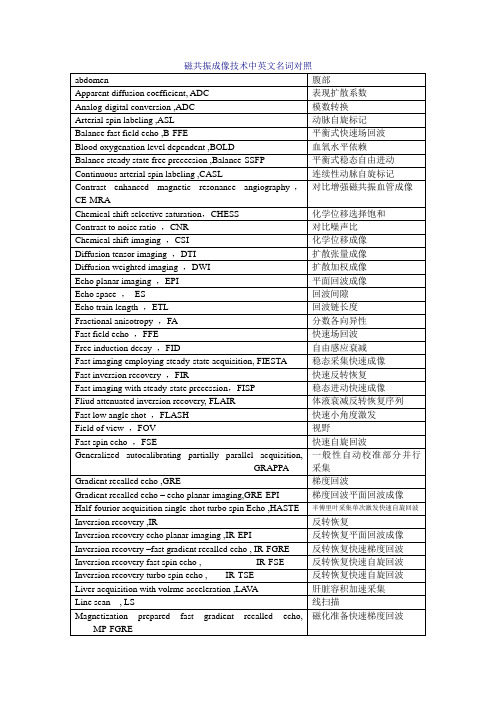
Emergency run-down unit ,ERDU
磁体急停单元
Hybrid magnet
混合型磁体
shiming
匀场
Passive shimming
被动匀场
Active shimming
主动匀场
Shimming coils
匀场线圈
Gauss meter
高斯计
Hall effect
快速小角度激发
Field of view,FOV
视野
Fast spin echo,FSE
快速自旋回波
Generalized autocalibrating partially parallel acquisition,GRAPPA
一般性自动校准部分并行采集
Gradient recalled echo ,GRE
反转恢复快速自旋回波
Inversion recovery turbo spin echo , IR-TSE
反转恢复快速自旋回波
Liver acquisition with volrme acceleration ,LAVA
肝脏容积加速采集
Line scan , LS
线扫描
Magnetization prepared fast gradient recalled echo,MP-FGRE
磁共振成像技术中英文名词对照
abdomen
腹部
Apparent diffusion coefficient, ADC
表现扩散系数
Analog-digital conversion ,ADC
模数转换
Arterial spin labeling ,ASL
mri工作原理
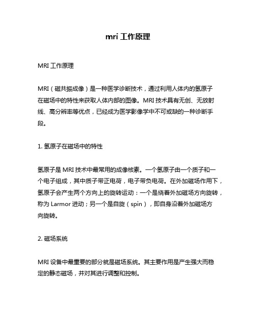
mri工作原理MRI工作原理MRI(磁共振成像)是一种医学诊断技术,通过利用人体内的氢原子在磁场中的特性来获取人体内部的图像。
MRI技术具有无创、无放射线、高分辨率等优点,已经成为医学影像学中不可或缺的一种诊断手段。
1. 氢原子在磁场中的特性氢原子是MRI技术中最常用的成像核素。
一个氢原子由一个质子和一个电子组成,其中质子带正电荷,电子带负电荷。
在外加磁场作用下,氢原子会产生两个方向上的旋转运动:一个是绕着外加磁场方向旋转,称为Larmor进动;另一个是自旋(spin),即自身沿着外加磁场方向旋转。
2. 磁场系统MRI设备中最重要的部分就是磁场系统。
其主要作用是产生强大而稳定的静态磁场,并对其进行调整和控制。
2.1 静态磁场静态磁场是MRI设备中最基本也是最重要的部分。
它由超导线圈或永久磁铁构成,能够产生强大的磁场。
静态磁场的稳定性和均匀性对成像质量有很大影响。
2.2 梯度线圈梯度线圈是MRI设备中的另一个重要组成部分。
它们能够产生额外的磁场,这些磁场在空间上是不同方向上的。
通过改变这些梯度磁场,可以使得不同位置的氢原子发生不同程度的Larmor进动,从而实现空间编码。
3. 频率编码与相位编码MRI成像过程中,需要对氢原子进行频率编码和相位编码。
频率编码是通过改变外加磁场强度来改变氢原子Larmor进动频率,从而实现位置信息的获取;相位编码则是通过改变梯度线圈产生的磁场来实现位置信息的获取。
4. 信号检测与处理MRI成像过程中所采集到的信号非常微弱,需要经过放大、滤波、数字化等处理才能得到可视化图像。
在信号处理过程中,还需要进行噪声抑制、伪影校正等操作以提高图像质量。
5. MRI成像模式MRI成像有多种模式,包括T1加权成像、T2加权成像、FLAIR成像等。
不同的成像模式对应着不同的信号强度和对比度,能够用于不同类型的疾病诊断。
6. MRI安全性MRI技术是一种无创且无放射线的医学诊断技术,但是在使用过程中仍需注意一些安全问题。
- 1、下载文档前请自行甄别文档内容的完整性,平台不提供额外的编辑、内容补充、找答案等附加服务。
- 2、"仅部分预览"的文档,不可在线预览部分如存在完整性等问题,可反馈申请退款(可完整预览的文档不适用该条件!)。
- 3、如文档侵犯您的权益,请联系客服反馈,我们会尽快为您处理(人工客服工作时间:9:00-18:30)。
How MRI worksThe body is largely composed of water molecules. Each water molecule has two hydrogen nuclei or protons. When a person goes inside the powerful magnetic field of the scanner, the magnetic moments of some of these protons change and align with the direction of the field. A radio frequency transmitter is briefly turned on, producing a further varying electromagnetic field. The photons of this field have just the right energy, known as the resonance frequency, to be absorbed and flip the spin of the aligned protons in the body. The frequency at which the protons resonate depends on the strength of the applied magnetic field. After the field is turned off, those protons which absorbed energy revert back to the original lower-energy spin-down state. They release the difference in energy as a photon, and the released photons are detected by the scanner as an electromagnetic signal, similar to radio waves.As a result of conservation of energy, the resonation frequency also dictates the frequency of the released photons. The photons released when the field is removed have an energy — and therefore a frequency — which depends on the energy absorbed while the field was active. It is this relationship between field-strength and frequency that allows the use of nuclear magnetic resonance for imaging. An image can be constructed because the protons in different tissues return to their equilibrium state at different rates, which is a difference that can be detected. Five different tissue variables — spin density, T1and T2relaxation times and flow and spectral shifts can be used to construct images.[2] By changing the parameters on the scanner, this effect is used to create contrast between different types of body tissue or between other properties, as in fMRI and diffusion MRI.The 3D position from which photons were released is learned by applying additional fields during the scan. This is done by passing electric currents through specially-wound solenoids, known as gradient coils. These fields make the magnetic field strength vary depend on the position within the patient, which in turn makes the frequency of released photons dependent on their original position in a predictable manner, and the original locations can be mathematically recovered from the resulting signal by the use of inverse Fourier transform.Contrast agents may be injected intravenously to enhance the appearance of blood vessels, tumors or inflammation. Contrast agents may also be directly injected into a joint in the case of arthrograms, MRI images of joints. Unlike CT, MRI uses no ionizing radiation and is generally a very safe procedure. Nonetheless the strong magnetic fields and radio pulses can affect metal implants, including cochlear implants and cardiac pacemakers. In the case of cochlear implants, the US FDA has approved some implants for MRI compatibility. In the case of cardiac pacemakers, the results can sometimes be lethal,[3]so patients with such implants are generally not eligible for MRI.Since the gradient coils are within the bore of the scanner, there are large forces between them and the main field coils, producing most of the noise that is heard during operation. Without efforts to damp this noise, it can approach 130 decibels (dB) with strong fields [4] (see also the subsection on acoustic noise).MRI is used to image every part of the body, and is particularly useful for tissues with many hydrogen nuclei and little density contrast, such as the brain, muscle, connective tissue and most tumors.Functional MRIFunctional MRI (fMRI) measures signal changes in the brain that are due to changing neural activity. The brain is scanned at low resolution but at a rapid rate (typically once every 2–3 seconds). Increases in neural activity cause changes in the MR signal via T*2 changes;[20] this mechanism is referred to as the BOLD (blood-oxygen-level dependent) effect. Increased neural activity causes an increased demand for oxygen, and the vascular system actually overcompensates for this, increasing the amount of oxygenated hemoglobin relative to deoxygenated hemoglobin. Because deoxygenated hemoglobin attenuates the MR signal, the vascular response leads to a signal increase that is related to the neural activity. The precise nature of the relationship between neural activity and the BOLD signal is a subject of current research. The BOLD effect also allows for the generation of high resolution 3D maps of the venous vasculature within neural tissue.While BOLD signal is the most common method employed for neuroscience studies in human subjects, the flexible nature of MR imaging provides means to sensitize the signal to other aspects of the blood supply. Alternative techniques employ arterial spin labeling (ASL) or weight the MRI signal by cerebral blood flow (CBF) and cerebral blood volume (CBV). The CBV method requires injection of a class of MRI contrast agents that are now in human clinical trials. Because this method has been shown to be far more sensitive than the BOLD technique in preclinical studies, it may potentially expand the role of fMRI in clinical applications. The CBF method provides more quantitative information than the BOLD signal, albeit at a significant loss of detection sensitivity.MRI versus CTA computed tomography (CT) scanner uses X-rays, a type of ionizing radiation, to acquire its images, making it a good tool for examining tissue composed of elements of a higher atomic number than the tissue surrounding them, such as bone and calcifications (calcium based) within the body (carbon based flesh), or of structures (vessels, bowel). MRI, on the other hand, uses non-ionizing radio frequency (RF) signals to acquire its images and is best suited for non-calcified tissue, though MR images can also be acquired from bones and teeth[29] as well as fossils.[30]CT may be enhanced by use of contrast agents containing elements of a higher atomic number than the surrounding flesh such as iodine or barium. Contrast agents for MRI have paramagnetic properties, e.g., gadolinium and manganese.Both CT and MRI scanners are able to generate multiple two-dimensional cross-sections (slices) of tissue and three-dimensional reconstructions. Unlike CT, which uses only X-ray attenuation to generate image contrast, MRI has a long list of properties that may be used to generate image contrast. By variation of scanning parameters, tissue contrast can be altered and enhanced in various ways to detect different features. (See Applications above.)MRI can generate cross-sectional images in any plane (including oblique planes). In the past, CT was limited to acquiring images in the axial (or near axial) plane. The scans used to be called Computed Axial Tomography scans (CAT scans). However, the development of multi-detector CT scanners with near-isotropic resolution, allows the CT scanner to produce data that can be retrospectively reconstructed in any plane with minimal loss of image quality. For purposes of tumor detection and identification in the brain, MRI is generally superior.[31][32][33] However, in the case of solid tumors of the abdomen and chest, CT is often preferred due to less motion artifact. Furthermore, CT usually is more widely available, faster, less expensive, and may be less likely to require the person to be sedated or anesthetized.MRI is also best suited for cases when a patient is to undergo the exam several times successively in the short term, because, unlike CT, it does not expose the patient to the hazards of ionizing radiation.核磁共振成像(MRI)的作品如何身体很大程度上是由水分子。