Coagulation disorders after traumatic brain injury
外科名词解释 (3)

灭菌法(sterilization):指用物理方法彻底消灭与手术区域或伤口的物品上所附带的微生物。
无菌术(asepticism):是针对人体和周围环境中各种感染来源所采取的一种预防措施,由灭菌法、消毒法和一定的操作规则及管理制度所组成。
目的是防止病原微生物在手术、换药、穿刺等过程中通过接触、空气或飞沫进入伤口或组织。
消毒(disinfection):系指杀灭病原微生物和其他有害微生物,并不要求清除或杀灭所有微生物(如芽胞)消毒法(disinfection):又称抗菌法,常指应用化学方法来消灭微生物,例如器械的消毒、手术室空气的消毒、手术人员的手、臂及病人皮肤的消毒。
容量失调:等渗体液的减少或增加,只引起细胞外液的变化,细胞内液容量无明显改变。
浓度失调:细胞外液中的水分有增加或减少,以及渗透微粒的浓度发生改变。
成分失调:细胞外液中其他离子浓度改变,但因渗透微粒的数量小,不会造成对渗透压的明显改变,仅造成成分失调。
功能性细胞外液:绝大部分的组织向液能迅速地与血管内液体或细胞内液进行交换并取得平衡。
无功能性细胞外液:另一部分组织向液仅有缓慢交换和取得平衡的能力,但在维持体液平衡方面的作用甚小,但胃肠消失液的大量丢失、可造成体液及成分的明显改变。
水中毒(Water intoxication):又称稀释性低血纳,指机体的摄水量超过了排水量倒置水分在体内潴留,引起血浆渗透压下降和循环血量增多高渗性缺水(hypertonic dehydration);又称原发性缺水。
水和钠同时缺失,但缺水多于缺钠,血清钠高于正常范围,细胞外液呈高渗状态。
低渗性缺水(hypotonic dehydration);又称慢性缺水或继发性缺水。
水和钠随同时缺失,但缺水少于缺钠,血清钠低于正常范围,细胞外液呈低渗状态。
等渗性缺水(isotonic dehydration):又称急性缺水或混合性缺水,水和钠呈比例的丧失,血清钠仍在正常范围,细胞外液的渗透压也保持正常。
病理生理学名词解释(发热应激休克DIC)部分词语配有英语注释
发热Fever即发热,是指在致热原的作用下,使体温调节中枢调定点上移而引起的调节性体温升高,超过正常值0.5C时的病理过程Fever refers to the regulatory elevation of core body temperature and 0.5 ℃ greater than normal value, due to the upregulation of the thermostat setpoint under the action of pyrogens.Set point即调定点,是指在下丘脑体温调节中枢内存在着与恒温器相类似的调定点Hyperthermia即过热,是指体温调节功能发生障碍,集体体温调节中枢不能将体温控制在与未上移的调定点相适应的水平,使体温被动性升高超过调定点时的病理过程。
Hyperthermia is characterized by an unchanged (normothermic) setting of the thermoregulatory center in conjunction with an uncontrolled increase in body temperature that exceeds the body’sability to lose heat.Pyrogenic activactor及发热激活物,是指能激活体内产内生致热原细胞,产生和释放内生致热原的物质。
The term pyrogen is used to describe any substance that causes fever.Exogenous pyrogen即外致热原,是指来自体外能引起人或动物发热的物质,使发热激活物的重要组成部分。
Exogenous pyrogens are derived from outside the patient; most are microbial products, microbial toxins, or whole microorganisms.EP即内生致热原,是指在发热激活物的作用下,产内生致热原细胞产生和释放的一类能引起体温升高的致热性细胞因子。
血液循环障碍英文课件:Coagulation disorders and DIC 2010

TF-Bearing Cell
IIa (Thrombin)
Normal Hemostasis
X
II
TF VIIa Xa Va
TF-Bearing Cell
VIII/vWF
IIa
VIIIa
Normal Hemostasis
X
II
TF VIIa Xa Va
TF-Bearing Cell
IIa VIII/vWF
Systemic process producing both thrombosis and
-Diagnosis -Treatment
hemorrhage
-Xigris
Also called consumption
coagulopathy and
defibrination syndrome1
-Treatment
Stimulates compensatory process of secondary fibrinolysis,
Plasminogen activators generate plasmin to digest fibrin (and fibrinogen) into fibrin(ogen) degradation products (FDPs).
-Pathophysiology -Etiology
Prolonged Activated partial thromboplastin time (aPTT)
-Clinical Manifestations Thrombocytopenia.
-Diagnosis
Increased fibrin formation
Gp Glycoprotein
急性创伤性凝血病发病机制的研究进展

急性创伤性凝血病发病机制的研究进展任小强【摘要】急性创伤性凝血病的发病机制十分复杂,涉及凝血系统的多个方面,其主要发病机制尚未完全明确.目前认为急性创伤性凝血病并不是一个简单的消耗性凝血障碍,而是促凝系统、抗凝系统、血小板、纤溶系统及内皮系统动态平衡的紊乱.进一步地改进复苏策略、改善创伤患者预后,需要对急性创伤性凝血病病理生理有更好的理解.【期刊名称】《医学综述》【年(卷),期】2014(020)018【总页数】3页(P3294-3296)【关键词】创伤;凝血障碍;病理生理;蛋白C【作者】任小强【作者单位】苏州大学附属第一医院急诊科,江苏苏州215006【正文语种】中文【中图分类】R554;R363.21急性创伤性凝血病(acute traumatic coagulopathy,ATC)是指严重创伤后出现的凝血功能紊乱,该病发展迅速,在创伤后不久,液体复苏前就已存在凝血异常,是一个内生性的凝血障碍[1-2]。
大出血是创伤死亡的主要原因,伴有凝血障碍的患者其多器官功能障碍发生率高,在重症加强护理病房或住院时间长,与正常者相比,死亡率将增加4倍[5]。
虽然损伤控制复苏策略的运用,但大出血仍然是患者创伤后24 h内死亡的主要机制。
有效地控制出血,恢复循环的稳定成为早期治疗创伤的关键。
目前对ATC发病机制的认识仍然不清楚,将对有效复苏策略的选择产生影响。
多种假设已经去解释如何以及为什么会出现止血的损坏,然而现在的证据不足。
现对ATC的病理生理学的理解做如下综述。
创伤性凝血病被认为是一个多种因素共同作用的结果。
严重的组织损伤和系统的灌注不足是ATC发展的先决条件。
Frith等[3]的一项大样本回顾性队列研究发现,并不是单一的因素与ATC有关,组织损伤和休克的程度共同引起临床相关的凝血病。
另一项对接受大量血液输注的战伤患者的回顾性研究发现,爆炸伤组与枪伤组相比,虽然有相同的格拉斯哥昏迷评分、创伤严重度评分、年龄、血红蛋白、死亡率,但爆炸组入院时国际标准化比率显著升高,有更高的碱缺失,有较大的凝血障碍发生率,对比碱缺失<-6 mmol/L的患者,爆炸组仍有较大的国际标准化比率[4]。
创伤性凝血病的早期诊断

创伤性凝血病的早期诊断姜亚静;侯军林【摘要】目的:分析创伤性凝血病的早期诊断依据,为该病的早期诊断与治疗提供参考依据。
方法:将我院2014年7月至2017年2月救治的77例多发伤患者纳入此次前瞻性对照研究。
按照创伤性凝血病发生与否,发生组(n=31)、未发生组(n=36)。
检测两组患者入院6h内、入院24h、入院48h、入院72h凝血酶原时间(PT)、活化部分凝血活酶时间(APTT),蛋白C(PC)、蛋白S(PS)、纤维蛋白原降解产物(FDP)、抗凝血酶Ⅲ(AT3)、D-二聚体(D-D)以及凝血指标、血小板聚集率变化并进行组间比较,归纳创伤性凝血病早期实验室指标变化趋势,总结诊断依据。
结果:发生组入院6h内、入院24h、入院48h、入院72h血浆PC、PS、FDP、AT3、D-D、血浆PT、国际标准化比值(INR)、APTT均高于未发生组;两组患者入院48h、入院72h 上述指标均较入院6h内下降,差异均有统计学意义(P<0.05)。
发生组入院6h内、入院24h、入院48h、入院72h血小板聚集率均高于未发生组;两组患者入院48h、入院72h血小板聚集率均较入院6h内升高,差异有统计学意义(P<0.05)。
结论:血浆PC、.PS、FDP、AT3、D-D以及凝血功能、血小板聚集率的显著变化,能够为创伤性凝血病的早期诊断提供重要参考。
【期刊名称】《现代仪器与医疗》【年(卷),期】2018(024)001【总页数】3页(P79-81)【关键词】创伤性凝血病;蛋白C蛋白S;纤维蛋白原降解产物;抗凝血酶Ⅲ;D二聚体;凝血功能;血小板聚集率;早期诊断【作者】姜亚静;侯军林【作者单位】[1]首都医科大学附属北京世纪坛医院检验科,北京100038;;[1]首都医科大学附属北京世纪坛医院检验科,北京100038;【正文语种】中文【中图分类】R446.11创伤性凝血病是机体遭受严重创伤后,在组织创伤、低灌注等多种因素影响下发生的全身抗凝及纤溶亢进,机体无法维持正常止血功能,患者病死率高达50%[1]。
颅脑损伤患者早期凝血纤溶功能变化与迟发性颅内出血及预后的相关性
·临床研究·2012年12月第9卷第36期中国医药导报CHINA MEDICAL HERALD[基金项目]国家自然科学基金(项目编号:81070922)。
[作者简介]丁亚楠(1979-),男,河北保定人;研究方向:神经外科。
颅脑损伤的致残率和病死率较高,而伤后迟发性颅内出血是影响患者病情及预后的重要因素之一。
研究发现,凝血功能紊乱是颅脑损伤后常见并发症,发生率高达3%~64%[1-2],它能激活凝血系统使血液处于高凝状态,并继发纤溶亢进,甚至导致弥漫性血管内凝血(DIC )[3-4]。
但目前临床上关于颅脑损伤(TBI )患者早期凝血纤溶功能变化与迟发性颅内出血的研究相对较少,本文回归性分析河北大学附属医院神经外科收治的142例TBI 患者的凝血纤溶指标,旨在探讨其与颅脑损伤程度及迟发性颅内出血的关系,现报道如下:1资料与方法1.1一般资料选择河北大学附属医院神经外科2011年1月~2012年6月收治的颅脑损伤患者142例。
入组标准:①已经初步排除血液、肝脏系统疾患和功能障碍,且均经CT 或MRI 证实颅脑损伤;②近期无服用抗凝药物史。
其中男98例,女44例;年龄29~71岁,平均(41.3±10.6)岁。
受伤到入院时间1~10h ,平均(4.2±1.5)h 。
致伤原因:车祸伤67例,坠落伤35例,钝器伤25例,其他15例。
1.2分组入院时按格拉斯哥(GCS )评分标准分为轻型组(13~15分)47例,中型组(9~12分)39例,重型组(3~8分)56例。
同时选择同期本院进行体检的健康体检者50例作为对照组,其中男26例,女24例。
治疗1个月后按是否出现迟发性颅内出血将患者分为无迟发性颅内出血组(93例)和迟发性颅内出血组(49例)。
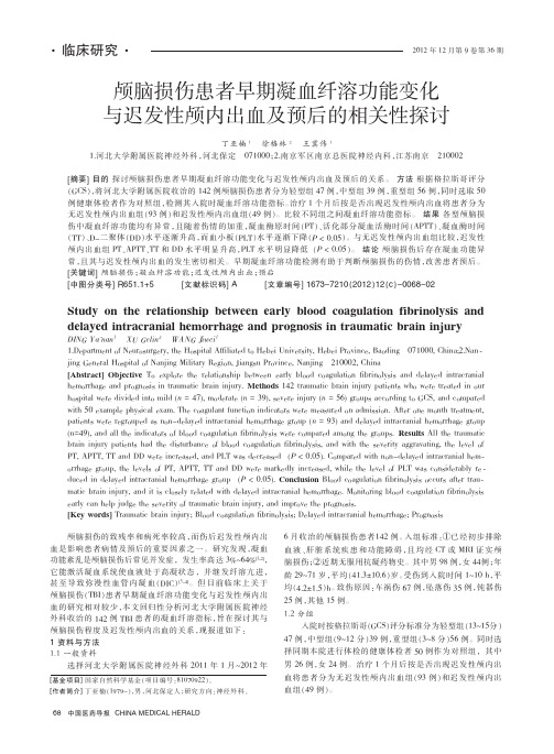
颅脑损伤患者早期凝血纤溶功能变化与迟发性颅内出血及预后的相关性探讨丁亚楠1徐格林2王冀伟11.河北大学附属医院神经外科,河北保定071000;2.南京军区南京总医院神经内科,江苏南京210002[摘要]目的探讨颅脑损伤患者早期凝血纤溶功能变化与迟发性颅内出血及预后的关系。
2023年欧洲指南:严重创伤出血和凝血障碍的管理(第6版)39项推荐

2023年欧洲指南:严重创伤出血和凝血障碍的管理(第6版)39项推荐1 早期复苏与防止再出血缩短间隔时间推荐1建议将严重创伤患者直接送往合适的创伤医疗中心进行救治(1B)尽量缩短创伤出现到出血控制的时间间隔(1B)局部出血管理推荐2建议局部按压以限制危及生命的出血(1B)在术前使用止血带来阻止开放性四肢损伤导致的危及生命的出血(1B)通气推荐3建议在存在气道阻塞、意识改变[格拉斯哥昏迷指数( GCS )≤8分]、低通气或低氧血症的情况下,立即进行气管插管或替代气道管理(1B)建议避免出现低氧血症(1A)建议避免高氧血症,除非存在即将出血的情况(2B)建议对创伤患者行正常通气(1B)建议对于存在脑疝迹象的创伤患者采取的救命措施-过度通气(2C)院前血液制品使用推荐4对院前血液制品的使用没有明确的赞成或反对的建议或意见2 出血的诊断与监测早期评估推荐5建议临床医生结合患者生理、损伤解剖类别、损伤机制和患者对初始复苏的反应来评估创伤性出血的程度(1C)建议使用休克指数(shock index,SI)来评估低血容量休克的严重程度和输血需求(1C)紧急干预推荐6建议有明显出血源、出现肢体失血性休克和疑似出血源的患者进行紧急止血(1 B)进一步检查推荐7建议对不明原因出血及不需要紧急止血的患者,立即进行进一步检查以确定出血源(1C)影像学检查推荐8建议,在可行且在不耽误转运的情况下,使用院前超声检查(PHUS)检测胸腹部损伤患者是否存在血/气胸、心包积血和/或腹腔游离积液(2B)建议对胸腹损伤患者使用包括FAST在内的床旁超声检查(POCUS)(1C)建议早期使用增强全身CT( WBCT ) 来检测和识别损伤类型及潜在的出血源(1 B)血红蛋白推荐9建议将重复Hb和/或Hct测定作为出血评估的实验室指标,因为正常范围内的初始值可能会掩盖早期出血表现(1B)血清乳酸盐和碱缺失推荐10建议将血乳酸作为估计和监测出血和组织低灌注程度的敏感指标,在没有乳酸测量的情况下,碱基缺失可能是一个合适的替代方案(1B)凝血功能监测推荐11建议采用传统的实验室方法( 凝血酶原时间( PT ) /国际标准化比值( INR )、Clauss纤维蛋白原水平和血小板计数和/或即时检验( POC ) PT / INR和/或黏弹性试验( viscoelastic method) ,进行早期、重复监测凝血功能(1C)血小板功能监测推荐12对于正在接受抗血小板治疗或疑似血小板功能异常的创伤患者,建议避免常规使用血小板功能床旁快速检测装置(point ofcare,POC)进行血小板功能监测(1C)3 组织氧合、容量、液体类型和体温容量置换和目标血压推荐13在创伤后的初始阶段,建议使用限制性容量置换以达到目标血压,目标收缩压为80~90 mmHg(平均动脉压为50~60 mmHg),直至严重出血得到控制且无颅脑损伤的临床指征(1B)建议严重创伤性脑损伤(traumatic brain injuries,TBI)(GCS≤8分)患者的平均动脉压维持在80mmHg以上(1C)血管升压药和强心药推荐14如果限制性容量替代策略未达到目标血压,我们建议在输注液体的同时给予去甲肾上腺素以维持目标动脉压(1C)建议在存在心肌功能障碍的情况下输注多巴酚丁胺(1C)液体类型推荐15建议对低血压性创伤出血患者开始使用0.9 %氯化钠或平衡晶体溶液进行液体治疗(1B)对于严重颅脑外伤患者避免使用低渗晶体溶液如乳酸林格液(1B)由于胶体对凝血功能的不良影响,建议限用胶体液(1C)红细胞水平推荐16若需红细胞输注,建议目标血红蛋白为70~90 g/L(1C)自体血回输推荐17建议在出现腹腔、盆腔或胸腔严重出血时考虑行自体血回输(2B)体温管理推荐18尽早采取措施减少热量散失,复温低体温患者,恢复和维持正常体温(1C)4 快速控制出血损伤控制性手术推荐19对于以失血性休克、持续出血、凝血功能障碍和/或合并腹部血管和胰腺损伤为临床表现的严重创伤患者,推荐损伤控制性手术(1B)应采取损伤控制性手术其他原因包括体温过低、酸中毒、难以触及的重大解剖损伤、长耗时的手术的严重创伤(1C)对于不存在上述任何情况的患者,建议进行初级确定性的外科手术治疗(1C)骨盆闭合和稳定推荐20建议在院前使用骨盆包扎带,以减少疑似骨盆骨折时危及生命的出血(1C)建议出血性休克合并骨盆环破裂的患者,尽早行骨盆环闭合和稳定的相关处理(1B)填塞、栓塞、手术及复苏性主动脉血管内球囊阻断术(REBOA)推荐21当正在出血和/或不能及时进行血管栓塞时,建议暂时性腹膜外填塞。
血液科病人颅内出血急救流程

血液科病人颅内出血急救流程英文回答:When it comes to the emergency management of intracranial hemorrhage (ICH) in hematology, prompt action is crucial. As a hematologist, I have encountered numerous cases of patients with ICH, and I follow a specific protocol to ensure the best possible outcome for my patients.First and foremost, upon receiving a patient with suspected ICH, I quickly assess their vital signs and level of consciousness. It is essential to stabilize the patient and ensure their airway, breathing, and circulation are intact. In severe cases, I may need to intubate the patient to secure their airway and provide adequate oxygenation.Next, I order a stat computed tomography (CT) scan of the head to confirm the diagnosis and determine thelocation and extent of the hemorrhage. This information iscrucial in guiding further management decisions. If the CT scan reveals a significant bleed, I consult with a neurosurgeon immediately to discuss the possibility of surgical intervention.While waiting for the neurosurgeon's evaluation, I initiate medical management to control the bleeding and prevent further complications. This typically involves administering medications to manage blood pressure and reduce intracranial pressure. For example, I may prescribe intravenous antihypertensive drugs such as labetalol or nicardipine to lower blood pressure and minimize the risk of rebleeding.In some cases, the patient may require transfusion of blood products to correct any coagulation abnormalities. For instance, if the patient has a low platelet count, I may administer platelet transfusions to improve clotting function.Throughout the management process, I closely monitor the patient's neurological status, including their level ofconsciousness, pupillary response, and motor function. Any deterioration in neurological status prompts immediate intervention, such as repeating the CT scan or adjusting medication dosages.Once the neurosurgeon evaluates the patient, they may recommend surgical intervention, such as evacuation of the hematoma or placement of a ventricular drain to relieve intracranial pressure. In such cases, I collaborate closely with the neurosurgeon to ensure optimal coordination of care.After the acute phase of ICH management, I focus on the patient's recovery and rehabilitation. This may involve physical and occupational therapy to regain motor function and improve activities of daily living. Additionally, I provide counseling and support to the patient and their family, as I understand the emotional impact such a traumatic event can have on them.中文回答:对于血液科的颅内出血(ICH)的急救管理,迅速行动至关重要。
- 1、下载文档前请自行甄别文档内容的完整性,平台不提供额外的编辑、内容补充、找答案等附加服务。
- 2、"仅部分预览"的文档,不可在线预览部分如存在完整性等问题,可反馈申请退款(可完整预览的文档不适用该条件!)。
- 3、如文档侵犯您的权益,请联系客服反馈,我们会尽快为您处理(人工客服工作时间:9:00-18:30)。
Acta Neurochir(Wien)(2008)150:165–175 DOI10.1007/s00701-007-1475-8Printed in The NetherlandsReview ArticleCoagulation disorders after traumatic brain injury B.S.Harhangi1,E.J.O.Kompanje2,F.W.G.Leebeek3,A.I.R.Maas4 1Department of Neurosurgery,Erasmus MC,Rotterdam,The Netherlands2Department of Intensive Care,Erasmus MC,Rotterdam,The Netherlands3Department of Hematology,Erasmus MC,Rotterdam,The Netherlands4Department of Neurosurgery,University Hospital Antwerp,Edegem,BelgiumReceived10April2007;Accepted1October2007;Published online2January2008#Springer-Verlag2008SummaryBackground.Over the past decade new insights in our understanding of coagulation have identified the promi-nent role of tissue factor.The brain is rich in tissue factor,and injury to the brain may initiate disturbances in local and systemic coagulation.We aimed to review the current knowledge on the pathophysiology,inci-dence,nature,prognosis and treatment of coagulation disorders following traumatic brain injury(TBI). Methods.We performed a MEDLINE search from 1966to April2007with various MESH headings,focus-ing on head trauma and coagulopathy.We identified441 eligible English language studies.These were reviewed for relevance by two independent investigators.A meta-analysis was performed to calculate the frequencies of coagulopathy after TBI and to determine the association of coagulopathy and outcome,expressed as odds ratios. Results.Eighty-two studies were relevant for the pur-pose of this review.Meta-analysis of34studies report-ing the frequencies of coagulopathy after TBI,showed an overall prevalence of32.7%.The presence of coagu-lopathy after TBI was related both to mortality(OR9.0; 95%CI:7.3–11.6)and unfavourable outcome(OR36.3; 95%CI:18.7–70.5).Conclusions.We conclude that coagulopathy follow-ing traumatic brain injury is an important independent risk factor related to prognosis.Routine determination of the coagulation status should therefore be performed in all patients with traumatic brain injury.These data may have important implications in patient management. Well-performed prospective clinical trials should be un-dertaken as a priority to determine the beneficial effects of early treatment of coagulopathy.Keywords:DIC;coagulopathy;meta-analysis;progno-sis;review;treatment;traumatic brain injury;head trauma. IntroductionDisturbances of the haemostatic mechanism are highly relevant in traumatic brain injury(TBI).Micro-hae-morrhages occur frequently in the brain parenchyma and a normal coagulation status is important to prevent progression of these to larger haematomas.Haemo-rrhage is a major cause of in-hospital mortality in patients with TBI[83].Coagulation abnormalities are not only a result from injury,but may also cause further secondary injury.Coagulation disorders in TBI are complex and can be characterised by a combination of coagulopathy and hypercoagulability.Hypercoagulability is the in-creased capacity of formation offibrin in the blood ves-sels.A hypercoagulable state may be generalised in the case of disseminated intravascular coagulation(DIC)or local with the development of microthrombi in the pen-All authors were involved in writing the review. Correspondence:B.Sanjay Harhangi,M.D.,Ph.D.,Department of Neurosurgery,Erasmus MC,P.O.Box2040,3000CA Rotterdam,The Netherlands.E-mail:b.s.harhangi@erasmusmc.nlumbra of a contusion.DIC is characterised by the wide-spread activation of coagulation,which results in the intravascular formation offibrin and ultimately throm-botic occlusion of small and midsize vessels[56,57,65]. In this review coagulopathy is defined as any coagula-tion abnormality in TBI as defined by the different cri-teria of the various studies.Coagulopathy may result from depletion of platelets and clotting factors following blood loss or consumption due to DIC,and may further be enhanced by dilution,acidosis and hypothermia[6]. The resulting disorders of the coagulation status may invoke both bleeding and ischaemia.Many studies have shown progression of parenchymal lesions,particularly in patients with coagulopathy[21,90,92–94,97].More-over,the presence of coagulation abnormalities is related to poorer outcome[66,69,87].Disorders of coagulation may be amenable to treatment,and adequate and prompt intervention may prevent secondary complications and poorer outcome[61].The aim of this review is to sum-marise the current literature on pathophysiology,inci-dence,nature,prognosis and treatment of coagulation disorders following TBI.Pathogenesis of coagulation disorders in TBIIn healthy individuals,coagulation andfibrinolysis are balanced to prevent excessive haemorrhage or thrombo-sis[15,56,71,79].Patients with traumatic brain injury (TBI)are at risk of developing abnormalities of both coagulation andfibrinolysis[43,76,85,101,105]. The nature of the coagulation abnormalities differs be-tween patients with isolated head injury and patients with multiple injuries.There is evidence that the extent of traumatised brain tissue,rather than traumatic shock or hypoxia,plays an important role in the occurrence of coagulation disorders after TBI[85].In the early seven-ties,the release of tissue factor(TF),formerly called thromboplastin or thrombokinase[11],from injured brain tissue was postulated as the cause[49].Tissue factor is a protein present in subendothelial tissue,plate-lets,and leukocytes necessary for the initiation of the coagulation cascade that eventually leads to thrombin formation from the zymogen prothrombin.TF is the main physiological initiator of coagulation[35]and its release therefore can also activate the coagulation system excessively in patients with head trauma.It is suggested that this activation depends on the amount of TF released from damage to brain tissue[7,39,49, 73].Recently,Gando and co-workers[33]demonstrated higher levels of TF in head injured patients than in non-head-injured trauma patients.However,this concerns plasma levels of soluble tissue factor antigen and it is yet unclear what these levels mean and its function is totally unknown.In addition,Pathak and co-workers [73]demonstrated increased TF activity in patients with TBI as compared to controls,highlighting the role of TF in coagulation disorders following TBI.Early exposure of TF to both factor VII and VIIa results in the FVIIa= TF complex.This complex forms and generates small amounts of factor Xa and factor IXa.Factor Xa in col-laboration with the membrane surface activates a small amount of prothrombin to thrombin.The generation of trace amounts of thrombin will activate platelets and factors V and VIII,that will thereby provide a suitable surface on which the prothrombinase complex can be assembled and which will lead to much more thrombin generation,required for the conversion offibrinogen to fibrin[55,58].This tissue factor-dependent activation of coagulation,formerly known as the extrinsic pathway, causes both micro-and macrovascularfibrin thrombiFig.1.Schematic diagram of traumaticbrain injury and coagulopathy166 B.S.Harhangi et al.formation.Control mechanisms,including tissue factor pathway inhibitor (TFPI),the protein C system,anti-thrombin and glycosamineglycans are active to counter fibrin formation and to localise fibrin formation to the site of injury,but are frequently insufficient after exten-sive TF exposure.DIC,instigated by TF activation,inhi-bits these antithrombotic mechanisms through cytokine release and upregulation causing defective physiological anticoagulation pathways.This may cause necrosis and haemorrhage in many organs,eventually leading to multi organ failure (MOF)[45,56,57,63,79,104].Current concepts of haemostasis and DIC,far beyond the scope of this review,were recently reviewed [56,58,79].A schematic diagram of the proposed mechanism of coagu-lation disorders following TBI is shown in Fig.1.Various studies have confirmed a correlation be-tween the presence of coagulation abnormalities and poorer outcome [66,69,87].The association between clotting abnormalities and delayed intracerebral haemor-rhage has become well established [21,90,92–94,97].Adequate control of haemorrhage remains an important aim in the management of TBI.Considerable contro-versies,on the diagnosis and treatment of coagulopathy after TBI still exist and it remains uncertain whether initiation of specific treatment in the early hypercoagu-lable phase may indeed prevent progression of lesions and improve outcome.Material and methodsWe performed a Pubmed search from 1966to April 2007.First of all,we defined appropriate keywords and subject headings.We combined different keywords and noted the number of studies observed with and without restric-tion to the English language (Table 1).Since the largest number of studies was observed when combining head trauma and coagulopathy (n ¼429,Table 1)we used these terms to identify relevant studies.To identify ad-ditional eligible studies,the other keyword combinations and the reference list of the selected studies were screened and 12additional studies were identified.Even-tually,this search strategy yielded 441hits eligible for this study.Two separate reviewers determined the rele-vance of all titles =abstracts and excluded non-relevant studies.We excluded among others,studies on pharma-cologic coagulopathy,chronic subdural haematoma,pre-existing coagulation disorders such as haemophilia and thrombotic thrombocytopenic purpura,spinal trauma and studies on ‘‘non-traumatic’’disorders.This selection resulted in retaining 90studies.Following a more de-tailed review we excluded an additional 8studies since coagulopathy was not studied.In total,this review was therefore based on 82studies.When available,frequen-cies of coagulopathy as defined and presented by the authors in relation to outcome were extracted from the studies to create two by two tables.These numbers were used to calculate crude odds ratios with 95%confidence intervals (95%CI).In the second stage,a summary (pooled)estimate is calculated as a weighted average of the effects estimated in the individual studies by using the Mantel Haenszel (MH)method,a widely recognised fixed effect method of meta-analysis.ResultsNone of the studies met the criteria for Class I evidence.Many studies had been performed retrospectively and some were limited to case reports.Twenty-six publica-Table 1.Pubmed search results KeywordsAll English HITS –review of titles =abstracts =references Cranio-cerebral trauma coagulopathy590414Exclusion of non-relevant studies,such as Cranio-cerebral trauma coagulation disorders 516348–chronic subdural haematomaHead trauma coagulation disorders 527358–pre-existing coagulation disorders Head trauma coagulopathy 606429–spinal traumaTBI coagulation disorders 14478–non traumatic disorders TBI coagulopathy173107–non clinicalClosed head injury and coagulation disorders 2618possibly relevant studies:90Closed head injury and coagulopathy 3729detailed review of full manuscripts included in systematic review:82222Coagulation disorders after traumatic brain injury 167tions represented prospective series,but some used a nested case control design,in which bias may play an important role.An overview of the studies from an epi-demiologic perspective is listed in Table 2.Table3shows the characteristics of the studies with frequencies of coagulation disorders,which vary between10and 97.5%with a mean of32.7%.When available,crude odds ratios were calculated for the association between coagulopathy after TBI as defined in the different stud-ies,and mortality or Glasgow Outcome Scale(GOS) 1–3(bad outcome),versus GOS4–5(good out-come).Data for the association between coagulopathy after TBI and the risk of mortality are presented in Table3A.Odds ratios in the studies vary between4.2 and161.Data for the association between coagulopathy after TBI and the risk of bad outcome are presented in Table3B.In these studies odds ratios vary between16 and58.All presented point estimates were statistically significant,although important modifiers(such as age and GCS)were not considered.When the crude data are pooled wefind an overall odds ratio for the risk of bad outcome(GOS1–3)of33.2(95%CI:15.9–69.1) and the risk of mortality of9.4(95%CI:7.6–11.6)Table2.Epidemiological classification of the studiesStudy design No.ofstudiesCase report10[1,23,28,49,62,64,74,100,110,111]Case-control10[9,34,45,51,75,84,85,96,114,117]Prospective30[10,13,14,18,19,22,29,30,38,39,43,48,50,52–54,59,63,67–69,89,92,93,99,101,107,108,113,115]Retrospective16[20,21,31,37,44,46,52,70,72,76–78,87,90,94,97,106]Cross-sectional3[8,105,109]Experimental1[104]Review5[42,47,81,88,91]Comments7[3–5,25,26,95,103]Total82Table3.Characteristics of the studies with frequencies of coagulopathy after traumatic brain injuryStudy No.of patients Coagulopathy No coagulopathy Prevalence of coagulopathy(%) Auer and Ott[9]40152537.5Avikainen[10]45153033Becker et al.[14]27171063Bredbacka and Edner[18]2015575Brohi et al.[19]107925682323.7Carrick et al.[20]1766011634.1Chang et al.[21]113219218.6Chiaretti et al.[22]6065410Gando et al.[34]1614287.5Goodnight et al.[39]26101638.5Hulka et al.[43]1595410534Hymel et al.[44]1474010727.2Kaufmann et al.[45]146842.8Kearney et al.[48]3631586.1Keller et al.[50]53203337.7Kumura et al.[51]100247624Kuo et al.[52]61441772.1Kushimoto et al.[54]4739883Miner et al.[63]87285932Olson et al.[69]26915411557.2Ordog et al.[70]180175597.2Patel et al.[72]852********.4Pfenninger et al.[75]50123824Piek et al.[76]73413559918.4Pondaag[77]46351176Selladurai et al.[87]1431083575.5Stein et al.[94]2536718626.5Stein et al.[92]33410223230.5Takahasi et al.[96]25101540Tan et al.[97]38112728.9Vavilala et al.[106]69333634.4Vecht et al.[107]4031977.5Vecht et al.[109]63350Vecht and Sibinga[108]126650Overall53571754360332.7168 B.S.Harhangi et al.after TBI and ing the Mantel-Haenszel method,the odds ratio for the risk of bad outcome (GOS 1–3)is 36.3(95%CI:8.7–70.5)and 9.0(95%CI:7.3–11.6)for the risk of mortality.DiscussionIncidence of coagulation disordersThe incidence of coagulopathy in TBI is high,but actual numbers vary considerably between studies.The wide variation in frequency is not striking because different study designs and definitions for coagulopathy have been used.The definitions of coagulopathy have been changed over time,and in addition,other techniques for measuring DIC have become available in the last de-cade.Recently the International Society of Thrombosis and Haemostasis (ISTH)simplified the laboratory diag-nosis of DIC by calculating a DIC score based on platelet count,elevated fibrin-related marker,prolonged prothrombin time and fibrinogen level [98].However,none of the studies included in our review have used this score,but instead were mainly based on other para-meters,including fibrinogen,fibrinogen degradation prod-ucts and antithrombin levels.Overall,in this review,one out of three patients with TBI have signs of coagulopa-thy (Table 3)and according to the literature it occurs in more than 60%of patients with severe TBI [8,24,42].In mild TBI coagulopathy is probably less than one percent [37].Kuo and colleagues [52]reported an inci-dence of coagulopathy in 100%of patients dying from head injury whereas Chiaretti and colleagues [22]pre-sented a frequency of 10%in patients with TBI.Studies from the late 1970’s already reported a decrease of platelets and fibrinogen during the first five days after injury,indicating consumptive coagulopathy [8,9],but could not confirm the presence of ter studies,utilising more sensitive laboratory techniques for detect-ing a hypercoagulable state,reported an increased fre-quency of DIC following head injury up to 76%[51,63,69,77,104].According to some authors 15–40%of patients with severe TBI meet the criteria for symp-Table 3B.Crude odds ratios for the risk of bad outcome according to GOS after TBI and coagulopathy StudyNo.of patients Coagulopathy GOS1-3No coagulopathy 3GOS1-3Coagulopathy GOS >3No coagulopathy GOS >3OR (95%CI)Bredbacka and Edner [18]201213416(1.3–200.9)Goodnight et al.[39]269411227(2.6–284.7)Kushimoto et al.[54]47174026n.a.Pfenninger et al.[75]509633216(3.3–77)Tan et al.[97]3810811923.8(2.6–218)Vavilala et al.[106]6929443258.3(13–253)Overall25086271212533.2(15.9–69.1)GOS Glasgow Outcome Scale,TBI traumatic brain injury,OR odds ratio,95%CI 95%confidence interval,n.a.not applicable.Table 3A.Crude odds ratios for the risk of mortality after traumatic brain injury with coagulopathy StudyNo.of patients Coagulopathy–no survival No coagulopathy–no survival Coagulopathy–survival No coagulopathy–survival OR (95%CI)Auer and Ott [9]40142123161(13.3–1943)Brohi et al.[19]107911490142733 6.5(4.7–9.1)Carrick et al.[20]176211339103 4.2(1.9–9.3)Hulka et al.[43]159********.9(3.5–22.9)Hymel et al.[44]147201220957.9(3.3–18.8)Keller et al.[50]5361143213.7(1.5–124.8)Kumura et al.[51]100145107119.9(5.9–67.1)Kuo et al.[52]611103317n.a.Miner et al.[63]8715713528.6(2.9–25.3)Olson et al.[69]26985116910412.2(6–24)Pondaag [77]46181171010.6(21.2–91.8)Selladurai et al.[87]143852233361(13.6–273)Takahasi et al.[96]259311236(3.2–405.9)Overall238543315441513839.4(7.6–11.6)OR odds ratio,95%CI 95%confidence interval,n.a.not applicable.Coagulation disorders after traumatic brain injury169tomatic DIC[18,41,63,94],however as indicated above different criteria for DIC were used.DIC seems to occur most frequently in patients with acute subdural haematoma,or parenchymal contusions[51].Haemo-static activation may be more prominent in cerebrovas-cular blood than in peripheral blood[67].The presence of local cerebral intravascular coagulation has been con-firmed in surgical specimens of human cerebral contu-sions,and cerebral contusions of rats and pigs[90].Severity of TBI and coagulopathy Hypercoagulability after injury is most prevalent during thefirst24h and more pronounced in women[86]. Children with a glasgow coma score(GCS)<14after TBI are at greater risk for coagulopathy and this risk is even more increased at lower GCS levels[50].Several studies have demonstrated thatfibrin degradation prod-uct(FDP)levels correlate positively with the degree of brain damage[46,51,69,102].Stein and colleagues [92]found that almost50%of patients with TBI devel-oped delayed brain injury,as defined by the occurrence of new intracranial lesions or progression of lesions ini-tially present on admission.In that study,abnormalities in the prothrombin time,partial thromboplastin time,or platelet count at admission were present in55%of pa-tients with evidence of delayed injury,versus only9%of patients without delayed injury.In particular,mean pro-thrombin time and partial thromboplastin time at admis-sion were significantly longer in patients developing delayed injury.More recently,Yokota and colleagues [116],found that V on Willebrand factor(vWF)and thrombomodulin(TM)are useful indicators of cerebral endothelial injury and that increased TM levels predict delayed brain injury.PrognosisThe presence of coagulopathy is not only related to the risk for developing delayed injury,but also to a poorer outcome.Continued bleeding is one factor,but not the only one responsible for a poorer outcome.As men-tioned above,DIC,which involves,by definition,‘‘in-travascular coagulation’’,causes multiple organ failure by thromboembolic ischaemia and may also be respon-sible for a bad outcome.Similarly,the DIC after TBI causes intravascular coagulation-induced cerebral is-chaemia and may be related to a bad prognosis.It is striking that the risk of mortality in patients with coa-gulopathy after TBI is approximately ten times higher than in these patients without coagulopathy(Table3A). The risk of bad outcome in patients with coagulopathy after TBI is even more than thirty times higher than in these patients without coagulopathy(Table3B).We are aware that bias plays an important role in these studies and the various prevalences cannot be compared due to different criteria of coagulopathy in each of the studies. Moreover,the parameters of coagulopathy in these stud-ies differ and this variation will lead to clinical hetero-geneity.This is an important limitation of our study. However,these results are consistent with thefindings of two similar studies[69,87]in which logistic regres-sion models with adjustment of different parameters were used to calculate the probability of a bad outcome after TBI and coagulopathy.Various large clinical stud-ies have demonstrated that DIC is an important predictor of poor outcome in head injury[46,63,94].This rela-tionship was absent in only one of the presented studies [67].Kushimoto and colleagues[53]studied the clinical significance offibrinolysis andfibrinogenolysis in patients with closed head injury and found that indices offibrinolytic activity likefibrinogen degradation pro-ducts(FDP),andfibrin degradation products(FbDP) were correlated to outcome.A limitation of this study is that they did not adjust for confounders such as gen-der,age or extent of brain tissue damage.The interpre-tation of different studies on the predictive value of coagulation parameters and outcome is complex due to differences in study design,population and definitions of DIC.Some studies have a relatively small sample size and short follow up periods whereas others only present cross-sectional data.Some authors claim that laboratory values for coagulation status may be a better predictor of mortality than midline shift or pupillary reactivity[52], but only few have investigated the predictive value of coagulopathy adjusted for other predictors.Olson and colleagues[69]and Selladurai and colleagues[87]stud-ied the predictive value of coagulopathy in TBI with logistic regression models.Both studies found that high FDP levels predict poor outcome independently of other variables and that prognosis worsens as the level of FDP increases.In patients with mild head injury,this high FDP level may lead to a rapid deterioration and eventu-ally to death.In patients with an intermediate level of severity of brain injury,the activated partial thrombo-plastin time(APTT)appeared to be of greater prognostic value.Increased plasma concentrations of FDP and plas-min-alpha2-plasmin inhibitor complex and decreasedfi-brinogen levels are associated with a high percentage of unfavourable outcome three months after injury.Similar170 B.S.Harhangi et al.results have also been reported in children[106].Very recently,results from the IMPACT(International Mission for Prognosis And Clinical Trial)study showed that the prothrombin time is a powerful independent prognostic factor after TBI[66].TreatmentGuidelines for the treatment of coagulopathy following TBI do not exist.No randomized studies have been performed to study any intervention such as infusion of fresh frozen plasma(FFP),antithrombotic drugs or antithrombotic coagulation factor concentrates after TBI. Therefore opinions on preferred approaches vary.These differences reflect the complexity of the problem and the strong heterogeneity in type,degree and phase of coag-ulation disturbances.In general,the therapy of coagula-tion disturbances should be aimed at treating the primary cause.Stein and colleagues[91]recently argued that the objectives of treatment should be to combat coagulation, to lyse existing clots,to replete clotting factors and re-verse hyperfibrinolysis.Fresh frozen plasma and platelet concentrates are recommended for patients with active bleeding.May and colleagues[60]have advocated the use of FFP as resuscitationfluid in patients with a GCS 7.However,Winter and co-workers[113]did notfind any benefit on outcome with the use of FFP.Keller and colleagues[50]propose administration of FFP to all children presenting the GCS<8in an attempt to combat coagulation disorders and prevent progression of lesions. According to Rubin and Colman[82]heparin anticoa-gulation followed by aggressive replacement with plate-lets and FFP is indicated as a treatment of patients with DIC in general.This treatment might be extrapolated to DIC in TBI.However,administration of these products may carry a risk of enhancing coagulation by adding procoagulant factors.Rubin and Colman[82]proposed administration of heparin in therapeutic doses,sufficient to overcome the coagulant forces that may have prod-uced a relative heparin-resistant state in the blood. Most clinicians treating TBI however will be extremely reluctant to administer heparin for fear of increasing intracranial haemorrhage and in view of the lack of clin-ical evidence we consider this approach,although theo-retically sound,debatable[27].DIC also occurs frequently in sepsis where activated protein C,a potent anticoagulant and profibrinolytic agent,has shown to improve coagulation disturbances and improve outcome in septic patients,who developed multiple organ failure[32].Nevertheless,an increased risk of bleeding has been observed with the administra-tion of protein C to patients with sepsis[16,32]and in children it may even cause serious bleeding events[36]. To date,no such studies have been performed in patients with TBI.Various other approaches have been explored to correct coagulopathy in TBI.The early administration of antithrombin III(AT III)to patients with TBI could inhibit or significantly shorten the time of coagulopathy. Grenander and colleagues[40]described a marginal re-duction of hypercoagulation parameters following ad-ministration of AT III but could not confirm a benefit on progression of lesions,on outcome or on length of ICU stay.Antifibrinolytic agents like aprotinin,tranexa-mic acid and[epsilon]-aminocaproic acid are commonly used in cardiac surgery.The role of antifibrinolytic agents in trauma patients with massive blood loss is yet not clear.The CRASH-2(Clinical Randomisation of Antifibrinolytic in Significant Haemorrhage)trial is a World Health Organization(WHO)supported large international multi-centre randomised placebo con-trolled trial of the effects of the antifibrinolytic agent tranexamic acid on death and transfusion requirements in adult trauma patients with significant haemorrhage.In this large study,recruitment is expected to be completed in December2009and will provide us more insight in the use of antifibrinolytic agents in trauma patients[80]. To our knowledge,no randomised data are available on the administration of antifibrinolytics after traumatic brain injury.Preliminary data indicate that recombinant factor VIIa(rFVIIa)provides a rapid and successful correction of coagulopathy in the head-injured patient which may suggest an alternative strategy for treating coagulopathy in TBI[64].Interestingly,treatment with rFVIIa within four hours after the onset of intracerebral haemorrhage limits the growth of the haematoma,re-duces mortality,and improves functional outcomes at 90days[61].Very recently,however,a completed phase 3,randomised,controlled trial involving821patient presented on the16th European Stroke Conference Glasgow,United Kingdom2007,showed that there was a significant reduction in the size of the intracere-bral haematoma but no effect on mortality and severe disability on day90(unpublished data).Since TBI is commonly associated with cerebral contusions and hae-morrhage[2]these results may be extrapolated to TBI as was reported recently[112].However,rFVIIa should be administered with caution due to the increased risk of thromboembolic complications[61].To date there is only one published randomised placebo-controlled trial of the use of rFVIIa in trauma[17].The authors showedCoagulation disorders after traumatic brain injury171。