Localized States and Resultant Band Bending in Graphene Antidot Superlattices
2022年教育部考试中心考研英语模拟试题(新题型4)
2022教育部考试中心考研英语模拟试题(新题型4)----新题型PartB汇编4、Directions:You are going to read a list of headings and a text about preparing in the academic community.Choose the most suitable heading from the list A-F for each numbered paragraph(41-45).The first and last paragraphs of the text are not numbered. There is one extra heading which you do not need to use. Mark your answers on ANSWER SHEET 1. (10 points)Until about two million years ago Africa’s vegetation had always been controlled by the interactions of climate; geology, soil, and groundwater conditions; and the activities of animals. The addition of humans to the latter group,however,has increasingly rendered unreal the concept of a fully developed “natural” vegetation—i.e., one approximating the ideal of a vegetational climax.(41).Early attempts at mapping and classifying Africa’s vegetation stressed this relationship:sometimes the names of plant zones were derived directly from climates.In this discussion the idea of zones is retained only ina broad descriptive sense.(42). In addition, over time more floral regions of varying shape and size have been recognized.Many schemes have arisen successively,all of which have had to take views on two important aspects: the general scale of treatment to be adopted, and the degree to which human modification is to be comprehended or discounted.(43).Quite the opposite assumption is now frequently advanced. An intimate combination of many species—in complex associations and related to localized soils, slopes, and drainage—has been detailed in many studies of the African tropics. In a few square miles there may be a visible succession from swamp with papyrus, the grass of which the ancient Egyptians made paper and from which the word“paper”originated,through swampy grassland and broad-leaved woodland and grass to a patch of forest on richer hillside soil,and finally to juicy fleshy plants on a nearly naked rock summit.(44). Correspondingly, classifications have differed greatlyin their principles for naming,grouping,and describing formations: some have chosen terms such as forest,woodland,thorn-bush, thicket, and shrub for much of the same broad tracts that others have grouped as wooded savanna (treeless grassy plain) and steppe (grassy plain with few trees).This is best seen in the nomenclature, naming of plants, adopted by two of the most comprehensive and authoritative maps of Africa’s vegetation that have been published: R. W. J. Keay’s Vegetation Map of Africa South of the Tropic of Cancer and its more widely based successor, The Vegetation Map of Africa,compiled by Frank White.In the Keay map the terms“savanna”and“steppe” were adopted as precise definition of formations, based on the herb layer and the coverage of woody vegetation; the White map, however, discarded these two categories as specific classifications.Yet any rapid absence of savanna as in its popular and more general sense is doubtful.(45).However,some100specific types of vegetation identified on the source map have been compressed into14broader classifications.[A] As more has become known of the many thousands of African plant species and their complex ecology, naming, classification,and mapping have also become more particular, stressing what was actually present rather than postulating about climatic potential.[B] In regions of higher rainfall, such as eastern Africa, savanna vegetation is maintained by periodic fires. Consuming dry grass at the end of the rainy season,the fires burn back the forest vegetation, check the invasion of trees and shrubs, and stimulate new grass growth.[C] Once, as with the scientific treatment of African soils, a much greater uniformity was attributed to the vegetation than would have been generally accepted in the same period for treatments of the lands of western Europe or the United States.[D] The vegetational map of Africa and general vegetation groupings used here follow the White map and its extensive annotations.[E] African vegetation zones are closely linked to climatic zones, with the same zones occurring both north and south of the equator in broadly similar patterns.As with climatic zones, differences in the amount and seasonal distribution of precipitation constitute the most important influence on the development of vegetation.[F]Nevertheless,in broad terms,climate remains the dominant control over vegetation.Zonal belts of precipitation,reflection latitude and contrasting exposure to the Atlantic and Indian oceans and their currents,give some reality to related belts of vegetation.[G]The span of human occupation in Africa is believed to exceed that of any other continent. All the resultant activities have tended, on balance, to reduce tree cover and increase grassland; but there has been considerable dispute among scholars concerning the natural versus human-caused development of most African grasslands at the regional level.答案41.F 42.A 43.C 44.G 45.D总体分析本文是一篇介绍非洲植被讨论的科普性文章。
广义联邦滤波器的全局最优性

(2)
T T , H k = [ H1 k , H 2k , · · · ,
T T HN ,V is a zero-mean white k] , V k = V Gaussian noise, whose covariance matrix is Rk , and Rk = diag {R1k , R2k , · · · , RN k }. Thus
With the development of information technology, decentralized filtering[1−3] and federated filtering[4−10] have been widely applied to multisensor information fusion. They have fast computing speed due to their suitable algorithm structure for parallel computing. Fault detection, fault isolation, and system reconfiguration can be achieved with the inherent analytic relation between the input variables and the state variables of the subsystem, so that the reliability of the whole system may be improved. Therefore, they are remarkably superior to the centralized Kalman filtering in computing efficiency and fault tolerance. In proving the optimality of federated filters, Carlson constructed an augmented system, where the variance upper bound technique was used to eliminate the correlation among several local filters. Then, the global optimal estimation was achieved with an uncorrelated fusion algorithm and the information sharing principle. However, in the high-dimensional situation, the computation amount of the above approach will increase sharply. Another approach is the minimum structure. A master filter only contains the common state, and the information sharing is limited to the common state between the master filter and the local filters. Considering the influence of the common state on the bias state, the common state after the information fusion is used to reset the common state of the local filters and also used as the observation feedback to correct the bias state of the local filters. Reference [7] analyzed the above approach theoretically, and pointed out that the minimum structure approach is based on the hypothetical condition that the state estimated error between the local filters and the master filter is uncorrelated. Usually this hypothetical condition is difficult to satisfy and only the suboptimal solution can be obtained. In this paper, based on the decentralized filtering algorithm and the information sharing principle, it is proved that the global filtering of federated filters is optimal only when the dimensions of the master filter and the local filters are totally equal. Meanwhile, the generalized federated filter is structured, where the dimensions of the master filter and the local filters are different, and the feedback correction by which the generalized federated filter may realize the global optimality is presented. The validity of the approach is theoretically proved and its effectiveness is demonstrated by its application to an integrated navigation
异位妊娠双语教学资料
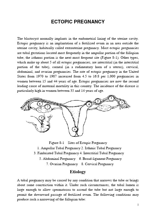
ECTOPIC PREGNANCYThe blastocyst normally implants in the endometrial lining of the uterine cavity. Ectopic pregnancy is an implantation of a fertilized ovum in an area outside the uterine cavity, habitually called extrauterine pregnancy. Most ectopic pregnancies are tubal gestations located most frequently in the ampullar portion of the fallopian tube; the isthmus portion is the next most frequent site (Figure 8-1). Other types, which make up about 5 of all ectopic pregnancies, are interstitial (in the interstitial portion of the tube), corneal (in a rudimentary horn of a uterus), cervical, abdominal, and ovarian pregnancies. The rate of ectopic pregnancy in the United States from 1970 to 1987 increased from 4.5 to 16.8 per 1,000 pregnancies in women between 15 and 44 years of age. Ectopic pregnancies are now the second leading cause of maternal mortality in this country. The incidence of the disease is particularly high in women between 35 and 14 years of age.Figure 8-1 Sites of Ectopic Pregnancy1. Ampullar Tubal Pregnancy2. Isthmic Tubal Pregnancy3. Fimbriated Tubal Pregnancy4. Interstitial Tubal Pregnancy5. Abdominal Pregnancy6. Broad-ligament Pregnancy7. Ovarian Pregnancy 8. Cervical PregnancyEtiologyA tubal pregnancy may be caused by any condition that narrows the tube or brings about some constriction within it. Under such circumstances, the tubal lumen is large enough to allow spermatozoa to ascend the tube but not large enough to permit the downward passage of fertilized ovum. The following conditions may produce such a narrowing of the fallopian tube:1.Previous pelvic inflammatory disease involving the tubal mucosa and producingpartial agglutination of opposing surfaces, for example, Chlamydia, gonorrheal salpingitis.2. Previous inflammatory processes of the external peritoneal surfaces of the tube, for example, puerperal and postabortal infections.3. Endometriosis of the tubal wall and lumen.4. Developmental abnormalities resulting in a segmental narrowing of the tubes or excessive length or kinking.5. Previous abdominal or tubal surgery with resultant scarring and adhesions. Failed tube ligation and a history of previous ectopic pregnancy also increase the risk for an ectopic pregnancy that implants in the fallopian tube. Women who have one ectopic pregnancy have10% to 20% chance that a subsequent pregnancy will also be ectopic. This is because salpingitis that leaves scarring is usually bilateral.6. Previous tubal sterilization.7. Use of contraception that prevents intrauterine pregnancy, such as intrauterine contraceptive devices (IUDs) or low-dose progesterone agents, is associated with increased risk of ectopic pregnancy.PathologyNatural History of Tubal PregnancyTubal Abortion The frequency of tubal abortion depends in part upon the implantation site. Tubal abortion is common in ampullary tubal pregnancy, whereas rupture is the usual outcome with isthmic pregnancy. The immediate consequence of tubal hemorrhage is further disruption of the connection between the placenta and membranes and the tubal wall. If placental separation is complete, all of the products of conception may be extruded through the fimbriated end into the peritoneal cavity. At this point, hemorrhage may cease and symptoms eventually disappear.Some bleeding usually persists as long as products remain in the oviduct. Blood slowly trickles from the tubal fimbria into the peritoneal cavity and typically pools in the recto uterine cul-de-sac. If the fimbriated extremity is occluded, the fallopian tube may gradually become distended by blood, forming a hematosalpinx. (Figure 8-2)After incomplete tubal abortion, pieces of the placenta or membranes may remain attached to the tubal wall and, after becoming surrounded by fibrin,give rise to a placental polyp. The process is similar to that in the uterus after an incomplete abortion.Tubal Rupture The invading, expanding products of conception may rupture the oviduct at any of several sites. Before sophisticated methods to measure chorionic gonadotropin were available, many cases of tubal pregnancy ended during the first trimester by intraperitoneal rupture. As a rule, whenever there is tubal rupture in the first few weeks, the pregnancy is situated in the isthmic portion of the tube. When the fertilized ovum is implanted well within the interstitial portion, rupture usually occurs later. (Figure 8-3)Rupture is usually spontaneous, but it may be caused by trauma associated with coitus or a bimanual examination. With intraperitoneal rupture, the entire conceptus may be extruded from the tube, or if the rent is small, profuse hemorrhage may occur without extrusion. In either event, the woman commonly shows signs of hypovolemia. If an early conceptus is expelled essentially undamaged into the peritoneal cavity, it may reimplant almost anywhere, establish adequate circulation, survive, and grow. This outcome is most unlikely, however, because of damage during the transition. The conceptus, if small, may be resorbed or, if larger may remain in the cul-de-sac for years as an encapsulated mass or even become calcified to form a lithopedion.Abdominal Pregnancy If only the fetus is extruded at the time of rupture, the effect upon the pregnancy will vary depending on the extent of injury sustained by the placenta. The fetus dies if the placenta is damaged appreciably, but if the greater portion the placenta retains its tubal attachment, further development ispossible. The fetus may then survive for some time, giving rise to an abdominal pregnancy. Typically, in such cases, a portion of the placenta remains attached to the tubal wall and the periphery grows beyond the tube and implants on surrounding structures.Broad-ligament Pregnancy When original zygote implantation is toward the mesosalpinx, rupture may occur at the portion of the tube not immediately covered by peritoneum, and the gestational contents may be extruded into a space formed between the folds of the broad ligament. This is designated an intraligamentous or broad-ligament pregnancy.Uterine changesThe uterus undergoes some of the changes associated with early normal pregnancy, including softening of the cervix and isthmus and an increase in size.The degree to which the endometrium is converted to deciduas is variable. The finding of uterine deciduas without trophoblast suggests ectopic pregnancy but is not absolute. In 1954, Arias-Stella described, as had others before him, these changes. Enlarged epithelial cells with nuclei that are hypertrophic, hyperchromatic, lobular, and irregularly shaped. Cytoplasm may be vacuolated and foamy, and occasional mitoses are found. These endometrial changes-the Arias-Stella reaction-are not specific for ectopic and may occur with a normal implantation. External bleeding---seldom severe---is seen commonly in cases of tubal pregnancy and is uterine in origin from degeneration and sloughing of the uterine deciduas. Soon after embryonic death, the decidua degenerates and is usually shed in small pieces. Occasionally it is cast off intact, as a decidual cast of the uterine cavity.Clinical ManifestationClinical Manifestations of a tubal pregnancy are diverse and depend on the site of implantation.In an early ectopic pregnancy, there often are no signs and symptoms. Once the ectopic pregnancy ruptures, however, classic manifestation is present. SymptomsAmenorrhea If implantation occurs in the distal end of the fallopian tube, which can contain the growing embryo longer, the woman may at first exhibit the usual early signs of pregnancy and consider herself to be normally pregnant. About a fourth of women do report amenorrhea; they mistake uterine bleeding that frequently occurs with tubal pregnancy for true menstruation.Abdominal Pain Within 3 to 5 weeks after a missed menstrual period, abdominal pain often develops. The nature, duration, and intensity of pain vary considerably with the length of gestation, site of implantation, and extent of blood loss. Pain isthe predominant symptom of tubal rupture and may be localized on one side or felt over the entire abdomen. The woman may complain of cramping or sharp, sudden, knifelike pain, often of extreme severity. Referred shoulder pain may be present when intraperitoneal bleeding has extended to the diaphragm and irritated the phrenic nerve.Vaginal Bleeding Vaginal bleeding, which occurs when the embryo dies and the deciduas begins to slough, often appears scant and dark brown, and may be intermittent or continuous.Shock and Syncope Depending on the amount of blood loss, the woman may or may not manifest syncope, hypotension, tachycardia, and other symptoms of shock. Hypovolemic shock is a major concern because systemic signs of shock may be rapid and extensive without external bleeding. Women with a ruptured ectopic pregnancy may often present with hypovolemia and shock.Pelvic Mass In some cases, there is gradual disintegration of tubal wall followed by slow leakage of blood into the lumen, peritoneal cavity, or both. Gradually, however, trickling blood collects in the pelvis, more or less walled off by adhesions, and a pelvic hematocele results.SignsGeneral Condition Before rupture, vital signs generally are normal. Blood pressure will fall, pulse rise and shock may present only if bleeding continues and hypovolemia becomes significant. After acute hemorrhage, the temperature may be normal or even low. Temperatures up to 38℃may develop, but higher temperatures are rare in the absence of infectionAbdominal Examination Exquisite tenderness on abdominal is demonstrable in most wo men with ruptured or rupturing tubal pregnancies. Gradually, the woman's abdomen becomes rigid from peritoneal irritation. Abdominal mass may be palpable in some women. With extensive infiltration of blood into the tubal wall, the mass may be firm.Pelvic Examination If blood is slowly seeping into the peritoneal cavity, the umbilicus may develop a bluish tinge (Cullen's sign). The woman may have continuing extensive or dull vaginal and abdominal pain; movement of the cervix on pelvic examination may cause excruciating pain. A tender mass is usually palpable in Douglas' cul-de-sac on vaginal examination. Because of placental hormones, in some cases, the uterus grows during the first3 months of a tubal gestation to nearly the same size as it would with a normal pregnancy. Its consistency may be similar as long as the fetus is alive.Diagnostic TestsTest of Human Chorionic Gonadotropin (hCG)Ectopic pregnancy cannot be diagnosed by a positive pregnancy test alone. The key issue, however, is whether the woman is preg nant. In virtually all cases of ectopic gestation, human chorionic gonadotropin (p-hCG) can be detected in serum, but usually at markedly reduced concentrations compared with normal pregnancy.Ultrasonography Vaginal sonography is more accurate than abdominal sonography in identifying an ectopic pregnancy. With sonographic absence of a uterine pregnancy, a positive pregnancy test, fluid in the cul-de-sac, and an abnormal pelvic mass, ectopic pregnancy is almost certain.Culdocentesis This is a simple technique for identifying hemopentoneum. The cervix is pulled toward the symphysis with a tenaculum, and a long 16-or18-gauge needle is inserted through the posterior fornix into the cul-de-sac. If present, fluid can be aspirated; however, failure to do so is interpreted only as unsatisfactory entry into the cul-de-sac and does not exclude an ectopic pregnancy, either ruptured or unruptured. Fluid-containing fragments of old clots, or bloody fluid that does not clot, are compatible with the diagnosis of hemoperitoneum resulting from an ectopic pregnancy. If the blood subsequently clots, it may have been obtained from an adjacent perforated blood vessel rather than from a bleeding ectopic pregnancy. Laparoscopy Laparoscopy is a definitive diagnosis method in most cases. A characteristic bluish swelling within the tube is the most common finding. It occasionally may be necessary to di agnose rupture of an ectopic pregnancy. Curettage Differentiation between threatened or incomplete abortion and a tubal pregnancy may be accomplished in many instances by office curettage. If an embryo, fetus, or placenta is identified, the diagnosis is apparent. When none of these is identified, tubal pregnancy is a probability and further follow-up is done using serial HCG levels and sonography.ManagementThe therapeutic goal of medical management is early diagnosis of ectopic pregnancy based on a detained health history, physical examination, and selected diagnostic tests. Once the diagnosis is made, surgery is usually necessary. In the past, treatment of an ectopic pregnancy most often was salpingectomy with or without ipsilateral oophorectomy. Medical procedures favoring tubal conservationare now being used more frequently. Earlier diagnosis of ectopic pregnancy through improved techniques has made this type of conservative management possible. If the woman has no history of infertility and no gross evidence of previous salpingitis, a salpingotomy, salpingostomy, or segmental resection and anastomosis may be performed.Postoperative management is directed toward maintaining homeostasis. In cases of ruptured ectopic pregnancy, intervention is aimed at combating shock. Methotrexate, a type of chemotherapy, has been successfully used as an alternative to surgery in some cases. It is a folic acid antagonist that interferes with deoxyribonucleic acid(DNA) synthesis and cell multiplication causing dissolution of the ectopic mass. Methotrexate, 0.4mg/k g·d intramuscularly, 5 days as a therapeutic period. Criteria for its use follow:1. Ectopic sac less than 3cm in diameter2. Tubal pregnancy before tubal abortion or tubal rupture3. No obvious intraperitoneal bleeding4. Blood HCG less than 2,000U/LNursing AssessmentThe initial assessment should focus on the classic triad: amenorrhea followed by abdominal pain and vaginal spotting. Abdominal pain, the most common symptom of ectopic pregnancy is often described as "crampy", "dull", or "restricting to the shoulder and back". The patient also should be questioned about any contraceptive methods, particularly the use of an IUD. A history of previous tubal damage caused by disease or developmental problems further supports the likelihood of a tubal pregnancy.Vital signs are assessed; however, these may not differ markedly from normal values unless tubal rupture and internal bleeding have occurred. During the pelvic examination, the patient is assessed for fullness in the cul-de-sac, cervical pain, and adnexal tenderness. The uterus is generally not enlarged beyond the size of 8 weeks' gestation. Laboratory analysis frequently reveals falling hematocrit and hemoglobin levels and leukocytosis.The amount of bleeding evident may be a poor indicator of the severity of the situation, because blood loss may be concealed in the pelvic cavity. Extensive blood loss leading to hypovolemic shock may be manifested by a rapid, thready pulse; tachypnea; and hypotension. The umbilicus may display a blue tinge (Cullen's sign), indicating bleeding in the peritoneal cavityNursing DiagnosisFluid volume deficit related to the following: bleeding from rupture at implantation site, exc essive fluid loss from surgery.Anticipatory grieving related to loss of pregnancy.Pain related to tubal rupture-peritonitis, intraperitoneal bleeding.Knowledge deficit related to lack of information about treatment and possible complications.Expected Outcomes1. The client will verbalize the pathophysiology of her condition and treatment alternatives.2. The client will demonstrate no signs or symptoms of complications.3. The client will discuss the impact of the loss on her and her family, progressing appropriately through the grieving process.Nursing InterventionsFor the patient with a suspected ectopic pregnancy, the nurse should explain the various diagnostic tests and provide support. When acute rupture of a fallopian tube occurs, the situation presents a surgical emergency requiring nursing care aimed at combating shock. An infusion is maintained so that blood or plasma expanders can be administered as needed to replace losses from the hemorrhage and surgery.Postoperatively, vital signs should be carefully monitored, fluid replacement administered, and intake and output recorded. Oral intake of foods and fluids should be avoided until bowel function has returned to normal. Early ambulation is encouraged. The nurse must accurately record and assess vaginal bleeding and perineal pad count, continuously monitoring the client for signs and symptoms of hemorrhage. The surgical site may require special care and dressings. Patients are often given broad-spectrum antibiotics prophylactically. Steroids are administered to decrease the postoperative inflammation that can contribute to the development of adhesions.Emotional care is directed toward facilitating effective coping by encouraging the patient and her family to verbalize their feelings, allowing them privacy to grieve the death of the fetus, and listening to their concerns about future chances for a successful pregnancy. Information about the causes of ectopic pregnancy may assist them in resolving feelings of guilt and self-blame.Nursing Evaluation1. The client verbalizes the pathophysiology of her condition and treatment alternatives.2. The client demonstrates no signs or symptoms of complications.3. The client discusses the impact of the loss on her and her family, progressing appropriately through the grieving process.。
遥感及其相关术语中英文对照
遥感及其相关术语中英文对照(不完全版)AA d d r e s s m a t c h i n g一种用来在两个使用地址的文件将进行关联的机制。
地理坐标和属性可以从一个地址转换成另一个。
举例来说,一个学生包含地址的文件可以映射到一个街道图层上,该图层包含了学生居住点的点图层的地址。
A D S弧段数字化系统。
一种数字化和编辑的简单系统,用来向图层上添加弧段和标签点。
A e r o p h o t o g r a p h航片A e r o p h o t o g r a p h i c a l S c a l e航空摄影比例尺A l l o c a t i o n在最大阻抗或资源容量范围内于网络终止拍到最近中心的弧段的过程。
A M/F M是英文A u t o m a t e d M a p p i n g/F a c i l i t i e s M a n a g e m e n t的缩写,是一种基于地理信息上的设备和生产技术管理的计算机图文交互系统,也是一种将图形技术与数据库管理技术相结合的计算机应用软件系统,采用A M/F M系统,能实现输配电网络系统的规划、建设、报装、调度、运行、检修和营业用电的计算机辅助管理,是目前在公共事业单位对分散设备(相对发电厂、钢厂等在地理上相对集中的集中设备而言)进行计算机辅助管理的先进、实用和理想的应用软件系统。
A M/F M系统是在地理信息系统(G I S)的基础上,根据设备工程管理的需要和生产技术管理的要求而开发的一种用于生产运行单位的新的信息管理系统,在很多场合也用A M/F M/G I S来代表A M/F M系统。
A n n o t a t i o n注释1.对图层特征物进行描述的文本,用来显示而不用于分析.2.在图层中用来标签其他特征物的一个特征类。
其信息包含一个字符串,字符串显示位置和文本特征信息(颜色,字体,大小等)。
又见T A T。
A N S I美国国家标准组织是一个全国性的标准化协调组织。
Hot Spot Tests for Crystalline Silicon Modules
John Wohlgemuth(1) and Werner Herrmann(2) (1) BP Solar International, Frederick, Maryland, US
(2)TUV Rheinland Group, Koln, Germany
Working Group 2 of IEC Technical Committee 82 on Photovoltaics is developing a revised Hot Spot Test as a modification to IEC 61215. Major features of the revision include 1) a new way of identifying low and high shunt cells by measuring a set of IV curves for a module with each cell shadowed in turn, 2) selection and testing of 3 low shunt cells and one high shunt cell, 3) providing modified procedures to determine the worst case shadowing for the selected cells and 4) testing of the low shunt cells for 1 hour and of the high shunt cell for a longer time (still to be determined).
Low Shunt Resistance Cells: o The worst case shadowing conditions occur when the whole cell (or a large fraction) is shadowed. o Often low shunt resistant cells are this way because of localized shunts. In this case hot spot
BoltzTraP. A code for calculation

PROGRAM SUMMARY
Manuscript Title: BoltzTraP. A code for calculating band-structure dependent quantities. Authors: Georg K. H. Madsen, David J. Singh Program Title: BoltzTrap Journal Reference: Catalogue identifier: Licensing provisions: none Programming language: Fortran90 Computer: The program should work on any system with a F90 compiler. The code has been tested with the Intel Fortran compiler. Operating system: Unix/Linux RAM: bytes Up to 2 Gb for low symmetry, small unit cell structures ∗ Corresponding author Email address: georg@chem.au.dk (Georg K. H. Madsen).
R
cRi SR (k) ,
SR (k) =
1 eik·ΛR n {Λ}
(1)
where R is a direct lattice vector, {Λ} are the n point group rotations. The idea of the Fourier expansion is to use more star functions than band energies, but to constrain the fit so ε ˜i are exactly equal to the band-energies, εi and use the additional freedom to minimize a roughness function.(1; 2; 3) The choice of the roughness function, ρR , was discussed by Pickett et al.(3) who found the following expression to be useful for suppressing oscillations between the data-points. ρR =
An introduction to entanglement measures
We present the basic principles underlying the theory and main results including many useful entanglement monotones and measures as well as explicit useful formulae. We do not, however, present detailed technical derivations. The majority of our review will be concerned with entanglement in bipartite systems with finite and infinite dimensional constituents, for which the most complete understanding has been obtained so far. The multi-party setting will be discussed in less detail as our understanding of this area is still far from satisfactory. It is our hope that this work will give the reader a good first impression of the subject, and will enable them to tackle the extensive literature on this topic. We have endeavoured to be as comprehensive as possible in both covering known results and also in providing extensive references. Of course, as in any such work, it is inevitable that we will have made several oversights in this process, and so we encourage the interested reader to study various other interesting review articles (e.g. [4, 5, 6, 7, 8, 9]) and of course the original literature.
专业英语(电子与信息工程类)翻译
1.As data networks advanced…also grew more complex.由于数据网络从面向终端的系统向分组交换、计算机与计算机连接的方向发展, 执行网络功能所必需的协议也变得愈来愈复杂。
2.An additional bit called a parity bit…during transmission.在每个字符的后面有时还包括一个称为奇偶校验位的附加位, 它们测试数据位在传输过程中是否被意外改变。
3.As already stated, with…downlink(FDD paired bands).如前所述, 在非对称通信量应用中, TD-SCDMA利用可用频谱的效率比其他3G标准高, 因为它在只利用一个频带(TDD单一频带)而不是两个独立的频带(FDD成对频带)进行上行与下行通信。
4.Although often simpler to implement, …digital modulation.虽然光纤系统的模拟调制易于实现, 但其效率较低, 且要求在接收端有比数字调制高得多的信噪比。
5.At present, the bandwidth…electronics ()is possible.目前传输100km的几吉赫兹的调制信号和传输300km的几百兆赫兹的调制信号都是可能的, 因此光纤系统的可用带宽并没有得到充分利用。
6.Both TD-SCDMA deployments-TSD…unpaired bands awarded.TD-SCDMA的两种部署——TSM和TDDCLR的数据速率、频谱利用率、覆盖率、移动性和可靠性等性能是一样的, 并基本上为所有取得非成对TDD频段牌照的运营商所采用。
7.Crossbar sw itching was carried…selection for all calls.纵横制交换由一个称为标志器的特定电路控制, 标志器提供整个号码的公共控制并选择所有呼叫的路由。
- 1、下载文档前请自行甄别文档内容的完整性,平台不提供额外的编辑、内容补充、找答案等附加服务。
- 2、"仅部分预览"的文档,不可在线预览部分如存在完整性等问题,可反馈申请退款(可完整预览的文档不适用该条件!)。
- 3、如文档侵犯您的权益,请联系客服反馈,我们会尽快为您处理(人工客服工作时间:9:00-18:30)。
a r X i v :1102.5135v 1 [c o n d -m a t .m e s -h a l l ] 25 F eb 2011Localized States and Resultant Band Bending in Graphene Antidot SuperlatticesMilan Begliarbekov 1,Onejae Sul 2,John J.Santanello 1,Nan Ai 1,Xi Zhang 1,Eui-Hyeok Yang 2,and Stefan Strauf 11Department of Physics and Engineering Physics,Stevens Institute of Technology,Hoboken NJ,USA and2Department of Mechanical Engineering,Stevens Institute of Technology,Hoboken NJ,USA We fabricated dye sensitized graphene antidot superlattices with the purpose of elucidating the role of the localized edge state density.The fluorescence from deposited dye molecules was found to strongly quench as a function of increasing antidot filling fraction,whereas it was enhanced in unpatterned but electrically back-gated samples.This contrasting behavior is strongly indicative of a built-in lateral electric field that accounts for fluorescence quenching as well as p-type doping.These findings are of great interest for light-harvesting applications that require field separation of electron-hole pairs.Graphene,a two dimensional monolayer of carbon atoms arranged in a hexagonal lattice has been recently isolated [1]and shown to exhibit excellent electrical [2,3],thermal [4],mechanical [5]and optical [6]properties.Electron transport has been studied extensively in single and few-layer graphene sheets [7,8],while optoelectronic properties and light matter interaction in nanostructured graphene gain increasingly more interest in the research community,in particular since the advent of first ultra-fast graphene photodetectors [9].Single layer graphene absorbs only 2.3%of the incident radiation in the visible spectrum [10],consequently,efficient photocarrier sepa-ration within graphene becomes particularly important.In order to create a built-in electrical field that facil-itates carrier separation silicon based technology relies on the pn-junction that is created by doping the silicon lattice.Physical doping of graphene has been previously achieved by addition of extrinsic atomic [11,12]or molec-ular [13,14]species either by adsorption or intercalation into the graphene lattice [12,15].A potentially simpler way to make graphene a viable material for optoelec-tronics can be achieved by utilizing lateral electric fields created by Schottky barriers near the source and drain metal contacts [9,16,17],as was previously done in car-bon nanotubes [18].In the presence of such metal con-tacts it was also observed that nanotube fluorescence can be significantly enhanced [19].While graphene does not display any exciton emission,quantum dots placed on unpatterned graphene were recently shown to undergo strong fluorescence quenching,which is indicative of en-ergy transfer from the quantum dot exciton oscillator into graphene [20].Such hybrids between graphene and light harvesting molecules can potentially overcome the low absorption efficiency of bare graphene.Nanostructured graphene offers further possibilities to explore light harvesting and carrier separation.Of par-ticular interest are the so called antidot superlattices,i.e.,lattices comprized of a periodic arrangement of perfora-tions in the underlying graphene structure.These super-lattices were predicted to posses a nonnegligible magnetic moment [21],a small band gap [22–25]that can be con-trolled by the antidot filling fraction [26,27],and Peierls type electron-hole coupling that leads to polaronic be-Figure 1:(a)The energetic shift (black diamonds)and broad-ening (blue triangles)of graphene’s G-band as a function of the antidot filling fraction.(b)Positive correlation of the en-ergetic shifts of the G’and G bands on different mono,bi,and tri layer samples,showing effective p-doping.(c)Example SEM images of antidot lattices with different filling fraction.havior [26].In a previous work,Heydrich et al.,showed that the introduction of an antidot superlattice results in the stiffening of the G-Band in Graphene’s Raman spec-trum,as well as an energetic shift of the G and G’-Bands commensurate with p-type doping [28].Furthermore,re-cent theoretical predictions show that the periphery of graphene possesses a nonnegligible density of states N edge that is spatially localized at the edges and is distinct from the bulk states N bulk that are present in graphene’s inte-rior regions.Consequently,antidot superlattices provide a natural framework for studying these states and their properties,since the edge states in these systems coex-ist with the bulk states,unlike in dot lattices,where the ratio of edge to bulk states is small.Here we report an electro-optical study of dye sensi-tized graphene antidot superlattices with the purpose of elucidating the role of the localized edge state den-sity on its light-harvesting properties.The amount of2p-type doping introduced by the edge states is quanti-fied for various antidot filling fractions using confocal µ-Raman spectroscopy and transport measurements.We show that the fluorescence from deposited dye molecules strongly quenches in linear proportion to the antidot fill-ing fraction,whereas it was enhanced in the presence of free carriers in unpatterned but electrically back-gated samples.This contrasting behavior is strongly indica-tive of a built-in lateral electric field that accounts for fluorescence quenching as well as p-type doping and the observed Raman signatures.Our study provides new in-sights into the interplay of localized edge states in antidot lattices and the resulting band bending,which are critical properties to enable novel applications of nanostructured graphene for light harvesting and photovoltaic devices.I.RESULTS AND DISCUSSION A.Antidot SuperlatticesGraphene flakes used in these experiments were pre-pared by micromechanical exfoliation of natural graphite onto a degenerately doped p ++Si wafer with a thermally grown 90nm SiO yer metrology was subse-quently performed using confocal µ-Raman spectrometryin order to identify mono,bi,and tri-layer graphene flakes [29,30].Following the initial characterization,various antidot superlattices were etched onto the flakes using electron beam lithography.Figure 1c shows two exem-plary lattices with different filling fractions F =φ/s of antidots,where φis the antidot diameter,and s is their separation.In accordance with previous experimental re-sults [28,31,32],the corresponding Raman spectra dis-play an energetic shift and linewidth narrowing of the G-band with increasing filling fraction,as shown in Fig 1a.The G band,which occurs at ~1580cm −1arises from doubly degenerate iTO and iLO phonon modes which possess E 2g symmetry.The observed stiffening (from 16.7cm −1to 6.6cm −1)can be understood in terms of the Landau damping of the phonon mode,while the en-ergetic shift arises from a renormalization of the phonon energy [31,33].Furthermore,the energetic shift of the G-band is positively correlated with the shift of the G’-band,as shown in Fig.1b,which is indicative of an ef-fective p-doping of the underlying graphene layer[34,35].In contrast,a negative correlation in the energetic shifts of the G and G’bands would imply n-doping.In order to correlate shift and stiffening of the G-band in antidot superlattices to an underlying carrier density,we fabricated electrically contacted devices without an antidot lattice,as shown schematically in ing the electrical field effect of the back gate,the sheet car-rier density ∆n s was modulated and the stiffening and energetic shift of the G-band in the unpatterned samples was used to estimate the edge state density in the anti-dot superlattice (see supporting online material).From these data the amount of p-doping in the antidot samplesFigure 2:(a)Schematic of the spatially resolved confocal µ-Raman experiment,showing the excitation beam (λ0=532nm)and the three optical signals,R6G Raman,R6G fluo-rescence and graphene Raman,that were monitored during these experiments in both,electrically gated and antidot de-vices.(b)An example of an electrically contacted graphene device used in these experiments.was determined to reach up to 4×1012cm −2at a filling fraction of two (top axis in Fig.1a),and was not found to depend on the number of graphene layers as shown in Fig.1b.The large amount of effective p-doping is rather remarkable since neither extrinsic dopants,nor an exter-nal gate potential were applied to the antidot samples.Furthermore,in order to investigate the microscopic origin of the observed p-doping we fabricated graphene-dye hybrids.Both,antidot flakes and electrically con-tacted devices were soaked in a 15nanomol solution of Rhodamine 6G (R6G),as shown schematically in Fig.2a.In these experiments,the R6G Raman peaks,the R6G fluorescence,and the Raman signal from graphene were monitored as a function of the antidot filling fraction F as well as different backgate and source-drain biases on the unpatterned flakes.In the subsequent discussion,we first focus on the R6G fluorescence signal.Figure 3a shows a scanning electron micrograph of a single bilayer graphene flake with three distinct antidot superlattices L1,L2,and L3,which was used to study the spatially resolved µ-fluorescence of the R6G dye.The rel-ative intensity of the broad fluorescence signal of the R6G molecule (recorded at λF L =577nm)normalized to the intensity of R6G fluorescence on the bare SiO 2substrate are identified by circles in Fig.3a.Our results indicate that the R6G fluorescence is moderately quenched on the unpatterned graphene substrate as compared to the flu-orescence on the bare SiO 2wafer.Remarkably,the flu-orescence becomes even stronger quenched in the region were the antidot superlattices are located.The amount of R6G fluorescence quenching increases with increasing filling fraction of the antidots as shown in Figures 3b-3e for filling fractions of zero (graphene),1/3(L3),1/2(L2),and 1(L1).The integrated intensity of the R6G fluorescence signal quenches up to a factor of five for the largest realized filling fraction,as shown in Fig 4.In contrast to the quenching fluorescence signal,the intensity of the Raman signals from both R6G and graphene were found to increase six-fold with increas-ing filling fraction,i.e.increasing density of edge states,Figure 3:(a)Scanning electron micrograph of the graphene flake,with nanopatterned areas outlined by the green boxes.The filling fractions for lattice L1,L2,and L3are 1,1/2,and 1/3respectively (all dots are 100nm in diameter),the colors correspond to the R6G fluorescence in the sampling region normalized to the fluorescence on the bare SiO 2wafer.SEM’s of the individual lattices are available in the supporting online materials.Several example spectra taken on (b)lattice 1,(c)lattice 2,(d)lattice 3,and (e)unpatterned graphene,are alsoshown.Figure 4:Integrated intensity of the fluorescence signal (pink triangles)right axis,and R6G Raman signals taken at 1390cm −1(black squares)and 1630cm −1(green stars)left axis,as a function of the antidot filling fraction;as shown in Fig. 4.In order to rule out any possi-ble influence of the carboxylic bonds at the edges of the antidots and the possible presence of oxygen groups on SiO 2,which could have been introduced during oxygen plasma etching,a control experiment was performed in which several antidot lattices were reduced using 1mmolL-ascorbic acid for 24hours.Reduction in ascorbic acid was previously shown to effectively remove oxygen groups from graphene [36,37].Our results (which are shown in the supporting online materials)indicate that no signifi-cant oxygen contamination occurs during the 10s etching process,and thus cannot be used to account for the ob-served enhancement of the Raman peaks.Phenomenologically,the fluorescence quenching may be understood as follows.The incident laser light creates electron-hole pairs in the R6G dye.In the absence of the graphene substrate,the e-h pairs radiatively recombine thereby giving rise to the fluorescence signal on the bare SiO 2wafer.It was previously shown that placing quan-tum dots on top of graphene results in an energy transfer from the dots into the underlying graphene layer [38],re-sulting in a suppression of blinking from the quantum dots.A similar effect is expected to occur for the R6G molecules on graphene,where the radiative recombina-tion of the excitons in the R6G molecule is suppressed.In our experiments,additional quenching of the fluorescence signal in the antidot regions was observed (as shown in Figs.3and 4).The additional quenching can thus be understood to arise from the extra states at the edges N edge ,that effectively prevent radiative recombination of the electron-hole pairs,and therefore quench the flu-orescence signal.The amount of quenching observed in our experiments is rather remarkable since increasing the antidot filling fraction decreases graphene’s surface areaFigure5:(a)Gate tunable R6Gfluorescence of an unpatterned,electrically contacted device,similar to the one shown in Fig.2b,and(b)intensities of several Raman peaks(green stars taken at1630cm−1and black squares taken at1390cm−1), graphene’s G-band(red circles)and R6Gfluorescence(pink triangles).The blue curve shows the source-drain current I sd,that was used to determine the sheet carrier density∆n s(top axis)measured in a separate transport experiment in the same sample prior to the addition of R6G.and introduces larger areas of SiO2into the excitationvolume on which thefluorescence is not quenched.The observed linear increase in carrier density withincreasingfilling fraction is in accordance with the theo-retical prediction of Whimmer et al.[21],who showedthat the ratio of edge states to bulk states is givenby N edgeE20sR,where is the reducedPlanck’s constant,v F is the Fermi velocity in graphene,αis a parameter that characterizes edge roughness,E0is the energy width of the band of edge states,s is the an-tidot separation,and R is the antidot radius.Therefore, decreasing s or alternatively increasing R gives rise to alinear increase in N edge.B.Gate-Tunable FluorescenceIn order to further elucidate the mechanism forfluo-rescence quenching and the nature of N edge we fabricatedelectrically contacted and back-gated grapheneflakes, which did not contain an antidot superlattice.Varryingthe backgate voltage,effectively moves the Fermi level in the device thereby affording the possibility of in-situelectron and hole doping of the grapheneflake according to∆n s=C g(V g−V Dirac)/e,where C g is the gate ca-pacitance,V g is the applied gate voltage,V Dirac is the location of the Dirac point,and e is the electron charge[3,39–41].Modulating the Fermi level with the back-gate creates a free sheet carrier density in the underlying graphene layer.The effect of free carriers on the R6G fluorescence and the R6G and graphene Raman is shown in Fig.5a,with the blue(red)traces corresponding to spectra from hole(electron)doped regions and the black trace was taken at the Dirac point.The intensities of several Graphene and R6G Raman peaks are plotted in Fig.5b together with the I sd−V bg trace(blue line), which illustrates that the current to the left of the mini-mum(the Dirac point)is due to hole conductivity,while the current to the right of the minimum corresponds to electron conductivity.As can be seen,the intensities of both the Raman peaks as well as thefluorescence signal can be either quenched or enhanced by the applied gate bias,and directly follow the free carrier density in the paring the values of∆n s(top axis in Fig. 5b)to N edge(top axis in Fig.4)it is evident that the enhancement of the Raman peaks achieved in antidot de-vices occurs at comparable concentrations of N edge and sheet carrier densities∆n s in unpatterned samples,as shown in Figs.6a and6b.Unlike the Raman peaks,the R6Gfluorescence is strongly quenched in the nanopat-terned samples,whereas it is enhanced in the electrically gated samples.The contrasting behavior of thefluores-cence signal is strongly indicative of the different nature of the carriers in the antidot superlattice as compared to unpatterned graphene,and can be used to establish a microscopic mechanism for the observedfluorescence quenching and p-doping in the nanostructured samples.In principle,two possible mechanisms could be respon-sible forfluorescence quenching:charge transfer from R6G into the trap states that are created by the ad-ditional edge state density or electricalfield dissociation of the radiative R6G exciton,which leads to a strong decrease in the exciton recombination rate due to the re-duced electron-hole wavefunction overlap in an electric field.Although charge transfer into trap states could account for the decrease of thefluorescence intensity,it cannot explain the observed stiffening and the energetic shift of the G-band phonon in graphene,both of whichFigure6:(a)Comparison of thefluorescence quenching in the nanopatterned samples as a function of edge state carrier den-sity to(b)the enhancement offluorescence in gated samples in which free carriers are injected into the conduction band;(c)A schematic of the band bending that occurs as a results of pinning the Fermi level at the localized density of states ρedge at the edges of the antidots(orange dashed lines). require an electricfield effect[31,32].In contrast,the field dissociation mechanism explains both phenomena, as well as the absence offluorescence quenching in un-patterned graphene under back-gate sweeping.Since the edge states create spatially localized carriers, which are immobile,they would not cause the G-band stiffening.However,their presence effectively pins the Fermi level at the edges,thereby bending the band struc-ture throughout the entire antidot superlattice,since no localized states exist in graphene’s basal plane and the Fermi level must remain continuous,as shown schemati-cally in Fig.6c.This band bending creates an effective potential,i.e.a built-in lateral electricalfield,that ac-counts for the dissociation of the R6G excitons,resulting in the observedfluorescence quenching.In contrast,the vertical back-gatefield of the unpatterned graphene de-vice does not lead to band bending,while the created free carrier density can effectively feed the carrier cap-ture into the R6G molecules,causing the observedfluo-rescence enhancement.The Raman signals are enhanced by the electricalfield mechanism providing free carriers in both cases.Quantitatively,the effect of the built-in electricalfield may be estimated tofirst order from the amount of p-doping that it introduces.In graphene,doping is com-mensurate with the movement of the Fermi level into the conduction or valence bands by the electricalfield.The band offset∆E F as a function of doping concentration n is given by∆E F= v F k F,where is the reduced Planck’s constant,v F is the Fermi velocity,and k F is the Fermi wave vector,which in graphene is given by k F=√6Science320,5881(2008).[11]I.Gierz,C.Riedl,U.Starke,C.R.Ast,and K.Kern,Nano Letters8,4603(2007).[12]Y.Wang,Y.Shao,D.W.Matson,J.Li,and Y.Lin,ACSNano4,1790(2010).[13]T.O.Wehling,K.S.Novoselov,S.V.Morozov,E.E.Vdovin,M.I.Katsnelson,A.K.Geim,and A.I.Licht-enstein,Nano Letters8,173(2008).[14]X.Dong,D.Fu,W.Fang,Y.Shi,P.Chen,and L.-J.Li,Small5,1422(2009).[15]B.Guo,Q.Liu,E.Chen,H.Zhu,L.Fang,and J.R.Gong,Nano Letters In Press(2010).[16]T.Mueller,F.Xia,M.Freitag,J.Tsang,and P.Avouris,Phys.Rev.B79,245430(2009).[17]T.Mueller,F.Xia,and P.Avouris,Nature Photonics4,297(2010).[18]P.Avouris,Materials Today9,46(2006).[19]G.Hong,S.M.Tabakman,K.Welsher,H.Wang,X.Wang,and H.Dai,J.Am.Chem.Soc.132,15920 (2010).[20]Z.Chen,S.Berciaud,C.Nuckolls,T.F.Heinz,and L.E.Brus,ACS Nano4,2964(2010).[21]M.Wimmer,A.R.Akhmerov,and F.Guinea,Phys.Rev.B82,045409(2010).[22]J.Bai,X.Zhong,S.Jiang,Y.Huang,and X.Duan,Nature Nanotechnol.5,190(2010).[23]X.Liang,Y.-S.Jung,S.Wu,A.Ismach,D.L.Olynick,S.Cabrini,and J.Bokor,Nano Lett.10,2454(2010).[24]M.Kim,N.S.Safron, E.Han,M.S.Arnold,andP.Gopalan,Nano Lett.10,1125(2010).[25]A.Sinitskii and J.M.Tour,J.Am.Chem.Soc.132,14730(2010).[26]V.M.Stojanovic,N.Vukmirovic,and C.Bruder,Phys.Rev.B82,165410(2010).[27]R.Petersen and T.G.Pedersen,Phys.Rev.B80,113404(2009).[28]S.Heydrich,M.Hirmer,C.Preis,T.Korn,J.Eroms,D.Weiss,and C.Schuller,Appl.Phys.Lett.97,043113(2010).[29]M.Begliarbekov,O.Sul,S.Kalliakos,E.-H.Yang,andS.Strauf,Appl.Phys.Lett.97,031908(2010).[30]A.C.Ferrari,J.C.Meyer,V.Scardaci,C.Casiraghi,zzeri,F.Mauri,S.Piscanec,D.Jiang,K.S.N.S., Roth,et al.,Phys.Rev.Lett97,187401(2006). [31]J.Yan,Y.Zhang,P.Kim,and A.Pinczuk,Phys.Rev.Lett98,166802(2007).[32]S.Pisana,zzeri, C.Casiraghi,K.S.Novoselov,A.K.Geim,A.C.Ferrari,and F.Mauri,Nature Mate-rials6,198(2007).[33]A. C.Ferrari,Solid State Communications143,47(2007).[34]A.Das,S.Pisana,B.Chakraborty,S.Piscanec,S.K.Saha,U.V.Waghmare,K.S.Novoselov,H.R.Krishna-murthy,A.K.Geim,A.C.Ferrari,et al.,Nature Nan-otechnology3,210(2008).[35]C.Stampfer,F.Molitor,D.Graf,K.Ensslin,A.Jungen,C.Hierold,and L.Wirtz,Appl.Phys.Lett.91,241907(2007).[36]J.Zhang,H.Yang,G.Shen,P.Cheng,J.Zhang,andS.Guo,mun.46,1112(2010).[37]B.Krauss,P.Nemes-Incze,V.Skakalova,L.P.Biro,K.von Klitzing,and J.H.Smet,Nano Lett.10,4544 (2010).[38]Z.Chen,S.Berciaud,C.Nuckolls,T.F.Heinz,and L.E.Brus,ACS Nano4,2964(2010).[39]N.Stander,B.Huard,and D.Goldhaber-Gordon,Phys.Rev.Lett.102,026807(2009).[40]M.Begliarbekov,O.Sul,N.Ai, E.-H.Yang,andS.Strauf,Appl.Phys.Lett.97,122106(2010).[41]Y.-W.Tan,Y.Zhang,K.Bolotin,Y.Zhao,S.Adam,E.H.Hwang,S.D.Sarma,H.L.Stormer,and P.Kim,Phys.Rev.Lett.99,246803(2007).[42]E.Hwang,S.Adam,and S.D.Sarma,Phys.Rev.Lett.98,186806(2007).。
