NITROGEN CYCLE - Western Connecticut State University
Western实验操作原理及方法 jst
蛋白免疫印迹(Western blotting)实验操作原理及方法一、实验目的:通过本实验,可以特异性的检测蛋白的表达水平。
二、实验原理:1、蛋白浓度测定:考马斯亮蓝(Coomassie Brilliant Blue)存在两种不同的颜色形式:红色和蓝色。
它和蛋白质通过范德华力结合,结合后燃料演的有红变蓝,最大光吸收由465 nm变成595 nm。
通过测定595 nm处光吸收的增加量可知与其结合蛋白质的量。
此反应大约2 min即可完成,并可稳定1小时。
2、SDS凝胶:丙烯酰胺(Acr)和甲叉丙烯酰胺(Bis)在催化剂过硫酸铵(ammonium persulfate, (NH4)2S2O8, APS)或核黄素(ribofavin, VB2)和加速剂N, N, N’,N’-四甲基乙二胺(N, N, N’,N’-ytetramethyl ethylenediamine, TEMED)的作用下,聚合成三维网状结构。
* Acr和Bis之比可以决定胶的孔径等因素。
浓缩胶为大孔胶,分离胶为小孔胶。
3、SDS-PAGE电泳:SDS是阴离子去垢剂,由于其带有大量负电荷,当与蛋白结合时,所带电荷大大超过了天然蛋白原有的负电荷,因而消除了蛋白之间原有的电荷差异,则电泳迁移率主要依赖于分子量,而与所带净电荷和形状无关。
三、实验方法及步骤:样品预处理:1、将细胞从平皿上刮下,离心后,用1 X PBS冲洗,12000rpm 1min离心,弃上清,细胞沉淀保存于-80℃。
2、把细胞沉淀取出,加M-PER或T-PER(Thermo,约为细胞体积的2~3倍),悬浮细胞。
10 mL M-PER或T-PER+50μL 200 mM Na3VO4+50μL 200 mM PMSF+1片IN(Roche,酶抑制剂复合物)3、将装有细胞悬液的Doff管固定在4℃层析柜中的脱色摇床上,max speed,剧烈摇1小时。
4、冰上,超声破碎(注意先用纯水清洗探头),AmL=35%超声10s,停顿5s,反复10次(探头不能碰到管壁,且始终保持在液面以下,尽量不要产生气泡)5、重新固定在脱色摇床上,4℃,1 h,max speed。
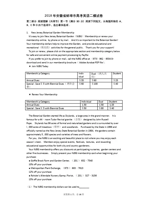
2018年安徽省蚌埠市高考英语二模试卷
B.$60.
C.$80.
D.$100.
【答案】
CБайду номын сангаас
C
A
【考点】
完形综合
阅读理解综合
【解析】
本文属于广告类阅读,主要向我们介绍了新泽西植物园的会员,讲述了如何加入会员以及会员所享受的优惠,使读者更了解NJBG的会员.
【解答】
(1)C.推理判断题.根据第一段第三句Your membership dollars help to improve the Garden,and provide educational and recreational(娱乐的)activities for the general public.可知会员费将被用来改善公园的设施,给大众提供教育和娱乐的活动.这些活动都属于服务类的行为,所以会员费将被用来提供更好的服务.故C项正确.
For you, the NJBG is an exciting and beautiful place to visit where you may enjoy each season’ s best.Members enjoy special events, festivals, lectures, and rewarding educational opportunities for both city and country gardeners.
15% off your purchase
(1)The NJBG membership dollars can be used to________
A.offer further education.
B.update online payment.
碧云天western blot步骤
Western实验步骤Western,也称Western blot、Western blotting、Western印迹,是用抗体检测蛋白的重要方法之一。
Western可以参考如下步骤进行操作。
1. 收集蛋白样品(Protein sample preparation)可以使用适当的裂解液,例如碧云天生产的Western及IP细胞裂解液(P0013)、RIPA裂解液等,裂解贴壁细胞、悬浮细胞或组织样品。
对于某些特定的亚细胞组份蛋白,例如细胞核蛋白、细胞浆蛋白、线粒体蛋白等,可以参考相关文献提取这些亚细胞组份蛋白,也可以使用试剂盒进行抽提,例如碧云天生产的细胞核蛋白与细胞浆蛋白抽提试剂盒(P0028)。
收集完蛋白样品后,为确保每个蛋白样品的上样量一致,需要测定每个蛋白样品的蛋白浓度。
根据所使用的裂解液的不同,需要采用适当的蛋白浓度测定方法。
因为不同的蛋白浓度测定方法对于一些去垢剂和还原剂等的兼容性差别很大。
如果使用碧云天生产的Western及IP细胞裂解液或RIPA裂解液,可以使用BCA蛋白浓度测定试剂盒(P0009/P0010/P0010S/P0011/P0012/P0012S)。
2. 电泳(Electrophoresis)(1) SDS-PAGE凝胶配制SDS-PAGE凝胶可以参考一些文献资料进行配制,也可以使用碧云天生产的SDS-PAGE凝胶配制试剂盒(P0012A)。
该试剂盒提供了除水和配胶器具外的所有试剂以及配制各种浓度SDS-PAGE的配方。
(2) 样品处理在收集的蛋白样品中加入适量浓缩的SDS-PAGE蛋白上样缓冲液。
例如2X或5X的SDS-PAGE蛋白上样缓冲液。
使用5X的SDS-PAGE蛋白上样缓冲液可以减小上样体积,在相同体积的上样孔内可以上样更多的蛋白样品。
5X的SDS-PAGE蛋白上样缓冲液可以参考相关文献资料配制,也可以使用碧云天生产的SDS-PAGE蛋白上样缓冲液(5X)(P0015)。
固相萃取-高效液相色谱检测牛肉中四环素类抗生素残留
固相萃取-高效液相色谱检测牛肉中四环素类抗生素残留陈士恩;程燕平;阿依木古丽;霍生东;申晓蓉;刘丽霞【期刊名称】《西北民族大学学报(自然科学版)》【年(卷),期】2011(032)001【摘要】利用Waters2695高效液相色谱仪对牛肉中四环素类抗生素残留进行测定.样品经5%高氯酸溶液提取、C18固相萃取柱净化、旋转浓缩、0.22μm微孔滤膜过滤,反相色谱分离,紫外检测.色谱柱为Waters Nova-parkC18柱(4μm,3.9mmi.d.×150mm),以磷酸二氢钠溶液-乙腈-甲醇(体积比为70:20:10)为流动相,流速为0.8 mL/min.紫外检测器检测波长为365nm,每次进样10μL.结果是土霉素、四环素、金霉素的检出限分别为0.036 5μg/mL0.043 0μg/mL、0.0558μg/mL,线性范围为0.1μg/mL~-1.0μg/mL.测定54份牛肉样品,只有一份牛肉的金霉素残留为132μg/kg,高于我国农业部规定的量高允许残留量标准(100μg/kg).【总页数】6页(P62-67)【作者】陈士恩;程燕平;阿依木古丽;霍生东;申晓蓉;刘丽霞【作者单位】西北民族大学生命科学与工程学院,甘肃兰州,730030;西北民族大学生命科学与工程学院,甘肃兰州,730030;西北民族大学生命科学与工程学院,甘肃兰州,730030;西北民族大学生命科学与工程学院,甘肃兰州,730030;西北民族大学生命科学与工程学院,甘肃兰州,730030;西北民族大学生命科学与工程学院,甘肃兰州,730030【正文语种】中文【中图分类】S851.4【相关文献】1.分子印迹固相萃取-高效液相色谱法测定牛奶中四环素类抗生素残留 [J], 付珍珍;曾月;李增威;何利;周康;刘书亮;邹立扣;敖晓琳;陈姝娟2.限进材料固相萃取-高效液相色谱在线联用检测牛奶中四环素类抗生素残留 [J], 杨旭;刘美娇;林深;徐丹;董襄朝3.固相萃取-高效液相色谱法测定有机肥中四环素类抗生素药物残留 [J], 唐春玲;张文清;夏玮;左萍萍;朱恩4.猪肉中四环素类抗生素残留的固相萃取-高效液相色谱荧光测定法 [J], 林荆;林奕帆;林立峰;赵建晖;吴文凡5.固相萃取-高效液相色谱法同时测定牛奶中3种四环素类抗生素残留量 [J], 蔡志斌;张英;刘丽因版权原因,仅展示原文概要,查看原文内容请购买。
技术磷酸化蛋白Western跑不出来好捉急,解螺旋给你支招
技术磷酸化蛋白Western跑不出来好捉急,解螺旋给你支招作者:解螺旋.大肚腩转载请注明来源:解螺旋,医生科研助手话说我做硕士课题的时候需要跑磷酸化的AMPK(p-AMPK),当时同组四五个孩子一起跑,效果都很不理想,要么是条带过淡,要么是根本出不来。
那几个月的时间我们疯狂的到处请教师兄师姐,在各种实验论坛上寻找改进方法,最终出来效果不错,文章也发了,在此总结几个Western跑磷酸化蛋白的要点,希望各位科研民工们少走弯路。
抗体工欲善其事必先利其器,抗体很关键。
这里我们要说的并不是最贵的就是最好的,如果条件允许,建议同时购买不同厂家的抗体,一起来试一试,因为有时候即使是最好的品牌,不同批次稳定性也不一样。
抗体从订购到到货有一定的时间,所以到货后请尽快分装好,准备做实验,我们当时因为实验进度问题,抗体到了3个多月才开始正式实验,这样拖了时间找厂家售后申诉也是很被动的。
我们那时候用CST的一抗,感觉效果不稳定,也可能跟抗体放久了有关。
最后买了另一家代理的抗体算是有了比较稳定的结果。
当然,还是选择购买口碑较好的公司的产品为佳,这样即使出了问题售后也会跟进。
如果经费有限,可以买买小包装的产品先试试,不少抗体公司还有5ul的抗体试用装免费申请。
再者,孵育一抗的时候,建议用新鲜抗体,什么意思呢?很多同学因为想图省事儿,很多时候配好孵育液,每次孵育好之后再次放进冰箱等待下次重复利用,一些比较好跑的蛋白到时没啥问题,但是磷酸化的蛋白建议每次都用新鲜的抗体原液,将条带剪下来后用杂交袋塑封好,按1:1000的比例加入抗体原液孵育,再这种情况下,100ul的抗体也是能用很久的,所以尽量每次都用新的抗体。
蛋白上样普遍反映磷酸化的条带很淡,有时候达不到制图标准,这点可以加大上样量来解决,另外,磷酸化蛋白一般与非磷酸化的蛋白一起跑一起制图,它本来就是要比非磷酸化的要淡一些,不要过分担心。
电泳不同kb的蛋白电泳时间有所差别,参数啥的要用脑子更改,不能一成不变,避免发生那种蛋白跑不见了的惨剧。
nox4分子量
nox4分子量
NOX4是一种酶,属于NADPH氧化酶家族的成员之一。
它在细胞中起着重要的作用,参与调节细胞内的氧化还原平衡和细胞信号传导等生物学过程。
NOX4的分子量是约65-70千道尔顿(kDa),其中kDa是千道尔顿的单位,用于表示蛋白质或分子的相对分子质量。
NOX4是一个由多个氨基酸组成的蛋白质,在人体细胞中通常由577个氨基酸残基构成。
在细胞中,NOX4主要定位在内质网(endoplasmic reticulum, ER)膜上,并通过将NADPH(还原型辅酶)转化为活性的超氧阴离子(O2•-)来催化化学反应。
这个过程被称为酶促电子传递,它是细胞内产生ROS(reactive oxygen species,活性氧物质)的重要途径之一。
NOX4可以与其他蛋白质相互作用,形成复合物,以便在细胞内特定位置执行其功能。
通过生成ROS,NOX4参与调节细胞内的氧化还原状态,维持正常的细胞生理功能。
此外,NOX4还参与调节细胞信号传导途径,如细胞增殖、凋亡和分化等。
在某些病理条件下,NOX4的过度活化可能会导致氧化应激和细胞损伤,与多种疾病的发展有关,如心血管疾病、炎症性疾病和肿瘤等。
因此,NOX4成为了药物开发和治疗相关疾病的潜在靶点。
总结起来,NOX4是一种分子量约为65-70kDa的酶,通过产生ROS参与调节细胞内的氧化还原平衡和信号传导。
它在多种生物学过程中发挥重要作用,并且与多种疾病的发展相关。
1。
Western一抗二抗去除液使用说明书
htpp://
Western 一抗二抗去除液
Western 一抗二抗去除液 Western 一抗二抗去除液(Stripping buffer),用于 Western 中转移了蛋白的膜的重复利用。 在 Western 中完成了一抗二抗结合和后续的化学发光检测后,有时还需要检测 tubulin、 actin 等表达量相对稳定的蛋白作为参照,或检测其它蛋白进行比较。通过使用一抗二抗去 除液,充分去除一抗二抗,可以非常方便地重新利用使用过的膜检测其它蛋白。和重新跑一 个 SDS-PAGE 胶相比,不仅省时省力,而且可以消除重新上样而带来的误差,使可比性更强。 使用 Western 一抗二抗去除液多次重复使用同一张膜,会导致蛋白信号的减弱。Western 一 抗二抗去除液,通常可以重复利用膜 3-5 次。
温馨提示 为了您的自身安全,使用试剂前,请做好防护,如穿实验服,带手套等。
储存温度 2~8℃保存。
包装清单 货号
HCY055
品名 Western 一抗二抗去除液说书包装 100mL 1份
1
注意事项 使用辣根过氧化物酶(HRP),任何一次封闭都应当使用 5%脱脂牛奶,如果使用碱性磷酸酯酶, 任何一次封闭都应当使用酪蛋白(casein)。 使用 ECL 等类似的化学发光试剂进行的 Western 检测适用本试剂。使用非化学发光试剂进行 的 Western 检测,例如 DAB,NBT/BCIP,不适用于本试剂。
使用说明 1. 在完成 Western 化学发光检测后,蒸馏水中漂洗 5min。 2. 弃蒸馏水,加入适量的 Western 一抗二抗去除液,至少需把膜完全覆盖。在摇床上漂洗 10min。 3. 弃 Western 一抗二抗去除液,并吸尽残余液体。加入 TBS、TBST 或 PBS 漂洗 3-4 次,每 次在摇床上漂洗 3-5min。 4.进行封闭等 Western 的后续操作。
211132938_GC-MS_法测定酊剂中4_种邻苯二甲酸酯类塑化剂
分析检测GC-MS法测定酊剂中4种邻苯二甲酸酯类塑化剂王 毅,任红燕,乔 彬(喀什地区食品药品检验所,新疆喀什 844000)摘 要:目的:探索一种新的、可靠的、用于检测酊剂中4种邻苯二甲酸酯类塑化剂含量的方法。
方法:样品采用正己烷超声提取,色谱柱为TM-5MS(30 m×0.25 mm×0.25 µm),载气为氦气,流速为1.2 mL·min-1,进样口温度为260 ℃,柱温为程序升温,电子轰击电离方式(EI),离子源温度为230 ℃,离子监测(SIM)模式。
结果:4种PAEs分离良好,在0~1.00 µg·mL-1内呈良好的线性关系,相关系数r>0.99,检出限为0.038~0.081 mg·kg-1,加样回收率在85.0%~104.7%,相对标准偏差在0.5%~6.0%。
结论:该法分离效果好、准确、灵敏度高,可同时检测塑料包装酊剂中4种邻苯二甲酸酯类塑化剂。
关键词:邻苯二甲酸酯类;塑化剂;酊剂Determination of 4 Phthalate Plasticizers in Tincture byGC-MSWANG Yi, REN Hongyan, QIAO Bin(Kashgar Regional Food and Drug Inspection Institute, Kashgar 844000, China) Abstract: Objective: To explore a new and reliable method for determining the content of four phthalate plasticizers in tinctures. Method: The samples were extracted by n-hexane ultrasound, TM-5MS(30 m×0.25 mm×0.25 µm), carrier gas was helium, flow rate 1.2 mL·m in-1, inlet temperature was 260 ℃, column temperature was programmed heating, electron impact ionization mode (EI), ion source temperature was set at 230 ℃, ion monitoring (SIM) mode. Result: The four PAEs were well separated and showed a good linear relationship in the range of 0~1.00 µg·mL-1. The correlation coefficient (r) was more than 0.99, the detection limit was 0.038~0.081 mg·kg-1, the recovery rate of sample addition was 85.0%~104.7%, and the relative standard deviation is between 0.5% and 6.0%. Conclusion: The method is effective,accurate and sensitive for the determination of 4 phthalate plasticizers in plastic packed tinctures.Keywords: phthalates; plasticizers; tincture邻苯二甲酸酯类(PAEs)增塑剂是一种塑料工业加工助剂,能够改善塑料制品的可塑性和柔韧性,广泛用于食品、玩具、医药行业等[1]。
Western及IP细胞裂解液(无抑制剂)使用说明
Western及IP细胞裂解液(无抑制剂)使用说明货号:LS00160规格:100mL保存:-20℃保存,12个月。
产品说明:多种成分均可以从细胞中提取总蛋白,如Triton、SDS、NP-40等,Western及IP细胞裂解液是采用一种非变性裂解方法来裂解细胞,并获得总蛋白的裂解液。
所获得的蛋白质可以用于PAGE电泳,Western,免疫沉淀(Immunol Precipitation,IP)和免疫共沉淀(co-IP)等,主要由Tris-HCl、NaCl、低浓度Triton X-100,低浓度sodium pyrophosphate等组成,不含蛋白酶、磷酸酶抑制剂,并维持原有的蛋白间相互作用。
用Western及IP细胞裂解液(无抑制剂)(Cell lysis buffer for Western and IP without inhibitors)得到的蛋白,可以用BCA蛋白定量试剂盒测定蛋白浓度。
由于含有较高浓度的Triton X-100等干扰物质,不宜用Bradford法测定由Western及IP细胞裂解液获得样本的蛋白浓度。
操作步骤(仅供参考):(一)贴壁培养细胞1、取Western及IP细胞裂解液室温溶解混匀,根据需要选择添加或不添加蛋白酶抑制剂。
2、去除贴壁细胞的培养液,用PBS、NS或无血清培养液清洗1次,低速离心,弃上清,留取沉淀。
3、按照6孔板每孔加入100~200µL裂解液的比例,加入Western及IP细胞裂解液。
移液器轻轻吹打,使裂解液和细胞充分接触。
通常裂解液作用于细胞1~5s内,细胞就会被裂解。
4、10000~12000g,离心3~5min(如果用冷冻离心机4℃离心效果更佳),取上清。
5、进行后续的SDS-PAGE、Western、免疫沉淀和免疫共沉淀等操作。
(二)悬浮培养细胞1、取Western及IP细胞裂解液室温溶解混匀,根据需要选择添加或不添加蛋白酶抑制剂。
细胞内叠氮化物反应探针的英文
细胞内叠氮化物反应探针的英文Intracellular Nitrogenase Reaction Probes: Applications and Advancements.Intracellular nitrogenase reaction probes have emerged as crucial tools in modern biochemistry, enabling researchers to monitor and understand the intricate nitrogen metabolism within cells. These probes, often fluorescently labeled, allow for real-time visualization of nitrogen fixation and associated processes, thereby providing insights into the dynamic nature of nitrogen metabolism.Background and Importance.Nitrogen is an essential element for all known forms of life, playing a pivotal role in amino acid synthesis, nucleic acid structure, and various other biological processes. However, nitrogen in its free form (N2) is unavailable for direct biological utilization due to itsinertness. Therefore, organisms rely on nitrogenases, a class of enzymes that catalyze the conversion of N2 into ammonia (NH3), a biologically usable form of nitrogen.Within cells, nitrogenase enzymes are often embedded within complex systems, involving multiple cofactors and electron transport chains. Monitoring these reactionswithin the cellular milieu is challenging due to the dynamic nature of the cellular environment and the often-subtle changes in substrate concentration. Intracellular nitrogenase reaction probes have been developed to overcome these challenges, providing a window into the intracellular world of nitrogen metabolism.Types of Intracellular Nitrogenase Reaction Probes.1. Fluorescent Probes: These probes are labeled with fluorescent molecules that change their emission properties upon interacting with nitrogenase or its intermediates. For example, fluorophores such as fluorescein or rhodamine can be conjugated to specific substrates or inhibitors of nitrogenase, allowing for the detection of enzymaticactivity through fluorescence microscopy or flow cytometry.2. Bioluminescent Probes: These probes emit light through a chemical reaction triggered by nitrogenase activity. Bioluminescent probes offer the advantage of being self-luminous, eliminating the need for external excitation sources.3. Radiolabeled Probes: Radiolabeled probes incorporate radioactive atoms (e.g., carbon-14 or tritium) into substrates or inhibitors of nitrogenase. The subsequent detection of radiolabeled products provides quantitative information about enzyme activity. However, the use of radiolabeled probes is limited due to safety concerns and the need for specialized equipment.Applications of Intracellular Nitrogenase Reaction Probes.1. Studying Nitrogen Fixation Pathways: By monitoring the activity of nitrogenase within cells, probes can reveal the preferred nitrogen fixation pathway utilized bydifferent organisms. This information is crucial for understanding the adaptability of microorganisms to varying nitrogen sources and environmental conditions.2. Analyzing Nitrogen Metabolism in Response to External Stimuli: Intracellular probes can be used to study how nitrogen metabolism is affected by external factors such as changes in nutrient availability, pH, or temperature. Such studies can provide insights into the mechanisms underlying cellular responses to environmental perturbations.3. Drug Discovery and Therapeutics: Nitrogenase inhibitors have been explored as potential therapeutics for treating diseases associated with abnormal nitrogen metabolism, such as cancer or certain infectious diseases. Intracellular probes can aid in the identification of effective inhibitors by allowing for high-throughput screening of candidate compounds.Future Directions.With the continuous advancement of biotechnology and imaging techniques, intracellular nitrogenase reaction probes are poised to make significant contributions to our understanding of nitrogen metabolism. Future research may focus on developing probes with improved sensitivity and specificity, enabling the detection of nitrogenase activity in single cells or even subcellular compartments. Additionally, the integration of probes with other omics technologies (e.g., genomics, proteomics, or metabolomics) could provide a comprehensive picture of nitrogen metabolism within cells, leading to new insights and potential therapeutic targets.In conclusion, intracellular nitrogenase reaction probes have emerged as invaluable tools for studying nitrogen metabolism within cells. Their ability to monitor enzymatic activity in real-time, combined with their versatility and sensitivity, makes them critical for advancing our understanding of nitrogen metabolism and its role in health and disease. As technology continues to evolve, these probes will play increasingly important roles in fundamental and applied research.。