MAPKAPK2
高通量药物筛选利器——HTRF,在激酶研究(kinase)中的应用

通过直接激发使镧系元素离子产生荧光是 不容易的,因为这些离子很难吸收光子。镧系 元素必须首先与有机分子形成复合物,有机分子收集光子并通过分子内非放射过程转移到 镧系元素上。稀土元素螯合物和穴状化合物是能量收集装置的典型代表,它们收集能量并 转移到镧系元素离子上,后者则发出其特征性的长寿命的荧光。
为了能够成功应用于生物学检测中,稀土元素复合物应该具有特定的性质,包括稳定 性、较高的发射光产率,并且能够与生物分子连接。除此之外,当直接在生物溶液中反应 时,能够耐受荧光淬灭就显得尤为重要。稀土元素螯合物稳定性较差,而且有的化合物可 竞争螯合物活性基团,当与 FRET 技术结合在一起时其灵敏度也受到限制。如果稀土元素 与穴状化合物结合,许多限制因素都可去除。
一般地,在 FRET 实验中使用的供体和受体是快速荧光基团,半衰期非常短。传统 FRET 技术的限制因素是由背景荧光引起的,其来自于样品成分,包括缓冲液、蛋白质、 化合物和细胞裂解液。检测到的荧光强度必须对这些自发荧光进行校正,极大地影响了实 验灵敏度,并使数据分析变得复杂。背景荧光非常短暂(寿命为纳秒级),可以利用时间 分辨荧光方法将其去除。
图 2 :试剂盒包装
产品特点 应用单克隆抗体保证批次的一致性 在 100 多种丝氨酸/苏氨酸激酶和 60 多种酪氨酸激酶上验证过 酶用量很少 ATP 浓度没有限制,因此可以用 ATP Km 进行筛选药物 每一个底物只有一个位点可以被磷酸化 针对酪氨酸激酶有特殊的试剂可以增强底物的稳定性 整个实验少于 2 小时 反应体系可以减少到 4ul
在保卫细胞信号转导中MAPK级联途径

浅谈在保卫细胞信号转导中MAPK级联途径摘要:叶片表面的气孔是由保卫细胞构成的特殊结构,是气体出入植物体的主要通道。
气孔可以通过保卫细胞控制植物与外界大气的气体交换,影响光合作用和蒸腾过程,对环境和内源信号进行感知从而对胁迫环境做出响应,以此减轻胁迫程度并提高植物的抗性。
而促分裂原活化蛋白激酶(MAPK)级联在这一过程中至关重要。
促分裂原活化蛋白激酶(MAPKs)形成具有蛋白激酶和MAPK激酶激酶的三层激酶级联,其信号转导到靶蛋白是以三级激酶级联的方式进行的。
在所有真核生物中,MAPK级联基因高度保守,并且它们在各生长发育及生理过程中发挥关键作用。
在生物和非生物胁迫反应过程中,MAPK级联功能通过将受体接收的细胞外信号与胞质事件和基因表达相连接而发挥作用。
本文以MAPK级联途径在保卫细胞信号转导为侧重点,将近年来MAPK级联的特异性的研究情况加以综述并分析亟待解决的问题,对以后的研究方向提出展望,以期为进一步的深入研究提供理论参考。
关键词MAPK级联;气孔;气孔运动;保卫细胞Brief Discussion on MAPK Cascades in Guard Cell Signal Transduction Abstract: Guard cells form stomata on the epidermis and continuously respond to endogenous and environmental stimuli to fine-tune the gas exchange and transpirational water loss, processes which involve mitogen-activated protein kinase (MAPK) cascades. MAPKsform three-tiered kinase cascades with MAPK kinases and MAPK kinase kinases, bywhich signals are transduced to the target proteins. MAPK cascade genes are highlyconserved in all eukaryotes, and they play crucial roles in myriad developmental andphysiological processes. MAPK cascades function during biotic and abiotic stress responses by linking extracellular signals received by receptors to cytosolic events and gene expression. In this review, we highlight recent findings and insights into M A P K- mediated guard cell signaling, including the specificity of MAPK cascades.The future research directions were also discussed,which could offer scientific references for its rational and efficientdevelopment.Key words: MAPK cascade, stomatal pore, stomatal movement, guard cell在光合作用过程中,通过由表皮中的保卫细胞围成的气孔,植物和大气之间进行气体交换和水分蒸腾[1]。
MicrosoftWord-4423-4426原野40doc

中国组织工程研究与临床康复第15卷第24期 2011–06–11出版Journal of Clinical Rehabilitative Tissue Engineering Research June 11, 2011 Vol.15, No.24 ISSN 1673-8225 CN 21-1539/R CODEN: Z LKHAH4423 1Department of Spine Surgery, Tangshan Second Hospital, Tangshan 063000, Hebei Province, China; 2Department of Pathology, Hebei Union University, Tangshan 063000, Hebei Province, ChinaYuan Ye★, Master, Associate chief physician, Department of Spine Surgery, Tangshan Second Hospital, Tangshan 063000, Hebei Province, China**************** Yuan Ye and Zhao Jing contributed equally to this paper. Correspondence to: Zhao Jing, Master, Associate professor, Master’s supervisor, Department of Pathology, Hebei Union University, Tangshan 063000, Hebei Province, China**************** Supported by: the Natural Science Foundation of Education Department of Hebei Province, No.Z2005102*; the Science and Technology Development Program of Hebei Province, No. 052761422* Received: 2011-03-07 Accepted: 2011-05-06磷酸化p38丝裂原活化蛋白激酶与转化生长因子β1在正常和腰椎间盘退变组织中的表达**★原野1,赵静2,赵杰2,李永民1,王旭1Phosphorylated p38 mitogen-activated protein kinase and transforming growth factor beta-1 expression in degenerative versus normal disc tissuesYuan Ye1, Zhao Jing2, Zhao Jie2, Li Yong-min1, Wang Xu1AbstractBACKGROUND:Studies show that p38 mitogen activated protein kinase (MAPK) signal pathway is highly correlated withinflammatory reactions during intervertebral disc degeneration.OBJECTIVE: To study the phosphorylated p38MAPK (p-p38MAPK) and transforming growth factor β1 (TGF-β1) expression inpatients with intervertebral disc degeneration.METHODS: Degenerative disc tissues resected by surgery from 36 patients with lumbar disc herniation were selected andnormal disc tissues from 10 cases of trauma were used as control group. The expression of p-p38MAPK and TGF-β1 wasevaluated with immunohistochemistry method and analyzed using pathological imaging analysis system.RESULTS AND CONCLUSION: The positive rate of p-p38MAPK and TGF-β1 expression in degenerative disc tissues wasgreater than normal disc tissue (P < 0.05). Results demonstrated that p-p38MAPK and TGF-β1 are highly expressed in theprocess of disc degeneration.Yuan Y, Zhao J, Zhao J, Li YM, Wang X. Phosphorylated p38 mitogen-activated protein kinase and transforming growth factorbeta-1 expression in degenerative versus normal disc tissues. Zhongguo Zuzhi Gongcheng Yanjiu yu Linchuang Kangfu.2011;15(24): 4423-4426. [ ]摘要背景:研究表明p38丝裂原活化蛋白激酶信号转导通路与椎间盘退变过程中炎症反应密切相关。
TNF信号通路的新检查点:MK2介导的RIPK1磷酸化

TNF信号通路的新检查点:MK2介导的RIPK1磷酸化订阅号APExBIOTNF是一种炎性细胞因子,当与其受体TNFR1结合时,可以驱动细胞因子的产生,细胞的存活或死亡。
TNFR1的刺激引起NF-κB,p38α及其下游效应激酶MK2的激活,从而促进靶基因的转录,mRNA的稳定和翻译。
来自英国Chester Beatty实验室ICR Breast Cancer Now Toby Robins研究中心的一篇研究显示MK2直接磷酸化RIPK1 的S321位点,抑制RIPK1的潜在细胞毒性,是TNF信号通路中的新的检查点。
肿瘤坏死因子(TNF)是主要的炎性细胞因子。
TNF通过调节炎症,细胞增殖、分化、存活和死亡来维持内稳态。
虽然TNF可导致细胞死亡,但大多数类型细胞的主要结局是细胞存活和促炎细胞因子的产生。
TNF / TNFR1诱导至少两种细胞信号传导复合物:受体相关的质膜复合物(复合物I),激活NF-κB和MAPK以及转录和翻译;胞质复合物(复合体II),其作用是启动细胞死亡。
TNFR1被刺激后,迅速募集TRADD,TRAF2, RIPK1和细胞凋亡抑制剂cIAP1和cIAP2,组装复合物I。
RIPK1和复合物I的其它组分被cIAP快速泛素化。
泛素(Ub)与RIPK1和复合物I组分的结合引起TAK1介导的IKK2,JNK,ERK 和p38α的活化。
复合物I的形成引起NF-κB和丝裂原活化蛋白激酶(MAPK)的激活,其最终导致细胞因子和促生存蛋白如cFLIP的产生,其是协调炎症反应所必需的。
复合物I形成之后,TNF还启动基于RIPK1的细胞质复合物的形成,即复合物II。
复合物II通过激活caspase-8和细胞凋亡,或通过RIPK3和MLKL,引起细胞坏死。
目前认为,一小部分RIPK1在30分钟至3小时内与复合物I解离,并与TRADD 一起与衔接蛋白FADD和procaspase-8结合形成复合体II。
MAPK信号通路

MAPK 信号通路2008-06-04 21:50 MAPK, 丝裂原活化蛋白激酶( mitogen-activatedprotein kinases,MAPKs )是细胞内的一类丝氨酸/苏氨酸蛋白激酶。
研究证实,MAPKs 信号转导通路存在于大多数细胞内,在将细胞外刺激信号转导至细胞及其核内,并引起细胞生物学反应(如细胞增殖、分化、转化及凋亡等)的过程中具有至关重要的作用。
研究表明,MAPKs 信号转导通路在细胞内具有生物进化的高度保守性,在低等原核细胞和高等哺乳类细胞内,目前均已发现存在着多条并行的MAPKs 信号通路,不同的细胞外刺激可使用不同的MAPKs 信号通路,通过其相互调控而介导不同的细胞生物学反应。
1 并行MAPKs 信号通路的组成及其活化特点在哺乳类细胞目前已发现存在着下述三条并行的MAPKs 信号通路 [1]。
1.1 ERK (extracellular signal-regulated kinase)信号通路1986 年由Sturgill 等人首先报告的MAPK 。
最初其名称十分混乱,曾根据底物蛋白称之为MAP2K 、ERK、MBPK 、RSKK 、ERTK 等。
此后,由于发现其具有共同的结构和生化特征,而被命名为MAPK 。
近年来,随着不同MAPK 家族成员的发现,又重新改称为ERK 。
在哺乳类动物细胞中,与ERK 相关的细胞内信号转导途径被认为是经典MAPK 信号转导途径,目前对其激活过程及生物学意义已有了较深入的认识。
研究证实,受体酪氨酸激酶、G 蛋白偶联的受体和部分细胞因子受体均可激活ERK 信号转导途径。
如:生长因子与细胞膜上的特异受体结合,可使受体形成二聚体,二聚化的受体使其自身酪氨酸激酶被激活;受体上磷酸化的酪氨酸又与位于胞膜上的生长因子受体结合蛋白2( Grb2)的SH2 结构域相结合,而Grb2 的SH3 结构域则同时与鸟苷酸交换因子SOS( Son of Sevenless)结合,后者使小分子鸟苷酸结合蛋白Ras的GDP 解离而结合GTP,从而激活Ras;激活的Ras进一步与丝/苏氨酸蛋白激酶Raf-1 的氨基端结合,通过未知机制激活Raf-1;Raf-1 可磷酸化MEK1 /MEK2 (MAP kinase/ERK kinase)上的二个调节性丝氨酸,从而激活MEKs ;MEKs 为双特异性激酶,可以使丝/苏氨酸和酪氨酸发生磷酸化,最终高度选择性地激活ERK1和ERK2(即p44MAPK 和p42MAPK )。
常见信号通路

JNK生理功能
参与细胞凋亡的调控 细胞存活 肿瘤的形成 机体的发育与分化
(三)p38信号转导通路
p38α:白细胞、肝、脾、骨髓中等高表达
p38β:脑和心脏中高泌器官中高表达
注: p38 α和 p38 β 具有不同的剪接体
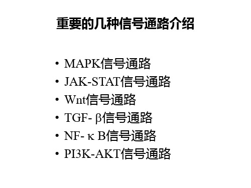
重要的几种信号通路介绍
• • • • • • MAPK信号通路 JAK-STAT信号通路 Wnt信号通路 TGF- 信号通路 NF- B信号通路 PI3K-AKT信号通路
MAPK信号通路 丝裂原活化蛋白激酶
MAPK信号级联反应
Stimulus
Growth factors, Mitogen, GPCR Raf, Mos, Tpl2
•
•
3个基因转录产物的选择性剪接产生10个JNK 亚型 (46kDa, 55kDa);
同一基因编码的46kDa和55kDa亚型无明显的 功能差异 。
JNK信号通路MKK和MKKK
MKK (MAP2Ks) • MKK4 ( SEK1/MEK4/JNKK1/SKK1 )
• 主要激活JNK,但对p38也有活化作用
(二)JNK信号转导通路
• 是已知的应答最多样刺激的细胞信号转 导途径之一 • JNK通过Thr-Pro-Tyr模体的磷酸化被激 活
JNK:
• • • 人的JNK由3个基因 ( jnk1, jnk 2和 jnk3)编码; JNK1和JNK2广泛地在多种组织表达,而 JNK3 主要在脑、心脏与睾丸组织中表达 JNK家族成员间的同源性超过80%;
激活p38途径的物理、化学应激:
• 氧化应激 (巨噬细胞 )
• 低渗压 (HEK293细胞 ) • 紫外线辐射 (PC12细胞 ) • 低氧 (牛肺动脉成纤维细胞 ) • 循环扩张 (肾小球膜细胞 )
所有的看家基因的列表

NM_000918 Homo sapiens procollagen-proline, 2-oxoglutarate 4-dioxygenase (proline 4-hydroxylase), beta polypeptide (protein disulfide isomerase; thyroid hormone binding protein p55) (P4H, mRNA 2719
*NM_000291 Homo sapiens phosphoglycerate kinase 1 (PGK1), mRNA 2727
*NM_005566 Homo sapiens lactate dehydrogenase A (LDHA), mRNA 2105
*NM_002954 Homo sapiens ribosomal protein S27a (RPS27A), mRNA 4156
NM_003753 Homo sapiens eukaryotic translation initiation factor 3, subunit 7 zeta, 66/67kDa (EIF3S7), mRNA 1363
NM_004541 Homo sapiens NADH dehydrogenase (ubiquinone) 1 alpha subcomplex, 1, 7.5kDa (NDUFA1), nuclear gene encoding mitochondrial protein, mRNA 2307
NM_004651 Homo sapiens ubiquitin specific protease 11 (USP11), mRNA 1950
NM_004888 Homo sapiens ATPase, H+ transporting, lysosomal 13kDa, V1 subunit G isoform 1 (ATP6V1G1), mRNA 928
paktool用法

paktool用法apktool是一款用于反编译和重新编译Android APK文件的工具。
以下是apktool的基本用法:1.安装apktool:首先需要下载并安装apktool。
可以在官网或其他可信来源下载最新版本的apktool。
2.解包APK文件:使用apktool解包APK文件,命令如下:phpapktool d <APK文件路径>其中,<APK文件路径>是你要解包的APK文件的路径。
3.编译APK文件:使用apktool重新编译APK文件,命令如下:phpapktool b <APK文件路径>其中,<APK文件路径>是你要编译的APK文件的路径。
编译后会在当前目录下生成一个新的APK文件。
4.签名APK文件:如果你需要将编译后的APK文件进行签名,可以使用以下命令:phpapksigner sign --key <密钥文件路径> --cert <证书文件路径> <APK文件路径>其中,<密钥文件路径>是你要使用的密钥文件的路径,<证书文件路径>是你要使用的证书文件的路径,<APK文件路径>是你要签名的APK文件的路径。
5.安装APK文件:最后,你可以使用以下命令将编译并签名后的APK 文件安装到设备上:phpadb install <APK文件路径>其中,<APK文件路径>是你要安装的APK文件的路径。
以上是apktool的基本用法,你可以根据需要进行进一步的探索和使用。
请注意,在使用apktool时需要遵守相关法律法规和隐私政策,不要用于非法用途。
- 1、下载文档前请自行甄别文档内容的完整性,平台不提供额外的编辑、内容补充、找答案等附加服务。
- 2、"仅部分预览"的文档,不可在线预览部分如存在完整性等问题,可反馈申请退款(可完整预览的文档不适用该条件!)。
- 3、如文档侵犯您的权益,请联系客服反馈,我们会尽快为您处理(人工客服工作时间:9:00-18:30)。
MAPK pathways in radiation responsesPaul Dent*,1,Adly Yacoub1,Paul B Fisher2,Michael P Hagan1and Steven Grant11Department of Radiation Oncology,Virginia Commonwealth University,Richmond,VA23298-0058,USA;2Department of Urology and Pathology,Columbia University,New York,NY10032,USAWithin the last15years,multiple new signal transduction pathways within cells have been discovered.Many of these pathways belong to what is now termed‘the mitogen-activated protein kinase(MAPK)superfamily.’These pathways have been linked to the growth factor-mediated regulation of diverse cellular events such as proliferation, senescence,differentiation and apoptosis.Based on currently available data,exposure of cells to ionizing radiation and a variety of other toxic stresses induces simultaneous compensatory activation of multiple MAPK pathways.These signals play critical roles in controlling cell survival and repopulation effects following irradiation, in a cell-type-dependent manner.Some of the signaling pathways activated following radiation exposure are those normally activated by mitogens,such as the‘classical’MAPK(also known as the ERK)pathway.Other MAPK pathways activated by radiation include those downstream of death receptors and procaspases,and DNA-damage signals,including the JNK and P38MAPK pathways. The expression and release of autocrine growth factor ligands,such as(transforming growth factor alpha)and TNF-a,following irradiation can also enhance the responses of MAPK pathways in cells and,consequently, of bystander cells.Thus,the ability of radiation to activate MAPK signaling pathways may depend on the expression of multiple growth factor receptors,autocrine factors and Ras mutation.Enhanced basal signaling by proto-oncogenes such as K-/H-/N-RAS may provide a radioprotective and growth-promoting signal.In many cell types,this may be via the PI3K pathway;in others, this may occur through nuclear factor-kappa B or multiple MAPK pathways.This review will describe the enzymes within the known MAPK signaling pathways and discuss their activation and roles in cellular radiation responses.Oncogene(2003)22,5885–5896.doi:10.1038/sj.onc.1206701 Keywords:signal transduction;kinase;phospatase;re-ceptor;autocrine ligand;RAS The‘classical’mitogen-activated protein kinase(MAPK)/ extracellular-regulated kinase(ERK)pathway‘MAP-2kinase’wasfirst reported by the laboratory of Dr Thomas Sturgill in1986(Sturgill and Ray,1986) (Figure1).This protein kinase was originally described as a42-kDa insulin-stimulated protein kinase activity whose tyrosine phosphorylation increased after insulin exposure,which phosphorylated the cytoskeletal protein MAP-2.Contemporaneous studies from the laboratory of Dr Melanie Cobb identified an additional44-kDa isoform of this enzyme,termed ERK1(extracellular signal regulated kinase)(Boulton and Cobb,1991). Since many growth factors and mitogens can activate these enzymes,the acronym for this enzyme was subsequently changed to denote mitogen-activated protein(MAP)kinase.Additional studies demonstrated that the p42(ERK2)and p44(ERK1)MAP kinases regulated another protein kinase activity(P90RSK) (Sturgill et al.,1988),and that they were themselves regulated by protein kinase activities designated MKK1/ 2(MAPK kinase;MAP2K),also termed MEK1/2 (mitogen-activated/extracellular-regulated kinase1/2) (Wu et al.,1992,1993;Haystead et al.,1993;Robbins et al.,1993).MKK1and MKK2were also regulated by reversible phosphorylation.The protein kinase respon-sible for catalyzing MKK1/2activation was initially described as the proto-oncogene RAF-1(Kyriakis et al., 1992;Dent et al.,1992;Haystead et al.,1993).This was soon followed by another MEK1/2activating kinase, termed MEKK1,which was a mammalian homologue with similarity to the yeast Ste11and Byr2genes (Lange-Carter et al.,1993).However,further studies have shown that the primary function of MEKK1is to regulate the c-Jun NH2-terminal kinase(JNK),rather than the ERK,pathway(Yan et al.,1994)(see the JNK pathway,below).MEK1,in addition to being regulated by RAF family members,can also be both positively and negatively controlled via phosphorylation at other sites.Cyclin-dependent kinases have been shown to phosphorylate T286and inhibit MEK1activity(Rossomando et al., 1994).In contrast,phosphorylation of MEK1at T292 and S298by PAK family enzymes downstream of Rac GTPase molecules enhances the interaction of MEK1 and ERK1/2,promoting activation of the ERK molecules(Frost et al.,1996;Coles and Shaw,2002; Eblen et al.,2002).*Correspondence:P Dent;E-mail:pdent@Oncogene(2003)22,5885–5896&2003Nature Publishing Group All rights reserved0950-9232/03$25.00/oncRAF-1is a member of a family of serine-threonine protein kinases termed RAF-1,B-RAF and A-RAF.All ‘RAF’family members can phosphorylate and activate MKK1/2,although the relative ability of each member to catalyze this reaction varies (B-RAF 4RAF-14A-RAF)(Bosch et al .,1997;Marais et al .,1997).Different growth factors,in a cell-type-dependent manner,have been shown to utilize either RAF-1or B-RAF to activate the ERK pathway (Tombes et al .,1998).Thus,the ‘RAF’kinases act at the level of an MAPK kinase (MAP3K).Of note,we have found in A431squamous carcinoma cells exposed to doses of B 2Gy that ionizing radiation activates RAF-1,but not B-RAF (Kasid et al .,1996;Schmidt-Ullrich et al .,1997;Suy et al .,1997).Other studies have shown in fibroblasts with a somatic deletion of exon 2within the RAF-1and B-RAF genes,generating nonfunctional proteins,that loss of RAF-1and B-RAF function reduced growth-factor-induced ERK1/2activation and enhanced apop-tosis (Wojnowski et al .,1997,1998,2000).This has also been noted following irradiation of these cells (Suy et al .,2001).Using an inducible deletion system in avian DT40B cells,the contribution of RAF-1and B-RAF to B-cell antigen receptor signaling has also been examined.Loss of RAF-1has no effect on BCR-mediated ERK activation,whereas B-RAF-deficient DT40cells display a reduced basal ERK activity as well as a shortened BCR-mediated ERK activation.The RAF-1/B-RAF double-deficient DT40cells show an almost complete block both in ERK activation and in the induction of the immediate-early gene products c-FOS and EGR-1(Brummer et al .,2002).These findings are in contrast to studies in embryonic fibroblasts where all RAF-1exons have been deleted (RAF-1À/Àcells),and in which growth factor signaling to ERK1/2has not been altered in the absence of RAF-1function (Huser et al .,2001).These contrasting findings argue that cells,particularly embryonic fibroblasts,mayutilize compensatory signaling from other MAP3K molecules when RAF-1expression is completely abol-ished.Several studies have demonstrated that the NH 2domain of RAF-1could reversibly interact with RAS in the plasma membrane,and that the ability of RAF-1to associate with RAS is dependent upon the RAS molecule being in the GTP-bound state (Moodie et al .,1993;Van Elst et al .,1993).Other findings have proved that the ability of RAF-1to be activated depends upon RAF-1translocation to the plasma membrane (Dent and Sturgill,1994;Leevers et al .,1994)(Figure 1).However,the regulation of RAF-1activity appears to be more complex than simple membrane translocation,with several additional mechanisms coordinately reg-ulating activity when in the plasma membrane environ-ment (Stokoe and McCormick,1997;Thorson et al .,1998;Tzivion et al .,1998;McPherson et al .,1999;Yip-Schneider et al .,2000;Light et al .,2002;Jaumot and Hancock,2001).Data from several laboratories has suggested that protein serine/threonine and tyrosine phosphorylation play a role in increasing RAF-1activity when in the plasma membrane environment (Fabian et al .,1993;Dent et al .,1995;Marais et al .,1995).Other studies have suggested that protein kinase C (PKC)isoforms can regulate RAF-1activity (Cai et al .,1997;Schonwasser et al .,1998).Phosphorylation of S338by PAK family enzymes,downstream of RHO GTPase and PI3K,has been shown to play a role in the RAF-1activation process (Sun et al .,2000),which may facilitate RAF-1tyrosine phosphorylation by Src family members (King et al .,2001).Other investigators have suggested that the lipid second messenger ceramide may be able to play a role in RAF-1activation (Zhang et al .,1997;Muller et al .,1998).The initial biochemical analyses of purified RAF-1,grown either in the presence or absence of active H-RAS V12,demonstrated near constitutive phosphorylation of S259and S621,with constitutive Y340-Y341phosphor-ylation when the protein was coexpressed with SRC (Fabian et al .,1993;Morrison et al .,1993).Site-specific modification of S259A resulted in a RAF-1protein that was B two-fold more active than the wild-type protein,suggesting that S259was a negative regulatory site on RAF-1(Morrison et al .,1993;Dent et al .,1995).More recent studies have shown that phosphorylation of RAF-1at S259by either AKT or the cAMP protein kinase (PKA)can inhibit RAF-1activity and its activation by upstream stimuli (Zimmermann and Moelling,1999;Yip-Schneider et al .,2000;Reusch et al .,2001;Dhillon et al .,2002).Phosphorylation of RAF-1at S43by PKA inhibits the interaction of RAF-1with RAS,thereby blocking RAF-1translocation to the plasma membrane and its RAS-dependent activation (Wu et al .,1993).In contrast to RAF-1,the B-RAF isoform does not contain an equivalent to S43,but contains multiple sites of AKT-mediated phosphoryla-tion in addition to the B-RAF equivalent of S259(Guan et al .,2000).B-RAF may also be activated by cAMP via the RAP1GTPase that has also been linked to SrcReceptor Homo-and Hetero-Figure 1Some of the characterized signal transduction pathways in mammalian cells.Growth factor receptors,for example,the ERBB family,can signal down through GTP-binding proteins into multiple intracellular signal transduction pathways.Predominant among these pathways is the MAP kinase superfamily of cascades (ERK1/2,ERK5,JNK,P38)as well as the PI3K pathwayRadiation and signalingP Dent et al5886Oncogenetyrosine kinase family members(York et al.,2000; Klinger et al.,2002).At the same time that RAF-1was shown to associate with RAS,it was found that growth factors,via their plasma membrane receptors,stimulate GTP for GDP exchange in RAS using guanine nucleotide exchange factors(Li et al.,1993;Olivier et al.,1993).Thus receptor signaling was linked to RAS,which was linked to the RAF/MEK/ERK pathway.Several groups have shown that the epidermal growth factor receptor(EGFR,also called ERBB1and HER1) is activated in response to irradiation of various carcinoma cell types(Balaban et al.,1996;Schmidt-Ullrich et al.,1997;Carter et al.,1998;Kavanagh et al., 1998;Bowers et al.,2001).Radiation exposure in the range of1–2Gy,via activation of the EGFR,can activate the ERK pathway to a level similar to that observed by physiologic,growth stimulatory,EGF concentrations(B0.1n m).Recent publications argue that radiation-induced free radicals play an important role in the activation of ERBB family receptors through the ERK pathway(Leach et al.,2001).The actions of ERBB receptor autocrine ligands have also been shown to play important roles in the activation of the ERK pathway after radiation expo-sure.Transforming growth factor-alpha(TGF-a)has been shown to mediate secondary activation of the EGFR and the downstream ERK and JNK pathways after irradiation of several carcinoma cell lines(Dent et al.,1999;Hagan et al.,2000).In these studies, radiation caused cleavage of pro–TGF-a in the plasma membrane that led to its release into the growth media. Increasing the radiation dose from2Gy up to10Gy enhanced both the secondary activation of ERBB1and the secondary activation of the ERK and JNK path-ways,suggesting that radiation can promote a dose-dependent increase in the cleavage of pro-TGF-a,which reaches a plateau at B10Gy.It should be noted that in contrast to secondary activation,primary activation of the receptor and signaling pathways appeared to reach a plateau at3–5Gy.In addition,signaling by RAS,ERK and P53,the activities of which can be increased following radiation exposure,has been shown in a variety of cell systems to increase the expression of autocrine factors such as HB-EGF and epiregulin(Baba et al.,2000).Thesefindings argue that the activation of ERBB family receptors and the ERK pathway by radiation will be influenced by both the RAS and P53status(mutant or wild type)of a given tumor cell.Thus,for example,in cells expressing a mutant K-RAS protein such as HCT116,loss of mutant RAS function lowers basal ERK activity and reduces epiregulin expression(Baba et al.,2000).This in turn correlates with reduced basal and radiation-induced ERK activation.In addition to playing a role in the activation of the ERK pathway,it is important to note that radiation-stimulated RAF-1may act upon substrates other than MEK1/2,such as the myosin-phosphatase-binding protein(Broustas et al.,2002).RAF-1has also been proposed to act as an inhibitor of apoptosis signaling kinase1(ASK1)by binding to ASK1:the inhibitory actions of RAF-1were reported to be independent ofRAF-1protein kinase activity(Chen et al.,2001a). Antisense oligonucleotides to RAF-1have been shownin vitro and in vivo to enhance the radiosensitivity of tumor cells(Gokhale et al.,1999),and it is most likelythat loss of RAF-1expression enhances the toxic effectsof radiation by promoting ASK1activation rather than altering ERK activity.ERK signaling also has the potential to regulate protein kinases within other growth factor-stimulated pathways.For example,ERK signaling negatively regulates the JNK pathway(see below)and positively regulates the P70S6kinase(Reardon et al.,1999; Contessa et al.,2002).ERK signaling has been shown to facilitate P70S6kinase activation by growth factors andby both UV and ionizing radiation.In the case of ionizing radiation,this has been linked to enhanced translation of certain promoter sequences.The JNK PathwayThe JNK pathway was discovered and described in theearly to mid-1990s(Hibi et al.,1993;Derijard et al., 1994).JNK1and JNK2were initially described bio-chemically to be stress-induced protein kinase activitiesthat phosphorylated the NH2-terminus of the transcrip-tion factor c-JUN;hence,the pathway is often called the stress-activated protein kinase(SAPK)pathway.Multi-ple stresses increase JNK1/2(and the subsequently discovered JNK3)activity including UV-and g-irradia-tion,cytotoxic drugs and reactive oxygen species(e.g.,H2O2).Phosphorylation of the NH2-terminal sites Ser63and Ser73in c-JUN increases its ability to transactivateAP-1enhancer elements in the promoters of many genes (Yang et al.,1998;Davis,1999).It has been recently suggested that JNK can phosphorylate the NH2-terminus of c-MYC,playing a role in both proliferativeand apoptotic signaling(Noguchi et al.,1999).In a manner similar to the previously described MAPK pathway,JNK1/2activities were regulated by dual threonine and tyrosine phosphorylation,which were found to be catalyzed by a protein kinase analogousto MKK1/2,termed stress-activated extracellular regu-lated kinase1(SEK1),also called MKK4(Derijardet al.,1995).An additional isoform of MKK4,termedMKK7,was subsequently discovered(Tournier et al., 1999).As in the case of MKK1and MKK2,MKK4andMKK7were regulated by dual serine phosphorylation. Recent studies have indicated that AKT can phosphor-ylate and inhibit the activity of MKK4(Park et al., 2002).In contrast to the ERK pathway,however,which appears to utilize primarily the three protein kinases ofthe RAF family and possibly MEKK1to activateMKK1/2,at least10protein kinases are known to phosphorylate and activate MKK4/7,including the Ste11/Byr2-homologues MKKK1-4,as well as proteinssuch as TAK-1and TPL-2(Lange-Carter et al.,1993; Radiation and signalingP Dent et al5887OncogeneYan et al .,1994;Schlesinger et al .,1998).The agonist and cell-type specificity of each MAP3K enzyme in the activation of the JNK pathway is currently under investigation (Figure 1).Upstream of the MAP3K enzymes is another layer of JNK pathway protein kinases,for example,Ste 20-homologues and low molecular weight GTP-binding proteins of the RHO family,in particular CDC42and RAC1(Figure 1)(Yustein et al .,2000;Graves et al .,2001;Chadee et al .,2002).It is not clear how growth factor receptors,for example,ERBB1,activate the Rho family low molecular weight GTP-binding proteins;one mechanism may be via the RAS proto-oncogene,whereas others have suggested via PI3K and/or PKC isoforms (Timokhina et al .,1998;Assefa et al .,1999).In addition,other groups have shown that agonists acting through the TNF-a receptor,via sphingomyelinase enzymes generating the lipid second messenger cera-mide,can activate the JNK pathway by mechanisms that may act through Rho family GTPases (Lu and Settleman,1999).There appear to be at least three distinct mechanisms by which ionizing radiation activates the JNK pathway.Initial reports demonstrated that radiation-induced ceramide generation and that the clustering of death receptors on the plasma membrane of cells played an important role in JNK activation (Rosette and Karin,1996;Verheij et al .,1996;Herr et al .,1997;Cremesti et al .,2001).This was linked to a proapoptotic role for JNK signaling following irradiation of cells.Other studies have argued that radiation-induced JNK activa-tion was dependent on the ataxia-telagectasia-mutated (ATM)and c-ABL proteins (Kharbanda et al .,1998;Bar-Shira et al .,2002;Zhang et al .,2002).Studies by our group of laboratories have shown that low-dose radia-tion activates JNK in two waves in carcinoma cells (Dent et al .,1999).The first wave of JNK activation was dependent on activation of the TNF-a receptor,whereas the second wave of JNK activity was dependent on the EGFR and autocrine TGF-a .Finally,it is also possible that radiation-induced JNK activation could be a secondary event to the activation of effector procas-pases;cleavage of the upstream activator MEKK1can lead to constitutive activation of this enzyme and the downstream JNK pathway,which in some cell types plays a key role in full commitment to apoptotic cell death (Widmann et al .,1997;Widmann et al .,1998;Schlesinger et al .,2002).The ERK and JNK pathways also appear to be in a dynamic balance with respect to radiation exposure,with the prosurvival ERK pathway acting to inhibit the proapoptotic JNK pathway (Carter et al .,1998;Rear-don et al .,1999;Vrana et al .,1999).Inhibition of the ERK pathway enhanced radiation-induced JNK path-way activation that was dependent,in part,on enhanced signaling through the RAS proto-oncogene.In carcino-ma cells,the enhanced JNK activation observed in the presence of ERK pathway inhibition was dependent on signaling from the EGFR.Thus,inhibition of ERK facilitates EGFR/RAS-dependent activation of the proapoptotic JNK pathway.The P38MAPK PathwayThe P38MAPK pathway was originally described as a mammalian homologue to a yeast osmolarity sensing pathway (Han et al .,1994).It was soon discovered that many cellular stresses activated the P38MAPK path-way,in a manner not dissimilar to that described for the JNK pathway (Lin et al .,1995).Rho family GTPases appear to play an important role as upstream activators of the P38MAPK pathway,a role facilitated via several MAP3K enzymes,for example,the PAK family (Holbrook et al .,1996),which regulate the MAP2K enzymes MKK3and MKK6(Lee et al .,2001b).At least four isoforms of P38MAPK exist;these are termed P38a ,b ,g and d (Kyriakis and Avruch,2001).There are several protein kinases downstream of P38MAPK enzymes that are activated following phosphorylation by P38isoforms including P90MAPKAPK2(Maizels et al .,2001)and MSK1/2(Deak et al .,1998).P90MAPKAPK2phosphorylates and activates heat-shock protein 27(HSP27),while MSK1/2can phosphorylate and activate transcription factors that regulate survival,such as CREB (Wiggin et al .,2002;Kato et al .,2001)Of note,recent studies from our group tend to argue that radiation-induced activation of CREB is dependent on the ERK pathway,rather than P38signaling (Amorino et al .,2002)(Figure 1).The role of P38MAPK signaling in cellular responses is diverse,depending on the cell type and stimulus.For example,P38MAPK signaling has been shown to both promote cell death as well as enhance cell growth and survival (Juretic et al .,2001;Liu et al .,2001;Yosimichi et al .,2001).The ability of ionizing radiation to regulate P38MAPK activity appears to be highly variable with different groups reporting either no activation (Kim et al .,2002),weak activation (Taher et al .,2000)or strong activation (Wang et al .,2000;Lee et al .,2002b).This is in contrast to the classical MAPK and JNK pathways,where radiation-induced activation has been observed by many groups,in diverse cell types,and in response to low-and high-radiation doses.In studies where P38MAPK activation has been observed following exposure to ionizing radiation,the P38g isoform has been proposed to play an essential role in causing radiation-induced G2/M arrest (Wang et al .,2000).In these studies,P38g signaling was dependent on expression of a functional ATM protein.In support of this finding,overexpression of constitu-tively active MKK6also enhanced cell numbers in the G2/M phase.Other groups have argued that P38a also plays a role in UV radiation-induced G2/M arrest (Bulavin et al .,2002).Collectively,these findings suggest that specific inhibitors of P38g may have therapeutic benefits.In this regard,our laboratories recently demonstrated that the novel cytokine MDA-7interleukin-24(IL-24)can enhance the activity of the P38pathway in melanoma cells,specifically P38a ,and that P38a activation was causal in the induction of GADD transcription factors and the death responsefollowingRadiation and signalingP Dent et al5888OncogeneMDA-7/IL-24expression(Sarkar et al.,2002).Further-more,expression of MDA-7has been shown to radio-sensitize lung carcinoma and glioma cells(Kawabe et al., 2002;Su et al.,2002).Kawabe et al.have recently argued that JNK phosphorylation plays a role in MDA-7-induced radiosensitivity,although whether other signaling pathways,such as P38,are also involved in radiosensitization remains to be determined.MDA-7 can promote G2/M arrest in a wide variety of tumor cell types independent of the P53status,which may play a role in radiosensitization,although this has been disputed.Based on the fact that P38a and g have the same upstream activators(MKK3and MKK6),it is very likely that MDA-7will activate P38g and promote either a basal or radiation-stimulated increase in cell numbers in G2/M phase,leading to enhanced radio-sensitivity.The MEK5–ERK5‘big MAP kinase’pathwayThe‘big MAP kinase’pathway wasfirst described in 1995(Zhou et al.,1995).The term‘big’derives from the fact that whereas the molecular masses of ERK1/2and JNK1/2are42/44and46/54kDa,respectively,ERK5 has a mass of B90kDa.The upstream activators of ERK5,the MEK5isoforms,have a molecular mass similar to other MAP2K molecules(English et al., 1995).Of note,however,whereas MEK1and MEK2have masses of44and46kDa,respectively,MEK5a and MEK5b have masses of40and50kDa,respec-tively,and appear to localize at different subcellular locations.The response of the MEK5–ERK5pathway to growth factors such as EGF appears to be very similar to that of the MEK1/2–ERK1/2pathway, including in many,but not all cell types,a dependency on RAS signaling(Kato et al.,1998;English et al.,1999; Kamakura et al.,1999).Furthermore,MEK5is also inhibited by the previously described MEK1/2inhibitors PD98059,U0126and PD184352(Figure2).It appears that PD184352is more specific at lower concentrations for MEK1/2than MEK5(Mody et al.,2001).The MAP3K enzymes recently shown to phosphorylate MEK5,MEKK2and MEKK3,had been previously linked to signaling through the JNK pathway(Sun et al., 2001).ERK5has been proposed to phosphorylate and activate the transcription factors MEF2C and SAP1a as well as c-MYC(Kato et al.,1997;Kamakura et al., 1999).In a manner similar to the ERK1/2pathway,the ERK5cascade has been proposed to play a key role in growth factor-stimulated cell growth and in cell survival processes.In growth-factor-deprived PC12cells,the ERK1/2and ERK5pathways appeared to each contribute B50%of a PD98059/U0126-inhibitable survival signal(Suzaki et al.,2002).RAF-1also appears competent at enhancing ERK5signaling by acting as a docking molecule(English et al.,1999):whether these findings are due to the fact that ERK5specifically interacts with RAF-1-associated proteins,such as HSP90,14-3-3,or that it associates with RAF-1in a nonspecific manner,remains to be determined.The ability of ionizing radiation to activate MEK5–ERK5is unknown.Based on the fact that EGFR/RAS signaling can promote MEK5–ERK5activation,and radiation also activates these molecules,it seems likelythat this pathway will be stimulated following exposureof cells to ionizing radiation.One report has shown indrug-resistant MCF-7cells that MEK5is overexpressedand plays a protective role against chemotherapeutic agents and death receptor activation(Weldon et al., 2002).Thus,it is possible that inhibition of MEK5–ERK5signaling may promote radiation-induced death. Phosphatidyl inositol3-kinase(PI3K)pathway andMAPK pathway interactions with ionizing radiationPI3K enzymes consist of two subunits,a catalytic P110 subunit and a regulatory and localizing subunit,P85. Several different classes of PI3K enzymes exist(Wy-mann and Pirola,1998;Vanhaesebroeck and Alessi, 2000).The P85subunit of PI3K enzymes contains a phosphotyrosine(SH2)-binding domain(Ching et al., 2001).The major catalytic function of the PI3K is in theP110subunit that acts to phosphorylate inositol phospholipids(PIP2:phosphatidyl inositol4,5bis-phosphate),in the plasma membrane at the3position within the inositol sugar ring.Mitogens such as TGF-aand heregulin stimulate tyrosine phosphorylation ofAG1478, CI1033CI1033, AG879Figure2Inhibitors of growth factor receptors and intracellularsignal transduction pathways.Since growth factor receptors,RAS proteins,and downstream pathways are often activated in tumorcells and by radiation,inhibitors have been developed to block thefunctionof these molecules,thereby slowing cell growth and promoting cell death responses following radiation exposure.Multiple inhibitors for the ERBB family receptors have been developed.Inhibitors of RAS farnesylation(and geranylgeranyla-tion)are in clinical trials,as are inhibitors of the MAPK/ERK pathway.It should be noted that MEK1/2inhibitors are capable of inhibiting the‘Big’MAP kinase pathway by blocking activation ofMEK5.PI3K inhibitors have been tested in vivo,but difficultieshave emerged with systemic toxicity to these reagents.Geldana-mycins are a class of agents that block the function of HSP90and downregulate protein expression of proteins that bind HSP90, including RAF family members ERBB2and AKTRadiation and signalingP Dent et al5889OncogeneERBB family receptors,providing acceptor sites for the SH2domain of p85(Lee et al .,2001a;Yu et al .,2001).Binding of P85to active ERBB receptors (predominantly ERBB3)results in P110PI3K activa-tion.Other studies have suggested in cells expressing mutant oncogenic H-RAS or which are stimulated by mitogens that utilize serpentine receptors that the P110subunit of PI3K can directly bind to RAS-GTP,leading to catalytic activation of the kinase (Rubio et al .,1997;Van-Weering et al .,1998;Gu et al .,2000)(Figure 1).The molecule inositol 3,4,5trisphosphate is an acceptor site in the plasma membrane for molecules that contain a plecstrin-binding domain (PH domain),in particular,the protein kinases PDK1and AKT (also called protein kinase B,PKB)(Filippa et al .,2000).PDK1is proposed to phosphorylate and activate AKT,as is SRC (Chen et al .,2001b).Signaling by PDK1to AKT and by PDK1and AKT downstream to other protein kinases,such as PKC isoforms,GSK3,mTOR,P70S6K and P90RSK ,has been shown to play a key role in mitogenic and metabolic responses of cells as well as protection of cells from noxious stresses (Cross et al .,1995;Alessi et al .,1996;Balendran et al .,2000;Dickson et al .,2001;Podsypanina et al .,2001).The antiapoptotic role of the PI3K/AKT pathway has been well documented by many investigators in response to numerous noxious stimuli,and in some cell types,the antiapoptotic effects of ERBB receptor signaling have been attributed to activation of the PI3K/AKT pathway (Daly et al .,1999;Kainulainen et al .,2000).ERBB signaling to PI3K/AKT has been proposed to enhance the expression of the mitochondrial antiapoptosis proteins BCL-XL and MCL-1and caspase inhibitor proteins such as c-FLIP isoforms (Leverrier et al .,1999;Kuo et al .,2001;Panka et al .,2001).Enhanced expression of BCL-XL and MCL-1will protect cells from apoptosis via the intrinsic/mitochondrial pathway,whereas expression of c-FLIP isoforms will block killing from the extrinsic pathway via death receptors (Suhara et al .,2001).In addition,AKT has been shown to phosphorylate BAD and human procaspase 9,thereby rendering these proteins inactive in apoptotic processes (Fujita et al .,1999;Li et al .,2001).Inhibitors of ERBB signaling have been shown to decrease the activity of the PI3K/AKT pathway in a variety of cell types and to increase the sensitivity of cells to a wide range of toxic stresses including cytotoxic drugs and radiation (Pianetti et al .,2001).Activation of AKT was shown to protect cells from death in the presence of ERBB receptor inhibition (Cuello et al .,2001).These findings strongly argue that PI3K/AKT signaling is a key cytoprotective response in many cell types downstream of ERBB family receptors.Data from several groups have argued that a key radioprotective pathway downstream of plasma mem-brane receptors is the PI3K pathway.Inhibition of P110PI3K function by use of the inhibitors LY294002and wortmannin radiosensitizes tumor cells expressing mu-tant RAS molecules or wild-type RAS molecules that are constitutively active (Gupta et al .,2000;Gupta et al .,2001;Gupta et al .,2002;Grana et al .,2002).It is possible that these inhibitors may exert a portion of their radiosensitizing properties by inhibiting proteins with PI3K-like kinase domains such as ATM,ATR and DNA-PK.Expression of a constitutively active P110PI3K molecule was able to recapitulate partially the expression of mutant H-RAS in protecting cells from radiation toxicity in these studies.In the same studies that PI3K P110inhibitors were shown to radiosensitize cells,‘P38inhibitors,’for example,SB203580,used at concentrations,which also have been shown to blunt PDK1and AKT activity (Lali et al .,2000;Rane et al .,2001;Zhang et al .,2001),did not radiosensitize cells.Recent data have also suggested that antisense PDK1oligonucleotides also do not radiosensitize cells (Naka-mura et al .,2002).Thus,LY294002and wortmannin may sensitize tumor cells to radiation through PI3K-dependent PDK1-independent pathway(s).Of note,in these cell lines and culture conditions,inhibition of the ERK pathway did not appear to alter the radio-sensitivity of cells.The cytotoxic effects of drugs,as well as radiation,can be magnified by the inhibition of ERBB receptors,paralleled by a reduced ability of cells to activate the ERK and PI3K pathways (e.g.,Munster et al .,2002).For example,expression of dominant-negative EGFR-CD533enhanced apoptosis and radiosensitized MDA-MB-231mammary carcinoma cells that were dependent upon,at least in part,inhibition of radiation-induced ERK signaling;neither basal activity nor activation of PI3K/AKT was blocked under these conditions (Con-tessa et al .,2002).Expression of this dominant-negative ERBB1molecule could also radiosensitize glioblastoma cells that correlated with both reduced basal ERK activity and radiation-induced ERK activation (Lam-mering et al .,2001a,b,c).In many cell types,ERK signaling does not appear to play a role in controlling radiosensitivity.In those cells where effects have been observed,the abilities of MEK1/2inhibitors to enhance cell killing by radiation were originally linked to a derangement of radiation-induced G2/M growth arrest and enhanced apoptosis (Abbott and Holt,1999;Park et al .,1999).However,activation of the ERK pathway following irradiation has been found to promote radiosensitivity in some cell types by abrogating the G2/M checkpoint (Warenius et al .,1996;Lee et al .,2002b).Indeed,data from the study by Abbott and Holt demonstrated that a consitutively active MEK1radiosensitized their cells,whereas domi-nant-negative MEK2or dominant-negative MEK1(S217A)caused radiosensitization.In agreement with this concept and of particular note,we have recently shown that the G2/M checkpoint abrogator UCN-01potently activates the ERK pathway (McKinstry et al .,2002).MEK1/2inhibitors synergize with UCN-01to promote cell death,but radiation does not further enhance the apoptotic response of these cells,indicating that MEK1/2inhibitors require an intact G2/M checkpoint to enhance radiation-induced apoptosis.The dual positive and negative nature of ERK signaling in the control of cell survival has also been observedforRadiation and signalingP Dent et al5890Oncogene。
