微点生物qLabs产品推荐手册
KODAK EKTACOLOR PRIME 迷你实验室化学品用法指南说明书

3Using KODAK EKTACOLOR PRIME Chemicals in MinilabsThis section lists the recommended steps, conditions, and replenishment rates for minilab processors using Process RA-4. The chemicals necessary for your minilab will depend on the type of processor and your production volume. Kodak supplies EKTACOLOR PRIME Chemicals which are adaptable for use in most minilabs, and are available in sizes specially designed for minilabs. For a listing of available sizes, see Section 1, KODAK EKTACOLOR Chemicals. Use these chemicals for processing Kodak Color Papers designed for Process RA-4, such as:•KODAK EDGE Paper•KODAK ROYAL Digital Paper•KODAK Photo Book Paper•KODAK PROFESSIONAL ENDURA Premier Paper •KODAK PROFESSIONAL ENDURA Premier Metallic PaperNote: For information on using KODAK SM Chemicals, see KODAK Publication No.Z-101, Using KODAK SM Chemicals in SM Minilabs.For information on using KODAK EKTACOLOR Processing Cartridge 111, for use in all models of Fuji Frontiers, and some models of Noritsu Digital Minilab equipment, using Processes CP-48S and CP-49E, see KODAK Publication No. CIS-229, Using the KODAK EKTACOLOR Processing Cartridge 111 with Digital Minilabs using Processes CP-48S and CP-49E.For information on using the KODAK EKTACOLOR Processing Cartridge 92/110 in a KODAK PROFESSIONAL RP 30 or SRP 30 Laser Printer, see Publication CIS-239, Using the KODAK EKTACOLOR Processing Cartridge 92/110, CAT No. 1440775, for Professional Color Papers in KODAK PROFESSIONAL RP 30 and SRP 30 Laser Printers. Table 3-1 Processing Steps and Conditions forProcess RA-4—Minilab Processors with Medium to High Production VolumeNote: For minilabs with process times shorter than 45 seconds in the developer and bleach-fix steps, it is acceptable to process KODAK EDGE, ROYAL Digital, ROYAL Luminous and Photo Book Paper at these shorter cycle times. For further information refer to/go/photochemicals and click on Technical Publications tab.Kodak does not recommend processing PROFESSIONAL Papers such as ENDURA Premier or ENDURA Premier Metallic Papers in developer cycle times shorter than 45 seconds due to potential decreases in the D-max areas of the image.Solution/StepTime*min:sec*Immersion time plus crossover time to the next tank. For best results, use the recommended times with crossover times of 6 seconds or less.Temperature°C(°F) EKTACOLOR PRIME SPDeveloper ReplenisherLORR0:4537.8±0.3(100.0±0.5) EKTACOLOR DeveloperReplenisher RT0:4535±0.3(95±0.5) EKTACOLOR PRIME SPBleach-Fix and ReplenisherLORR or EKT ACOLORBleach-Fix and Replenisher0:4530to36(86to97)EKTACOLOR PRIMEStabilizer and ReplenisherLORR††Use four countercurrent-flow stabilizer tanks with equal times in all tanks (0:23in each tank). With three countercurrent-flow tanks, use a replenishment rate of 390mL/m2 (36mL/ft2); with two countercurrent-flow tanks, use 780mL/m2 (72mL/ft2). If your minilab uses a countercurrent-flow wash instead of a stabilizer, use a wash-water temperature of 30to40°C (86to104°F). For wash times of 1:30 or longer, the wash-flow rate should be between 2160and 10,800mL/m2 (200and 1000mL/ft2). The actual rate depends on the number of tanks and the wash time (see Section 2, Wash Rates for Process RA-4).1:3030to37(86to99) Dry As neededNot over96(205)AgitationThe recirculation rates for the developer and bleach-fix should be 0.50 to 0.75 tank volumes/minute. The recirculation rate for the stabilizer should be 0.67 to 1.0 tank volumes/minute. With multiple tanks, the recirculation rate should be the same in each tank. Low-volume and slow-transport speed processors may require higher agitation to maintain process activity.Good agitation is important during the first few seconds of the developer and bleach-fix steps. If initial agitation is poor in the developer, development may be uneven. Poor initial agitation in the bleach-fix may not stop development uniformly, which can cause magenta streaks and non-uniformity. Excessive developer carryover into the bleach-fix will aggravate this problem.FiltrationProcessing solutions and wash water may contain some insoluble materials. If you don’t filter out these materials, they can stick to the paper, tank walls, rollers, and lines, and possibly damage the paper. Use the filters designed for your processor or those recommended by the manufacturer. Usually, filters with a porosity of 10 to 30 microns are effective for solutions and wash water. For incoming water supplies, use a filter with a porosity of 15 microns.CHEMICAL OPTIONS FOR YOUR MINILAB KODAK EKTACOLOR PRIME LORR Chemicals are supplied as a single-part concentrates for easy mixing, and recommended for minilabs with medium to high production volumes. The lower replenishment rates of PRIME Chemicals mean that waste-solution volume, packaging waste, and the need for solution mixing are all minimized. Most minilab equipment using Process RA-4 can use KODAK EKTACOLOR PRIME LORR Chemicals but the chemicals you will need and use will depend on the type of processor and your production volume. Minilabs operating in low utilization conditions may require other chemical choices.Using Production Volume to Determine Chemical ChoiceChoosing which chemicals are correct to use in your minilab is a simple exercise. You will need only two pieces of information:1. Volume of the developer tank2. Number of prints processed in an average dayIf the developer tank volume is relatively large and the number of prints per average day is relatively low, your processor is operating for a significant amount of time without sufficient replenishment of fresh chemicals. This can lead to oxidation of the solutions and considerable evaporation from the tank. Both conditions can adversely affect print quality.To manage this impact, see Periodic Low-Volume Situations later in this section.The table shows which developer and bleach-fixto choose based on the developer-tank volume and the number of4x6-inch (10.2x15.2-cm) prints typically processed in a day.Note: If your lab prints 3.5x5-inch (8.9x12.7-cm) prints, multiply the number of prints you make each day times 0.73 to convert them into 4x6-inch (10.2x15.2-cm) sized equivalent. Use that number to determine your developer. Minilabs with Medium- to High-Production Volume KODAK EKTACOLOR PRIME SP DeveloperReplenisher LORRKODAK EKTACOLOR PRIME SP Bleach-FixReplenisher LORRKODAK EKTACOLOR PRIME Stabilizer Replenisher LORRMinilabs with Very Low-Production Volume(or equipment with a roller-transport design) KODAK EKTACOLOR RA Developer Replenisher RT (or EKTACOLOR PRIME Developer Replenisher LU in certain regions)KODAK EKTACOLOR RA Bleach-Fix ReplenisherKODAK EKTACOLOR PRIME Stabilizer andReplenisher LORRTable 3-2 Developer Options for Process RA-4Number of 4 x 6-Inch (10.2 x 15.2 cm) Prints Per DayTank Volume–Litres1252503755007501000125018752500510152025304050Use EKTACOLOR PRIME SP Developer Replenisher LORR Use EKTACOLOR RA Developer Replenisher RTReplenishment RatesThe specified replenishment rates are starting-pointrecommendations. The actual rates depend on the type of processor, amount of paper processed, and other variables of the processing system. The rates are given in millilitres per square metre and in millilitres per square foot. To convert the rate to millilitres per minute, multiply the rate in mL/m 2 by the processor speed in m 2/min (or mL/ft 2 by the processor speed in ft 2/min).The bleach-fix replenishment rates assume minimum developer carryover. If carryover is greater than normal, increase the bleach-fix replenishment rate to maintain the bleach-fix chemical balance and pH level. Otherwiseproblems such as retained silver may occur. Retained silver will be evident on KODAK Control Strips, Process RA-4, as a yellow patch that appears significantly brown. See your equipment manual for specifications and adjustments for squeegees or squeegee rollers.Table 3-3 Replenishment for MinilabsDryingThe maximum drying temperature for KODAK EDGE and KODAK PROFESSIONAL ENDURA Premier Papers is 96°C (205°F).Replenishment for EDGE, ROYAL, Photo Book P apers mL/M 2 (mL/ft 2)**Starting point rates only, exact rate dependant upon utilization and image contentReplenishmentfor ENDURA Premier and ENDURA Premier Metallic P apersmL/M 2 (mL/ft 2)*Products for Mid to High Utilization PRIME SP DeveloperReplenisher LORR 80 (7.5)83 (7.7)PRIME SP Bleach-Fix Replenisher LORR 54 (5)54 (5)PRIME SP Stabilizer and Replenisher LORR (4 tanks)195 (18)195 (18)Products for Low Utilization EKTACOLOR Developer Replenisher RT170 (15.8)174 (16.2)EKTACOLOR Bleach-Fix and Replenisher 215 (20)215 (20)PRIME Stabilizer and Replenisher LORR (4 tanks)195 (18)††For three countercurrent-flow tanks, use a replenishment rate of 390 mL/m2 (36 mL/ft2); with two countercurrent-flow tanks, use 780 mL/m2 (72 mL/ft2).195 (18)†Periodic Low-Volume SituationsFrom time to time, a minilab will experience low-volume periods. EKTACOLOR PRIME LORR Chemicals are tolerant of low-volume periods that last for four to eight weeks.However, low-volume operation for longer periods may lead to unacceptable performance. We recommend using EKTACOLOR RA Developer Replenisher RT and EKTACOLOR Bleach-Fix Replenisher, which have higher replenishment rates, until production returns to normal.To control the effects of low-volume processing, you can also take the steps described below.When the number of prints processed is very low, you can observe two changes in process quality:1. D-min, especially the yellow D-min, increases by asmuch as 6density points.2. The LD (Speed) or Black Patch (D-max) process-control parameter will fall below aim by as much as10density points.Note: These conditions can also result from a processor malfunction—for example, if an air leak develops in the recirculation line or the replenishment rate is too low.You can take a number of steps to minimize these conditions. Be sure to return to normal operation when production volume returns to normal.•The yellow D-min increase described above is most commonly caused by the stabilizer solution. Replacing the stabilizer tank solution will reduce the yellow D-min. In many cases, changing only the first tank or the first two tanks will be sufficient. Routine dumping of the stabilizer every two to four weeks will minimize yellow D-min problems.•If high yellow D-min persists, increase the replenishment rate for the EKTACOLOR PRIME Stabilizer LORR to reduce the problem. Increase the rate from 18mL/ft2 (195mL/m2) to 23mL/ft2 (248mL/m2) until production increases.•When production volume is low, the LD speed parameter typically moves below aim. Small printer adjustments to maintain print density can accommodate minor shifts. However, if the low LD speed falls outside the lower action limit, you should increase the developer replenishment. Typically an increase from the nominal 7.5mL/ft2 (80 mL/m2) to 8.5mL/ft2 (91 mL/m2) will bring the process back into control. Also increase the bleach-fix rate to 6mL/ft2 (64mL/m2).Note: Before increasing the rates, verify that the processor meets specifications for replenisher delivery, solution time and solution temperature. Also check to be sure that the developer recirculation is working properly. For more information on operating a minilab at low utilization, see Publication CIS-246, Operating Minilabs at Low Levels of Utilization: Process C-41 and Process RA-4.。
biontech lpx成分
Biontech LPX 成分Biontech LPX 是一种新型的基因疗法药物,它的主要成分是一种特殊的脂质纳米颗粒。
这种脂质纳米颗粒是由多种生物大分子组成,包括磷脂、胆固醇和 PEG 脂质。
这些成分在制备 Biontech LPX 时起到了非常重要的作用,它们为药物提供了良好的稳定性和生物相容性,使得 Biontech LPX 能够有效地传递基因药物到靶细胞内。
1. 磷脂磷脂是构成细胞膜的主要成分,它具有双亲性的化学性质,既能够与水相互作用,又能够与脂肪相互作用。
在 Biontech LPX 中,磷脂主要起到了包裹和保护 mRNA 分子的作用。
由于 mRNA 分子本身非常容易被降解,所以需要通过包裹在磷脂纳米颗粒内来提高其稳定性和细胞摄取率。
2. 胆固醇胆固醇是一种重要的生物大分子,在细胞膜的结构和功能上发挥着非常重要的作用。
在 Biontech LPX 中,胆固醇的加入可以增加纳米颗粒的稳定性和生物相容性,使得纳米颗粒能够更好地在体内循环并传递基因药物到靶细胞内。
3. PEG 脂质PEG 脂质是一种水溶性的聚合物,它具有良好的生物相容性和生物降解性。
在 Biontech LPX 中,PEG 脂质的加入可以增加纳米颗粒在体内的循环时间,并减少免疫反应的发生,从而提高基因药物的传递效率和安全性。
总结Biontech LPX 是一种基因疗法药物,其主要成分是一种特殊的脂质纳米颗粒。
这种纳米颗粒由磷脂、胆固醇和 PEG 脂质组成,它们能够为基因药物的传递提供良好的稳定性和生物相容性。
通过了解 Biontech LPX 的成分和作用机制,我们可以更好地理解其在基因治疗领域的应用前景和潜在的临床效果。
Biontech LPX 成分对于药物的效果有着重要影响,值得相关研究者和医学工作者进一步深入研究和探讨。
Biontech LPX 的发展和应用将为基因疗法领域的发展带来新的希望和机遇。
Biontech LPX 成分的研究和开发将有助于推动基因疗法领域的进步,为更多患者带来健康和幸福。
JetQuick 生物学DNA纯化试验用品指南说明书

JetQuick™ Genomic DNA Purification Kits USER GUIDEFor the purification of genomic DNA from blood, body fluids, and mamma-lian cellsCatalog Numbers A30703, A30704, A30705, and A30706Publication Number MAN0001742Revision A.0For Research Use Only. Not for use in diagnostic procedures.The information in this guide is subject to change without notice.DISCLAIMERTO THE EXTENT ALLOWED BY LAW, LIFE TECHNOLOGIES AND/OR ITS AFFILIATE(S) WILL NOT BE LIABLE FOR SPECIAL, INCIDENTAL, INDIRECT, PUNITIVE, MULTIPLE, OR CONSEQUENTIAL DAMAGES IN CONNECTION WITH OR ARISING FROM THIS DOCUMENT, INCLUDING YOUR USE OF IT. Revision history: Revision history of Pub. no. MAN0001742Important Licensing Information: These products may be covered by one or more Limited Use Label Licenses. By use of these products, you accept the terms and conditions of all applicable Limited Use Label Licenses.Corporate entity: Life Technologies Corporation | Carlsbad, CA 92008 USA | Toll Free in USA 1 800 955 6288Trademarks: All trademarks are the property of Thermo Fisher Scientific and its subsidiaries unless otherwise specified. Triton is a trademark of Union Carbide Corporation.©2016 Thermo Fisher Scientific Inc. All rights reserved.Contents■Product information (5)Product description (5)Contents and storage (5)Required materials not supplied (6)■Methods (8)Procedural guidelines (8)Before you begin (8)Before first use of the kit (8)Before each use of the kit (8)Purify gDNA from blood and body fluids using centrifugation (9)(Optional) Prepare concentrated lysates (9)Lyse the samples (9)Bind the DNA to the membrane (10)Wash the DNA on the membrane (10)Elute the DNA (10)Purify gDNA from blood and body fluids using vacuum (11)Lyse the samples (11)Bind the DNA to the membrane (12)Wash the DNA on the membrane (12)Elute the DNA (12)Purify gDNA from mammalian cells (13)Prepare the cells (13)Lyse the samples (13)Bind the DNA to the membrane (14)Wash the DNA on the membrane (14)Elute the DNA (14)■APPENDIX A Troubleshooting (16)■APPENDIX B Safety (18)Chemical safety (19)Biological hazard safety (20)JetQuick™ Genomic DNA Purification Kit User Guide 3ContentsDocumentation and support (21)Customer and technical support (21)Limited product warranty (21)4 JetQuick™ Genomic DNA Purification Kit User GuideProduct informationIMPORTANT! Before using this product, read and understand the information in the“Safety” appendix in this document.Product descriptionJetQuick™ Genomic DNA Purification Kits are designed for rapid and efficientpurification of genomic DNA (gDNA) from blood, body fluids (amniotic fluid, saliva,sperm, lymph...), and mammalian cells. Their silica membrane-based technologyallows for isolation of DNA of 20–50 kb in size and that is suitable for a wide range ofdownstream applications, including PCR, restriction enzyme digestion, and Southernblotting.Contents and storageTable 1 JetQuick™ Blood and Cell Culture DNA Midiprep Kit (Cat. Nos. A30703 and A30704)[1]For long-term storage, store in single-use aliquots at –25°C to –15°C.JetQuick™ Genomic DNA Purification Kit User Guide 5Table 2 JetQuick ™ Blood and Cell Culture DNA Maxiprep Kit (Cat. Nos. A30705 and A30706)[1]For long-term storage, store in single-use aliquots at –25°C to –15°C.Required materials not suppliedUnless otherwise indicated, all materials are available through .MLS: Fisher Scientific ( ) or other major laboratory supplier.Product informationRequired materials not supplied6JetQuick ™ Genomic DNA Purification Kit User GuideProduct informationRequired materials not suppliedJetQuick ™ Genomic DNA Purification Kit User Guide7MethodsProcedural guidelines•Perform all steps at room temperature (20–25°C) unless otherwise noted.•Use disposable, individually wrapped, sterile plasticware.•Use only sterile, new pipette tips and microcentrifuge tubes.•Maintain a sterile environment when handling DNA to avoid any contamination from DNases.•Do not vortex samples for more than 5–10 seconds at each vortexing step to avoid extensive shearing of gDNA.•Loosely attach a cap to the Receiver Tubes to allow ventilation during centrifugation.•To minimize DNA degradation, perfom lysate preparation steps quickly and avoid repeated freezing and thawing of the samples.•Wear a laboratory coat, disposable gloves, and eye protection when handling blood samples.Before you begin•Reconstitute Buffer K2 and Buffer KX with 100% ethanol as instructed on the labels, mix well, then store at room temperature.•Resuspend Protease in double-distilled water to a final concentration of20 mg/ml, then store the reconstituted enzyme in single-use aliquots at –20°C.•Prepare White Blood Cell (WBC) Buffer (10 mM Tris-HCl, pH 8.5; 5 mM MgCl 2;320 mM sucrose; 1% Triton ™ X-100) if you use blood samples >10 mL.•Heat water bath or heat block at 70°C.•Prewarm an appropriate volume of Elution Bufferat 70°C.Before first use of the kitBefore each use of the kit8JetQuick ™ Genomic DNA Purification Kit User GuidePurify gDNA from blood and body fluids using centrifugationIf you are using blood samples >10 mL, follow the following procedure to concentrate the samples. Otherwise, proceed directly to “Lyse the samples“ on page 9.1.Place the whole blood sample into a sterile 50-mL harvesting tube.2.Add an equal volume of WBC Buffer to the sample.3.Mix thoroughly by inverting the tube several times.4.Centrifuge for 2 minutes at 5000 × g .5.Aspirate the supernatant with a pipette.Do not disturb the light-red white blood cell pellet.6.Resuspend the pellet in 9.5 mL of PBS 1X.7.Mix by pulse-vortexing.Proceed immediately to “Lyse the samples“ on page 91.Transfer the sample to a clean, sterile 50-mL harvesting tube.2.Add the following volume of Protease to the sample, then mix well by vortexing.3.(Optional) Add the following volume of RNase A, then mix well by vortexing.4.Add the following volume of BufferK1, then mix well by vortexing.5.Incubate for 10 minutes at 70°C to degrade the proteins.(Optional) Prepare concentrated lysatesLyse the samplesMethodsPurify gDNA from blood and body fluids using centrifugationJetQuick ™ Genomic DNA Purification Kit User Guide96.Add the following volume of ethanol, then mix well by vortexing.IMPORTANT! Mix the sample immediately to prevent nucleic acid precipitation due to high local alcohol concentration.Place a JetQuick ™ Spin Column in a 50-mL Receiver tube.1.Apply the sample to the JetQuick ™ Spin Column.2.Centrifuge for 3 minutes at 2000 × g .3.Discard the flow-through.1.Add 10 mL of Buffer KX.2.Centrifuge for 2 minutes at 5000 × g .3.Discard the flow-through.4.Add 10 mL of Buffer K2.5.Centrifuge for 2 minutes at 5000 × g .6.Discard the flow-through.7.Centrifuge for 10 minutes at 5000 × g to remove any residual liquid.8.Discard the Receiver Tube.1.Place the JetQuick ™ Spin Column on a clean 50-mL Elution Tube.2.Add prewarmed Elution Bufferaccording to the following table.3.Incubate for 5 minutes at room temperature.4.Centrifuge for 2 minutes at 5000 × g .5.(Optional) Perform a second elution in another Elution Tube to recover moreDNA The Elution Tube contains the purified DNA.Bind the DNA to the membraneWash the DNA on the membraneElute the DNAMethodsPurify gDNA from blood and body fluids using centrifugation10JetQuick ™ Genomic DNA Purification Kit User GuideStore the purified DNA:•at 4°C for immediate use.•at –20°C in aliquots for longer-term storage.Purify gDNA from blood and body fluids using vacuum1.Transfer the sample to a clean, sterile 50-mL harvesting tube.2.Add the following volume of Protease to the sample, then mix well by vortexing.3.(Optional)Add the following volume of RNase A, then mix well by vortexing.4.Add the following volume of BufferK1, then mix well by vortexing.5.Incubate for 10 minutes at 70°C to degrade the proteins.6.Add the following volume of ethanol, then mix well by vortexing.IMPORTANT! Mix the sample immediately to prevent nucleic acid precipitation due to high local alcohol concentration.Lyse the samplesMethodsPurify gDNA from blood and body fluids using vacuumAttach vacuum manifold to a vacuum source. Attach JetQuick ™ Spin Column to the vacuum manifold.1.Apply the sample to the JetQuick ™ Spin Column.2.Apply vacuum (-200 to -650 mbar) until all liquid is pulled through the column.3.Turn off the vacuum source.1.Add 10 mL of Buffer KX.2.Apply vacuum (-200 to -650 mbar) until all liquid is pulled through the column.3.Turn off the vacuum source.4.Add 10 mL of Buffer K2.5.Apply vacuum (-200 to -650 mbar) until all liquid is pulled through the column.6.Turn off the vacuum source.7.Apply vacuum (-200 to -650 mbar) to remove any residual liquid.8.Turn off the vacuum source.1.Place the JetQuick ™ Spin Column on a clean 50-mL Elution Tube.2.Add prewarmed Elution Buffer according to the following table.3.Incubate for 5 minutes at room temperature.4.Centrifuge for 2 minutes at 5000 × g .5.(Optional) Perform a second elution in another Elution Tube to recover moreDNA The Elution Tube contains the purified DNA.Store the purified DNA:•at 4°C for immediate use.•at –20°C in aliquots for longer-term storage.Bind the DNA to the membraneWash the DNA on the membraneElute the DNAMethodsPurify gDNA from blood and body fluids using vacuumPurify gDNA from mammalian cellsThe following table lists the amount of starting material recommended for each purification kit.1.Harvest the cells in a 50-mL harvesting tube.•For cells grown in monolayer: remove the growth medium for the culture plate and harvest the cells by trypsinization or use a cell scraper according to established protocols.•For cells grown in suspension: collect the cells directly in the harvesting tube.2.Centrifuge for 5 minutes at 300–350 × g to pellet the cells.3.Discard the supernatant.4.Resuspend the pellet in 1X PBS according to the following table.1.Transfer the sample to a clean, sterile 50-mL harvesting tube.2.Add the following volume of Protease to the sample, then mix well by vortexing.3.(Optional)Add the following volume of RNase A, then mix well by vortexing.Prepare the cellsLyse the samplesMethodsPurify gDNA from mammalian cells4.Add the following volume of Buffer K1, then mix well by vortexing.5.Incubate for 10 minutes at 70°C to degrade the proteins.6.Add the following volume of ethanol, then mix well by vortexing.IMPORTANT! Mix the sample immediately to prevent nucleic acid precipitation due to high local alcohol concentration.Place a JetQuick ™ Spin Column in a 50-mL Receiver tube.1.Apply the sample to the JetQuick ™ Spin Column.2.Centrifuge for 3 minutes at 2000 × g .3.Discard the flow-through.1.Add 10 mL of Buffer KX.2.Centrifuge for 2 minutes at 5000 × g .3.Discard the flow-through.4.Add 10 mL of Buffer K2.5.Centrifuge for 2 minutes at 5000 × g .6.Discard the flow-through.7.Centrifuge for 10 minutes at 5000 × g to remove any residual liquid.8.Discard the Receiver Tube.1.Place the JetQuick ™ Spin Column on a clean 50-mL Elution Tube.2.Add prewarmed Elution Buffer according to the following table.3.Incubate for 5 minutes at room temperature.Bind the DNA to the membraneWash the DNA on the membraneElute the DNAMethodsPurify gDNA from mammalian cells4.Centrifuge for 2 minutes at 5000 × g .5.(Optional) Perform a second elution in another Elution Tube to recover moreDNA The Elution Tube contains the purified DNA.Store the purified DNA:•at 4°C for immediate use.•at –20°C in aliquots for longer-term storage.MethodsPurify gDNA from mammalian cellsTroubleshootingAppendix A TroubleshootingPurify gDNA from mammalian cellsSafety WARNING! GENERAL SAFETY. Using this product in a manner not specifiedin the user documentation may result in personal injury or damage to the instrument or device. Ensure that anyone using this product has received instructions in general safety practices for laboratories and the safety information provided in this document.·Before using an instrument or device, read and understand the safety information provided in the user documentation provided by the manufacturer of the instrument or device.·Before handling chemicals, read and understand all applicable Safety Data Sheets (SDSs) and use appropriate personal protective equipment (gloves, gowns, eye protection, etc). To obtain SDSs, see the “Documentation and Support” section in this document.Chemical safetyWARNING! GENERAL CHEMICAL HANDLING. To minimize hazards,ensure laboratory personnel read and practice the general safety guidelines for chemical usage, storage, and waste provided below, and consult the relevant SDS for specific precautions and instructions:·Read and understand the Safety Data Sheets (SDSs) provided by thechemical manufacturer before you store, handle, or work with any chemicals or hazardous materials. To obtain SDSs, see the “Documentation and Support” section in this document.·Minimize contact with chemicals. Wear appropriate personal protective equipment when handling chemicals (for example, safety glasses, gloves, or protective clothing).·Minimize the inhalation of chemicals. Do not leave chemical containers e only with adequate ventilation (for example, fume hood).·Check regularly for chemical leaks or spills. If a leak or spill occurs, follow the manufacturer's cleanup procedures as recommended in the SDS.·Handle chemical wastes in a fume hood.·Ensure use of primary and secondary waste containers. (A primary waste container holds the immediate waste. A secondary container contains spills or leaks from the primary container. Both containers must be compatible with the waste material and meet federal, state, and local requirements for container storage.)·After emptying a waste container, seal it with the cap provided.·Characterize (by analysis if necessary) the waste generated by the particular applications, reagents, and substrates used in your laboratory.·Ensure that the waste is stored, transferred, transported, and disposed of according to all local, state/provincial, and/or national regulations.·IMPORTANT! Radioactive or biohazardous materials may require special handling, and disposal limitations may apply.Appendix B SafetyChemical safetyBiological hazard safetyWARNING! BIOHAZARD. Biological samples such as tissues, body fluids,infectious agents, and blood of humans and other animals have the potential to transmit infectious diseases. All work should be conducted in properly equipped facilities using the appropriate safety equipment (for example,physical containment devices). Safety equipment also may include items for personal protection, such as gloves, coats, gowns, shoe covers, boots,respirators, face shields, safety glasses, or goggles. Individuals should be trained according to applicable regulatory and company/ institutionrequirements before working with potentially biohazardous materials. Follow all applicable local, state/provincial, and/or national regulations. The following references provide general guidelines when handling biological samples in laboratory environment.·U.S. Department of Health and Human Services, Biosafety in Microbiologicaland Biomedical Laboratories (BMBL), 5th Edition, HHS Publication No. (CDC)21-1112, Revised December 2009; found at:/biosafety/publications/bmbl5/BMBL.pdf·World Health Organization, Laboratory Biosafety Manual , 3rd Edition,WHO/CDS/CSR/LYO/2004.11; found at:www.who.int/csr/resources/publications/biosafety/Biosafety7.pdfAppendix B SafetyBiological hazard safetyDocumentation and supportCustomer and technical supportVisit /support for the latest in services and support, including:•Worldwide contact telephone numbers•Product support, including:–Product FAQs–Software, patches, and updates•Order and web support•Product documentation, including:–User guides, manuals, and protocols–Certificates of Analysis–Safety Data Sheets (SDSs; also known as MSDSs)Note: For SDSs for reagents and chemicals from other manufacturers,contact the manufacturer.Limited product warrantyLife Technologies Corporation and/or its affiliate(s) warrant their products as set forthin the Life Technologies' General Terms and Conditions of Sale found on LifeTechnologies' website at /us/en/home/global/terms-and-conditions.html. If you have any questions, please contact LifeTechnologies at /support.JetQuick™ Genomic DNA Purification Kit User Guide 21For support visit /support or email ************************ 15 April 2016。
逗点生物技术实验室样本处理耗材说明书

Sample Prep Parts实验室样本处理耗材About us关于我们深圳逗点生物技术有限公司成立于2006年,是一家集研发、生产、国内外销售、OEM服务于一体的高新技术企业集团。
公司秉承更好滤芯、更好样本前处理的愿景,不断开拓创新,打造了多孔塑料、分离材料、精密注塑三大技术平台,是分子诊断与色谱质谱样本前处理工具、实验室耗材的领先制造商。
公司已通过ISO 9001:2015质量管理体系认证和ISO 13485:2016医疗器械质量管理体系认证,并被评为国家高新技术企业、深圳市高新技术企业,全资子公司逗点医疗是专业医疗器械和体外诊断试剂生产企业。
筛板/滤芯 (6)实验室筛板/滤芯UHMW-PE筛板 (7)疏水性筛板 (10)亲水性筛板 (12)固相萃取筛板 (13)亲和层析筛板 (14)核酸提取筛板 (15)Oligo合成筛板 (16)多肽固相合成筛板 (16)溶剂过滤头滤芯 (17)微流控芯片滤芯 (18)CPG Frits (18)其他定制筛板 (19)医用滤芯遇水封闭滤芯 (21)湿化瓶滤芯 (23)制氧机滤芯 (23)4Tip TM系列吸头滤芯 (24)4Tip TM Pro吸头滤芯 (25)4Tip TM 吸头滤芯 (26)4Tip TM 特殊功能吸头滤芯 (27)带筛板空柱 (28)固相萃取空柱针筒型固相萃取空柱 (29)串联型固相萃取空柱 (30)无沿型固相萃取空柱 (30)固相萃取玻璃空柱 (30)固相萃取多孔板 (30)固相萃取空柱工具套装 (31)蛋白层析空柱针筒型亲和层析空柱 (32)串联型亲和层析空柱 (32)中压层析空柱 (33)长体亲和层析空柱 (34)旋盖式离心微量蛋白纯化空柱 (34)扣盖式离心微量蛋白纯化空柱 (35)离心式蛋白纯化空柱 (35)亲和层析空柱工具套装 (36)固相合成空柱第一代Oligo合成空柱 (37)高loading Oligo合成空柱 (37)无沿合成空柱 (37)biocomma ® 筛板/滤芯选用纯净的超高分子量聚乙烯(Ultra-high molecular weight polyethylene, UHMW-PE)、聚丙烯(PP)、聚四氟乙烯(PTFE)等原材料,经独特的工艺加工而成,广泛应用于生物医药、生命科学、临床诊断、化学分析、样本处理、气体过滤等领域。
9076-16 快速试剂盒-B2-微球蛋白 使用说明书

DIAGNOSTIC AUTOMATION, INC.23961 Craftsman Road, Suite D/E/F,Calabasas, CA 91302Tel: (818) 591-3030 Fax: (818) 591-8383See external label 2°C-8°C Σ=96 tests Cat # 9076-16CHEMILUMINESCENCEENZYME IMMUNOASSAY (CLIA)BETA-2 MICROGLOBULIN (B2MG)B2-MicroglobulinCat # 9076-16Enzyme Immunoassay for the Quantitative Measurement ofBeta-2 Microglobulin (B2MG) Human Serum.INTRODUCTION OF CHEMILUMINESCENCE IMMUNOASSAY Chemiluminescence Immunoassay (CLIA) detection using Microplate luminometers provides a sensitive, high throughput, and economical alternative to conventional colorimetric methodologies, such as Enzyme-linked immunosorbent assays (ELISA).ELISA employs a label enzyme and a colorimetric substrate to produce an amplified signal for antigen, haptens or antibody quantitation. This technique has been well established and considered as the technology of choice for a wide variety of applications in diagnostics, research, food testing, process quality assurance and quality control, and environmental testing. The most commonly used ELISA is based on colorimetric reactions of chromogenic substrates, (such as TMB) and label enzymes.Recently, a chemiluminescent immunoassay has been shown to be more sensitive than the conventional colorimetric method(s), and does not require long incubations or the addition of stopping reagents, as is the case in some colorimetric assays. Among various enzyme assays that employ light-emitting reactions, one of the most successful assays is the enhanced chemiluminescent immunoassay involving a horseradish peroxidase (HRP) labeled antibody or antigen and a mixture of chemiluminescent substrate, hydrogen peroxide, and enhancers.The CLIA Kits are designed to detect glow-based chemiluminescent reactions. The kits provide a broader dynamic assay range, superior low-end sensitivity, and a faster protocol than the conventional colorimetric methods. The series of the kits covers Thyroid panals, such as T3, T4, TSH, Hormone panals, such as hCG, LH, FSH, and other panals. They can be used to replace conventional colorimetric ELISA that havebeen widely used in many research and diagnostic applications. Furthermore, with the methodological advantages, Chemiluminescent immunoassay will play an important part in the Diagnostic and Research areas that ELISAs can not do.The CLIA Kits have been validated on the MPL2 microplate luminometer from Berthold Detection System, Lus2 microplate luminometer from Anthos, Centro LB960 microplate luminometer from Berthold Technologies, and Platelumino from Stratec Biomedical Systems AG. We got acceptable results with all of those luminometers.INTRODUCTION OF B2 MG IMMUNOASSAYBeta-2-microglobulin (β2-MG) is expressed by the nucleated cells of the body and on many tumor lines. Human β2-MG is a low molecular weight protein (MW 11600) consisting of a single polypeptide chain of 99 amino acids. It is identical to the small chain of the HLA-A, -B, and -C major histocompatibility complex antigens. In structure and amino acid sequence, it resembles the CH3 region of IgG, though it is antigenically distinct.β2-MG is eliminated via the kidneys. After filtration through the glomeruli, it is reabsorbed and catabolized by the proximal tubular cells through endocytosis. It is found at low levels in the serum andurine of normal individuals. Typically only trace amounts of β2-MGare excreted in the urine and higher rates are interpreted as evidence of tubular dysfunction. Urinary excretion is markedly increased in tubulointerstitial disorders, and where aminoglycosides and anti-inflammatory compounds are present. β2-MG is also excreted in increased amounts in the urine of patients with upper urinary tract infections10 and connective-tissue diseases such as rheumatoid arthritis and Sjogren’s syndrome.Elevated serum concentrations in the presence of normal glomerular filtration rate suggest increased β2-MG production or release. In patients with rheumatoid arthritis, systemic lupus erythematosus, sarcoidosis and some viral diseases including cytomegalovirus, non-A and non-B hepatitis and infectious mononucleosis, the β2-MG serum level changes in relation to disease activity.TEST PRINCIPLEThe B2 MG EIA test is a solid phase two-site immunoassay. One monoclonal antibody is coated on the surface of the microtiter wells and another monoclonal antibody labeled with horseradish peroxidase is used as the tracer. The B2 MG molecules present in the standard solution or serum are "sandwiched" between the two antibodies. Following the formation of the coated antibody-antigen-antibody-enzyme complex, the unbound antibody-enzyme labels are removed by washing. The horseradish peroxidase activity bound in the wells is then assayed by adding the substrate reagents and undergoing the chemiluminescent reactions. The intensity of the emitting light from the associated well is proportional to the amount of enzyme present and is directly related to the amount of B2 MG antigen in the sample. By reference to a series of B2 MG standards assayed in the same way, the concentration of B2 MG in the unknown sample is quantified.MATERIALS AND COMPONENTSMaterials provided with the test kits:1. Anti-B2 MG antibody coated 96 well microtiter plate.2. Sample Diluent, 100 ml.3. Enzyme conjugate reagent, 22 ml.4. B2 MG reference standards, containing 0, 0.5, 2.0,5.0, 10 and20 µg/ml B2 MG, 1:100 prediluted liquid, ready for use.5. 50x Wash Buffer Concentrate, 15 ml6. Chemiluminescence Reagent A, 6.0 ml7. Chemiluminescence Reagent B, 6.0 mlMaterials required but not provided:1. Distilled water.2. Precision pipettes: 0.5~10µl, 0.05~ 0.2ml,1.0ml3. Disposable pipette tips.4. Glass tube or flasks to mix Reagent A and B.5. Microtiter well luminometer.6. Vortex mixer or equivalent.7. Absorbent paper.8. Graph paper.REAGENT PREPARATION1. To prepare substrate solution, make a 1:1 mixing of Reagent A with Reagent B right before use. Mixgently to ensure complete mixing. Discard excess after use.2. Dilute 1 volume of Wash Buffer (50x) with 49 volumes of distilled water. For example, Dilute 15 ml ofWash Buffer (50x) into 735 ml of distilled water to prepare 750 ml of washing buffer (1x). Mix well before use.ASSAY PROCEDUREImportant Note:The B2 MG standards have already been prediluted and are ready for use. Please DO NOT dilute again!1. Patient serum and control serum should be diluted, 101 fold, before use. Prepare a series of smalltubes (such as 1.5 ml microcentrifuge tubes)and mix 10 µl serum with 1.0 ml Sample Diluent.2. Secure the desired number of coated wells in the holder. Dispense 5µl of B2MG standards, dilutedspecimens, and diluted controls into appropriate wells. Dispense 200 µl Sample Diluent. Gently mix for 20 seconds.3. Incubate at 37°C for 30 min.4. Remove the incubation mixture by emptying the plate content into a waste container.5. Rinse and flick the microtiter wells 5 times with washing buffer(1X).6. Strike the wells sharply onto absorbent paper to remove residual water droplets.7. Dispense 200µl of enzyme conjugate reagent into each well.Gently mix for 10 seconds.8. Incubate at 37°C for 30 min.9. Remove the contents and wash the plate as described in step 4, 5 and 6 above.10. Dispense 100µl Chemiluminescence substrate solution into each well. Gently mix for 5 seconds.11. Read wells with a chemiluminescence microwell reader 5 minuters later. (between 5 and 20 min. afterdispensing the substrates).Important Note:1. The wash procedure is critical. Insufficient washing will result in poor precision and falsely elevatedabsorbance readings.2. It is recommended that no more than 32 wells be used for each assay run, if manual pipetting is used,since pipetting of all standards, specimens and controls should be completed within 5 minutes. A full plate of 96 wells may be used if automated pipetting is available.3. Duplication of all standards and specimens, although not required, is recommended. CALCULATION OF RESULTS1. Calculate the average read relative light units (RLU) for each set of reference standards, control, andsamples.2. We recommend using proper software to calculate the results. The best curve fitting used in theassays are 4-parameter regrassion or cubic spline regaression. If the software is not available,construct a standard curve by plotting the mean RLU obtained for each reference standard against B2 MG concentration in µg/ml on linear graph paper, with RLU on the vertical (y) axis and concentration on the horizontal (x) axis.3. Using the mean absorbance value for each sample, determine the corresponding concentration of B2MG in µg/ml from the standard curve.EXAMPLE OF STANDARD CURVEResults of a typical standard run are shown below. This standard curve is for the purpose of illustration only, and should not be used to calculate unknowns. It is required that running assay together with a standard curve each time. The calculation of the sample values must be based on the particular curve, which is runing at the same time.B2 MG (µg/ml) Relative Light Units (RLU)(105)0 0.050.5 0.632 2.135 4.2410 7.7720 9.52EXPECTED VALUES AND SENSITIVITYHealthy individuals are expected to have B2MG values below 2.0 µg/mL.REFERENCES1. Berggard I and Beam AG: 1968. Isolation and properties of a low molecular weight ß2-globulinoccurring in human biological fluids. J Biol Chem 243: 4095-4103.2. Grey HM, Kubo RT, Colon SM, Poulik MD, Cresswell P, Springer T, Turner M and Strominger JL: 1973. he small subunit of HL-A antigens is ß2-microglobulin. J Exp Med 138: 1608-1612.3. Nakamuro K, Tanigaki N and Pressman D: 1973. Multiple common properties of human ß2-icroglobulin and the common portion fragment derived from HL-A antigen molecules. Proc Natl AcadSci 70: 2863-2865.4. Evrin PE and Wibell L: 1972. The serum levels and urinary excretion of ß2-microglobulin in apparentlyhealthy subjects. Scand J Clin Lab Invest 29: 69-74.5. Crisp AJ, Coughlan RJ, Mackintosh D, Clark B and Panayi, GS: 1983. ß2-microglobulin plasma levelsreflect disease activity in rheumatoid arthritis. J Rheumatol 10: 954-956.Date Adopted : Reference No.2007-07-21 DA-B2-Microglobulin-2009DIAGNOSTIC AUTOMATION, INC.23961 Craftsman Road, Suite D/E/F, Calabasas, CA 91302Tel: (818) 591-3030 Fax: (818) 591-8383ISO 13485-2003Revision Date: 02/16/09。
SuperFlex平台产品培训教学课件
792mm
主要性能参数
项目
Hale Waihona Puke 名称型号原理
615mm
最大测试速度 首个结果报告时间
试剂
670mm
最小样本量 进样模式
加样方式
操作方式
参数
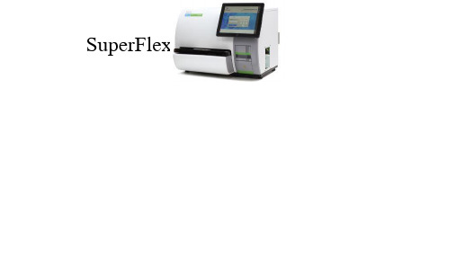
全自动化学发光免疫分析仪 SuperFlex
基于吖啶酯的直接化学发光 50测试/小时 5分钟
单人份一体化试剂条 20ul
样本架:2×12孔,原管上机,急诊优先 一次性Tip头
线性范围:0.3-300 ng/mL 样本用量: 50ul
标本类型:血清、血浆(肝素锂) 单个报告时间: 13分钟
CKMB
平台优势总结
市场理念:突破传统,引领POCT新时代
原理先进,媲美大型发光 吖啶酯直接化学发光 专利磁珠
智能化、人性化操控 全中文界面 高清触摸屏操作 一键运行,云端服务
按照应用场景的分类 美国国家临床生化科学院
高级阶段 广义POCT
小到极致:手持式、手机,手表、蓝牙插件 场景扩展:医院外、医院内、临床科、检验科 小型平台:自动化、通量、向大发光看齐
即时检测
目前医院内POCT的项目群
血气/电解质
……
血糖监测
血糖(葡萄糖) 糖化血红蛋白 ……
感染性疾病
HBV、HCV、HIV、TP 流感……
肉眼判读
量子点 时间分辩 上转发光.
化学发光
小平台概念-准、小 通量、试剂多 操作更快捷 质控管理
“定性”
“定量”
更好的定量
自动化
小化学发光
未来
基于膜的荧光层析
手持或小仪器 单通道
全定量判读
自动化程度
微点生物qLabs产品推荐手册

qLabs®电化学凝血检测仪项目推荐书第一部项目概述及背景一、项目概述凝血功能的正常和稳定是人体重要的生理基础,但是在抗凝治疗、瓣膜置换术、心脏支架书后,以及严重感染和腹泻、中毒、外伤、血管损伤及免疫紊乱等多种因素的作用下,容易引起机体凝血功能异常。
如果不能及时检测并有效的采取治疗措施,往往可能引起一系列致命的出血性或栓塞性后果,例如血管内弥散性凝血、深静脉血栓、急性肺栓塞等等。
凝血功能检测是评估人体凝血系统是否正常的重要方法,尤其是以凝血酶原时间PT、活化部分凝血活酶时间APTT、凝血酶时间TT和纤维蛋白原FIB组合的凝血四项最为重要和最为常用,对于评估患者凝血功能有着极为重要的价值。
凝血功能异常相关疾病常常起病隐匿、进展迅速,因此需要快速的诊断并及时救治对挽救病人的健康和生命十分重要。
而传统检验科凝血四项检测往往需要最少花1~2小时,时间较慢,无法满足相关疾病的快速救治需求。
深圳微点生物股份有限公司推出的全球首款qLabs®快速凝血四联电化学检测仪,qLabs微流控-电化学检测平台利用国际领先的微流控-电化学技术把传统的检验项目集成在一个芯片上,只需要1滴指尖血,4~7分钟时间即可得到凝血四项结果。
因此,qLabs®快速凝血四联电化学检测仪对于急诊、急救的患者是非常好的选择;其次,由于凝血功能的正常与否关系到手术患者能否正常止血,因此在所有外科手术术前均需进行凝血四项的检测,在术前、书中及术后快速的评估患者凝血功能,避免在手术中出现因凝血功能障碍导致的出血不止的风险,快速床旁凝血四项检测就非常有意义;再次,在血液净化、体外循环手术、大出血患者、拔牙、麻醉复苏的患者中,均需要及时的检测凝血功能从而制定合理的治理方案。
第二部微点生物qLabs电化学检测平台的性能及参数一、微点生物qLabs电化学检测仪的介绍qLabs®电化学检测仪是配套本公司生产的电化学检测卡共同使用的专用检测平台,主要定位于凝血类诊断项目。
浙江卓胜产品手册说明书

浙江卓胜产品手册ZSup Products Catalogue 您实验室的可靠伙伴Your reliable partner in laboratory浙江卓胜医疗科技有限公司2018年成立,技术和运营团队是由模具制造、高分子材料、生物医学、物联网专家组成,在符合GMP要求的十万级净化车间内生产高品质实验室耗材,并承接高精密注塑模具制造、定制生物耗材,努力为客户提供一站式解决方案。
Zhejiang ZSup Medical Technology Co., Ltd. was established in 2018 by expert teams of mold manufacturing, polymer materials, biomedicine, and IOT. We produce high-quality laboratory consumables in GMP qualified 100,000-level cleanliness level purification workshop. We also undertake manufacture of high-precision injection mold, customized bio supplies. We strive to provide one-stop solutions to our cus-tomers.●高精密模具制造:技术团队具有丰富的生物实验室耗材高精密模具生产经验,专业生产薄壁、多腔、高速、高精密注塑模具;Manufacture of high-precision mold: Technical team own excellent experiences in producing of high-precision molds for bio supplies, particularly for thin-walled, multi-cavity, high-speed injection molds.●专业注塑生产:一流质控管理下的国际质量标准高端生物实验、生产用耗材,含Manufacture of injection molding products: Top level production of laboratory supplies by world class QC and QA management, including●通用耗材:离心管、吸头、PCR、细胞培养系列产品等;General consumables: centrifuge tubes, tips, PCR, cell culture products, etc.;●定制耗材:各类自动化专用生物实验耗材;Customized consumables: all kinds of automated special biological experi-ment consumables;●OEM定制:共享设备、模具、生产线,提供产品贴牌生产。
- 1、下载文档前请自行甄别文档内容的完整性,平台不提供额外的编辑、内容补充、找答案等附加服务。
- 2、"仅部分预览"的文档,不可在线预览部分如存在完整性等问题,可反馈申请退款(可完整预览的文档不适用该条件!)。
- 3、如文档侵犯您的权益,请联系客服反馈,我们会尽快为您处理(人工客服工作时间:9:00-18:30)。
qLabs®电化学凝血检测仪
项目推荐书
第一部项目概述及背景
一、项目概述
凝血功能的正常和稳定是人体重要的生理基础,但是在抗凝治疗、瓣膜置换术、心脏支架书后,以及严重感染和腹泻、中毒、外伤、血管损伤及免疫紊乱等多种因素的作用下,容易引起机体凝血功能异常。
如果不能及时检测并有效的采取治疗措施,往往可能引起一系列致命的出血性或栓塞性后果,例如血管内弥散性凝血、深静脉血栓、急性肺栓塞等等。
凝血功能检测是评估人体凝血系统是否正常的重要方法,尤其是以凝血酶原时间PT、活化部分凝血活酶时间APTT、凝血酶时间TT和纤维蛋白原FIB组合的凝血四项最为重要和最为常用,对于评估患者凝血功能有着极为重要的价值。
凝血功能异常相关疾病常常起病隐匿、进展迅速,因此需要快速的诊断并及时救治对挽救病人的健康和生命十分重要。
而传统检验科凝血四项检测往往需要最少花1~2小时,时间较慢,无法满足相关疾病的快速救治需求。
深圳微点生物股份有限公司推出的全球首款qLabs®快速凝血四联电化学检测仪,qLabs微流控-电化学检测平台利用国际领先的微流控-电化学技术把传统的检验项目集成在一个芯片上,只需要1滴指尖血,4~7分钟时间即可得到凝血四项结果。
因此,qLabs®快速凝血四联电化学检测仪对于急诊、急救的患者是非常好的选择;其次,由于凝血功能的正常与否关系到手术患者能否正常止血,因此在所有外科手术术前均需进行凝血四项的检测,在术前、书中及术后快速的评估患者凝血功能,避免在手术中出现因凝血功能障碍导致的出血不止的风险,快速床旁凝血四项检测就非常有意义;再次,在血液净化、体外循环手术、大出血患者、拔牙、麻醉复苏的患者中,均需要及时的检测凝血功能从而制定合理的治理方案。
第二部微点生物qLabs电化学检测平台的性能及参数
一、微点生物qLabs电化学检测仪的介绍
qLabs®电化学检测仪是配套本公司生产的电化学检测卡共同使用的专用检测平台,主要定位于凝血类诊断项目。
检测仪的工作原理是用动态电流法检测凝血试剂卡的电流信号,通过信号处理算法分析并生成特定项目的检测结果。
目前可以开展的项目有PT/INR,APTT以及PT/APTT二合一及凝血四项(PT/APTT/FIB/TT)的检测。
qLabs®电化学检测仪由测试模块、液晶显示屏、上盖、下盖以及可选配的条码模块组成。
检测仪内置存储可以保存200条检测结果,并通过数据线(qLabs® eCable)上传数据,或通过专用的打印机(qLabs® eStation)打印检测报告。