ApisTransfector-H(CN)
Study_on_the_pharmacological_activities_and_chemic

ReviewStudy on the pharmacological activities and chemicalstructures of Viburnum dilatatumZhiheng Gao, Yufei Xi, Man Wang, Xiaoxiao Huang*, Shaojiang Song*Key Laboratory of Computational Chemistry-Based Natural Antitumor Drug Research &Development, Liaoning Province, School of Traditional Chinese Materia Medica, ShenyangPharmaceutical University, Shenyang 110016, ChinaAbstractViburnum dilatatum (jiami in Chinese), belonging to the Caprifollaceae family, is widely distributed in Japan and China. Phytochemical investigations of Viburnum dilatatum (V. dilatatum) have resulted in the isolation of triterpenoids, phenolic glycosides essential oil, norisoprenoids, etc. Research results have shown that the chemical constituents of V. dilatatum possess various pharmacological activities, including antihyperglycemic, antioxidant activity and antiulcer effects. This study reviewed the chemical constituents and pharmacological activities of V. dilatatum to provide practical and useful information for further research and development of this plant.Keywords: Viburnum dilatatum; pharmacological activity; chemical structures1 IntroductionViburnum dilatatum (called jiami in Chinese, gamazumi in Japanese and snowball tree in English), beloinging to family Caprifoliaceae, is a deciduous low tree distributed widely in the hills of northern China and Japan [1]. There are many types of chemical constituents in Viburnum dilatatum (V. dilatatum), including triterpenoids, * Author to whom correspondence should be addressed. Address:School of Traditional Chinese Materia Medica, Shenyang Pharmaceutical University, 103 Wenhua Rd., Shenyang 110016, China; Tel.: +86-24-43520793 (Xiaoxiao Huang); +86-24-43520707 (ShaojiangSong);E-mail:*******************(XiaoxiaoHuang); ****************(ShaojiangSong).Received: 2021-04-16 Accepted: 2022-08-28phenolic glycosides and norisoprenoids [2-4]. The leaves have been utilized as a traditional Chinese medicine, and phenolic compounds have been reported as the main active chemical component of the leaves. Many researchers have analyzed the functions of these medicinal components and found that these components have good antioxidant antihyperglycemic and antiulcer effects. For example, the gamazumi crude extract obtained from the squeezed juice of the fruit prevented oxidative injury in rats [5]. This review described the chemical structures and pharmacological activities of V. dilatatum, so as to help readers understand comprehensively the research progress of V. dilatatum and provide help for the development of V. dilatatum.2 Chemical constituents and structuresPrevious reports have indicated that the main chemical constituents of V. dilatatum are phenolic glycosides and triterpenoids.2.1 Phenolic glycosidesThirteen phenolic glycosides were isolated and identified from V. dilatatum by extensive spectroscopic methods, namely p -hydroxyphenyl-6-O -trans-caffeoyl-β-D -glucoside (1) [6], p -hydroxyphenyl-6-O -trans-caffeoyl-β-D -alloside (2) [6], 4-allyl-2-methoxyphenyl-6-O -β-D -apiosyl(1→6)-β-D -glucoside (3) [6], 1-(4’-hydroxy-3’-methoxypheny1)-2-[2’’-hydroxy-4’’-(3’’’-hydroxypropyl)]-1,3-propanediol-l-O -β-D -glucopyranoside (erythro isomer) (4-7) [7], neochlorogenic acid methyl ester (8-9) [7], cryptochlorogenic acid methyl ester (10-11) [7], cyanidin-3-sambubioside (Cy-3-sam) (12) [8], cyanidin-3-glucoside (Cy-3-glc) (13) [8], 5-O -caffeoyl-4-methoxyl quinic acid (4-MeO-5-CQA) (14) [8], chlorogenic acid (5-CQA) (15) [8], quercetin (16) [8], 2-(glucopyranosyloxy)-benzyl-3-(glucopyranosyloxy)-benzoate (17) [9] and jiamizioside E (18) [10]. These structures are shown in Fig. 1.Fig. 1 Phenolic glycosides isolated from V . dilatatumContinued fig. 12.2 TriterpenoidsThere were about seventeen triterpenoids isolated and characterized from V. dilatatum , such as viburnols A (19) [11], viburnols B (20) [11], viburnols C (21) [11], viburnols D (22) [11], viburnols E (23) [11], viburnols F (24) [12], viburnols G (25) [12], viburnols H (26) [12], viburnols I (27) [12], viburnols J (28) [12],viburnols K (29) [12], viburnudienone B 2methyl ester (30) [13], viburnenone H 2 (31) [13],v i b u r n e n o n e B 2 m e t h y l e s t e r (32) [13], viburnudienone B 1 methyl ester (33) [13], viburnenone H 1 (34) [13], and viburnenone B 2 methyl ester (35) [13]. The structures are shown in Fig. 2.Continued fig. 23 Pharmacological activities3.1 Antioxidant activityOxidative stress caused by free radicals and their derivatives leads to disturbances in redox homeostasis. Reactive oxygen species (ROS) are not only endogenously produced during intracellular metabolic processes but also generated by exogenous stimuli such as UV radiation, pollutants, smoke and drugs. The cell triggers its defense systems or undergoes apoptosis when intracellular oxidative status increases. It influences numerous cellular processes including core signaling pathways, which are associated with development of systematic and chronic disorders, such as aging and cancer. Therefore, it is critical to remove cellular oxidants and restore redox balance.solution of V. dilatatum (GSS) had strong antioxidant activity in vivo and prevent stress-induced oxidative damage by the XYZ-dish method and the澳electron spin resonance (ESR) method [14]. The experimental result showed that the concentrations of lipid peroxide in plasma, liver and stomach in the GSS group were reduced. Furthermore, the activities of plasma lactic dehydrogenase, amylase and creatine phosphokinase are ordinarily increased by stress. However, these activities in the GSS group decreased to that in the control group. It was concluded that gastric ulcer formation, increase of lipid peroxidation in plasma and tissues and elevation of plasma enzymatic activities were confirmed in rats with water immersion restraint stress. It was also found that intake of GSS could protect the stomach and other tissues from oxidative damage.Kim et al. identified and isolated two major anthocyanins by NMR and LC-ESI-MS/MS, namely, cyanidin 3-sambubioside (I) and kuromanin (II) [15]. By the electron spin resonance method, the superoxide anion radical scavenging activities of I and II were evaluated with the IC 50 values of 17.3 and 69.6 µM, and their activities on hydroxyl radicals were evaluated with the IC 50 values of 4.3 and 53.2 mM. As the positive control, the IC 50 values of ascorbic acid were 74.2 µM on superoxide anion radicals and 3.0 mM on hydroxyl radicals, respectively. The above results suggested that these anthocyanins with radical scavenging properties might be the key compounds contributing to the antioxidant activity and physiological effects of V . dilatatum fruits.Woo et al. determined the free radical scavenging capacity of VD (the leaves of V. dilatatum ) [16]. Anti-oxidant activity of the extracts was assessed by the ability to scavenge 2,2-diphenyl-1-picrylhydrazyl (DPPH) or 3-ethylbenzothiazoline-6-sulfonic acid (ABTS) radicals. Butylated hydroxytoluene (BHT), a synthetic antioxidant, or α-tocopherol, was used as the positive control in these assays. The experimental result showed that VD inducedincrease in radical scavenging activity. In addition, lipid peroxidation inhibitory activity was determined via measurement of MDA (Malondialdehyde) levels using mouse liver tissue homogenate treated with various concentrations of the extracts. The concentration-dependent decrease in MDA levels observed was consistent with radical scavenging activities of the extracts. To examine whether VD extracts could protect mam-malian cells from oxidative stress, cultures of a human mammary gland-derived epithelial cell line MCF-7 were treated with each extract prior to challenging them with tBHP. The intracellular ROS (Reactive oxygen species) production was determined with the relative intensity of dichlorofluorescein fluorescence. While intracellular ROS formation was significantly promoted by tBHP treatment, the augmented ROS level was significantly reduced after the treatment with VD extracts.3.2 Antihyperglycemic effectIwai et al. used an oral glucose tolerance test on the diabetic rats [17]. They found that the elevation of plasma glucose level after oral administration of 2 g/kg glucose was suppressed by the repeated administration of the freeze-dried powder of V. dilatatum fruit juice (CEV). The α-glucosidase inhibitory activities of isolated compounds from CEV were also measured. Cyanidin 3-sambubioside and 5-caffeoyl quinic acid A showed inhibitory activity. These results suggested that V. dilatatum fruit had the antihyperglycemic effects.4 ConclusionV. dilatatum is distributed widely in the hills of northern China and Japan. Currently, the studies on V. dilatatum have been conducted at home and abroad, but few studies focus on its chemical components and pharmacological activities. Previousphytochemical investigations showed that the constituents of V. dilatatum included triterpenoids, phenolic glycosides, norisoprenoids and other compounds. This study describes thirteen phenolic glycosides and seventeen triterpenoids and their different degrees of antihyperglycemic, antioxidant activity and antiulcer effects, aiming to provide a reference for further studies on V. dilatatum and pharmaceutical development.References[1] Jeffrey B, Harborne A. Colour atlas of medicinal plantsof Japan. Phytochemistry, 1981, 20: 1467.[2] Miyazawa M, Hashidume S, Takahashi T, et al. Aromaevaluation of gamazumi (Viburnum dilatatum) by aroma extract dilution analysis and odour activity value.Phytochem Anal, 2012, 23: 208-213.[3] Kurihara T, Kikuchi M. Studies on the constituentsof flowers. IV. On the components of the flower of Viburnum dilatatum Thunb. J Health Sci, 1975, 95: 1098-1102.[4] Machida K, Kikuchi M. Norisoprenoids from Viburnumdilatatum. Phytochemistry, 1996, 41: 1333-1336. [5] Iwai K, Onodera A, Matsue H. Mechanism of preventiveaction of Viburnum dilatatum Thunb (gamazumi) crude extract on oxidative damage in rats subjected to stress. J Sci Food Agric, 2010, 83: 1593-1599.[6] Machida K, Nakano Y, Kikuchi M. Phenolic glycosidesfrom Viburnum dilatatum. Phytochemistry, 1991, 30: 2013-2014.[7] Machida K, Kikuchi M. Phenolic compounds fromViburnum dilatatum. Phytochemistry, 1992, 31: 3654-3656.[8] Kim MY, Iwai K, Matsue H. Phenolic compositions ofViburnum dilatatum Thunb. fruits and their antiradical properties. J Food Compos Anal, 2005, 18: 789-802. [9] Lu D, Yao S. Phenolic glycoside from the roots ofViburnum dilatatum. Nat Prod Commun, 2009, 4: 945-946.[10] Wu B, Zeng X, Zhang Y. New metabolite fromViburnum dilatatum. Nat Prod Commun, 2010, 5: 1097-1098.[11] Machida K, Kikuchi M. Viburnols: Novel triterpenoidswith a rearranged dammarane skeleton from Viburnum dilatatum. Tetrahedron Lett, 1996, 37: 4157-4160. [12] Machida K, Kikuchi M. Viburnols: Six noveltriterpenoids from Viburnum dilatatum. Tetrahedron Lett, 1997, 38: 571-574.[13] Machida K, Kikuchi M. Studies on the Constituents ofViburnum Species. XIX. Six New Triterpenoids from Viburnum dilatatum Thunb. Chem Pharm Bull, 1999, 47: 692-694.[14] Iwai K, Onodera A, Matsue H, et al. Antioxidant activityand inhibitory effect of Gamazumi (Viburnum dilatatum THUNB.) on oxidative damage induced by water immersion restraint stress in rats. Int J. Food Sci Nutr, 2001, 52: 443-451.[15] Kim MY, Iwai K, Onodera A, et al. Identification andAntiradical Properties of Anthocyanins in Fruits of Viburnum dilatatum Thunb. J Agric Food Chem, 2003, 51: 6173-6177.[16] Woo YJ, Lee HJ, Jeong YS, et al. Antioxidant Potentialof Selected Korean Edible Plant Extracts. Bio Med Res Int, 2017, 2017: 1-9.[17] Iwai K, Kim MY, Akio O, et al. Alpha-glucosidaseinhibitory and antihyperglycemic effects of polyphenols in the fruit of Viburnum dilatatum Thunb. J Agric Food Chem, 2006, 54: 4588-4592.。
高效液相色谱-荧光检测器法检测单克隆抗体注射液中吐温80的含量

Vol.7 No.1Feb. 2021生物化工Biological Chemical Engineering第 7 卷 第 1 期2021 年 2 月高效液相色谱-荧光检测器法检测单克隆抗体注射液中吐温80的含量张博慧,贾戴辉,许俊彦*(宝船生物医药科技(上海)有限公司药物分析部门,上海 201203)摘 要:目的:采用高效液相色谱-荧光检测器法(HPLC-FLD)测定单克隆抗体注射液中吐温80的含量,并进行方法学验证。
方法:按设定的色谱条件对检测单克隆抗体注射液中吐温80含量的HPLC-FLD 法进行方法学验证,包括专属性、精密度、准确度、线性和范围、耐用性,并采用该方法对不同单克隆抗体注射液进行检测。
结果:经验证,该方法具有专属性;重复性验证试验中,保留时间和含量的RSD 值分别为0.5%和0.4%;中间精密度验证试验中,保留时间和含量的RSD 值分别为0.4%和2.2%;准确度验证试验中,回收率分别为105%、98%和108%;线性相关系数为0.999 1,检测范围为0.05~0.80 mg/mL;不同批次反应线圈对检测结果无影响,不同品种单克隆抗体注射液吐温80含量检测结果偏差较小。
结论:该方法具有专属性,线性关系良好,精密度和准确度较好,测定结果稳定可靠,适用于单克隆抗体注射液中吐温80含量的检测。
关键词:高效液相色谱-荧光检测器;吐温80;单克隆抗体注射液中图分类号:R927.2 文献标识码:ADetermination of Tween80 Content in Monoclonal Antibody Injection by HPLC-FLDZHANG Bohui, JIA Daihui, XU Junyan *(Drug Analysis Department, Dragonboat Biopharmaceutical, Co., Ltd, Shanghai 201203)Abstract: Objective: To establish a method for the determination of Tween 80 in monoclonal antibody injection byHigh-performance liquid chromatography fluorescence detector. Methods: Under designed condition, the method was verified for specificity, precision, accuracy, linearity, range and durability. The method was also used to determination of Tween 80 content of different monoclonal antibody injections. Results: The HPLC-FLD method showed good specificity. In reproducibility test, the RSD of retention time and content were 0.5% and 0.4% respectively. In intermediate-precision test, the RSD of retention time and content were 0.4% and 2.2% respectively. The recovery rates were 105%, 98% and 108% respectively. The correlation Coefficient was 0.999 1 and the range was from 0.05 to 0.8 mg/mL. The different batches of reaction coils had no effect on the detection results. The Tween 80 contents in different mAb injections showed little deviation. Conclusion: The HPLC-FLD method showed good specificity, precision, accuracy and linearity, and the test result was stable and reliable, which was suitable for determination of Tween 80 content of different monoclonal antibody injections.Keywords: High-performance Liquid Chromatography-Fluorescence Detector; Tween 80; monoclonal antibody injection吐温80又名聚山梨酯80,其化学名为聚氧乙烯20山梨醇酐单油酸酯,作为助溶剂、乳化剂和稳定剂,常用于治疗性单克隆抗体注射液制剂中[1]。
东洋纺反转录试剂盒
高效率逆转录试剂盒 First Strand cDNA Synthesis Kit
ReverTra Ace -α-
(Code No. FSK-100,FSK-101)
使用说明书
科研用
TOYOBO CO., LTD. Life Science Department OSAKA JAPAN
目录
※ 除Positive Control RNA外,所有组分请均保存在-20℃条件下;Positive Control RNA请保存于-80℃条件下。
※ Positive Control RNA 由于运输过程中不能一直保证 -80℃条件,在运输过程中可能 降解。建议用户选择人、鼠来源的 Total RNA 作为 Control RNA。
G3PDH mRNA
Primer F 559
intron
Size(bases)
1634
1237
1110 Primer R
90 129 90 92 k-193 104
图 2. Control Primer F,R 的 location
[Positive Control RNA]
本 试 剂 盒 作 为 Positive Control 用 RNA , 添 附 有 Human G3PDH (Glyceraldehyde 3-Phosphate Dehydrogenase)遗传基因的 in vitro 转录产物。 (在 3’末端添加了 22mer 的 Poly(A)tail)(请参照图 3)。
※ 组分中的 5×RT Buffer 在溶解时,可能会出现白色沉淀现象,但不影响其品质。此时, 请使用振荡器等仪器使其混合均匀,等完全溶解后再使用。
[各引物的序列]
Gateway LR Clonase II Enzyme Mix

Gateway®LR Clonase II Enzyme MixCat. No. 11791-020 Size: 20 reactionsCat. No. 11791-100 Size: 100 reactionsStore at -20°C (non-frost-free freezer) Gateway® TechnologyThe Gateway® Technology is a universal cloning method that takes advantage of the site-specific recombination properties of bacteriophage lambda (1) to provide a rapid and highly efficient way to move DNA sequences into multiple vector systems. The Gateway® Technology is schematically represented below.att B1-gene-att B2 u att P1-ccd B-att P2 att L1-gene-att L2 u att R1-ccd B-att R2(expression clone) (pDONR™) (entry clone) (destination vector) The att B u att P reaction is mediated by Gateway® BP Clonase II enzyme mix; the att L u att R reaction is mediated by Gateway® LR Clonase II enzyme mix. ccd B is the F plasmid-encoded gene that inhibits growth of E. coli (2,3) and “gene” represents any DNA segment of interest (e.g. PCR product, cDNA, genomic DNA). DescriptionGateway® LR Clonase™ II enzyme mix is a proprietary enzyme and buffer formulation containing the bacteriophage lambda recombination proteins Integrase (Int) and Excisionase (Xis), the E. coli-encoded protein Integration Host Factor (IHF) (1), and reaction buffer provided in a single mix for convenient reaction set up. Gateway® LR Clonase™ II enzyme mix catalyzes in vitro recombination between an entry clone (att L-flanked “gene”) and an att R-containing destination vector to generate an att B-containing expression clone. Store Gateway® LR Clonase™ II enzyme mix at -20ºC (non-frost-free freezer) for up to 6 months. For long-term storage, store at -80ºC.Components Supplied20 rxns 100 rxnsP lGateway® LR Clonase II Enzyme Mix 40 P l 200P lP l 200 Proteinase K Solution (2 P g/P l) 40P lpENTR™-gus Positive Control (50 ng/P l) 20P l 20 Quality ControlLR Clonase II enzyme mix is functionally tested in a 1 hour recombination reaction followed by a transformation assay.Part No. 11791.II.pps Rev. Date: 10 Jun 2004 This product is distributed for laboratory research only. CAUTION: Not for diagnostic use. The safety and efficacy of this product in diagnostic orother clinical uses has not been established.For technical questions about this product, call the Invitrogen Tech-Line SM U.S.A. 800 955 6288Page 2 General Recommendations and Guidelinesx pENTR™-gus is provided for use as a positive control in the LR reaction and is an entry clone containing the Arabidopsis thaliana E-glucuronidase (gus)gene (4). Refer to our Web site () for a map andsequence of pENTR™-gus.x We recommend using plasmid DNA purified with the PureLink™ HQ Mini Plasmid Purification Kit (Catalog no. K2100-01). Mini-prep (alkaline lysis) DNA preparations are adequate for Gateway® cloning reactions; however, in general, such DNA cannot be quantitated by UV absorbance due tocontaminating RNA and nucleotides. Estimate concentrations by gelelectrophoresis in comparison with standard DNA (e.g. DNA Mass Ladder, Catalog no. 10068-013 or 10496-016).x For LR recombination reactions, the most efficient substrates are supercoiled att L-containing entry vectors and supercoiled att R-containing destination vectors. For large (>10 kb) entry clones or destination vectors, linearizing the entry clone or destination vector may increase the efficiency by up to 2-fold.x To increase the number of colonies containing the desired expression clone, increase the incubation time from the recommended 1 hour to 2 hours-overnight. Longer incubations are recommended for plasmids t10 kb toincrease the yield of colonies.x We recommend using 50-150 ng entry clone per 10 P l reaction. Highest colony yields are typically obtained using 150 ng entry clone and 150 ngdestination vector. Do not use >150 ng entry clone as you may obtaincolonies containing multiple DNA molecules (often with an associated“small colony” phenotype). Using <50 ng entry clone will generate fewer colonies.Page 3 ProceduresLR ReactionLR Clonase II enzyme mix is supplied as a 5X solution. If you wish to scale the reaction volume, make sure the LR Clonase II enzyme mix is at a final concentration of 1X. For a positive control, use 100 ng (2 P l) of pENTR™-gus.1. Add the following components to a 1.5 ml microcentrifuge tube at roomtemperature and mix:Entry clone (50-150 ng) 1-7 P lP lDestination vector (150 ng/P l) 1TE buffer, pH 8.0 to 8 P l2. Thaw on ice the LR Clonase II enzyme mix for about 2 minutes. Vortex theLR Clonase™ II enzyme mix briefly twice (2 seconds each time).3. To each sample (Step 1, above), add 2 P l of LR Clonase II enzyme mix to thereaction and mix well by vortexing briefly twice. Microcentrifuge briefly.4. Return LR Clonase II enzyme mix to -20q C or -80q C storage.5. Incubate reactions at 25q C for 1 hour.6. Add 1 P l of the Proteinase K solution to each sample to terminate thereaction. Vortex briefly. Incubate samples at 37q C for 10 minutes. Transformation1. Transform 1 P l of each LR reaction into 50 P l of One Shot® OmniMAX™ 2 T1Phage-Resistant Cells (Catalog no. C8540-03). Incubate on ice for 30 minutes.Heat-shock cells by incubating at 42q C for 30 seconds. Add 250 P l of S.O.C.Medium and incubate at 37q C for 1 hour with shaking. Plate 20 P l and 100 P l of each transformation onto selective plates. Note: Any competent cells witha transformation efficiency of >1.0 u 108 transformants/P g may be used.2. Transform 1 P l of pUC19 DNA (10 ng/ml) into 50 P l of One Shot®OmniMAX™ 2 T1 Phage-Resistant Cells as described above. Plate 20 P l and 100 P l on LB plates containing 100 P g/ml ampicillin.Expected ResultsAn efficient LR recombination reaction will produce >5000 colonies if the entire LR reaction is transformed and plated.Page 4 References1. Landy, A. (1989) Ann. Rev. Biochem. 58, 913.2. Bernard, P. and Couturier, M. (1992) J. Mol. Biol.226, 735.3. Miki, T., Park, J.A., Nagao, K., Murayama, N., and Horiuchi, T. (1992) J. Mol. Biol. 225, 39.4. Kertbundit, S., Greve, H.D., Deboeck, F., Montagu, M.V., and Hernalsteens, J.P. (1991)Proc. Natl. Acad. Sci. USA, 88, 5212.Limited Use Label License No. 19: Gateway® Cloning ProductsThis product and its use is the subject of one or more of U.S. Patent Nos. 5,888,732, 6,143,557, 6,171,861, 6,270,969, 6,277,608, and 6,720,140 and/or other pending U.S. and foreign patent applications owned by Invitrogen Corporation. The purchase of this product conveys to the buyer the non-transferable right to use the purchased amount of the product and components of the product in research conducted by the buyer (whether the buyer is an academic or for profit entity). The purchase of this product does not convey a license under any method claims in the foregoing patents or patent applications, or to use this product with any recombination sites other than those purchased from Invitrogen Corporation or its authorized distributor. The right to use methods claimed in the foregoing patents or patent applications with this product for research purposes only can only be acquired by the use of Clonase™ purchased from Invitrogen Corporation or its authorized distributors. The buyer cannot modify the recombination sequence(s) contained in this product for any purpose. The buyer cannot sell or otherwise transfer (a) this product, (b) its components, or (c) materials made by the employment of this product or its components to a third party or otherwise use this product or its components or materials made by the employment of this product or its components for Commercial Purposes. The buyer may transfer information or materials made through the employment of this product to a scientific collaborator, provided that such transfer is not for any Commercial Purpose, and that such collaborator agrees in writing (a) not to transfer such materials to any third party, and (b) to use such transferred materials and/or information solely for research and not for Commercial Purposes. Notwithstanding the preceding, any buyer who is employed in an academic or government institution may transfer materials made with this product to a third party who has a license from Invitrogen under the patents identified above to distribute such materials. Transfer of such materials and/or information to collaborators does not convey rights to practice any methods claimed in the foregoing patents or patent applications. Commercial Purposes means any activity by a party for consideration and may include, but is not limited to: (1) use of the product or its components in manufacturing; (2) use of the product or its components to provide a service, information, or data; (3) use of the product or its components for therapeutic, diagnostic or prophylactic purposes; or (4) resale of the product or its components, whether or not such product or its components are resold for use in research. Invitrogen Corporation will not assert a claim against the buyer of infringement of the above patents based upon the manufacture, use or sale of a therapeutic, clinical diagnostic, vaccine or prophylactic product developed in research by the buyer in which this product or its components was employed, provided that none of (i) this product, (ii) any of its components, or (iii) a method claim of the foregoing patents, was used in the manufacture of such product. Invitrogen Corporation will not assert a claim against the buyer of infringement of the above patents based upon the use of this product to manufacture a protein for sale, provided that no method claim in the above patents was used in the manufacture of such protein. If the purchaser is not willing to accept the limitations of this limited use statement, Invitrogen is willing to accept return of the product with a full refund. For information on purchasing a license to use this product for purposes other than those permitted above, contact Licensing Department, Invitrogen Corporation, 1600 Faraday Avenue, Carlsbad, California 92008. Phone (760) 603-7200.Limited Use Label License No. 23: GUS Control VectorThe GUS positive control vector in these products is claimed in patents and patent applications (See U.S. Patent No. 5,599,670 and Great Britain Patent No. 2,197,653) licensed to Invitrogen by Cambia Biosystems, L.L.C. ("CBL"). Use of the GUS gene is restricted to use as a positive control. Any other use may require a license from CBL.©2004 Invitrogen Corporation. All rights reserved.。
Roche_Xtreme GeneHP_protocal

0910.06479774001ቢ
The recommended starting concentration is a 3:1. For most cell types, these X-tremeGENE HP DNA Transfection Reagent to DNA ratios provide excellent transfection efficiency. Further optimization may increase transfection efficiency in your particular application. In addition to varying the ratio, other parameters may also be evaluated, such as the amount of transfection complex added. For additional optimization guidelines, see Section 3, Troubleshooting and visit . Plasmid DNA • For best results, accurately determine the plasmid DNA concentration using 260-nm absorption; estimates of DNA by measuring gel band density are not recommended. Determine DNA purity using a 260 nm/280 nm ratio (the optimal ratio is 1.8). • Prepare the plasmid DNA solution using sterile TE (Tris/EDTA) buffer or sterile water at a concentration of 0.1 to 2.0 µg/µl. • Use high quality DNA preparation kits to obtain endotoxin-free DNA. Cell Culture Conditions • Minimize intra- and inter-experimental variance in transfection efficiency using cells that are regularly passaged, proliferating well in a log-growth phase, and plated at a consistent density. • For best results, accurately quantify cell concentration using a hematocytometer or automated system. • Cells must be healthy and free of Mycoplasma. • Cells should have a low passage number to achieve best results. Other Media Additives In some cell types, antimicrobial agents (e.g., antibiotics and fungicides) commonly included in cell-culture media may adversely affect the transfection efficiency of X-tremeGENE HP DNA Transfection Reagent. If possible, exclude additives in initial experiments. Once high-efficiency conditions have been established, these components can be added back while monitoring transfection results. Cell growth and/or transfection efficiency may be affected by variations in serum quality and medium formulations. Verification of Vector Function Optimize transfection conditions using a known positive-control reporter gene construct before transfecting cells with a new vector construct: • Determine transfection efficiency using a reporter gene assay, such as -Gal*, Luciferase*, or SEAP*. • Sequence flanking vector insert regions to verify the integrity of your new construct. 2.2 Transfection Procedure Adherent Cells: Plate cells approximately 24 hours before transfection making sure cells are at the optimal concentration in the appropriate cell culture vessel. Suspension Cells: Plate freshly passaged cells at optimal concentration.
三羟甲基氨基甲烷盐酸盐质谱裂解碎片

三羟甲基氨基甲烷盐酸盐质谱裂解碎片1. 引言1.1 背景介绍三羟甲基氨基甲烷盐酸盐(简称TMAH)是一种广泛应用于化学分析领域的试剂,其在有机物质质谱分析中具有重要的作用。
TMAH可以将有机物质中的酯类、醇类、酚类等化合物甲基化,使其具有更适合在质谱仪中进行分析的性质。
TMAH的盐酸盐形式是其常见的实验室试剂,广泛用于有机物质的分析化学实验中。
TMAH盐酸盐的质谱裂解碎片特征对于分析化学领域起着至关重要的作用。
了解TMAH盐酸盐在质谱中的裂解碎片特征可以帮助分析师更准确地识别未知化合物、推断其结构以及进行定量分析。
对TMAH盐酸盐质谱裂解碎片特征进行深入研究具有重要的理论和应用意义。
本文将从TMAH盐酸盐的合成与性质、质谱技术在TMAH盐酸盐质谱裂解中的应用、TMAH盐酸盐质谱裂解碎片特征分析、相关反应机制研究以及质谱技术在TMAH盐酸盐质谱裂解中的局限性等方面展开详细讨论,旨在深入探讨TMAH盐酸盐在质谱分析中的重要性以及相关研究领域存在的问题和挑战。
【2000字】.1.2 研究目的本研究旨在通过对三羟甲基氨基甲烷盐酸盐质谱裂解碎片进行详细分析,揭示其分子结构特征及相关反应机制,为进一步探索其生物活性和药理作用奠定基础。
具体研究目的包括:1. 探究三羟甲基氨基甲烷盐酸盐质谱裂解碎片的形成机理,揭示其分子内部的键合情况和反应路径;2. 分析三羟甲基氨基甲烷盐酸盐质谱裂解碎片的碎片特征,解析其质谱图谱中各离子峰的起源及关联;3. 探讨质谱技术在三羟甲基氨基甲烷盐酸盐分子结构鉴定中的优势和局限性,为其质谱分析提供科学依据;4. 为进一步深入研究三羟甲基氨基甲烷盐酸盐在药物开发和医学应用中的潜在作用提供理论支持和实验基础。
1.3 研究意义三羟甲基氨基甲烷盐酸盐是一种重要的有机化合物,具有广泛的应用价值。
其在药物合成、生物医药和化学工业领域具有重要的地位。
对三羟甲基氨基甲烷盐酸盐进行质谱分析,可以更深入地了解其结构和性质,为其应用提供重要的信息和参考。
FuGENE HD转染试剂说明书
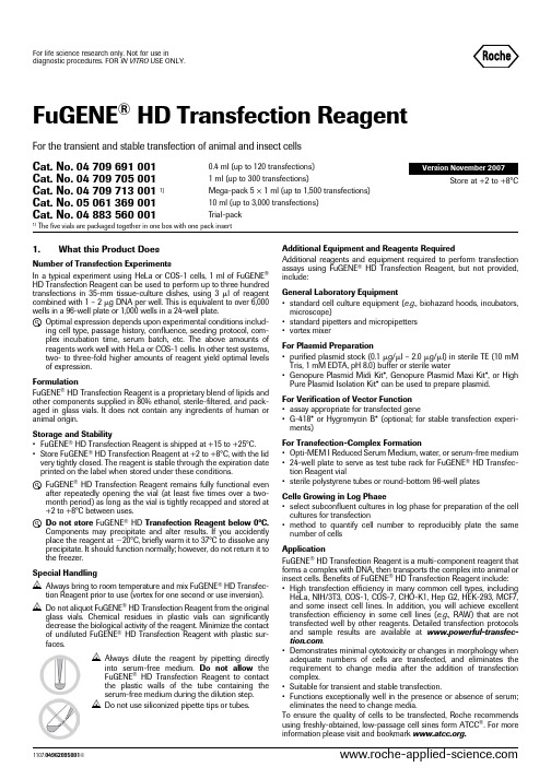
FuGENE ®HD Transfection Reagent1.What this Product DoesNumber of Transfection ExperimentsIn a typical experiment using HeLa or COS-1 cells, 1 ml of FuGENE ®HD Transfection Reagent can be used to perform up to three hundred transfections in 35-mm tissue-culture dishes, using 3 l of reagent combined with 1 – 2 g DNA per well. This is equivalent to over 6,000wells in a 96-well plate or 1,000 wells in a 24-well plate.L Optimal expression depends upon experimental conditions includ-ing cell type, passage history, confluence, seeding protocol, com-plex incubation time, serum batch, etc. The above amounts of reagents work well with HeLa or COS-1 cells. In other test systems,two- to three-fold higher amounts of reagent yield optimal levels of expression.FormulationFuGENE ® HD Transfection Reagent is a proprietary blend of lipids and other components supplied in 80% ethanol, sterile-filtered, and pack-aged in glass vials. It does not contain any ingredients of human or animal origin.Storage and Stability•FuGENE ® HD Transfection Reagent is shipped at +15 to +25°C.•Store FuGENE ® HD Transfection Reagent at +2 to +8°C, with the lid very tightly closed. The reagent is stable through the expiration date printed on the label when stored under these conditions.L FuGENE ® HD Transfection Reagent remains fully functional evenafter repeatedly opening the vial (at least five times over a two-month period) as long as the vial is tightly recapped and stored at +2 to +8°C between uses.L Do not store FuGENE ® HD Transfection Reagent below 0°C.Components may precipitate and alter results. If you accidently place the reagent at Ϫ20°C, briefly warm it to 37°C to dissolve any precipitate. It should function normally; however, do not return it to the freezer.Special HandlingN Always bring to room temperature and mix FuGENE ® HD Transfec-tion Reagent prior to use (vortex for one second or use inversion).N Do not aliquot FuGENE ® HD Transfection Reagent from the originalglass vials. Chemical residues in plastic vials can significantly decrease the biological activity of the reagent. Minimize the contact of undiluted FuGENE ® HD Transfection Reagent with plastic sur-faces.N Always dilute the reagent by pipetting directlyinto serum-free medium. Do not allow the FuGENE ® HD Transfection Reagent to contact the plastic walls of the tube containing the serum-free medium during the dilution step.N Do not use siliconized pipette tips or tubes.Additional Equipment and Reagents RequiredAdditional reagents and equipment required to perform transfection assays using FuGENE ® HD Transfection Reagent, but not provided,include:General Laboratory Equipment•standard cell culture equipment (e.g., biohazard hoods, incubators,microscope)•standard pipetters and micropipetters •vortex mixerFor Plasmid Preparation•purified plasmid stock (0.1 g/l – 2.0 g/l) in sterile TE (10 mM Tris, 1 mM EDTA, pH 8.0) buffer or sterile water•Genopure Plasmid Midi Kit*, Genopure Plasmid Maxi Kit*, or High Pure Plasmid Isolation Kit* can be used to prepare plasmid.For Verification of Vector Function •assay appropriate for transfected gene•G-418* or Hygromycin B* (optional; for stable transfection experi-ments)For Transfection-Complex Formation•Opti-MEM I Reduced Serum Medium, water, or serum-free medium •24-well plate to serve as test tube rack for FuGENE ® HD Transfec-tion Reagent vial•sterile polystyrene tubes or round-bottom 96-well platesCells Growing in Log Phase•select subconfluent cultures in log phase for preparation of the cell cultures for transfection•method to quantify cell number to reproducibly plate the same number of cellsApplicationFuGENE ® HD Transfection Reagent is a multi-component reagent that forms a complex with DNA, then transports the complex into animal or insect cells. Benefits of FuGENE ® HD Transfection Reagent include:•High transfection efficiency in many common cell types, including HeLa, NIH/3T3, COS-1, COS-7, CHO-K1, Hep G2, HEK-293, MCF7,and some insect cell lines. In addition, you will achieve excellent transfection efficiency in some cell lines (e.g., RAW) that are not transfected well by other reagents. Detailed transfection protocols and sample results are available at .•Demonstrates minimal cytotoxicity or changes in morphology when adequate numbers of cells are transfected, and eliminates the requirement to change media after the addition of transfection complex.•Suitable for transient and stable transfection.•Functions exceptionally well in the presence or absence of serum;eliminates the need to change media.To ensure the quality of cells to be transfected, Roche recommends using freshly-obtained, low-passage cell sines form ATCC ®. For more information please visit and bookmark .For the transient and stable transfection of animal and insect cellsCat. No. 04 709 691 0010.4 ml (up to 120 transfections)Version November 2007Cat. No. 04 709 705 001 1 ml (up to 300 transfections)Store at +2 to +8°CCat. No. 04 709 713 001 1)Mega-pack 5 × 1 ml (up to 1,500 transfections)Cat. No. 05 061 369 00110 ml (up to 3,000 transfections)Cat. No. 04 883 560 001Trial-pack1)The five vials are packaged together in one box with one pack insertFor life science research only. Not for use in diagnostic procedures. FOR IN VITRO USE ONLY.2.How to Use this Product2.1Before you BeginRequired Amount of FuGENE® HD Transfection ReagentFor initial optimization experiments, transfect a monolayer of cells that is 80 – 90% confluent in a six-well culture dish, using 3:2, 4:2, 5:2, 6:2, 7:2, and 8:2 ratios of FuGENE® HD Transfection Reagent (l) to DNA (g), respectively. For most cell types, these FuGENE® HD Transfection Reagent:DNA ratios provide excellent transfection levels.L Subsequent optimization may further increase efficiency in your particular application. In addition to varying the volume of FuGENE® HD Transfection Reagent, other parameters may be evaluated (see section 2.6, Parameters for Optimization, and sec-tion 3, Troubleshooting).Plasmid DNA•It is critical to accurately determine the plasmid DNA concentration using 260-nm absorption (estimates of DNA content based on the intensity of gel bands are not sufficiently accurate). Determine the DNA purity using a 260 nm/280 nm ratio; the optimal ratio is 1.8.•Prepare the plasmid DNA solution in sterile TE (Tris/EDTA) buffer or sterile water at a concentration between 0.1 g/l and 2.0 g/l. Cell Culture ConditionsMinimize both intra- and inter-experimental variance in transfection efficiency by using cells that are regularly passaged, proliferating well (best when in a log-growth phase), and plated at a consistent den-sity. FuGENE® HD Transfection Reagent is different from FuGENE® 6 Transfection Reagent regarding the optimal density of cells required for maximal expression with minimal negative effect; FuGENE®6 Transfection Reagent is formulated to work at low cell densities, whereas FuGENE® HD Transfection Reagent is formulated to work at higher cell densities. Cells must be healthy and free of mycoplasma and other contaminants.L If you have used FuGENE® 6 Transfection Reagent in the past, we suggest that you increase the plating density for initial tests using FuGENE® HD Transfection Reagent. For most cell lines, use the reagent at cell-plating densities at least twice that used with FuGENE® 6 Transfection Reagent to yield maximum protein expression. For most cell lines, cultures should be 80 – 90% con-fluent at the time of transfection. For contact-inhibited cell lines such as NIH/3T3, optimal results are obtained when cells are plated at lower densities.Other Media AdditivesIn some cell types, antimicrobial agents (e.g., antibiotics and fungicides) that are commonly included in cell-culture media may adversely affect the transfection efficiency of FuGENE® HD Transfection Reagent. If possible, exclude additives for initial experi-ments. Once high-efficiency conditions have been established, these components can be added back while monitoring your transfection results. Cell growth and/or transfection efficiency may be affected by variations in sera quality or media formulations.Verification of Vector FunctionOptimize transfection conditions with a known positive-control reporter gene construct prior to transfecting cells with a new vector construct:•Determine transfection efficiency with a reporter gene assay (CAT*,-Gal*, Luciferase*, SEAP*, or hGH*).•Sequence across the flanking vector insert regions to verify the integrity of your new construct.2.2Preparation of Cells for Transfection2.3Overview of Initial Transfection ExperimentAdherent and Suspension Cells in a Six-well Plate or 35-mm Culture DishFor initial optimization, test FuGENE® HD Transfection Reagent:DNA ratios of 3:2, 4:2, 5:2, 6:2, 7:2, and 8:2 (l for FuGENE® HD Transfection Reagent, and g for DNA, respectively). The preparation of the com-plex for a single well of a six-well plate, or a 35-mm culture dish, is described in section 2.4. These ratios will function very well for com-monly used adherent cells and suspension cells. For your particular cell line and culture conditions you may find that ratios of 9:2, 10:2, 11:2, or 12:2 result in even greater expression. Try these ratios if you find the highest expression levels in the 8:2 ratio well.N Prepare the transfection complex in diluent that does not contain serum (e.g., Opti-MEM I Reduced Serum Medium), even if the cells are transfected in the presence of serum. For some cell lines, the complex may be formed in DMEM or sterile water.L For additional optimization tips, see section 2.6 and visit /fugene/hdRatio OverviewPreparation of a transfection complex that is sufficient for a 35-mm culture dish, or one well of a six-well plate, at six different ratios: Tab. 1: Preparation of transfection complex for a 35-mm culture dish.Cell Type ProcedureAdherentcellsOne day before the transfection experiment, trypsinizethe monolayer, adjust cell concentration, and plate thecells in the chosen cell-culture vessel. For most celltypes, plating 3 – 6 × 10⁵ cells in 2 ml of medium in a35-mm culture dish (or six-well plate) overnight willachieve the desired density of Ͼ80% confluency atthe time of transfection. For cell lines with specialcharacteristics, such as contact-inhibited NIH/3T3cells, a lower plating density should be used. If usingculture plates of a different size, adjust the total num-ber of cells, starting volume of FuGENE® HD Transfec-tion Reagent, and the starting mass of DNA inproportion to the relative surface area (Table 2). SuspensioncellsUse freshly passaged cells at a concentration of 5×10⁵/ml to 1 × 10⁶/ml in 2 ml of medium in a 35-mmculture dish (or six-well plate). If using culture platesof a different size, adjust the total number of cells,starting volume of FuGENE® HD TransfectionReagent, and the starting mass of DNA in proportionto the relative volume (Table 2).Tubelabel(ratio)Diluent(l)FuGENE® HDTransfectionReagent (l)DNA(g)Comments3:210032Add the entire volume toa 35-mm culture dish oreach well of a six-wellplate, or 2 – 15 l to eachwell of a 96-well plate.Suggested volumes fordifferent culture vesselsare included in Table 2. 4:2100425:2100526:2100627:2100728:2100822.4Transfection ProcedureNotes:L As with any experiment, include appropriate controls. Preparewells with cells that remain untransfected, cells with transfection reagent alone, and cells with DNA alone.L For stable transfection experiments, the complex-containingmedium should be left unchanged until the cells need to be pas-saged. At that time, include the appropriate selection antibiotics (G 418* or Hygromycin B*).L To prepare transfection complexes for different-sized containers orparallel experiments, proportionally change the quantity of all components according to the total surface area of the cell culture vessel being used (Table 2).L For ease-of-use when transfecting small volumes, as in 96-wellplates containing 0.1 ml culture medium per well, prepare 100 l of transfection complex and add 2 – 15 l to each well depending upon the cell type.L The optimal ratio of transfection reagent:DNA and the optimaltotal amount of complex may vary with cell line, cell density, day of assay, and gene expressed.L After performing the optimization experiment where several ratioswere tested, select a ratio in the middle of the plateau for future experiments.2.5Cotransfection ExperimentsSuggestionsFor cotransfection experiments with FuGENE ® HD Transfection Reagent, maintain the same total reagent:total DNA ratio as that used for a single plasmid in your system. Thus, the total amount of the plas-mid DNA should be equal to the amount of plasmid used in a single plasmid transfection.ᕡAllow FuGENE ® HD Transfection Reagent, DNA, anddiluent to adjust to +15 to +25°C. Vortex for one second or invert the FuGENE ® HD Transfection Reagent vial to mix.ᕢDilute DNA with appropriate diluent, for example, Opti-MEM IReduced Serum Medium, serum-free medium (without anti-biotics or fungicides), or sterile water to a concentration of 2g plasmid DNA/100 l Opti-MEM (0.02g/l).L For insect cells, use sterile water as diluent. For other celllines, try sterile water or serum-free medium as an alterna-tive diluent.ᕣPlace 100 l diluent, containing 2 g DNA into each of six ster-ile tubes labeled 3:2, 4:2, 5:2, 6:2, 7:2, and 8:2.Recommendation : Use sterile polystyrene tubes or round-bottom, 96-well plates to form the transfection complex.L Due to manufacturer variability with release agents for96well plates, we suggest using tissue culture treated 96well plates to reduce variablity.ᕤForm the transfection complex by adding FuGENE ® HDTransfection Reagent to tubes containing diluted DNA :Pipet the FuGENE ® HD Transfection Reagent (3, 4, 5, 6, 7, or 8l) directly into the medium containing the diluted DNA with-out allowing contact with the walls of the plastic tubes.N To avoid adversely affecting transfection efficiency, do notallow undiluted FuGENE ® HD Transfection Reagent to come into contact with plastic surfaces (such as the walls of the tube that contains the serum-free medium) other than pipette tips. Do not use siliconized pipette tips or tubes.ᕥMix and incubate the transfection complex :Vigorously tap the tube or vortex for one to two seconds to mix the contents. If using a 96-well plate, place the plate on a rotat-ing shaker for 5 – 10 seconds. Incubate the transfection reagent:DNA complex for 15 minutes at room temperature.For some ratios and cell types, incubation is not necessary for optimal complex formation, while a longer incubation time is better for other cell types. Determine this for your particular cell line and the ratio you use.ᕦAdd the transfection complex to cell s:Remove culture vessel from the incubator. Removal of growth medium is not necessary. Add the transfection complex to the cells in a drop-wise manner or add below the surface of the medium. Swirl the wells or flasks to ensure distribution over the entire plate surface. Use of a rotating platform shaker for 30 seconds at low speed provides adequate mixing for 96-well plates.Once the FuGENE ® HD Transfection Reagent:DNA complex has been added to the cells, there is no need to remove and replace with fresh medium (as is necessary with some other transfection reagents).L In our experience, the exposure of most common laboratorycell types (COS-1, CHO-K1, HEK-293, HeLa, Hep G2, MCF-7) to the transfection complex until performance of the gene expression assay (24–48 hours later) does not affect the results. If you desire to transfect cells that are in serum-free medium during the transfection process, then replace the medium with serum-containing medium 3 – 8 hours after transfection, unless the cells normally grow in serum-free medium. ᕧIncubate cells and assay the results :Following transfection, incubate the cells for 18 – 72 hours prior to measuring protein expression. The length of incubation depends upon the transfected vector construct, the cell type being transfected, the cell medium, cell density, and the type of protein being expressed. After this incubation period, measure protein expression using an assay that is appropriate for your system.L If you observe low transfection levels or more than10 – 30% cell death, refer to section 3, Troubleshooting and /fugene/hd2.6Parameters for Optimization2.7Transfection of Adherent Cells Adapted for Suspension Growth•In some cases, adherent cells may be adapted for suspension growth, thus enabling the production of transiently transfected cells on a very large scale.•HEK-293 cells grown in suspension in serum-free medium that did not contain heparin or dextran sulfate produced significant amounts of protein following transfection.2.8Guidelines for Preparing FuGENE® HD Transfection Reagent:DNA Complex for Various Culture Vessel SizesThe starting volume and mass to add to the different culture vessels is based upon preparing a 100-l transfection complex as described in sec-tions 2.3 and 2.4. For best results, prepare a 100-l complex at different ratios and add varying amounts of each ratio when optimizing. The amounts below are based on the 100-l complex as prepared in sections 2.3 and 2.4.Suggested seeding density for adherent cells = 30,000 – 70,000 cells per cm2Suggested seeding density for suspension cells = 250,000 – 500,000 cells per mlTab. 2: Refer to the table below when setting up your transfection reac-tions. T hese are suggested seeding densities and are media, passage level, laboratory, and cell-line dependent. It is critical that log phase cultures are selected for subculture for the transfection experiments, and that cultures are seeded at the proper density for the transfection experiment. Observe cultures and plate them so that the monolayer is 80–90% confluent at the time of trans-fection. This must be determined empirically. For some cell lines, 60–80% con-fluency is sufficient. However, a contact-inhibited cell line, such as NIH/3T3, should be plated at lower confluence due to its growth characteristics.1) Scale up total volume for larger vessels.3.TroubleshootingParameter to beoptimizedProcedureFuGENE® HD Transfection Reagent:DNA ratio Form the transfection complex at several ratios: 3:2, 4:2, 5:2, 6:2, 7:2, 8:2, 10:2, and 12:2 (l FuGENE® HD Transfec-tion Reagent: g DNA).In some systems, altering the ratio of FuGENE® HD Transfection Reagent to DNA can increase the level of protein expression.L It has been reported that for some plasmid preparations, a ratio of 2:2 yielded optimal results. This is unusual and may reflect some property of the plasmid preparation rather than a characteristic of the FuGENE® HD Transfec-tion Reagent.Amount of transfectioncomplex addedTry adding 200%, 150%, 75%, 50%, and 25% of the amount of 100-l transfection complex suggested in Table 2.Number of cells plated Plating more cells will overcome negative growth effects of excess transfection complex. For cells with special growth characteristics, such as NIH/3T3 cells, do not use this as the first parameter for optimization.Incubation time for the transfection complex to form Vary the length of incubation time for transfection-complex formation: add the complex to the cells immediately after the components are combined and mixed, and then at several intervals up to 40 minutes (i.e., 0, 15, 25, and 40 min-utes). We have observed that in some cell lines, the transfection-complex incubation time tends to have no effect on results when using higher ratios; however, results using lower-ratio-complexes varied depending on the incubation time for complex formation.Special tips for sensitive cell lines •Reduce the time of exposure to the transfection complex (2–3 hours maximum), then replace the medium.•Use the lower ratios, and allow the complex to form for a longer period of time (determine empirically for your cell line), then add lower amounts of the complex (50% or less of what was originally tested).Culture vessel Surfacearea(cm2)TotalvolumeofmediumSuggested seeding densitySuggested amount ofthe 100-l trans-fection complex toadd to each well (l)Final amount of FuGENE® HDTransfection Reagent (l) ineach well following addition ofsuggested amount of 100-ltransfection complexCells/wellAdherent cellsCells/wellSmall or suspensioncellsUsing the3:2 ratioUsing the8:2 ratio totalvolumevolume forlargerlow high low high96-well plate(1 well)0.30.110,00020,00025,00050,00050.150.424-well plate(1 well)1.90.550,000125,000250,000500,000250.752.012-well plate(1 well)3.8 1.0100,000250,000375,000750,00050 1.54.0 35-mm dish82200,000500,000500,0001,000,000100 3.08.06-well plate(1 well)9.42200,000600,000500,0001,000,000100 3.08.0 60-mm dish215500,0001,400,0001,250,0002,500,000250 1)7.520.0 10-cm dish55101,500,0003,500,0002,500,0005,000,000500 1)15.040.0 T-25 flask256700,0001,700,0001,500,0003,000,000300 1)9.024.0 T-75 flask75202,000,0005,000,0005,000,00010,000,000900 1)27.072.0Low trans-fection efficiency Poor quality orinsufficient quantityof nucleic acidsVerify the amount, purity, and sequence of nucleic acid.Perform a control transfection experiment with a commercially available transfection-grade plasmidpreparation.Chemical contaminants may be in the plasmid preparation. Avoid phosphate buffers until you havetested them in your system.L Endotoxins are reported to be cytotoxic to some very sensitive cell lines.Insufficient numberof cellsUse adherent cells that are at least 80% confluent. Low cell density results in fewer cells available totake up transfection complex, and excess complex may be cytotoxic; in addition, fewer cells yieldless protein.Too many cells orcells post log phaseWhen confluent cultures are subcultured, or cells are plated at too high a density, the cells fail to dividein the culture being transfected. This results in suboptimal expression.Suboptimal FuGENE®HD TransfectionReagent:DNA ratio,complex incubationtime, total amount oftransfection complexadded, or cell densityOptimize the FuGENE® HD Transfection Reagent:DNA ratio, complex incubation time, amount ofcomplex added to cells, and cell density, according to the following procedure:Day before transfection:Prepare two 96-well plates of cells at high and low seeding densities (see Table 2 for suggestions).Day of transfection:•Form 200 l of transfection complex at ratios of 2:2, 3:2, 4:2, 5:2, 6:2. 7:2, and 8:2 (l transfectionreagent:g DNA) following the protocol in this pack insert (sections 2.3, 2.4) and doubling theamounts of all components.•As soon as the complexes are combined and mixed, add 10, 5, or 2.5 l of each complex to one of3columns of cells in each 96-well plate (i.e., columns 2, 3, and 4). Leave all outer wells empty ascontrols.•Continue to incubate the complexes at room temperature. After an additional 10 – 15 minutes, add 10,5, or 2.5 l of each complex to the next 3 columns (5, 6, and 7) of cells in each 96-well plate.•Continue to incubate the complexes at room temperature. After an additional 10 – 15 minutes, add 10,5, or 2.5 l of each complex to the next 3 columns (8, 9, and 10) of cells in each 96-well plate.•Assay the plates 1–2 days later. Select the ratio, amount of complex, and time of transfection-complexincubation that resulted in optimal expression.•If optimal transfection occurs at the higher ratios, repeat this process using ratios of 6:2, 7:2, 8:2, 10:2,12:2, and 14:2. Add 5, 10, and 15 l of complex. We have never successfully transfected cells using theratio of 2:2, but it has been reported that some plasmid preparations transfect at this ratio.See section 2.6, Optimization of FuGENE® HD Transfection Reagent:DNA ratio, for more information andvisit /fugene/hdFuGENE® HD Trans-fection Reagent wasaliquotedCheck that FuGENE® HD Transfection Reagent is stored in the original container. If the reagent wasaliquoted into plastic containers, there is a high chance of inactivation. Make sure the reagent isimmediately mixed with the dilute DNA either by vortexing or pipetting up to 10 – 15 times.FuGENE® HD Trans-fection Reagent cameinto contact withplastic or wasinadequately mixedRepeat transfection, carefully pipetting FuGENE® HD Transfection Reagent directly into the serum-freemedium, being careful not to touch the sides of the container while adding the FuGENE® HD Transfec-tion Reagent to the diluted DNA. If the FuGENE® HD Transfection Reagent is added too gently, it maylayer on top of the medium, thus making contact with the plastic.Transfection complexwas formed in serum-containing mediumCheck original bottle of medium used for complex formation. Repeat experiment using new bottle ofOpti-MEM that does not contain any additives (e.g., serum, antibiotics, growth enhancers, heparin,dextran sulfate, etc.). Try forming the complex in sterile water or plain DMEM.Media and mediacomponentsDifferent media and media components may influence the level of transfection efficiency andsubsequent growth of the transfected cells, as well as expression of the recombinant protein. Some lotsof sera have been reported to interfere with optimal transfection.Quality and/or lot-to-lot differences that affect transfection experiments have been noted in both seraand media. Check that the medium and/or serum is from the same lot that worked previously. Try newlots or a different vendor.Culture may becontaminated withmycoplasmaCultures contaminated with mycoplasma have been shown to have decreased transfection efficacy.Determine if culture is contaminated with mycoplasma; use the Mycoplasma Detection Kit* orMycoplasma PCR ELISA* to assess contamination.Inconsistent results Ratio or amount oftransfection complexis at the edge ofperformance plateauInitial experiments should be completed to determine the ratios, amount of complex to be added, andlength of time for complex formation for optimal performance. In our experience, we have found the pla-teau to be relatively broad. We recommend that future experiments be performed with ratios, incubationtime, and amounts of complex that were in the middle of the plateau. If conditions are selected at theedge of the plateau, very small procedural differences may cause large differences in the resulting pro-tein expression. Increased consistency may be achieved by shifting parameters away from the edge ofthe plateau to the middle of the plateau.Transfection complexformation:timing,amounts, and ratioFormation of the complex involves a multifaceted interaction between the transfection reagent and DNAas well as biological parameters. Differences in any of the components or techniques may result ininconsistencies. If results do not meet your expectations, then repeat the optimization experimentselecting areas near the plateau found in previous experiments. For current experiments, determine ifyou should use a different ratio, length of time, or amount of complex for more consistent transfectionresults.Extensive testing of the FuGENE® HD Transfection Reagent is performed on two cell lines: one easy totransfect and one very difficult to transfect. All reagent lots must pass this rigorous testing before wemake it available to you. However, we cannot test all cell lines, media, sera, and vectors; in your labora-tory, you may find slight differences in the optimal ratio, amount of complex, or time for complex forma-tion for some lots of FuGENE® HD Transfection Reagent.Cells For consistent results, cells must be properly maintained. Cells change with passage level, passage conditions, media, and sera. For some cell lines, these changes have little to no effect on transfectionexperiments, but for other cell lines, these changes have profound effects. Each cell type may have a dif-ferent optimal transfection condition. Optimal values for a single cell type may also change slightly withvector construct and type of protein expressed.Observation Possible cause RecommendationSigns of cytotoxicity Transfected protein iscytotoxic or isproduced at highlevelsReduced viability or slow growth rates may be the result of high levels of protein expression, as the cell’smetabolic resources are directed toward production of the heterologous protein. The expressed proteinmay also be toxic to the cell at the level expressed.To analyze cytotoxicity, prepare experimental controls as described below.Prepare extra control wells containing:ቢ Cells that are not transfectedባ Cells treated with DNA alone (e.g., without FuGENE® HD Transfection Reagent)ቤ Cells treated with FuGENE® HD Transfection Reagent alone (no DNA added)ብ Cells transfected with a non-toxic or secreted protein.Compare experimental transfected cells to cells in the control wells (described above). Considerrepeating the experiment with a secreted reporter gene such as SEAP, hGH, or a standard -gal controlvector. Cells expressing SEAP should show little to no evidence of cytotoxicity.Too much transfec-tion complex fornumber of cellsIncrease the number of cells plated, and/or decrease the total amount of complex added to the cells. Trydifferent ratios and allow the complexes to form for different time intervals. Add different amounts ofcomplex; for example, make the complex as usual but add 75%, 50%, or 25% of the usual amounts toeach well. See Suboptimal FuGENE® HD:DNA Ratio in "Low transfection efficiency" section of this tablefor details or optimization protocol.Culture may becontaminated withmycoplasmaDetermine if culture is contaminated with mycoplasma; use the Mycoplasma Detection Kit* orMycoplasma PCR ELISA* to assess contamination.Cells may not behealthyAssess physiological state of cells and the incubation conditions (e.g., check incubator CO2, humidity,and temperature levels). Observe cells prior to each passage for morphology and absence of contami-nants. Make sure cells do not overgrow. Routinely passage cells prior to reaching confluency. Make surethat culture media and additives are within expiration date and have been stored properly.Diluent is toxic to thecellsDMEM is toxic to some insect cell lines. For these cells, prepare the transfection complex in sterilewater. You may also try forming the complex in the medium in which the cells are growing, providingthat the medium does not contain serum, heparin, or dextran sulfate.Plasmid preparationcontaminated withendotoxinEndotoxin is reported to be cytotoxic to sensitive cell lines.If above tests provenegative, FuGENE®HD TransfectionReagent may not beoptimal for your cells.Try FuGENE® 6 Transfection Reagent*, DOTAP Liposomal Transfection Reagent*, DOSPER LiposomalTransfection Reagent*, or X-tremeGENE Q2 Transfection Reagent*.High protein-expression levelsHigh expression levels of certain intracellular proteins (e.g., Green Fluorescent Protein [GFP]) may becytotoxic to some cell types. Cell proliferation, toxicity, and cell death may be monitored using Apoptosisand Cell Proliferation products from Roche Applied Science (visit /apoptosis for more information).Media and mediacomponentsTest different media and optimize the level of each medium component for these cytotoxic effects.Although it is not usually necessary to remove the transfection complex following the transfection step,it may be necessary to feed your cells with fresh media for extended growth periods. This is particularlyimportant if the transfected cells are allowed to continue to grow for 3 – 7 days to provide maximalprotein expression.。
WHO_TRS_937__annex8_eng
© World Health OrganizationWHO Technical Report Series, No. 937, 2006Annex 8Proposal to waive in vivo bioequivalence requirements for WHO Model List of Essential Medicines immediate-release, solid oral dosage forms Introduction1. Background2. WHO revisions to the criteria for Biopharmaceutics Classifi cation Systemclassifi cation3. WHO extensions to the scope of application of the biowaiver4. WHO additional criteria for application of the biowaiver procedure5. Explanation of the tables6. Biowaiver testing procedure according to WHOIntroductionThis proposal is closely linked to the Multisource (generic) pharmaceutical products: guidelines on registration requirements to establish interchange-ability (WHO Technical Report Series, No. 937, Annex 7). It aims to give national authorities suffi cient background information on the various orally administered active pharmaceutical ingredients (APIs) on the WHO Model List of Essential Medicines (EML), also taking into account local usage of the API, to enable them to make an informed decision as to whether generic formulations should be subjected to in vivo bioequivalence (B E) studies or whether a biowaiver can be granted. In light of scientifi c work and dis-cussion in the last decade, some of the criteria used to evaluate the API in terms of potential for a biowaiver have been revised to allow a broadened scope of application. The result is that many APIs on the EML can now be considered for the biowaiver procedure, subject to the usage and risks in the national setting.1. Background1.1Initiatives to allow biowaivers based on the BiopharmaceuticsClassifi cation SystemIn 1995 the American Department of Health and Human Services, US Food and Drug Administration (HHS-FDA) instigated the B iopharmaceutics391Classifi cation System (BCS), with the aim of granting so-called biowaiv-ers for scale-up and post-approval changes (SUPAC) (/cder/ guidance/cmc5.pdf). A biowaiver means that in vivo bioavailability and/or bioequivalence studies may be waived (i.e. not considered necessary for product approval). Instead of conducting expensive and time-consuming in vivo studies, a dissolution test could be adopted as the surrogate basis for the decision as to whether two pharmaceutical products are equivalent. At that time the biowaiver was only considered for SUPAC to pharmaceutical products.More recently, the application of the biowaiver concept has been extended to approval of certain orally administered generic products (/ cder/guidance/3618fnl.htm).Within the context of the documents cited above, only APIs with high solu-bility and high permeability and which are formulated in solid, immediate-release (IR) oral formulations can be approved on the basis of the biowaiver procedure. A major advantage of the biowaiver procedure is the simplifi ca-tion of the product approval process and the reduction of the time required, thus reducing the cost of bringing new products to market.1.2What is the Biopharmaceutics Classifi cation System?The Biopharmaceutics Classifi cation System (BCS) was proposed in 1995 by Amidon et al.1 It is a scientifi c framework which divides APIs into four groups, according to their solubility and permeability properties.1.3 Classifi cation of active pharmaceutical ingredients accordingto the Biopharmaceutics Classifi cation SystemAccording to the HHS-FDA defi nitions in the documents cited above, the four possible categories for an API according to the BCS are:•BCS class I: “high” solubility – “high” permeability•BCS class II: “low” solubility – “high” permeability•BCS class III: “high” solubility – “low” permeability•BCS class IV: “low” solubility – “low” permeability.Depending on the classifi cation, the oral availability of the API may be expected to range from being heavily dependent on the formulation and manufacturing method (e.g. Class II APIs: poorly soluble yet highly perme-able) to being mostly dependent on the API permeability properties (e.g.Class III APIs: highly soluble yet poorly permeable).1Amidon GL, Lennemas H, Shah VP, Crison JR. A theoretical basis for a biopharmaceutic drug classifi cation: the correlation of in vitro drug product dissolution and in vivo bioavailability. Phar-maceutics Research, 1995, 12:413–420.3921.4How is high or low solubility currently defi ned by the Departmentof Health and Human Services, US Food and Drug Administration?The aqueous solubility of a drug substance is considered as high according to the HHS-FDA BCS criteria when:• the ratio of the highest orally administered dose (in mg) to the solubility (mg/ml) is 250 ml or lower.—This criterion is met over the pH range 1–7.5 at 37 °C.According to HHS-FDA guidances, the determination of the equilibrium solubility should be carried out with the shake-fl ask method (other methods such as acid or base titration are permitted when their ability to predict the equilibrium solubility is justifi ed). The experiments should be carried out at a temperature of 37 ± 1°C. Further, a suffi cient number of pH conditions should be chosen to cover the pH range of 1–7.5 and each determination should be carried out at least in triplicate. The buffer solutions given in the United States Pharmacopeia (USP) are appropriate for the tests, but other buffers are also allowed for these experiments. The pH value of each buffer solution should be checked before and after each experiment. Degradation of the API due to pH or buffer composition should be reported together with other stability data.The reason for the 250-ml cut-off criterion for the dose:solubility ratio is that in pharmacokinetic bioequivalence studies, the API formulation is to be ingested with a large glass of water (8 ounces corresponds to about 250 ml). If the highest orally administered dose can be completely dissolved in this amount of water, independent of the physiological pH value (hence the determination over the pH range 1–7.5), solubility problems are not expected to hinder the uptake of the API in the small intestine.The other important parameter for the BCS is the intestinal permeability of the API.1.5How is high or low permeability currently defi ned by the Departmentof Health and Human Services, US Food and Drug Administration?According to HHS-FDA a drug is considered highly permeable, when 90 % or more of the orally administered dose is absorbed in the small intestine.Permeability can be assessed by pharmacokinetic studies (for example, mass balance studies), or intestinal permeability methods, e.g. intestinal perfusion in humans, animal models, Caco 2 cell lines or other suitable, validated cell lines. In vivo or in situ animal models or in vitro models (cell lines) are only considered appropriate by HHS-FDA for passively trans-ported drugs. It should be noted that all of these measurements assess the fraction absorbed (as opposed to the bioavailability, which can be reduced substantially by fi rst-pass metabolism).393HHS-FDA suggests use of two different methods for determining the per-meability classifi cation if results with one method are inconclusive.1.6Which pharmaceutical formulations can currently be consideredfor a biowaiver according to the Department of Health andHuman Services, US Food and Drug Administration?To be considered bioequivalent according to the HHS-FDA biowaiver pro-cedure, a pharmaceutical product:• should contain a Class I API;• should be rapidly dissolving, meaning it should release at least 85% of its content in 30 minutes in three different media (pH 1.2, pH 4.5 and pH6.8, composition see “Multisource document”)1 in a paddle (50 rpm) orbasket (100 rpm) apparatus at 37 °C and a volume of 900 ml;• should not contain excipients which could infl uence the absorption of the API;• should not contain an API with a narrow therapeutic index; and• should not be designed to be absorbed from the oral cavity.The reasoning for the above-mentioned dissolution restrictions is that whena highly soluble, highly permeable API dissolves rapidly, it behaves like asolution in the gastrointestinal tract. If this is the case, the pharmaceutical composition of the product is insignifi cant, provided that excipients which infl uence the uptake across the gut wall are excluded from the formulation.The API is not prone to precipitation after its dissolution due to its good solu-bility under all pH conditions likely to be found in the upper gastrointestinal tract. The high permeability ensures the complete uptake (> 90%) of the API during its passage through the small intestine. The rapid dissolution of the product guarantees that the API is available long enough for the uptake in the small intestine (the passage time in the small intestine is approximately four hours) and negates any slight differences between the formulations.Pharmaceutical products containing an API with a narrow therapeutic index should always be tested with in vivo methods, because the risk to the patient resulting from a possible incorrect bioequivalence decision using the bio-waiver procedure is considered too high with these kinds of APIs.As the BCS is only applicable to APIs which are absorbed from the small intestine; drugs absorbed from other sites (e.g. from the oral cavity) are not eligible for a biowaiver.It is clear that the HHS-FDA requirements for the classifi cation of APIs and eligibility criteria for the biowaiver are very strict. During the last decade,1Multisource (generic) pharmaceutical products: guidelines on registration requirements to establish interchangeability (WHO Technical Report Series, No. 937, Annex 7).394several publications and continuing scientifi c discussions have suggested that the original HHS-FDA criteria for application of the biowaiver pro-cedure could be relaxed without substantially increasing the risk to public health or to the individual patient. On the basis of these publications and dialogue, WHO has proposed revised BCS criteria and additional consid-erations for the eligibility of a pharmaceutical product for the biowaiver procedure in the “Multisource document”.12.WHO revisions to the criteria for BCS classifi cationWHO revisions to the BCS criteria are as follows:•WHO high-solubility defi nitionWhen an API shows a dose:solubility ratio of 250 ml or lower at 37 °C over a pH range of 1.2–6.8, it can be classifi ed as “highly soluble”. The decrease in pH from 7.5 in the FDA guidances to 6.8 refl ects the need to dissolve the drug before it reaches the mid-jejunum to ensure absorption from the gastrointestinal tract.• Furthermore, the dose that is to be used for the calculation is the highestdose indicated in the Model List of Essential Medicines (EML). In some countries, products may be available at doses exceeding the highest dose on the EML. In such cases, the classifi cation given in the tables at the end of this Annex may no longer be appropriate and the dose:solubil-ity ratio and the permeability will have to be reassessed at the product dose.•WHO permeability defi nitionWhen an API is absorbed to an extent of 85% or more, it is considered to be “highly permeable”. The permeability criterion was relaxed from 90% in the FDA guidance to 85% in the WHO “Multisource document”.Some examples of APIs now included in BCS Class I that were previ-ously considered to be in Class III are paracetamol, acetylsalicylic acid, allopurinol, lamivudine and promethazine.Application of these revised criteria has changed the classifi cation of some APIs in the list. Thus, the classifi cations in the tables attached to this docu-ment supersede those in previous publications. As new APIs appear on the EML, it will be necessary to classify them according to the revised BCS;so it is therefore anticipated that the tables will be revised regularly. In addition, some APIs have not yet been suffi ciently characterized to assign them a BCS classifi cation. As the tables evolve, it is anticipated that more concrete information will be generated for these APIs as well.1Multisource (generic) pharmaceutical products: guidelines on registration requirements to establish interchangeability (WHO Technical Report Series, No. 937, Annex 7).395the basket apparatus (applies to pharmaceutical products containingClass III APIs);—rapidly dissolving (release of > 85% of the labelled amount of drug in 30 minutes) in standard media at pH 1.2, 4.5 and 6.8, at a rota-tional speed of 75 rpm in the paddle apparatus or 100 rpm in the bas-ket apparatus (applies to pharmaceutical products containing Class IAPIs and/or Class II APIs which are weak acids and meet the 250 mldose:solubility requirement at pH 6.8).(4)Considerations relating to excipientsThe national authority should be aware that some excipients can infl uencemotility and/or permeability in the gastrointestinal tract. Therefore, the ex-cipients used in the multisource product formulation should be scrutinized.In this regard, the national authority can draw on the experience relat-ing to formulations which have been approved on the basis of humanbioequivalence studies in their own or in other jurisdictions.If the multisource product under consideration contains excipients thathave been used before in similar amounts in other formulations of thesame API, it can be reasonably concluded that these excipients will haveno unexpected consequences for the bioavailability of the product. If,however, the formulation contains different excipients, or amounts ofthe same excipients that are very different from usual, the national au-thority may choose to declare the biowaiver procedure inapplicable.A list of usual and acceptable excipients can be found at the following website: /cder/iig/iigfaqWEB.htm; formulations of some productscan be found on the web sites of some national drug regulatory authorities.5.Explanation of the tablesThe decision of a national authority to allow a biowaiver based on the BCS should take into consideration the solubility and permeability char-acteristics as well as the therapeutic use and therapeutic index of the API, its pharmacokinetic properties, the similarity of the dissolution profi les of the multisource and the comparator products in standard buffers with a pH of 1.2, pH 4.5 and pH 6.8 at 37 °C. Data related to the excipients compo-sition in the multisource product are also required. A systematic approach to the biowaiver decision has been established by the International Pharma-ceutical Federation (FIP) and published in the Journal of Pharmaceutical Sciences (/cgi-bin/jhome/68503813).The relevant documents can also be downloaded from the FIP web site at: http://www.fi/. These monographs provide detailed information which should be taken into account whenever available in the biowaiver consideration.3985.1Which active pharmaceutical ingredients are included in thetables?The substances listed in the 14th WHO Model List of Essential Medicines (EML) of March 2005 have been evaluated and classifi ed according to the revised criteria given above.5.2Where do the data come from?The solubility and permeability values were found in the publicly available literature, such as Martindale’s, the Merck Index and scientifi c journals.Please note that the doses used for the calculation of the dose:solubility ratio are those stated in the EML.The indications given in the tables are reproduced directly from the EML. If the EML specifi es the dosage form (e.g. sublingual tablet) this is indicated under “comments”.5.3“Worst case” approach to the Biopharmaceutics Classifi cationSystemThe drugs listed in the EML were classifi ed according to the criteria explained above. Where no clear classifi cation could be made, the “worst case” was as-sumed. For example if a substance is highly soluble, but absolute bioavailability data were not available, the test conditions for BCS Class III substances have been proposed. The same procedure was adopted for fi xed combinations, for example amoxicillin and clavulanic acid, the testing procedure was always fi xed according to the “worst” BCS classifi cation, in this example clavulanic acid (BCS Class III/1), because amoxicillin is a BCS Class I drug. This com-bination would therefore be tested according to BCS Class III requirements.The results of the revised classifi cation can be found in Tables 1–3.5.4Why are there three Tables?Table 1 lists all APIs on the EML that are administered orally, with the excep-tion of the APIs listed as complementary. Table 2 summarizes the APIs listed as complementary in the EML and Table 3 lists the APIs for which no classifi cation had previously been assigned, or that had been introduced with the 14th EML (March 2005), together with a more detailed explanation of their classifi cation.5.5 Risk assessmentTo minimize the risks of an incorrect biowaiver decision in terms of public health and risks to individual patients, the therapeutic indications of the API, known pharmacokinetic variations, food effects, etc. should be evalu-ated based on local clinical experience, taking into account the indications399for which the API is prescribed in that country as well as specifi c pharmaco-kinetic population variations (for example CYP polymorphisms). Known potential risks are listed under “potential risks” in the tables. The absence of an entry under “potential risks” should not, however, be misconstrued as meaning that there are no risks associated with the use of the medicine. 6.Biowaiver testing procedure according to WHODepending on the BCS classifi cation of the API, based on solubility and permeability characteristics listed in the accompanying tables, the testing procedure is defi ned in section 9.2.1 of the “Multisource document”1:6.1For pharmaceutical products containing BiopharmaceuticsClassifi cation System Class I (highly soluble, highlypermeable) APIsFor rapidly dissolving (as defi ned above) pharmaceutical products contain-ing BCS Class I APIs, more than 85% dissolution of the labelled amount is required within 30 minutes in standard media at pH 1.2, 4.5 and 6.8 using the paddle apparatus at 75 rpm or the basket apparatus at 100 rpm. The dis-solution profi les of the comparator and the multisource products should be compared by an f> 50 or an equivalent statistical criterion.2If within 15 minutes more than 85% of the API are released from the compar-ator and the multisource formulation under the above-mentioned conditions the products will be considered very rapidly dissolving. In this case the prod-ucts are deemed to be equivalent and a profi le comparison is not required.6.2For pharmaceutical products containing BiopharmaceuticsClassifi cation System Class III (highly soluble, lowpermeability) APIsA biowaiver can be considered only if both the multisource and the com-parator product are very rapidly dissolving. Eighty-fi ve per cent or more dissolution of the labelled amount of the API should be achieved within15 minutes in standard media at pH 1.2, 4.5 and 6.8 using the paddle ap-paratus at 75 rpm or the basket apparatus at 100 rpm.Generally, the risks of an inappropriate biowaiver decision should be more critically reviewed (e.g. site-specifi c absorption, induction/competition at the absorption site, excipient composition and therapeutic risks) for prod-ucts containing BCS Class III APIs than for BCS Class I drugs.1Multisource (generic) pharmaceutical products: guidelines on registration requirements to establish interchangeability (WHO Technical Report Series, No. 937, Annex 7).4006.3For pharmaceutical products containing APIs with highsolubility at pH 6.8 but not at pH 1.2 or 4.5 and with highpermeability (by defi nition, BCS Class II compoundswith weak acidic properties)These are eligible for a biowaiver provided that the multisource product:• is rapidly dissolving, i.e. 85% or more dissolution of the labelled amount of the API should be achieved within 30 minutes in standard media at pH 6.8 using the paddle apparatus at 75 rpm or the basket apparatus at 100 rpm; and• the multisource product exhibits similar dissolution profiles, as deter-mined with the f2 value or equivalent statistical evaluation, to those ofthe comparator product in buffers at all three pH values (pH 1.2, 4.5 and6.8).For multisource products containing BCS Class II APIs with dose:solubility ratios of 250 ml or less, at pH 6.8, the excipients should also be critically evaluated in terms of type and amounts of surfactants in the formulation.Further details of eligibility for the biowaiver and appropriate test proce-dures can be found in sections 5 and 9 of the “Multisource document”.11Multisource (generic) pharmaceutical products: guidelines on registration requirements to establish interchangeability (WHO Technical Report Series, No. 937, Annex 7).401405c h l o r p h e n a -m i n e h yd r o ge n m a l e a t e 4 m g h i g hB A 25-59%, fi r s t p a s s 3/19.2.1.2C Y P 2D 6 p o l y -m o r p h i s m a n t i a l l e r g i ce x t e n t of fi r s t -p a s s m e t a b o l i s m u n c e r t a i nc h l o r p r o m a z i n e h yd r o c h l o r i de 100 m gh i g hl o w39.2.1.2 p s y c h o t h e r a p e u -t i c m e d i c i n e c i p r o fl o x a c i nh y d r o c h l o r i d e 250 m g h i g hB A 70–82%, p o s s i b l e fi r s t p a s s , h i g h i nC a c o -2 c e l l s3/19.2.1.2 a n t i b a c t e r i a le x t e n t of fi r s t - p a s s m e t a b o l i s m u n c e r t a i nc l o f a z i m i n e100 m gi n s u f fi c i e n t l i t e r a t u r e l o w 4/3N o t e l i g i b l e f o r b i o w a i v e r a t p r e s e n t a n t i l e p r o s y m e d i c i n ec l o m i f e n e c i t r a t e50 m g h i g h i n s u f fi c i e n t l i t e r a t u r e 3/19.2.1.2o v u l a t i o n i n d u c e rc l o m i p r a m i n e h yd r o c h l o r i de 25 m g h i g h66% e x c r e t e d i n t h e u r i n e , t h e r e m a i n d e r b e i n g e l i m i -n a t e d i n t h e f a e c e s 3/19.2.1.2p s y c h o t h e r a p e u -t i c m e d i c i n el a c k o f a b s o l u t e b i o a v a i l a b i l i t y d a t ac l o x a c i l l i n (a s s od i u m s a l t )1000 m g h i g hl o w 39.2.1.2a n t i b a c t e r i a lc ode i n e p h o s p h a t e 30 m g h i g h l o w39.2.1.2r i s k o f a b u s eo p i o i d a n a l g e s i c ,d i a r r h oe a i n a d u l t sd a p s o n e100 m gl o w (w e a k b a s e ) h i g h 2N o t e l i g i b l e f o r b i o w a i v e rG 6P D d e fi -c i e n c ya n t i l e p r o s y m e d i c i n ed i a ze p a m 5 m g h i g hh i g h19.2.1.1p s y c h o t h e r a p e u -t i c m e d i c i n e s c o r e d t a b l e tB A , B i o a v a i l a b i l i t y ; G 6P D , g l u c o s e -6-p h o s p h a t e d e h y d r o g e n a s e .409g l y c e r y l t r i n i t r a t e 500 μgh i g hs u b l i n g u a l a p p l i c a t i o n ,p e r m e a b i l -i t y i n t h e o r a l c a v i t y m o r e i m p o r t a n t t h a n G I p e r m e a b i l i t y3/1N A hl o c a l a b s o r p t i o n a n t i a n g i n a l m e d i c i n e s u b l i n g u a l a p p l i c a t i o ng r i s e o f u l v i n 250 m gl o w (n e u t r a l ) h i g h2N o t e l i g i b l e f o r b i o w a i v e r a n t i f u n g a lh a l o p e r i d o l2 m gb o r d e r l i n e < 0.01 m g /m l 2l o w 4/3N o t e l i g i b l e f o r b i o w a i v e rp s y c h o t h e r a p e u -t i c m e d i c i n eh y d r a l a z i n e h y d r o c h l o r i d e50 m g h i g hl o w 39.2.1.2a n t i h y p e r t e n s i v e m e d i c i n eh y d r o c h l o r o -t h i a z i d e 25 m g h i g h l o w 39.2.1.2a n t i h y p e r t e n s i v e m e d i c i n e , d i u r e t i c a n d u s e d i n h e a r t f a i l u r es c o r e d t a b l e ti b u p r o f e n 400 m gl o w , w e a k a c i d (p K a 4.4,5.2)h i g h 29.2.1.3N S A I D , a n t i m i -g r a i n e m e d i c i n ei n d i n a v i r s u l f a t e 400 m g l o w l o w (?)4/2N o t e l i g i b l e f o r b i o w a i v e r C Y P 450 3A 4, f o o d e f f e c t (–)a n t i r e t r o v i r a lu n k n o w n w h e t h e r p o o r B A i s d u e t o p o o r s o l u b i l i t y o r p o o r s o l u b i l i t y a n d p o o r p e r m e a b i l i t yD :S , D o s e :s o l u b i l i t y r a t i o ; B A , b i o a v a i l a b i l i t y .426T a b l e 3C o m p o u n d s i n t r o d u c e d t o t h e W H O M o d e l L i s t o f E s s e n t i a l M e d i c i n e s s i n c e M a r c h 2005 f o r w h i c h n o c e r t a i n c l a s s i fi c a t i o n h a d b e e n p r e v i o u s l y r e p o r t e d (t h e s e c o m p o u n d s a l s o a p p e a r i n T a b l e 1 a n d T a b l e 2)M e d i c i n e aH i g h e s t o r a l s t r e n g t h a c c o r d i n g t o W H O E s s e n t i a l M e d i c i n e s L i s t a S o l u b i l i t y bP e r m e a b i l i t y c B C S c l a s s dD i s s o l u t i o n t e s t (f o r b i o w a i v e r )e P o t e n t i a l r i s k s fI n d i c a t i o n (s )a c c o r d i n g t o W H O E s s e n t i a l M e d i c i n e s L i s t (E M L )aC o m m e n t s a n d s p e c i a l d o s a g e f o r m i n d i c a t i o n s aa m l o d i p i n e 5 m gs l i g h t l y s o l u b l e (1),D :S 5 m lB A a b s60–65%,e x c r e t i o n o f d r u g m e t a b o -l i t e s i n u r i n e 90–95% (2)19.2.1.1a n t i h y p e r t e n s i v e m e d i c i n eB A a b s < 85% a s c r i b e d t o fi r s t -p a s s m e t a b o l i s ma m o d i a q u i n e(b a s e )200 m g45 m g /m l 2,D :S 4.4 m lB A > 75% (3)3/19.2.1.2C Y P 2C 8p o l y m o r p h i s m ,i n c r e a s e d r i s k f o r a g r a n u l o c y -t o s i s a n d h e p a -t o t o x i c i t y (4)a n t i m a l a r i a la m o x i c i l l i n + c l a v u l a n i c a c i d 500 m g + 125 m gf r e e l y s o l u b l e i n w a t e r (1),D :S 1.25 m la b s o r p t i o n > 73% (5)1 + 3/19.2.1.2a n t ib ac t e r i a lt e s t s b a s e d o n c l a v u l a n i c a c i d c l a s s i fi c a t i o na r t e s u n a t e 50 m gv e r ys l i g h t l y s o l u b l e (6),D :S 500 m l ;(w e a k a c i d ,p K a ~ 6.4)B A a b s 82% (1),B A a b s 88% (7),B A a b s 61% (8)4/2N o t e l i g i b l e f o r b i o w a i v e ra n t i m a l a r i a lp e r m e a b i l i t y d e p e n d s o n s e v e r i t y o f d i s e a s eD :S , D o s e : s o l u b i l i t y ; B A , B i o a v a i l a b i l i t y .427a z i t h r o m y c i n 500 m gp r a c t i c a l l y i n s o l u b l e i n w a t e r (1)< 0.01m g /m l , D :S 50 000 m lB A a b s 16% (9);B A 37%(10, 11); 4/2N o t e l i g i b l e f o r b i o w a i v e ra n t ib ac t e r i a l u n k n o w n w h e t h e r p o o r B A i sd ue t o p o o r s o l u b i l i t y o r p o o r s o l u b i l i t y a n d p o o r p e r m e a b i l i t yc a l c i u m f o l i n a t e 15 m gs p a r i n g l y s o l u b l e i n w a t e r (P h . E u r . 5.2); v e r y s o l u b l e (U S P 28); D :S 15 m l a n d 0.015 m l , r e s p e c -t i v e l yB A a b s 92% 25 m g (12, 13);B A a b s 73.4%(15 m g ) (14);f u l l y a b s o r b e d ;A UC a n d t 1/2s i m i l a r a f t e r i.v . & p .o (15)19.2.1.1 a n t i c y t o t o x i c m e d i c i n el e v o d o p a (l ) + c a r b i d o p a (c )(l ) 250 m g + (c ) 25 m g(l ) h i g h +(c ) s o l u b l e 1 i n 500 o f w a t e r , f r e e l y s o l u b l e i n 3 M H C l (1)(l ) h i g h +(c ) B A 58% (16); B A a b s88% (d o g s ) (17)(l ) 1 +(c ) 3/19.2.1.2n a r r o w t h e r a p e u t i c i n d e xa n t i p a r k i n s o n m e d i c i n et e s t s b a s e d o n c a r b i d o p a c l a s s i fi c a t i o nc e fi x i m e 400 m gs l i g h t l ys o l u b l e (2),D :S 400 m l22–54% (2)4N o t e l i g i b l e f o r b i o w a i v e ra n t ib ac t e r i a lD :S , D o s e : s o l u b i l i t y ; B A : B i o a v a i l a b i l i t y ; P h .E u r ., E u r o p e a n P h a r m a c o p o e i a ; U S P , U n i t e d S t a t e s P h a r m a c o p o e i a ; A U C , a r e a u n d e r t h e c u r v e ; i.v ., i n t r a v e n o u s .。
- 1、下载文档前请自行甄别文档内容的完整性,平台不提供额外的编辑、内容补充、找答案等附加服务。
- 2、"仅部分预览"的文档,不可在线预览部分如存在完整性等问题,可反馈申请退款(可完整预览的文档不适用该条件!)。
- 3、如文档侵犯您的权益,请联系客服反馈,我们会尽快为您处理(人工客服工作时间:9:00-18:30)。
第3页 共4页
建议继续培养 24-48 小时或采用表达更好的质粒和 状态良好的细胞用于转染 建议转染时汇合度减低到 30-50%,同时延长转染后 观察时间 建议采用 OD260/OD280 在 1.8 左右,无蛋白和 RNA,无内毒素 DNA 建议减少转染时的细胞汇合度,48 小时甚至更长时 间观察结果 建议步骤 3(1)*采用含血清的全培养基 建议采用表 2 推荐的每孔稀释用溶液体积 建 议 选 用 OPTI-MEM 或 无 血 清 DMEM 稀 释 ApisTransfectorTM-H 和 DNA 建议采用表 2 推荐的每孔培养基量 建议步骤 3(1)*的全培养基量加大 建议采用其他可靠质粒做阳性对照,比较转染结果 建议减少细胞数量 建议预实验优化 建议彻底清洁所有细胞培养相关用品
提前一天将细胞种植在 6 孔板,以转.染.时.细胞密度在 50-70%左右为宜。
2.转染复合物的制备
⑴取 1 个 EP 管,将 3μg 的 DNA 用 50μl 的 OPTI-MEM 或无血清 DMEM 稀释,充分混匀,制成 DNA 稀释 液。
⑵再取 1 个 EP 管,将 7.5μl 的 ApisTransfectorTM-H 用 50μl 的 OPTI-MEM 或无血清 DMEM 稀释,充分混 匀,制成 ApisTransfectorTM-H 稀释液。室温静置 5 分钟。
注意:①对本试剂,采用含血清的全培养基有助于提升转染效率。②请轻柔混匀避免破坏转染复合物。
第2页 共4页
⑵移除细胞上原有的培养基,加入上述含转染复合物的培养基,培养 4-6 小时后更换培养基,继续培养 36-48 小时。
注意:①如完全培养基用量较大,操作不便,可直接将转染复合物滴加到含培养基和细胞的培养容器中, 前后摇晃培养容器混匀,培养 4-6 小时后更换培养基,继续培养 36-48 小时。②如细胞没有毒性等不良情 况,转染后 4-6 小时可不必更换培养基。
四 优化条件的应用
根据预实验的最优化条件,按培养器皿表面积比例应用到其他培养容器。对大培养皿,可采用较少的培养基 总量以增加转染复合物与细胞接触的机会,提高转染效率。
五 其他事项
1. 更好地优化。表 1 的用量由于各实验室的差异会有所不同。为更好地优化,建议用不同浓度的 DNA 配合不 同的剂量的 ApisTransfectorTM-H 进行梯度实验。以 6 孔板为例,质粒浓度可以设 0.5μg、0.75μg、1μg、2μg、 4μg 等梯度,同时转染试剂可以设 DNA(μg):转染试剂量(μl)为 1:2、1:2.5、1:3 几个梯度。
六 常见问题与解决方案
问题
可能原因
转染 24 小时 DNA 与转染试剂比例不佳 建议预实验优化
解决方案
后效率低 细胞毒性
表达体系不佳
细胞密度太高
DNA 纯度不高
转染后培养时间不够 转染时培养基不合适 稀释用溶液体积不合适 稀释用溶液含血清或蛋白
培养基用量不合适 质粒表达系统毒性大 细胞密度太大 DNA 与转染试剂比例不佳 细胞污染
250μl
37.5μl
250μl
15ml
注意:①DNA 和转染试剂的比例严重影响转染效率,请优化后确定具体用量。一般来说,对 HEK 293T 细胞,DNA(μg)和转染试剂(μl)比例为 1:2 到 1:3 较好;②如用于同时转染多种质粒,DNA 的用量指每种质 粒用量的总和,这时如需得到较高转染效率,建议将 DNA 的用量和转染试剂的用量同步提高 2-4 倍。
%以上,同时有很低的细胞毒性。
本品适合在 HEK293T 细胞中进行高效快速转染或进行大量染的实验。
二 转染前重要提示
1.本品在转染过程中可以添加抗生素,抗生素的添加与否不影响转染效率和毒性。
2.本品毒性很低,转.染.时.细胞密度(汇合度,confluent) 较普通试剂低,同时,稀释液的用量也较少,有利 于细胞转染后更长时间的培养和提升转染效率。
DNA 的量 稀释液体积
用量
稀释液体积
总量
0.3
0.2
0.25μg
5μl
0.63μl
5μl
100μl
1.9
1
1μg
15μl
2.5μl
15μl
500μl
3.8
2
2μg
25μl
5μl
25μl
1ml
10
5
3μg
50μl
7.5μl
50μl
2ml
21
10
8μg
125μl
20μl
125μl
5ml
58
30
15μg
5. 细胞密度。细胞密度是决定转染效率和细胞毒性的重要因素之一,为求最佳转染效果,建议转染时的细胞密 度从 30%到 70%进行不同细胞梯度转染,确定最佳转染数量。
6. 转染用 DNA。推荐采用:高纯度和无内毒素 DNA。高纯度 DNA:OD260/OD280 在 1.8 左右,无蛋白和 RNA。 采用高纯度和无内毒素 DNA 将提高转染效率。
第1页 共4页
ApisTransfectorTM-H 使用手册 (用于将 DNA 转染入 HEK293T 细胞)
Cat. No. BJ0003H-10 Size:1.0ml
常温运输,储存于 4℃
一 产品介绍
ApisTransfectorTM 是阿匹斯生物公司研发合成的纳米聚合物转染试剂,该试剂采用纳米技术合成,是最新 一代转染试剂。由于纳米技术的应用,ApisTransfectorTM-H 在 HEK293T 细胞中转染效率最高的可以达到 95
九 质量保证
北京阿匹斯生物技术有限公司对 ApisTransfectorTM-H 转染试剂的每批产品实行严格质量检验,并进行转染 验证,以确保产品质量。请用户使用前务必认真阅读本手册。
十 使用限制
本转染试剂仅限科研用途。
第4页 共4页
Copyright © Beijing APIS Biosystem Co., Ltd (China). 2010-2013. All rights reserved. 版权所有 © 北京阿匹斯生物技术有限公司 2010-2013。保留全部权利。
七 储存与安全
本品常温运输,储存于 4℃,有效期 12 个月。 本品使用安全,未发现任何生物、化学毒性。如不慎沾染,用清水冲洗即可。
八 其他相关试剂
ApisTransfectorTM-D:用于将 DNA 高效低毒转染入动物细胞。 ApisTransfectorTM-R:用于将 siRNA 转染入动物细胞。 ApisTransfectorTM-in vivo:用于动物体内转染。
细胞培养容器
96-well 24-well 12-well 6-well/35-mm 60 mm/T25 flask 100 mm/T75 flask
表 1 不同细胞培养容器转染用量
表面积相对于 表面积(cm2)
24-well 比率
每孔加入 每孔 DNA 每孔 ApisTransfectorTM-H ApisTransfectorTM-H 每孔培养基
2. 稀释用液的选择。推荐采用 OPTI-MEM 或无血清 DMEM。
3. 培养时间。应用本转染试剂后,由于不同细胞和不同的表达体系的差异,一般来说,观察转染效果的最佳时 间是转染后 2-4 天。
4. 转染用细胞。建议采用传代在 50 代以内,生长旺盛的细胞,细胞生长的培养基没有特殊要求,可以采用含 血清培养基或加入抗生素。
⑶将 ApisTransfectorTM-H 稀释液分别加入到对应 DNA 稀释液中(请.遵.照.加.入.先.后.顺.序.,否.则.影.响.转.染.效. 率.),加.入.后.立.即.充分混匀(可用振荡器振荡或用加样器吹吸 10 次以上),室温静置 15 分钟。转染复合 物制备完成。
3.转染
⑴将 1.9ml 完全培养基(含 10%血清和抗生素)加入到转染复合物中,轻.柔.混匀。
3.优化的必要性。由于 DNA 和转染试剂的用量比值是决定转染效率的重要因素,同时由于各实验室质粒的 定量误差,质粒纯化程度不同以及细胞状态不同,造成不同细胞和实验室的最优实验条件是有差异的,为 取得最高的转染效率,初次应用时,建议先进行优化。最优条件确定后,实验的结果将非常稳定。
三 最优条件实验(以 6 孔板为例) 1.提前一天细胞铺板