病理考试名词解释与简答题最全
病理学名词解释(综合版最全版)

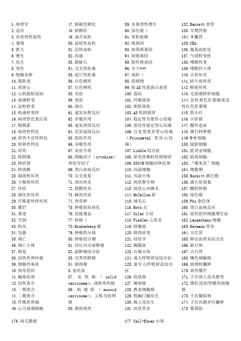
1.病理学2.适应3.亚致死性损伤4.萎缩5.肥大6.增生7.化生8.变性9.细胞水肿10.脂肪变11.虎斑心12.心肌脂肪浸润13.玻璃样变14.淀粉样变15.粘液样变性16.病理性色素沉着17.脂褐素18.病理性钙化19.营养不良性钙化20.转移性钙化21.坏死22.核固缩23.核碎裂24.核溶解25.凝固性坏死26.干酪性坏死27.坏疽28.液化性坏死29.纤维素性样坏死30.糜烂31.窦道32.空洞33.机化34.包裹35.凋亡36.凋亡小体37.修复38.创伤性神经瘤39.细胞外基质40.肉芽组织41.瘢痕疙瘩42.创伤愈合43.一期愈合44.二期愈合45.纤维性骨痂46.心力衰竭细胞47.肺褐色硬化48.槟榔肝49.混合血栓50.延续性血栓51.层状血栓52.再通53.静脉石54.交叉性栓塞55.逆行性栓塞56.白色梗死57.红色梗死58.炎症59.变质60.渗出61.速发短暂反应62.穿胞作用63.速发持续反应64.迟发延续反应65.趋化作用66.吞噬作用67.炎症介质68.细胞因子(cytokine)和化学因子69.类白血病反应70.完全愈复71.渗出性炎72.假膜性炎73.蜂窝织炎74.肉芽肿75.肿瘤的异质化76.直接蔓延77.转移(78.Krukenberg瘤79.肿瘤的分级80.肿瘤的分期81.异位内分泌肿瘤82.副肿瘤综合症83.交界性肿瘤84.癌肉瘤5.角化珠87.实性癌(solidcarcinoma):或称单纯癌86.粘液癌(mucoidcarcinoma):又称为胶样癌88.癌前病变89.非典型性增生90.原位癌(91.肾胚胎瘤92.癌基因93.病毒癌基因94.原癌基因95.隐性癌基因96.分子***97.端粒(98.促癌物99.致AS性脂蛋白表型100.脂纹101.纤维斑块102.粥样斑块103.AS性固缩肾104.稳定性劳累性心绞痛105.恶化性稳定型心绞痛106.自发型变异型心绞痛(Prinzmetal变异心绞痛)107.Liddle综合症108.原发性颗粒性固缩肾109.833+B细胞同种抗体110.风湿细胞111.风湿小体112.疣状赘生物113.疣状心内膜炎114.McCallum斑115.绒毛心116.Roth点117.Osler小结118.Fiedler心肌炎119.肺腺泡120.肺肉质变121.硅结节122.胸膜斑123.石棉小体124.成人呼吸窘迫综合征125.新生儿呼吸窘迫综合征126.疣状癌127.瘢痕癌128.燕麦细胞癌129.假幽门腺化生130.肠上皮化生131.疣状胃炎132.Barrett食管133.早期胃癌134.革囊胃135.CEA136.胞浆疏松化137.气球样变性138.嗜酸性变139.嗜酸性小体140.点状坏死141.碎片状坏死142.桥接坏死143.毛玻璃样肝细胞144.急性黄色肝萎缩或急性红色肝萎缩145.假小叶146.小肝癌147.髓外造血148.淋巴样肿瘤149.R-S细胞150.镜影细胞151.霍奇金细胞152.陷窝细胞153.“爆米花”细胞154.细胞期155.Burkitt淋巴瘤156.满天星现象157.髓样肿瘤158.绿色瘤159.Ph1染色体160.类白血病反应161.恶性组织细胞增生症ngerhans细胞163.Heymann肾炎164.大红肾165.肺出血肾炎综合征166.新月体167.大白肾168.嫌色细胞癌169.纳博特囊肿170.真性糜烂171.子宫颈上皮内瘤变172.微灶浸润型鳞状细胞癌173.子宫腺肌病174.子宫内膜异位囊肿175.葡萄胎176.绒毛膜癌177.Call-Exner小体178.前列腺特异性抗原179.硬化性腺病180.小叶原位癌181.APUD细胞,APUD瘤182.Simond综合征183.Sheehan综合征184.纤维性甲状腺炎185.许特尔细胞瘤186.砂粒体187.嗜铬细胞瘤188.沃-弗189.卫星现象190.噬神经细胞现象191.鬼影细胞192.PrP病193.缺血性脑病194.层状坏死195.Verocay小体196.神经原纤维缠结197.Hirano小体198.结核结节199.原发综合征200.开放性愈合201.结核瘤202.干性结核性胸膜炎203.伤寒细胞204.髓样肿胀期205.凹空细胞206.隐性梅毒207.树胶样肿208.白色肺炎209.Kaposi肉瘤210.嗜酸性脓肿211.假结核结节212.象皮肿213.干线性肝硬化214.腊样变性215.阿米巴肿216.含铁小结217.无反应性结核218.肿瘤219.肿瘤的实质220.肿瘤的间质221.肿瘤的异型性222.间变223.间变性肿瘤224.肿瘤的演进225.癌脐226.心内膜下心肌梗死227.心肌纤维化228.高血压脑病229.细支气管230.呼吸性细支气管231.小气道232.慢性阻塞性肺病233.慢性支气管炎234.肺气肿235.继发性颗粒固缩肾236.VonHippel-Lindan(VHL)病237.腹膜假粘液瘤238.迟发性神经原死亡239.软脑膜癌病240.老年斑241.含铁血黄素1.:是一门研究疾病的病因、发病机制、病理改变(包括代谢、机能和形态结构的改变)和转归的医学基础学科。
病理学名词解释及问答题总结(第六章)

病理学名词解释及问答题总结(第六章)心血管系统疾病1. 动脉粥样硬化(atherosclerosis,AS)①与血脂异常及血管壁成分改变有关;②累及弹力型及弹力肌型动脉;③内膜脂质沉积及灶性纤维性增厚、深部成分坏死、崩解,形成粥样物、管壁变硬、管腔狭窄2. arteriosclerosis(动脉硬化)①动脉壁增厚、失去弹性;②类型:动脉粥样硬化、细动脉硬化、动脉中层钙化3. 冠状动脉性心脏病(coronary heart disease,CHD)因冠状动脉狭窄所致心肌缺血而引起,只有当冠状动脉粥样硬化而引起的心肌缺血、缺氧的功能性和器质性病变时,才称CHD4. angina pectoris(心绞痛)①心肌急剧的、暂时性缺血;②胸骨后压榨性或紧缩性疼痛感;③放射至心前区或左上肢;④持续数分钟;⑤用药、休息可缓解5. myocardial infarction(心肌梗死)①冠状动脉供血中断;②心肌坏死;③剧烈而持久的胸骨后疼痛;④有突出的血清心肌酶活力增高和心电图改变6. subendocardial myocardial infarction(心内膜下心肌梗死)①累及心室壁内侧1/3的心肌,波及肉柱及乳头肌;②多发性、小灶性、不规则分布的坏死7. transmural myocardial infarction(透壁性心肌梗死)①病灶较大,最大径大于2.5cm,累及心室壁全层;②最常见于左冠状动脉前降支供血区,其次是右冠状动脉供血区8.ventricular aneurysm(室壁瘤)在梗死急性期或愈合期,多见于左心室前壁近心尖处,在心室内压作用下梗死区局限性向外膨隆,可继发附壁血栓、乳头肌功能不全、心律紊乱及左心衰竭9. 冠状动脉性猝死(sudden coronary death)①多见于男性青、壮年;②常在某种诱因作用下或在睡眠中发病;③发病后迅速死亡;④冠状动脉病变及相应心肌病变10. primary hypertension(原发性高血压)①原因未明;②体循环动脉血压持续升高;③独立性全身性疾病;④基本病变为全身细动脉和肌性小动脉硬化;⑤常引起心、脑、肾和眼底病变11. concentric hypertrophy of heart(心脏向心性肥大)①压力性负荷增加;②左心室代偿性肥大;③心腔不扩张,甚至略缩小12. hypertensive encephalopathy(高血压脑病)①高血压患者;②脑血管病变及痉挛,血压骤升;③中枢神经系统功能障碍:头痛、呕吐、视力障碍及意识模糊;④脑水肿或伴点状出血13. accelerated hypertension(急进型高血压)①多见于青中年;②血压显著升高,舒张压常>17.3kPa(130mmHg);③病变进展迅速,病变:增生性小动脉硬化和细动脉纤维蛋白样坏死;④较早出现肾衰竭14. aneurysm(动脉瘤)指动脉壁因局部病变(薄弱或结构破坏)而向外膨出,形成永久性的局限性扩张,常见于弹性动脉及其主要分支。
植物病理学名词解释-简答题

植物病理学名词解释-简答题一、名词解释1.病状:植物得病后本身所表现的病态。
病征:病原物在植物病部表现出的特征性结构体。
症状:植物生病后的不正常表现。
病原:引起植物发病的原因。
2.喷菌现象在徒手切片中看到有大量细菌从病部喷出,这种现象称为喷菌现象(bacteria exudation,BE) 。
为细菌病害所特有,是区分细菌病害和其它病害的最简便的手段之一。
3.钝化温度(Thermal lnactivation Point,TIP) 将病组织汁液处理10min使病毒丧失活性的最低温度,用摄氏度表示。
4.病害循环病害循环(disease cycle) 也称作侵染循环(infection cycle):5.整体产果(Holocarpic)。
低等真菌繁殖时, 营养体全部转为繁殖体时叫整体产果分体产果(Eucarpic)。
高等真菌繁殖时, 营养体部分转为繁殖体时叫分体产果。
6.系统侵染:7.综合防治综合防治(综合治理IPM)(植物病害管理PDM):从农业生产的全局和农业生态系的总体观念出发,根据有害生物和环境之间的相互关系,充分发挥自然控制因素的作用,因地制宜地协调应用生物的、物理的、化学的等必要措施,将有害生物控制在经济损害允许水平之下,以获得最佳的经济、生态和社会效益。
同时要把防治过程中可能产生的有害副作用减少到最低限度。
8.局部侵染:系统侵染:9生理小种(Physiological race) :指在种内或变种内或专化型内,在形态上没有差异,但生理特性(培养性状、生理生化、病理、致病力或其他特性)有差异的生物型或生物型群。
9.单分体病毒;10侵染性病害:病原生物侵染植物引起的病害非侵染性病害:由植物自身及非生物因素引起的植物病害。
11、同宗配合:单个菌株生出的雌雄性器官能交配,自身亲合,如卵菌。
异宗配合: 异宗配合:同一菌株上生出的雌雄性器官不能交配,必须和另一有亲和力菌株上的才能交配,担子菌以此为主(接合、子囊两种方式都有)。
病理生理 名词解释 考试必备

病理生理名词解释考试必备AB实际碳酸盐值,指在隔绝空气的条件下,在实际PaCO2,体温和血氧饱和度条件下测得的血浆HCO3¯浓度,受呼吸和代谢两方面的影响AG阴离子间隙,是一个计算值,指在血浆中未测定的阴离子US与未测定的阳离子UC的差值。
AG=UC-UA,波动范围12±2 mmol/LBE碱剩余,是指在标准条件下,用碱或酸滴定全血标本至PH7.4所需的酸或碱的量,酸的量为BE正值,碱的量为BE负值,正常-3、0-+3、0DIC:弥散性血管内凝血是在多种病因下凝血过程强烈激活,广泛微血栓形成,导致凝血因子与PLT大量消耗,激发纤溶功能增强,出现凝血功能障碍并以出血为特征的临床综合症。
FDP:纤溶酶可水解肽链上各单位的赖氨酸-精氨酸,逐步将整个纤溶蛋白和纤维蛋白原分成很多可溶性的小肽片段,总称纤维蛋白原降解产物。
II型呼衰(respiratory failure typeII)伴有高碳酸血症的低氧血症型呼吸衰竭,因外呼吸功能障碍,在海平面水平,静息状态下,动脉血氧分压降低8kpa(60mmHg)下,伴动脉血二氧化碳分压增高超过6、67kpa(50mmHg)病理过程I型呼衰(respiratory failure type I)低氧血症型呼吸衰竭,指由于外呼吸功能障碍,以致在海平面水平,静息状态下,动脉血氧分压降低至8kpa(60mmHg)以下,不伴有动脉血二氧化碳增高的病理过程。
MDF:心肌抑制因子,是胰腺外分泌细胞损伤后释放的蛋白水解酶水解组织蛋白产生的小分子多肽,作用①抑制心肌收缩②抑制单核吞噬细胞功能③收缩腹腔内脏器官血管。
MOSF:多系统器官衰竭,指在炎症创伤,感染和休克时,原无器官功能障碍的患者同时或在短时间内相继出现2个以上器官系统的功能障碍甚至衰竭,往往是休克病人死亡的重要原因P aCO2(动脉血CO2分压)是指血浆中呈物理溶解状态的CO2产生的张力,正常值33-46mmHg,平均值为40 mmHg,反映呼吸因素的指标。
病理学名词解释大题

1化生:为适应环境变化,一种已分化组织转变为另一种分化组织的过程。
2坏死:以溶酶性变化为特点的活体内局部组织中细胞的死亡3凋亡:是指机体细胞在某些因素作用下,通过细胞内基因及其产物的调控而发生的一种程序性的细胞死亡。
4肉芽组织:是新生的富含毛细血管的幼稚阶段的纤维结缔组织,由纤维母细胞、毛细血管及一定数量的炎性细胞等有形成分组成的。
栓塞:在循环血液中出现的不溶于血液的异常物质,随血流运行阻塞5血栓:在活体的心血管内,血液发生凝固或血液中某些有形成分析出、凝集形成固体质块的过程。
形成的固体质块称为血栓。
6梗死:局部组织因血流中断引起的缺血性坏死7、肉芽肿:炎症局部巨噬细胞及其衍生物增生形成境界清楚的结节状病灶为特征。
8异型性:无论在瘤细胞形态还是在组织学结构上,肿瘤都与其来源的正常组织有不同程度的差异,这种差异性称为异型性。
(反映了肿瘤分化程度)9:癌前病变:指具有癌变潜在可能性的病变,这些病变长期存在有可能转变为癌。
-10、假小叶:是肝硬化的特征性病变,是指由广泛增生的纤维在组织将原来的肝小叶和再生的肝细肝细胞团。
11、结核结节:是在细胞免疫的基础上形成的,由于上皮样细胞、朗格汉斯细胞加上外周局部聚集的淋巴细胞和少量反应性增生的纤维母细胞构成。
12、原发综合征:肺的原发病灶、淋巴管炎和肺门淋巴结结核称为~13风湿小体:风湿性肉芽肿,也称阿少夫小体。
多见于心肌间质、心内膜下及皮下结缔组织,是一种肉芽肿型病变,形状略呈梭行形。
镜下中心见纤维素样坏死,周围有Aschoff细胞,外周有少量淋巴细胞、浆细胞浸润。
心衰细胞: 左心衰竭肺淤血时,有些巨噬细胞吞噬了红细胞并将其分解,胞浆内形成含铁血黄素,此时这种细胞称为心力衰竭细胞。
又称心衰细胞。
15. 原位癌(carcinoma in situ):原位癌一般指粘膜鳞状上皮层内或皮肤表皮层内的重度非典型增生几乎*累及或累及上皮的全层(上皮内瘤变Ⅲ级)但尚未侵破基底膜而向下浸润生长者。
病理学期末试题(含答案)

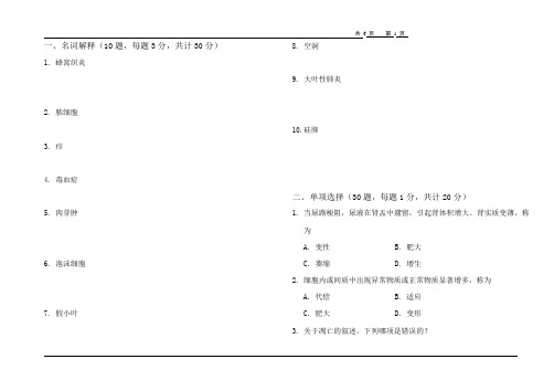
一、名词解释(10题,每题3分,共计30分)1. 蜂窝织炎2. 脓细胞3. 疖4. 毒血症5. 肉芽肿6. 泡沫细胞7. 假小叶8. 空洞9. 大叶性肺炎10. 硅肺二、单项选择(30题,每题1分,共计20分)1.当尿路梗阻,尿液在肾盂中潴留,引起肾体积增大、肾实质变薄,称为A. 变性B. 肥大C. 萎缩D. 增生2.细胞内或间质中出现异常物质或正常物质显著增多,称为A. 代偿B. 适应C. 肥大D. 变形3.关于凋亡的叙述,下列哪项是错误的?A. 是基因调控的程序化细胞死亡B. 凋亡细胞核固缩、核染色质边集C. 凋亡细胞周围可有巨噬细胞存在D. 凋亡细胞周围可有中性粒细胞存在4.病毒性肝炎点状坏死后再生属于A. 完全再生B. 不完全再生C. 纤维性修复D. 瘢痕性修复5.完成瘢痕修复的物质基础是A. 上皮组织B. 肉芽组织C. 纤维蛋白网架D. 毛细血管网6.临床上一次抽取腹水量不能过大,是为了防止A. 静脉性充血B. 减压性充血C. 心力衰竭D. 肺淤血7.槟榔肝的典型病变是A. 肝小叶结构破坏B. 肝细胞坏死C. 门静脉分支扩张淤血D. 肝血窦扩张淤血,肝细胞脂肪变性8.血液从血管或心腔逸出,积聚在体腔内,称A. 出血B. 内出血C. 外出血D. 破裂性出血9.关于白色血栓的叙述,哪像不正确?A. 多发生在血流缓慢的心瓣膜上B. 多见于静脉血栓的起始部C. 不易脱落引起远端血管栓塞D. 主要由血小板和少量纤维蛋白组成10.血栓转归中不会发生的是A. 钙化B. 溶解C. 化生D. 机化11.至少多少毫升的空气一次性进入机体静脉就可以导致患者猝死A. 50B. 60C. 80D. 10012.男,14岁,右股深部巨大血管瘤,术后情况良好,伤口一期愈合。
拆线后下床活动5分钟后,突然晕倒,抢救无效死亡。
应考虑A. 脑血管意外B. 心肌梗死C. 脂肪栓塞D. 肺动脉栓塞13.易发生出血性梗死的器官是A. 心B. 肾C. 脾D. 肠14.贫血性梗死不常见于A. 脾梗死B. 肾梗死C. 心肌梗死D. 肺梗死15.下列哪一项不是炎症时实质细胞常出现的变质性变化?A. 细胞水肿B. 黏液变性C. 脂肪变性D. 凝固性坏死16.炎症时引起局部疼痛的主要因素是A. 炎症局部动脉扩张B. 炎症局部静脉阻塞C. 局部充血渗出D. 炎症渗出物和炎症介质的作用17.急性炎症时,组织变红的主要原因是A. 血管扩张,血流加速B. 炎症灶内血栓形成C. 炎性细胞浸润D. 组织间隙水肿18.炎症反应最重要的功能是A. 杀灭侵入人体的病原微生物B. 产生免疫反应C. 将炎性细胞输送到炎症灶D. 吞噬损伤因子19.不符合急性炎症时白细胞渗出叙述的是A. 白细胞黏附于血管内皮细胞是白细胞从血管游出的前提B. 游出血管外的白细胞可分泌胶原酶降解血管基底膜,进入周围组织C. 白细胞介素6具有阳性趋化作用D. 趋化因子通过与白细胞表面的特异性G蛋白偶联受体相结合而发挥作用20.急性蜂窝织炎的常见致病菌是A. 金黄色葡萄球菌B. 溶血性链球菌C. 大肠杆菌D. 多为混合感染21.浆液性炎常见于下列哪种疾病?A. 大叶性肺炎B. 小叶性肺炎C. 毒蛇咬伤D. 阑尾炎22.金黄色葡萄球菌感染最常引起A. 浆液性炎B. 化脓性炎C. 蜂窝织炎D. 脓肿23.炎症在局部蔓延时不会形成A. 糜烂B. 溃疡C. 瘘管D. 炎性假瘤24.对维持肿瘤生长起重要作用的肿瘤间质成分是A. 结缔组织B. 血管C. 淋巴细胞D. 间质细胞25.属于良性肿瘤的是A. 淋巴管瘤B. 精原细胞瘤C. Krukenberg瘤D. 黑色素瘤26.良性肿瘤对机体影响最大的因素是A. 生长部位B. 生长速度C. 组织来源D. 体积大小27.高分化鳞癌最典型的病理特点是A. 癌细胞核分裂象多见B. 形成癌巢C. 出现角化珠D. 细胞间可见细胞间桥28.动脉粥样硬化粥瘤的粥样物质的主要成分是A. LDLB. VLDLC. ox-LDLD. 胆固醇29.冠心病心肌梗死后肉眼能辨认病灶的最早时间是A. 1~2小时B. 4小时C. 5小时D. 6小时30.Mc Callum斑常见于A. 左心房后壁B. 左心房前壁C. 右心房D. 左心室三、简答题(4题,每题10分,共计40分)1 比较坏死和凋亡(20分)2. 简述胃良恶性溃疡的区别(12分)3.简述类白血病反应(8分)答案及评分标准一、名词解释(10题,每题3分,共计30分)1.蜂窝织炎:疏松结缔组织的弥漫性化脓性炎症,常发生于皮肤肌肉和阑尾。
病理生理学重点 名词解释与简答题

病理生理学重点名词解释与简答题(Key terms of pathophysiology Interpretation and brief answer)First, the noun explanationThe basic pathological process: mainly refers to a variety of diseases may occur, common, complete sets of functions, metabolism and structural changes.Health: health means not only sickness or illness, but also a complete, physical, mental, and social state of being.Disease: disease is under certain conditions caused by the cause of damage, due to the body's self regulatory disorder and the occurrence of abnormal life process.Etiology: a cause of disease that is characteristic of the disease and is known as the cause of the disease.Incentives: factors that contribute to the development of disease are called triggers.Brain death (brain death): the permanent cessation of the function of the organism as a whole is a permanent loss of the function of the brain. At present, the standard of brain death above the foramen magnum is generally used.Hypotonic dehydration: also called hyponatremia of low volume, characterized by losing Na+ more than dehydration, serum Na+ concentration <130mmol/L, plasma osmotic pressure <280mmol/L, accompanied by a decrease in extracellular fluid volume.Hypertonic dehydration: also known as low volume hypernatremia, characterized by more water loss than loss of Na+, serum Na+ concentration of >150mmol/L, plasma osmolality, >310mmol/L, extracellular fluid volume and extracellular fluid volume are reduced.Water intoxication: high solubility hyponatremia, characterized by decreased serum sodium, serum Na+ concentration, <130mmol/L, plasma osmotic pressure <280mmol/L, but the total body sodium is normal or increased, the patient has water retention, so that the volume of fluid increases significantly.Dehydration fever: because of the decrease of water from the skin, the heat is affected, leading to elevated body temperature, called dehydration fever.Edema: excessive fluid that is called edema in the interstitial space or inside the body cavity.Hyperkalemia: serum potassium levels are higher than 5.5mmol/LHypokalemia: serum potassium levels below 3.5mmol/L.Disturbance of acid-base balance: the excessive acid base overload, serious deficiency or regulatory disturbance caused by pathological conditions lead to the PH abnormal condition caused by the destruction of acid-base homeostasis in the environment.Metabolic acidosis: a type of acid-base disturbance characterized by an increase in extracellular fluid H+ or a primary decrease in HCO3- and a decrease in PH.Respiratory acidosis: a CO2 disorder or excessive intake causes a disturbance of acid-base balance characterized by a primary increase in plasma [H2CO3] and a decrease in PH.Metabolic acidosis: extracellular liquid increase or loss of H+, [HCO3-] in plasma increased and PH increased primaryacid-base disorders characterized by type.Respiratory alkalosis: a type of acid-base disturbance characterized by hyperventilation of the lungs and primary decrease in plasma [H2CO3] and elevated PH.Mixed acid-base disturbance: there are two or three simple types of acid-base disturbances in the same patient.Hypoxia (hypoxia): pathological changes in cellular metabolism, function, and morphological structure resulting from reduced oxygen supply or impaired oxygen use.Cyanosis (cyanosis): when the uniform concentration of deoxygenated hemoglobin in the blood capillary of more than 5g/dl, the skin and mucous membranes were purple.Type four hypoxia: hypoxia hypoxia, blood type hypoxia, circulatory hypoxia, tissue hypoxia.Enterogenous cyanosis: methemoglobinemia due to eating leadsto a large number of hemoglobin oxidation caused by.Shock: this word by Shock, and English shock, various strong risk factors acting on the body, the circulation decreased dramatically, tissue and organ perfusion of microcirculation is seriously insufficient, and systemic critical pathological process of vital organs function, metabolic disorder serious.MODS (multiple organ dysfunction syndrome): refers to the severe trauma, infection, shock, the original organ dysfunction of patients at the same time or within a short period of time have appeared more than two organ system dysfunction that homoiostasis must rely on clinical intervention to maintain syndrome.Systemic inflammatory response (SIRS): a systemic inflammatory response syndrome that results from uncontrolled, self sustained enlargement and self destruction caused by infection or non infectious agents acting on the body.Compensatory anti-inflammatory response syndrome (CARS): when an infection or trauma occurs, the organism produces an endogenous anti-inflammatory response that is associated with an increased immune response and an increased susceptibility to infection.DIC (disseminated intravascular bleeding): a role in the pathogenic factor, the coagulation system was activated and induced by micro thrombosis, or secondary fibrinolysis, resulting in bleeding, shock, organ dysfunction and microangiopathic hemolytic anemia in a pathological process.FDP fibrin degradation products: plasmin hydrolyze various fragments produced by Fbg and Fbn.Split cell: a special variant of the shape of red blood cells,Called schistocyte, shape, star, moon shaped helmet, referred to as the red cell debris. The fragment is high in brittleness and susceptible to hemolysis.Heart failure: cardiac pump dysfunction caused by various causes, and a negative or relatively reduced cardiac output that fails to meet the metabolic needs of tissue.Myocardial tension (muscle) derived dilatation:The source of expansion: reduce the myocardial tension due to stroke volume, the ventricular end diastolic volume increased, pre load led to the increase of the initial length of the muscle fibers increased, then enhance myocardial contractility, metabolism increases stroke volume, heart cavity enhanced myocardial contractility with this expansion called heart source expansion.Myogenic expansion: long-term volume overload and heart failure caused by dilated cardiomyopathy, mainly caused by excessive sarcomere stretch, the heart cavity enlargement, heart cavity with the excessive stretch and decreased myocardial contractility with expansion called myogenic expansion.Respiratory failure: a pathological process in which the arterial blood oxygen pressure is below normal and accompanied by or without elevated carbon dioxide partial pressure due to severe external respiratory dysfunction.Restrictive hypoventilation: alveolar ventilation caused by restricted expansion of the alveoli during inspiration. Obstructive hypoventilation: a ventilatory disorder caused by narrowing or obstruction of the airways.Functional shunt: part of alveolar ventilation lesions significantly reduced blood flow and weight, not less, even because of inflammatory hyperemia to increase blood flow (such as lobar pneumonia, early) ratio of alveolar minute ventilation and pulmonary blood flow per minute was significantly reduced, so that the flow through this part of alveolar venous blood without sufficient artery can mixed with arterial blood, the similar arteriovenous short circuit, called functional shunt, also known as blood doping.Dead space ventilation: under certain etiological factors, partial alveolar blood flow can be reduced, VA/Q can be significantly increased, less alveolar blood flow and ventilation, alveolar ventilation can not be fully utilized, become dead cavity like ventilation.Pulmonary encephalopathy: because of respiratory failure caused by brain dysfunction, patients can show a series of neuropsychiatric symptoms, such as directional, memory disorders, confusion, headache, drowsiness, coma and so on.ARDS (acute respiratory distress syndrome): respiratory dysfunction caused by alveolar capillary membrane injury caused by various reasons and the occurrence of acute respiratory failure syndrome clinical characteristics, is characterized by progressive dyspnea and hypoxemia.Hepatic encephalopathy: neuropsychiatric syndrome secondary to severe liver disease.False neurotransmitter: phenylethanolamine and octopamine in chemical structure and normal neurotransmitter norepinephrine and dopamine are similar, but can not complete the authenticity of neurotransmitter function, known as false neurotransmitter.Hepatorenal syndrome: acute renal tubular necrosis associated with functional renal failure and severe hepatitis in decompensated cirrhosis.Acute renal failure due to GFR decreased rapidly, or renal tubular degeneration and necrosis in a pathological process caused by acute severe, often oliguria, azotemia, hyperkalemia, metabolic acidosis and water intoxication syndrome.Chronic renal failure:Any disease of renal unit destruction, in the months and years or longer after the remaining nephron cannot fully remove metabolic waste and environment, thus to maintain the constant in the gradual emergence of metabolic waste retention and water electrolyte and acid-base balance disorders and renal endocrine dysfunction.The levels of non protein nitrogen (NPN) in blood, including uremia, creatinine, uric acid, and so on, are called azotemia. The NPN in normal blood is 25~35mg%, and the urea nitrogen is 10-15mg%.Renal hypertension: hypertension caused by renal parenchymal disease is called renal hypertension.Uremia: refers to acute or chronic renal failure to severe stage, metabolites and toxins accumulate in the body, water, electrolyte and acid-base balance disorders and some endocrine disorders caused by a series of systemic function and metabolic disorder syndrome.Two, Jane answer1. briefly describe the diagnostic criteria for brain deathAccording to the determination of brain death is the irreversible coma and brain response; second, stop breathing for 15 minutes, artificial respiration still no spontaneous breathing; the cranial nerve reflex; the pupil or fixed; the brain waves disappeared; the complete cessation of cerebral blood circulation (cerebral angiography).2. which type of dehydration is prone to loss of fluid shock? Why?Hyponatremia with low volume (hypotonic glue) is liable to cause fluid shock. (1) the extracellular fluid Bian through thepressure drop, no thirst, drinking water reducing. (2) the extracellular fluid Bian through pressure reduction, ADH reflex secretion decreased, no significant decrease in urine volume.(3) the extracellular fluid transferred to the intracellular fluid, and the extracellular fluid decreased further.3. to analyze the influence of water intoxication on the body.Extracellular fluid was diluted due to excessive water, so the blood sodium concentration decreased and osmotic pressure decreased. In addition, the kidney can not discharge too much water in time, the water is transferred to the relatively high osmotic pressure cells, causing edema. The result is that both the intracellular and external fluid volume are increased, and the osmotic pressure is decreased. Because the cell liquid capacity greater than the extracellular fluid retention capacity, so most of the moisture accumulation in the cells, therefore in mild water intoxication, interstitial water retention degree is still not enough to cause recessive edema of the concave. Acute water intoxication, the nerve cells in the brain edema and intracranial pressure, the cerebral symptoms appeared earliest and prominent, can occur in various neuropsychiatric symptoms, such as gaze, aphasia, confusion, lethargy, irritability and other directional arrhythmia, and papilloedema, severe cases can occur due to cerebral hernia caused by respiratory and cardiac arrest patients with mild, chronic or water poisoning is slow, symptoms are often not obvious, many covered by the symptoms and signs of primary disease, can have drowsiness, headache, nausea, vomiting,muscle spasms and pain symptoms such as weak and feeble.4. to discuss the mechanism of edema caused by imbalance of fluid exchange inside and outside the blood vessel.The force that drives the flow out of the intravascular fluid is the average effective hydrostatic pressure. The force that causes the fluid to flow back into the capillaries is effective colloid osmotic pressure. Normally, the tissue fluid is slightly larger than the reflux. Lymph reflux, tissue fluid reflux, the remaining part of the lymphatic system through the backflow into the blood circulation, normal adults in quiet state, enzyme hours about 120ml liquid through the lymphatic system into the blood circulation. The interstitial fluid increased hydrostatic pressure, and the rate of lymph formation was accelerated. In addition, the permeability of the lymphatic wall is higher and the protein is easy to pass. Therefore, lymph reflux can not only send a little more of the tissue fluid back into the body, but also the proteins that leak out of the capillaries, and the large molecules produced by cell metabolism absorb the circulation of the body. One or more of these factors at the same time or one after another disorders may be an important cause of edema.5. hypokalemia and hyperkalemia can cause muscle paralysis. What is the mechanism of this?Hypokalemia is mainly due to hyperpolarized block. The ratio of [K+ /[K+]e] the increase of hypokalemia, and muscle cell negative resting potential, the resting potential and threshold potential interval increases, cell excitability anddecreased, even not serious when excited, i.e. cells in the hyperpolarized state block. Hyperkalemia results in inactivation of the fast sodium channel because of the small resting potential of the skeletal muscle and is unable to be excited by extracellular depolarization.6. what kinds of acid-base disorders are caused by severe vomiting? To analyze the mechanism of its occurrence.Severe vomiting easily leads to metabolic alkalosis:H+ lost the gastric juice, the digestive tract of HCO3- H+ is not neutralized and absorbed into the blood; the loss of Cl- in gastric juice, can cause hypochloremic alkalisis; K+ lost the gastric juice, can cause hypokalemic alkalosis; the loss caused by a large amount of gastric juice, effective circulating blood volume reduction, secondary aldehyde solid ketone the increase caused by metabolic alkalosis.7. what types of acid-base disturbances can occur in the oliguria stage of acute renal failure? Why? What are the changes in the acid-base balance index?Can occur because of metabolic acidosis, renal function and acid alkali, in severe renal failure, can not be fixed in acid by urinary excretion, especially sulfuric acid and phosphoric acid accumulation in the body, resulting in the increase of H+ concentration HCO3- concentration decreased, due to reduced HCO3-, so AB, SB and BB significantly decreased. Negative BE increased, PH decreased by Paco2 secondary respiratory compensation, down AB<SB.8. what type of hypoxia can be caused by hemorrhagic shock?.Hemorrhagic shock, due to a large number of blood loss and lack of tissue blood, resulting in inadequate oxygen supply, can lead to circulatory hypoxia.9. what kind of hypoxia can pulmonary edema cause? Its hypoxia mechanism?Can cause hypoxic hypoxia, due to pulmonary edema, ventilation function of fertilizer limited caused by alveolar PO2 decreased, pulmonary ventilation dysfunction by alveolar diffuse into the blood oxygen reduction, PaO2 and lack of oxygen, caused by hypoxic hypoxia.10. to analyze the mechanism of red blood cell growth during chronic hypoxia.When the number of red blood cells in chronic hypoxia is mainly due to the increase in renal production and release of erythropoietin EPO; hypoxia can increase the activity of HIF-1 in cytoplasm, HIF-1 gene and EPO 3 enhancer binding, enhanced the expression of EPO gene, the EPO increased, the molecular weight of EPO was 34000 of the egg white sugar, can promote stem cell the original red blood cell differentiation, and promote the differentiation, proliferation and maturation, accelerate the synthesis of hemoglobin, the bone marrow reticulocyte and red blood cells released into the blood.11. try to describe the mechanism of shortness of breath inpeople on the upper plateau.The air in the plateau is thin and low in oxygen,At the beginning of the plateau, hypoxia induced PAO2 below 60mmHg and stimulated the peripheral chemoreceptor of the carotid body and the aortic body. The impulses were introduced into the medulla via the sinus nerve and the vagus nerve, which caused the respiration to accelerate gradually.12. can arterial blood pressure be used as a marker for the occurrence of shock? Why?No, because the shock early sympathoadrenomedullary excitement, heart rate, contractility increased, blood transfusion and infusion through their own, as well as increased renal reabsorption of sodium and water, increase the turn output, so that the increased cardiac output, plus the peripheral resistance increased, so the blood pressure was not obvious.13. to describe the characteristics and compensatory significance of microcirculatory changes in early stage of shock.Is the early stage of shock shock compensatory, the sympathetic nervous excitement, increased catecholamine, some organs of small vascular contraction or spasm, especially micro artery, after arterioles and precapillary sphincter contraction, the precapillary resistance increased, capillary closure, true capillary network reduced blood flow, blood flow slows down; beta adrenergic receptor the stimulated arteriovenousanastomoses are open, blood through the direct pathway and open the arteriovenous anastomosis reflux, microcirculation of non nutritive blood flow increase, reduce nutrient flow, severe hypoxic ischemic tissue. The above changes of microcirculation, on the one hand, cause ischemia and hypoxia of organs such as skin, abdomen, viscera and kidneys, on the other hand, they have compensatory significance to the whole. The main manifestations are: blood redistribution, autotransfusion, and self infusion.14. the mechanism of microcirculatory changes in shock stage II.The mechanism of microcirculatory changes in progressive stage is related to the effects of prolonged microvascular contraction and ischemia, hypoxia, acidosis, and various humoral factors.Acidosis: hypoxia leads to the decrease of tissue oxygen partial pressure, accumulation of CO2 and lactic acid and acidosis. Acidosis causes a decrease in the responsiveness of vascular smooth muscle to catecholamines and causes vasodilation.The local vasodilator metabolites increased: long-term ischemia and hypoxia, acidosis stimulates the release of histamine from mast cells increased, ATP decomposition products of adenosine accumulation, kinins generation increases, can cause vascular smooth muscle relaxation and telangiectasia.The changes of blood rheology: shock progression blood flow decreased obviously, micro slow blood flow in erythrocyte aggregation and easy; the effect of histamine on capillary permeability, plasma extravasation, blood viscosity increased; perfusion pressure decreased, resulting in leukocyte rolling, adhesion and adhesion to endothelial cells, blood blocked postcapillary resistance increased.IV: the role of endotoxin: the relaxation of vascular smooth muscle, resulting in persistent hypotension.15. what is the most common cause of multiple organ dysfunction syndrome (MODS)? Its pathogenesis?80% of the MODS patients had significant shock when they entered the hospital.Infectious causes, such as sepsis and severe infection, can affect approximately 70% of MODS, especially sepsis caused by severe infections.Non infectious causes such as major surgery and severe trauma, major surgery and severe trauma, whether or not infections are pure, can occur in MODS.Pathogenesis: (1) the general inflammatory response is out of control. Promoting the imbalance between anti-inflammatory and anti-inflammatory mediators. Other factors that cause organ dysfunction: organ microcirculation perfusion disorder, high metabolic state, ischemia-reperfusion injury.What is the mechanism of 16.DIC?The release of tissue factor, the activation of exogenous coagulation system, the initiation of blood coagulation system, the damage of vascular endothelial cells, the imbalance of coagulation and anticoagulant regulation, the destruction of blood cells, the activation of platelets, and the introduction of procoagulant substances into the blood.What is the mechanism by which 17.DIC leads to shock?Because of the large amount of micro thrombus in blood vessel, the microcirculation is blocked and the blood volume is reduced obviously. Extensive bleeding can reduce the volume of blood. The injury of cardiac muscle leads to decrease of cardiac output. The activation of coagulation factor XII may activate the kallikrein system, complement system and fibrinolytic system, and produce some vasoactive substances, such as bradykinin and complement components. The complement component can make the basophils and mast cells release histamine, etc. the peptide and histamine can relax the microvascular smooth muscle, increase the permeability, reduce the peripheral resistance and reduce the blood volume. Some components of FDP can enhance the action of histamine, bradykinin and promote the expansion of microvessels.18. try to explain the compensatory response of the heart when the heart function is incomplete.Heart rate increased, heart tension dilated, myocardial contraction increased, ventricular remodeling19. cardiac insufficiency after the type of compensatory cardiac hypertrophy and its mechanism, sarcomere replication.Hypertrophy of the heart can be caused by a variety of reasons. When partial myocardial cells are lost, reactive myocardial hypertrophy may occur in the residual myocardium. Prolonged overload can cause excessive cardiac hypertrophy,According to the cause of overload and cardiac reaction of different forms can be divided into overload cardiac hypertrophy: concentric hypertrophy, cardiac function in long-term excessive load pressure, systolic wall tension increase myocardial sarcomere parallel type hyperplasia, thickening of myocardial cells. Eccentric hypertrophy, heart in the long-term excessive load capacity, diastolic wall tension increase myocardial sarcomere was series hyperplasia, myocardial cell growth, ventricular volume increases; while the heart enlargement and systolic wall stress increases, and thus stimulate the proliferation of sarcomere parallel.20. briefly describe the mechanism and significance of heart rate reduction during heart failure.The mechanism of heart rate is: due to decreased cardiac output, heart rate caused by the intracardiac residual blood volume is increased; reduce the heart pump blood, ventricular end diastolic volume and pressure, can stimulate the right atrium and vena cava volumereceptor, via vagal afferent fibers in the vagus nerve to pivot, inhibition, sympathetic stimulation. If the combined hypoxia can stimulate the aortic body and carotidbody chemoreceptor, reflex excitability causes heart rate to accelerate. Significance: cardiac output is the product of stroke volume and heart rate, in a certain range, can improve the heart rate, cardiac output, and can improve the diastolic blood pressure, blood perfusion to the coronary artery, to maintain arterial blood pressure, which is of positive significance to the blood supply to vital organs.21. what is the mechanism of dyspnea caused by heart failure? What types of dyspnea do you have?Mechanism: lung congestion and pulmonary edema lead to lower lung compliance. To breathe the same amount of air, it is necessary to increase the work done by the respiratory muscle and consume more energy, so the patient feels labored. The bronchial mucosal hyperemia, swelling and airway secretions lead to airway resistance increases; the pulmonary capillary pressure and interstitial edema of pulmonary interstitial pulmonary capillary pressure increased, stimulation of fat receptors, causing reflex rapid shallow breathing. Types include exertional dyspnea, dyspnea, and paroxysmal nocturnal dyspnea.22. the severe hypokalemia (or excessive anesthetics, sedatives, hypnotics, severe pneumothorax, pleural effusion, obstruction of trachea, bronchial asthma, chronic bronchitis) the pathogenesis of respiratory failure caused by.The main effects of hypokalemia on the body is the membrane potential caused by the abnormal effect of nerve muscle, muscle relaxation of skeletal muscle weakness and even paralysis,leading to respiratory muscle contraction diastolic dysfunction caused by restrictive ventilation due to insufficient pulmonary dysfunction, which is outside the alveolar gas and gas exchange in extracellular potassium ion concentration in cell metabolism disorder caused by the disturbance of acid-base balance disorders, hypokalemia decreased, intracellular potassium concentration gradient transfer to the outside of the cell and extracellular hydrogen ion transfer to the cells increased, the hydrogen potassium exchange; the renal ammonia (hydrogen ions).23. to discuss the pathogenesis of pulmonary heart disease.The alveolar hypoxia and CO2 retention caused by blood H+ concentration is too high, can cause pulmonary arteriolar constriction, the pulmonary arterial pressure increased, thereby increasing the right ventricular afterload; pulmonary arteriole contraction and hypoxia can cause non muscular arterioles in muscle, pulmonary vascular smooth muscle cells to fibroblast cell hyperplasia, collagen and elastin synthesis increase, lead to pulmonary vascular wall thickening and lumen narrowing, hardening, resulting in chronic pulmonary hypertension lasting and stable due to the long-term hypoxia; compensatory polycythemia can increase blood viscosity, will also increase the resistance of pulmonary blood flow and increased right heart load; and some abdominal diseases such as pulmonary arteritis, small the destruction of a large number of pulmonary capillary and pulmonary embolism can be a cause of pulmonary hypertension; the hypoxia and acidosis family cardiac contractility and dyspnea, Forced expiratory intrathoracic pressure can cause abnormal increase of cardiaccompression effect of cardiac diastolic function, abnormal inspiratory intrathoracic pressure is reduced, the negative pressure outside the heart increases, increased right ventricular systolic load to right heart failure.24. to discuss the pathogenesis of pulmonary encephalopathyEffects of hypoxia, acidosis and hypercapnia on cerebral blood vessels:1, hypoxia and acidosis to cerebral vascular dilation; 2, hypoxia and acidosis leads to vascular endothelial injury: one is to increase vascular permeability, aggravate brain edema, while promoting intravascular coagulation; 3, hypoxia and acidosis reduced the formation of ATP: dysfunction of sodium pump, cell edema, cerebral vascular compression increased intracranial pressure, intracranial pressure, cerebral vascular compression, increased hypoxia, and forming a vicious spiral.Two. The effects of hypoxia, acidosis and hypercapnia on neurons:1, on the one hand, hypoxia can directly inhibit brain function, on the other hand can cause dysfunction of sodium pump, make the brain edema, increased intracranial pressure; 2, hypercapnia easily lead to intracellular acidosis, on the one hand, the glutamic acid decarboxylase activity increased, the increasing production of GABA, inhibit the function of central nervous system,On the other hand, it can enhance phospholipase activity, decompose membrane phospholipids, dissolve lysosomes, and aggravate the damage of nerve cells.What is the cause of elevated blood ammonia in 25. hepatic encephalopathy? What are the toxic effects of ammonia intoxication on the brain?.The reasons for the increase of blood ammonia include: the decrease of urea synthesis and the lack of ammonia clearance. Toxicity: 1, ammonia to neurotransmitters in the brain changes, brain cell energy metabolism and ammonia the interference effect on the nerve cell membrane pump: NA-K interference on the nerve cell membrane; competition of K ion and ion distribution of membrane.26. what are the metabolic changes in acute renal failure and oliguria? Explain its mechanism.The change of urine: oliguria, anuria, low proportion of urine, hematuria, proteinuria, urinary tube; functional ARF, renal function is not damaged, the oliguria is mainly due to GFR significantly reduced by, and the organic ARF and glomerular and renal tubular dysfunction.Water intoxication: ARF, for reasons of oliguria, catabolism caused by increased intake of water, in the water too much, resulting in water retention, dilutional hyponatremia and cell edema at.The hyperkalemia: potassium with the urine reduced urine volume;。
临床医学病理学问答题、名词解释练习题及答案

临床医学病理学问答题、名词解释练习题及答案*萎缩的定义和病理性萎缩有哪些?★~P7萎缩:已发育正常的组织和器官,实质细胞体积减少,而致组织或器官缩小。
一、生理性:退化-动脉导管、脐带血管;胸腺,老年性萎缩。
二、病理性:1、全身性营养不良性萎缩,如:长期饥饿者、恶性肿瘤者2、神经性萎缩,如:脊髓前角灰貭炎者3、废用性萎缩,如:肢体骨折石膏固定后,肢体长期不活动4、压迫性萎缩,如:尿路结石,尿排泄不畅,而积在肾盂,引起肾实质发生压迫性萎缩5、内分泌性萎缩,如:垂体功能低下引起肾上腺、甲状腺、性腺等器官萎缩6、缺血性萎缩,如:冠状动脉粥样硬化引起心肌萎缩、脑动脉硬化引起脑萎缩*化生的定义,常见类型?★~P8化生:为适应环境变化,一种以分化成熟的组织转变为另一种分化成熟的组织的过程常见类型:1.上皮组织化生(1)鳞状上皮化生~常见部位:气管、支气管黏膜,如:长期吸烟者、慢性炎症者(2)肠上皮化生~常见部位:胃体、胃窦部,如:胃溃疡、慢性萎缩性胃炎2.间叶组织化生:结缔组织化生为骨,软骨或脂肪组织如:骨化性肌炎,纤维组织增生,并发生骨化生*心脏萎缩有哪些改变,以心肌为例?★~P81.大体改变:(1)心肌褐色(2)心尖锐尖(3)包膜皱缩(4)冠状动脉蛇样弯曲2.镜下改变:(1)心肌纤维变细窄(2)肌间距变宽(3)心肌细胞中可见脂褐素颗粒*病理性钙化的概念,种类~p.17正常机体内仅在骨和牙齿内含有固体钙盐,如在骨和牙齿以外的其它组织内有固体钙盐沉积,称之病理性钙化种类-营养不良性钙化(变性,坏死组织或义务的钙盐沉积)转移性钙化(全身性钙,磷代谢障碍引起血钙或血磷增高,导致钙盐在未受伤组织内沉积)。
*坏死的结局★(1)溶解吸收(2)分离排出(3)机化(4)包裹,钙化*坏疽比较:★*细胞凋亡与坏死的区别*再生性修复与纤维性修复的区别由耗损的同种实质细胞再生进行修复,可完全恢复原有细胞、组织的结构和功能。
由结缔组织来修补填空缺损,并形成瘢痕,只能恢复组织的完整性,不能完全恢复原有的结构和功能。
