
Particle-in-cell
Particle-in-cell(PIC方法,质点网格法)计算二维非定常可压缩理想流动问题的欧拉-拉格朗日混合方法,简称PIC法,它特别适用于计算具有多种介质和大变形流动的问题。
在流体动力学中,通常可用欧拉和拉格朗日两种不同坐标系来求解流体动力学问题,即所谓欧拉法和拉格朗日法。
欧拉法可用于求解流体大畸变问题,但精度不高,而且在各个区域进行物质输运时会产生严重的物质扩散,使界面和自由面的位置不能精确确定。
拉格朗日法正好相反,计算精度较高,能精确确定界面和自由面,但不能处理流体大畸变和在各种介质之间有剪切间断的滑移现象。
针对这种情况,美国F.H.哈洛等人于1955年成功地把欧拉法和拉格朗日法结合起来,提出了质点网格法。
基本要点:PIC法的基本要点是,把含有多种介质的流动所通过的区域用欧拉法分成有限个网格,每个网格中的每种流体,用一组特定的离散化拉格朗日质点表示。
只包含一种流体质点的格子称为纯单元,两种流体质点同时存在的格子称为混合单元,不存在任何流体质点的格子称为空单元。
每个质点具有一定的质量,每个网格单元内的质点数目和质点分布都以流体流动的初始状态为依据,而且这些质点具有一定的速度和能量。
计算开始后,质点在欧拉网格之间迁移,表示流体在运动。
在每个时间步长内,计算分两步:第一步用欧拉法计算,即忽略偏微分方程中的输运效应,用差分方法计算由压力分布所引起的欧拉网格上速度(或动量)和能量的变化。
若一个网格内含有多种流体,就应按一定的规则把能量的改变量适当分配给不同的质点。
第二步是质点迁移计算,它是在第一步的基础上,按一定的加权平均方法计算出每个质点的速度和在时间步长结束时的新位置。
一个质点从一个网格迁移到另一个网格,就把所携带的质量以及相应的动量和能量从原来的网格输送到新的网格中去。
这一步实质上是对第一步计算中忽略的输运效应计算的补偿。
在具有激波间断的流动中,处理激波间断是一个难题(见激波数值处理)。
PIC法由于有非线性的耗散效应,不仅可以减少差分格式所引起的起伏现象,而且起着类似于人工粘性的作用。
因此,PIC法能自动处理流动中的激波间断。
但在低速流动和固壁条件的计算中,这个耗散效应很弱,为了使计算稳定,还须引入人工粘性。
要得到较好的计算结果,除应考虑满足一定的稳定性条件外,还须考虑每个单元内的质点数目和分布以及它们的内能等。
方法的推广:在PIC法基础上,人们提出了流体网格法(fluid-in-cell method),简称FLIC 法。
它和PIC法一样采用欧拉网格,不同的只是在第二步计算中不计算质点的迁移,而计算连续流体的迁移,即先算出通过网格边界的质量输送量,得出每个网格的新密度,再算出通过网格的质量所携带的动量和能量的输送量,最后得到每个网格的新速度和能量。
FLIC 法还有一套局部网格单元的计算格式,能计算一些边界形状比较复杂的问题。
计算二维不可压缩粘性流动的PIC法后来还发展成为所谓标记网格法(marker-and-cell method),简称MAC法。
此法仍然采用欧拉矩形网格单元,对纳维-斯托克斯方程则用差分近似,而把压力和速度分量作为基本未知量。
此外,这种方法还在网格中布置适量的标记点,但这种标记点和PIC法中的质点不同,本身并不带有质量。
在每一个时间步长上,只用PIC法中确定质点速度的方法来确定每个标记的速度,并在整个计算中跟踪每个标记,以判定网格里有哪种流体存在。
因此MAC法能用于计算多种流体和带有自由面的问题。
近年来,在研究爆炸和高
速碰撞的现象中还发现介质会经历从固体(弹性、塑性、断裂)到流体的各个阶段,因而在计算时必须考虑固体强度效应,为此在PIC、FLIC和MAC等方法的基础上又导出一种计算流体-弹塑性流动的方法(computational method of hydro-elastic-plastic flow)其中最典型的是HELP编码(HELP code)计算方法。
HELP编码包括三个步骤:第一步计算压力效应;第二步计算输运效应;第三步计算应力偏量效应。
此外在多种介质界面和自由表面上引入一些没有质量的标记点,用以确定界面和自由面的位置。
此外,近年来还广泛采用其他一些类型的欧拉-拉格朗日的混合方法。
最常用的是任意拉格朗日-欧拉方法(Arbitrary
lagrangian-Eulerian Method),简称ALE法。
它将离散化的方程建立在既非欧拉,又非拉格朗日的任意活动的网格上,以达到不断重分网格,适应大变形计算的目的。
质点网格法以及由它演变出来的其他方法都具有数值模拟的特点。
这些方法大都直接来源于对物理问题的描述,受数学方程的约束较小,因此能广泛应用于流体动力学的各个领域,如爆炸、燃烧高速碰撞以及物理-化学流体动力学、液体动力学等方面,甚至还可应用于电流体动力学、磁流体力学和相对论流体力学等方面。
但是,对上述的研究还很不够,许多问题还只处于实验阶段。
PIC数值模拟方法,即Particle-In-Cell, 可用于分子原子尺度运动的计算机数值模拟,在等离子体研究等领域有着广泛的应用。
Particle-In-Cell模拟的发展 - 物理考虑和计算技术

王 虹 宇∗
鞍 山 师 范 学 院 物 理 系 , 辽 宁 , 鞍 山 ,114007
姜巍
大 连 理 工 大 学 物 理 系 , 辽 宁 , 大 连 ,116024
Abstract
粒子云网格(Particle In cell)方法是研究等离子体行为的关键性理论工具之一。它可以看成方 便而粗糙的流体力学模拟和严格但困难的动理学方程求解的一种有效的折中。从七十年代建立 了PIC技术的基本框架以来,这种技术被应用到不断扩展的领域中,从机理研究到具体设备的设 计都从中受益。 尽管PIC方法的基本数学框架并没有实质性的变化,但随着使用领域的推广,问题背景的变化 对PIC模拟技术的细节提出了越来越多的要求:实际工程模拟和设备设计要求模拟方法提供更快 的速度和更好的适应性;新物理现象的机理研究要求模拟必须可以考虑更多的物理效应甚至化学 问题;极端条件下的物理问题的理解要求模拟具有更高的精度或者使用全新的物理理论;多尺度 问题的存在要求模拟中使用更强有力的数学方法;最后,部分问题的极端复杂性需要使用混合模 拟来克服,而这要求PIC方法和其他模拟手段的无缝对接。在现在的模拟技术中,PIC模拟方法向 上连接到连续模拟(流体力学等),而向下重叠了纯粹的分子动力学模拟(MD),在这个庞大的领 域中,提供了各种复杂问题的分析方法。 在另外一方面,从上世纪末期开始,简单而廉价的并行技术,如集群(cluster)成为计算机技 术的亮点之一。集群技术的出现使得高性能计算机变成了能够普及的产品。传统上巨大计算量 的PIC模拟技术从中获得了极大的帮助。在近几年,“桌面上的超级计算”进一步地提供了PIC模 拟方法的发展希望。相应地,这类非传统的高性能计算技术常常意味着以前的模拟程序框架被彻 底地重建(即使数学上没有本质区别)以便发挥硬件的性能。这也对PIC模拟技术提出了新的挑 战。 本文将概述近年来上述两方面的重要进展,并分析现阶段存在的关键性问题及可能的突破口。
细胞分选技术

Label
1. Antibody Cocktail
Layer & Spin
Collect
6. Collect almost entire volume 7. Wash to remove Density Medium
4. Layer over SpinSep® Density 2. Anti-Biotin TAC (mouse Medium only)
M1
Fil e : P rep Ch i n a Dem o 0 60 1 01 1.0 11 S am pl e ID: 0 .5 , P an NK PE sel e cti o n Ga te : G 2 T ota l E ve n ts: 1 00 00 Ma rker % G ate d % T ota l A ll 1 00 .00 9 1.9 0 M1 9 7.2 9 8 9.4 1
Dense Particle(1mm)
间接标记小鼠的细胞
Biotinylated Antibody AntiBiotin AntiParticle
Unwanted Cell
Tetrameric Antibody Complex (TAC)
Dense Particle(1mm)
SpinSep 的分选过程
室温下 20分钟 用缓冲液 稀释
标记
离心
收集
的密度分离液DM-L
• 为了最大限度回收淋巴细胞,StemCell专门研制了 一种适合RosetteSep™的密度分离液—— DM-L • DM-L 的密度:1.081 g / mL • DM-L 的渗透压与人外周血的渗透压相同(isoosmolar) • 适用于以下的淋巴细胞分选: – CD3+ T cells - CD8+ T cells – CD4+ T cells - B cells
Zeta电位
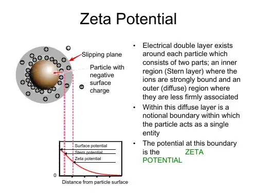
entity
• The potential at this boundary
is the
ZETA
POTENTIAL
0 Distance from particle surface
Zeta Potential
• The magnitude of the zeta potential gives an indication of the potential stability of the colloidal system
• The higher the ionic strength, the more compressed the double layer becomes
• The valency of the ion will also influence double layer thickness
• A trivalent ion such as Al3+ will compress the double layer to a greater extent compared to a monovalent ion such as Na+
• If all the particles have a large negative or positive zeta potential they will repel each other and there is DISPERSION STABILITY
• If the particles have low zeta potential values then there is no force to prevent the particles coming together and there is DISPERSION INSTABILITY
细胞器之间相互作用在非酒精性脂肪性肝病发生发展中的作用
2 DOI:10.3969/j.issn.1001-5256.2023.01.028细胞器之间相互作用在非酒精性脂肪性肝病发生发展中的作用刘天会首都医科大学附属北京友谊医院肝病中心,北京100050通信作者:刘天会,liu_tianhui@163.com(ORCID:0000-0001-6789-3016)摘要:细胞器除了具有各自特定的功能外,还可与其他细胞器相互作用完成重要的生理功能。
细胞器之间相互作用的异常与疾病的发生发展密切相关。
近年来,细胞器之间相互作用在非酒精性脂肪性肝病(NAFLD)发生发展中的作用受到关注,特别是线粒体、脂滴与其他细胞器之间的相互作用。
关键词:非酒精性脂肪性肝病;细胞器;线粒体;脂肪滴基金项目:国家自然科学基金面上项目(82070618)RoleoforganelleinteractioninthedevelopmentandprogressionofnonalcoholicfattyliverdiseaseLIUTianhui.(LiverResearchCenter,BeijingFriendshipHospital,CapitalMedicalUniversity,Beijing100050,China)Correspondingauthor:LIUTianhui,liu_tianhui@163.com(ORCID:0000-0001-6789-3016)Abstract:Inadditiontoitsownspecificfunctions,anorganellecanalsointeractwithotherorganellestocompleteimportantphysiologicalfunctions.Thedisordersoforganelleinteractionsarecloselyassociatedthedevelopmentandprogressionofvariousdiseases.Inrecentyears,theroleoforganelleinteractionshasattractedmoreattentionintheprogressionofnonalcoholicfattyliverdisease,especiallytheinteractionsbetweenmitochondria,lipiddroplets,andotherorganelles.Keywords:Non-alcoholicFattyLiverDisease;Organelles;Mitochondria;LipidDropletsResearchfunding:NationalNaturalScienceFoundationofChina(82070618) 细胞器可以通过膜接触位点与其他细胞器相互作用,完成物质与信息的交换,形成互作网络[1]。
Odyssey CLX 操作指南1

导入图片 ........................................................................................................................... 18 泳道设置 ........................................................................................................................... 19 设定 Marker....................................................................................................................... 20 创建新 marker ................................................................................................................... 20 自动识别条带 ................................................................................................................... 21 手动编辑条带 ................................................................................................................... 21 单通道信号归一化 ..............................................................................................................21 查看表格 ........................................................................................................................... 22
纳米颗粒对细胞的生物学效应研究进展

纳米颗粒对细胞的生物学效应研究进展王培欢;刘洪臣【摘要】Nanomaterial,with one dimension in the range of 1 to 100 nm at least,possesses unique properties and functions of nanoscale.And nanoparticles are three-dimensional nanoscale materials.Nanoparticles have a broad application prospect because of their unique physical and chemical properties and special effects.Meanwhile,the biological safety ofnanoparticles has attracted more and more attention.Nanoparticles can affect organisms in multiple levels,such asanimals,cells,subcells,proteins,genes and so on.The effect of nanoparticles on organisms is an extremely complex biological process,and the current research is mostly focused on the level of cells.Nanotechnology has promoted the improvement of traditional dental materials and made great progress in the clinical treatment of oral diseases.With the increasing application of nanomaterials in the field of stomatology,the chances of exposure to nanoparticles in patients with oral diseases are greatly increased.Studies have shown that a variety of dental nanomaterials are potentially toxic to the central nervous system,and the biological effects of different nanoparticles on osteoblasts,dental pulp cells and periodontal ligament cells will become the focus of future research.The study of the biological effects of nanoparticles on cells is beneficial to the safe application of nanoparticles in the field of life sciences.In this review,we summarized the biological effects,possible mechanisms and influencingfactors of nanoparticles on cells.%纳米材料是指至少有一个维度的直径范围在1-100nm之间且具有纳米尺度独特性质和功能的物质,而三维均在纳米尺度的纳米材料被称为纳米颗粒.纳米颗粒以其特有的理化性质和特殊效应展现出广阔的应用前景,与此同时其生物安全性也越来越多的引起人们的关注.纳米颗粒可以在动物、细胞和亚细胞、蛋白和基因等多水平对生物体产生作用,纳米颗粒对生物体作用的方式、途径和机制是一个极其复杂的生物学过程,目前的研究多集中于细胞水平.纳米技术推动了传统口腔材料的改良,使口腔临床治疗取得了巨大进步.随着纳米材料在口腔医学领域的应用日益增多,口腔患者接触到纳米颗粒的机会大大增加.研究表明,多种口腔纳米材料对中枢神经系统具有潜在毒性,而不同纳米颗粒对成骨细胞、牙髓细胞及牙周膜细胞等的生物学效应将成为未来研究的重点.开展纳米颗粒对细胞生物学效应的研究,有利于纳米颗粒材料在生命科学领域中的安全应用.本文结合相关文献就纳米颗粒对细胞的生物学效应、可能机制及影响因素做一论述.【期刊名称】《中华老年口腔医学杂志》【年(卷),期】2018(016)002【总页数】5页(P120-124)【关键词】纳米颗粒;细胞;生物学效应;自噬;凋亡;口腔医学【作者】王培欢;刘洪臣【作者单位】解放军总医院口腔医学研究所北京 100853;解放军总医院口腔医学研究所北京 100853【正文语种】中文【中图分类】R783.1纳米材料是指至少有一个维度的直径范围在1-100nm之间且具有纳米尺度独特性质和功能的物质,而三维均在纳米尺度的纳米材料被称为纳米颗粒[1]。
注射剂中的颗粒物英文788ParticulateMatter
<788> PARTICULATE MATTER IN INJECTIONSThis general chapter is harmonized with the corresponding texts of the European Pharmacopoeia and/or the Japanese Pharmacopoeia. These pharmacopeias have undertaken not to make any unilateral change to this harmonized chapter.Portions of the present general chapter text that are national USP text, and therefore notpart of the harmonized text, are marked with symbols () to specify this fact.Particulate matter in injections and parenteral infusions consists of mobile undissolved particles, other than gas bubbles, unintentionally present in the solutions.For the determination of particulate matter, two procedures, Method 1 (Light Obscuration Particle Count Test) and Method 2 (Microscopic Particle Count Test), are specified hereinafter. When examining injections and parenteral infusions for sub-visible particles Method 1 is preferably applied. However, it may be necessary to test some preparations by the light obscuration particle count test followed by the microscopic particle count test to reach a conclusion on conformance to the requirements.Not all parenteral preparations can be examined for sub-visible particles by one or both of these methods. When Method 1 is not applicable, e.g. in case of preparations having reduced clarity or increased viscosity, the test should be carried out according to Method 2. Emulsions, colloids, and liposomal preparations are examples. Similarly, products that produce air or gas bubbles when drawn into the sensor may also require microscopic particle count testing. If the viscosity of the preparation to be tested is sufficiently high so as to preclude its examination by either test method, a quantitative dilution with an appropriate diluent may be made to decrease viscosity, as necessary, to allow the analysis to be performed.The results obtained in examining a discrete unit or group of units for particulate matter cannot be extrapolated with certainty to other units that remain untested. Thus, statistically sound sampling plans must be developed if valid inferences are to be drawn from observed data to characterize the level of particulate matter in a large group of units.METHOD 1. LIGHT OBSCURATION PARTICLE COUNT TESTUse a suitable apparatus based on the principle of light blockage which allows an automatic determination of the size of particles and the number of particles according to size. The definition for particle-free water is provided in Reagent Specifications under Reagents, Indicators and Solution section.The apparatus is calibrated using dispersions of spherical particles of known sizes between 10 µm and 25 µm. These standard particles are dispersed in particle-free water.Care must be taken to avoid aggregation of particles during dispersion. System suitability can be verified by using the USP Particle Count RS (<11>).General precautionsThe test is carried out under conditions limiting particulate matter, preferably in a laminar-flow cabinet.Very carefully wash the glassware and filtration equipment used, except for the membrane filters, with a warm detergent solution and rinse with abundant amounts of water to remove all traces of detergent. Immediately before use, rinse the equipment from top to bottom, outside and then inside, with particle-free water.Take care not to introduce air bubbles into the preparation to be examined, especially when fractions of the preparation are being transferred to the container in which the determination is to be carried out.In order to check that the environment is suitable for the test, that the glassware is properly cleaned and that the water to be used is particle-free, the following test is carried out: determine the particulate matter in 5 samples of particle-free water, each of 5 ml, according to the method described below. If the number of particles of 10 µm or greater size exceeds 25 for the combined 25 ml, the precautions taken for the test are not sufficient. The preparatory steps must be repeated until the environment, glassware and water are suitable for the test.MethodMix the contents of the sample by slowly inverting the container 20 times successively. If necessary, cautiously remove the sealing closure. Clean the outer surfaces of the container opening using a jet of particle-free water and remove the closure, avoiding any contamination of the contents. Eliminate gas bubbles by appropriate measures such as allowing to stand for 2 min or sonicating.For large-volume parenterals, single units are tested. For small-volume parenterals less than 25 ml in volume, the contents of 10 or more units is combined in a cleaned container to obtain a volume of not less than 25 ml; the test solution may be prepared by mixing the contents of a suitable number of vials and diluting to 25 ml with particle-free water or with an appropriate particle-free solvent when particle-free water is not suitable. Small-volume parenterals having a volume of 25 ml or more may be tested individually. Powders for parenteral use are reconstituted with particle-free water or with an appropriate particle-free solvent when particle-free water is not suitable.The number of test specimens must be adequate to provide a statistically soundassessment. For large-volume parenterals or for small-volume parenterals having a volume of 25 ml or more, fewer than 10 units may be tested, based on an appropriate sampling plan.Remove four portions, each of not less than 5 ml, and count the number of particles equal to or greater than 10 µm and 25 µm. Disregard the result obtained for the first portion, and calculate the mean number of particles for the preparation to be examined.EvaluationFor preparations supplied in containers with a nominal volume of more than 100 ml, apply the criteria of test 1.A.For preparations supplied in containers with a nominal volume of less than 100 ml, apply the criteria of test 1.B.For preparations supplied in containers with a nominal volume of 100 ml, apply the criteria of test 1.B [Note: Test 1.A is used in the Japanese Pharmacopoeia]If the average number of particles exceeds the limits, test the preparation by the Microscopic Particle Count Test.Test 1.A — Solutions for parenteral infusion or solutions for injection supplied in containers with a nominal content of more than 100 mL.The preparation complies with the test if the average number of particles present in the units tested does not exceed 25 per mL equal to or greater than 10 µm and does not exceed 3 per mL equal to or greater than 25 µm.Test 1.B — Solutions for parenteral infusion or solutions for injection supplied in containers with a nominal content of less than 100 ml.The preparation complies with the test if the average number of particles present in the units tested does not exceed 6000 per container equal to or greater than 10 µm and does not exceed 600 per container equal to or greater than 25 µm.METHOD 2. MICROSCOPIC PARTICLE COUNT TESTUse a suitable binocular microscope, filter assembly for retaining particulate matter and membrane filter for examination.The microscope is equipped with an ocular micrometer calibrated with an objective micrometer, a mechanical stage capable of holding and traversing the entire filtration area of the membrane filter, two suitable illuminators to provide episcopic illumination in addition to oblique illumination, and is adjusted to 100 ± 10 magnifications.The ocular micrometer is a circular diameter graticule (see Figure 1) and consists of a large circle divided by crosshairs into quadrants, transparent and black reference circles 10 µm and 25 µm in diameter at 100 magnifications, and a linear scale graduated in 10 µm increments. It is calibrated using a stage micrometer that is certified by either a domestic or international standard institution. A relative error of the linear scale of the graticule within ± 2 per cent is acceptable. The large circle is designated the graticule field of view (GFOV).Two illuminators are required. One is an episcopic brightfield illuminator internal to the microscope, the other is an external, focusable auxiliary illuminator adjustable to give reflected oblique illumination at an angle of 10° to 20°.The filter assembly for retaining particulate matter consists of a filter holder made of glass or other suitable material, and is equipped with a vacuum source and a suitable membrane filter.The membrane filter is of suitable size, black or dark gray in color, non-gridded or gridded, and 1.0 µm or finer in nominal pore size.General precautionsThe test is carried out under conditions limiting particulate matter, preferably in a laminar-flow cabinet.Very carefully wash the glassware and filter assembly used, except for the membrane filter, with a warm detergent solution and rinse with abundant amounts of water to remove all traces of detergent. Immediately before use, rinse both sides of the membrane filter and the equipment from top to bottom, outside and then inside, with particle-free water.In order to check that the environment is suitable for the test, that the glassware and the membrane filter are properly cleaned and that the water to be used is particle-free, the following test is carried out: determine the particulate matter of a 50 ml volume of particle-free water according to the method described below. If more than 20 particles 10 µm or larger in size or if more than 5 particles 25 µm or larger in size are present within the filtration area, the precautions taken for the test are not sufficient. The preparatory steps must be repeated until the environment, glassware, membrane filter and water are suitable for the test.MethodMix the contents of the samples by slowly inverting the container 20 times successively. If necessary, cautiously remove the sealing closure. Clean the outer surfaces of the container opening using a jet of particle-free water and remove the closure, avoiding any contamination of the contents.For large-volume parenterals, single units are tested. For small-volume parenterals less than 25 ml in volume, the contents of 10 or more units is combined in a cleaned container; where justified and authorized, the test solution may be prepared by mixing the contents of a suitable number of vials and diluting to 25 ml with particle-free water or with an appropriate particle-free solvent when particle-free water is not suitable. Small-volume parenterals having a volume of 25 ml or more may be tested individually.Powders for parenteral use are constituted with particle-free water or with an appropriate particle-free solvent when particle-free water is not suitable.The number of test specimens must be adequate to provide a statistically sound assessment. For large-volume parenterals or for small-volume parenterals having a volume of 25 ml or more, fewer than 10 units may be tested, based on an appropriate sampling plan.Wet the inside of the filter holder fitted with the membrane filter with several milliliter of particle-free water. Transfer to the filtration funnel the total volume of a solution pool or of a single unit, and apply vacuum. If needed add stepwise a portion of the solution until the entire volume is filtered. After the last addition of solution, begin rinsing the inner walls of the filter holder by using a jet of particle-free water. Maintain the vacuum until the surface of the membrane filter is free from liquid. Place the membrane filter in a Petri dish and allow the membrane filter to air-dry with the cover slightly ajar. After the membrane filter has been dried, place the Petri dish on the stage of the microscope, scan the entire membrane filter under the reflected light from the illuminating device, and count the number of particles that are equal to or greater than 10 µm and the number of particles that are equal to or greater than 25 µm. Alternatively, partial membrane filter count and determination of the total membrane filter count by calculation is allowed. Calculate the mean number of particles for the preparation to be examined.The particle sizing process with the use of the circular diameter graticule is carried out by transforming mentally the image of each particle into a circle and then comparing it to the 10 µm and 25 µm graticule reference circles. Thereby the particles are not moved from their initial locations within the graticule field of view and are not superimposed on the reference circles for comparison. The inner diameter of the transparent graticule reference circles is used to size white and transparent particles, while dark particles are sized by using the outer diameter of the black opaque graticule reference circles.In performing the microscopic particle count test do not attempt to size or enumerate amorphous, semi-liquid, or otherwise morphologically indistinct materials that have the appearance of a stain or discoloration on the membrane filter. These materials show little or no surface relief and present a gelatinous or film-like appearance. In such cases the interpretation of enumeration may be aided by testing a sample of the solution by the light obscuration particle count test.EvaluationFor preparations supplied in containers with a nominal volume of more than 100 ml, apply the criteria of test 2.A.For preparations supplied in containers with a nominal volume of less than 100 ml, apply the criteria of test 2.B.For preparations supplied in containers with a nominal volume of 100 ml, apply the criteria of test 2.B. [Note: Test 2.A is used in the Japanese Pharmacopoeia]Test 2.A — Solutions for parenteral infusion or solutions for injection supplied in containers with a nominal content of more than 100 mL.The preparation complies with the test if the average number of particles present in the units tested does not exceed 12 per mL equal to or greater than 10 µm and does not exceed 2 per mL equal to or greater than 25 µm.Test 2.B — Solutions for parenteral infusion or solutions for injection supplied in containers with a nominal content of less than 100 mlThe preparation complies with the test if the average number of particles present in the units tested does not exceed 3000 per container equal to or greater than 10 µm and does not exceed 300 per container equal to or greater than 25 µm.。
optical tweezers
我们在几何光学模型下计算其在微米级的介质球上的作用力大小,也被称为“光镊”。
这可作为描述激光阱作用于活体细胞及其细胞器的操作系统的简单模型。
梯度力和散射力被定义为在几何光学条件下的多光束复合形态。
计算使用输入强度分布为TEM00和TEM01模和小球整个截面折射率各异的受力情况。
强大的均匀势阱可能与力的变量因子小于2球体横截面有关。
对于10MW功率的激光和1.2相对折射率计算其中梯度阱最弱的方向俘获力量可达〜1.2*10 ^ -6达因dyne(向后)。
结果表明,良好的俘获要求高收敛光束来自高数值孔径的目标。
给出一个对照,使用明场或差分干涉光对比光学相衬光学阱单光束梯度辐射压力光阱。
INTRODUCTIONThis paper gives a detailed description of the trapping of micron-sized dielectric spheres can serve as first simple models of living cells in biological trapping experiments and also as basic particles in physical trapping experiments. Optical trapping of small particles by the forces of laser radiation pressure has been used for about 20 years in the physical sciences for the manipulation and study of micron and submicron dielectric particles and even individual atoms. These techniques have also been extended more recently to biological particles.本文给出了一种微米大小的介质球俘获的详细描述,可以作为活细胞的第一个简单的模型生物俘获实验和基本粒子物理俘获实验。
基于微米级颗粒临界沉积
化工进展Chemical Industry and Engineering Progress2023 年第 42 卷第 12 期基于微米级颗粒临界沉积/剥离标准的研究进展邵宏勋,谢俊,桂玉双,李润东(沈阳航空航天大学能源与环境学院,辽宁 沈阳110136)摘要:基于气固两相流动的微颗粒沉积一直是一个棘手的问题,作为一种常见的物理现象,带来的影响更多是负面的,如砂粒导致涡轮叶片的磨损,飞灰颗粒引起锅炉换热面结垢腐蚀等一系列问题。
尽管吹灰方案的不断优化已经很大程度减少了飞灰沉积带来的负面影响,但仍有一些不可控或意外的问题存在。
因此,归纳分析颗粒黏附行为规律对工程颗粒沉积的抑制具有重要意义。
本文以锅炉飞灰沉积为主要背景,阐述了以惯性撞击为主导机制的颗粒黏附机理与特性,回顾了近年来对临界沉积标准的研究工作,重点剖析了沉积因素对颗粒黏附/反弹判据标准的影响,总结了颗粒剥离的主要判断准则,通过横向对比相关判据存在的问题与联系,说明了颗粒黏附判据标准的研究现状与存在的问题,为实现锅炉沉积的有效抑制及颗粒剥离理论的推广提供技术理论支撑。
关键词:微米颗粒;沉积模型;临界捕集速度;恢复系数;临界剪切速度中图分类号:TK-9;TQ021 文献标志码:A 文章编号:1000-6613(2023)12-6141-16Research progress of critical deposition/stripping standards based onmicron-sized particlesSHAO Hongxun ,XIE Jun ,GUI Yushuang ,LI Rundong(College of Energy and Environment, Shenyang Aerospace University, Shenyang 110136, Liaoning, China)Abstract: It is a thorny problem for micro-particle deposition based on gas-solid flow. Particle deposition is a common physical phenomenon but with more negative impact, such as the wear of turbine blades caused by sand particles, scale corrosion caused by fly ash particles on boiler heat exchange surface and so on. Although the continuous optimization of soot blowing scheme has greatly reduced the negative impact of fly ash deposition, there are still some uncontrollable problems. Therefore, it is of great significance to summarize and analyze the law of particle adhesion behavior to suppress particle deposition in engineering. Based on the main background of boiler fly ash deposition, this paper expounded the mechanism and characteristics of particle adhesion dominated by inertial impact, reviewed the research work on critical deposition criteria in recent years, focused on the analysis of the influence of deposition factors on the criteria of particle adhesion/rebound, and summarized the main criteria of particle detachment. By comparing the problems and connections of relevant criteria horizontally, the research status and existing problems of particle adhesion criteria was explained, which provided technical and theoretical support for the effective suppression of boiler deposition and the promotion of particle detachment theory.综述与专论DOI :10.16085/j.issn.1000-6613.2023-0084收稿日期:2023-01-17;修改稿日期:2023-03-13。
晶胞中粒子数的计算方法
晶胞中粒子数的计算方法The calculation of the number of particles in a unit cell is an important concept in the field of solid-state physics and materials science. It provides key information about the structure and properties of a material at the atomic level. The number of particles in a unit cell can vary depending on the type of crystal structure and the nature of the atoms involved. For example, a simple cubic structure contains one particle per unit cell, while a face-centered cubic structure contains four particles per unit cell.晶胞中粒子数的计算方法是固态物理和材料科学领域的一个重要概念。
它提供了关于材料在原子水平上的结构和性质的关键信息。
晶胞中的粒子数可以根据晶体结构的类型和涉及的原子的性质而变化。
例如,简单立方结构中每个晶胞含有一个粒子,而面心立方结构中每个晶胞含有四个粒子。
In order to calculate the number of particles in a unit cell, one must first identify the type of crystal structure being considered. The three main types of crystal structures are simple cubic, body-centered cubic, and face-centered cubic. Each of these structures has a different number of particles per unit cell, which can be determinedusing the formula: nparticles = nunit x Z, where nparticles is the number of particles in the unit cell, nunit is the number of atoms per unit cell, and Z is the number of lattice points within the unit cell.要计算晶胞中的粒子数,首先必须确定所考虑的晶体结构类型。