肾脏的局部解剖(英)
合集下载
肾脏的局部解剖英参考幻灯片
11
Costodiaphragmatic recess of the pleural
cavity
12
Renal hilum, Renal sinus and Renal pedicle
Renal pyramid Renal sinus
cortex Renal column calyx
Renal hilum
50
Question
16
Hydronephrosis
17
renal artery
T11
18
Renal artery
19
Vascular renal segment
Superior (apical)
Anterior superior (upper)
Anterior inferior (middle)
Inferior (lower)
papilla
Renal pedicle
13
14
15
From anterior to posterior renal vein renal artery renal pelvis
From above downwards the renal artery renal vein renal pelvis
2
Position
?Retroperitoneal ?Upper poles
T12 vertebra Lower poles
L3 vertebra ?Right is lower than left
3
Cadaveric kidneys
4
Surface Projection of kidney
5
40
Relation
肾切除术Donor Nephrectomy
优点: (1)大大的缩短了肾脏的热缺血时
间,一般不超过5分钟; (2)减少了供肾及血管、输尿管损
伤的发生率;
供肾原位灌注整块切取法优缺点
优点: (3)如肾脏为多支血管,不仅不影
响灌注,还有利于移植时的血管重 建(4)能充分利用其他器官,用 该法可同时切取肝脏、胰腺和小肠。
供肾原位灌注整块切取法优缺点
间不超过10分钟。
三、手术步骤
1.常规进入腹腔后,助手将全部肠管推 向头侧,暴露盆腔入口的后腹膜,提 起剪开后可见下腔静脉和腹主动脉下 段,并在两血管远端分叉为髂总动静 脉。在腹主动脉远断先带入一根粗索 线并结扎,然后在距离其上2cm处另带 线一根,暂不打结。提起腹主动脉在 两线间剪开血管前壁,将特制的带气 囊的动脉灌注管插入20cm,用第二根 线固定。
缺点: 切取过程中需持续灌注,灌洗液用 量较大。一般需要3000-4000毫升。
供肾切取后离体灌注法 :
可以分双肾分别切取和同时 切取法两种。
双肾分别切取法
进腹后将两肾分别切下。因左 肾动脉略短,切左肾动脉应带 一块主动脉壁,右肾静脉应带 一块下腔静脉壁,避免损伤血 管分支。取下后离体灌注。
ቤተ መጻሕፍቲ ባይዱ
双肾分别切取
肾脏的局部解剖
3.肾的被膜(renal capsule): 从外向内依次为肾周筋膜 (perirenal fascia )、肾脂肪 囊(adeps renis )、及肾包膜 (renal capsular)。
4 . 血 管 (vessel) : 肾 静 脉 (renal vein )及肾动脉(renal artery )
病人体位
二、手术切口(incision)
根切口的类型有:经腰部第11肋间 隙切口,经腰部第12肋骨下缘切口 和经腹部腹膜内切口。
肾脏解剖ppt课件
肾单位:双肾约200万个 肾单位=肾小体+肾小管 肾单位+集合管共同完成泌尿 功能
3
1.肾单位结构: 肾小囊
肾 肾小球
肾小球
小 体 肾小囊
入球小A
近曲小管
出球小A
近端小管
髓袢降支粗段
肾 小
髓袢细段
髓袢降支细段 髓袢升支细段
管
远端小管 髓袢升支粗段 远曲小管
肾小管
4肾单位Βιβλιοθήκη 组成肾小球远曲小管
近曲小管
集
合
髓
管
袢
5
(二)球旁器(近球小体)
球旁细胞:分泌肾素 致密斑:感受小管液NaCl含量变化,调节肾素释放 球外系膜细胞:吞噬和收缩功能
球外系 膜细胞
球旁细胞 致密斑
6
二 尿的生成
肾小球的滤过作用 肾小管、集合管的
重吸收选择性 肾小管、集合管的
分泌排泄作用
上一页 下一7 页
尿生成过程 血浆在肾小球的滤过 肾小管和集合管的选择性重吸收和分泌
8
1.肾小球滤过 血液流经肾小球毛细血管时,除蛋白质以外的血
浆成分被滤过进入肾小囊腔而形成超滤液(原尿)的 过程。
9
二.滤过膜的结构:
毛细血管内皮细胞:
窗孔 阻止血细胞通过。
基膜:
网孔 阻止血浆蛋白通过。
脏层肾小囊上皮细胞
裂隙(Φ4-11nm) 防止蛋白质的漏出。
机械屏障: 这种滤过作用因蛋白质分子量不同而异的屏障作用
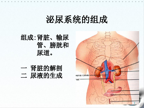
泌尿系统的组成
组成:肾脏、输尿 管、膀胱和 尿道。
一 肾脏的解剖 二 尿液的生成
1
一、肾脏的基本解剖
(一)大体解剖 位于腹膜后,第12胸椎至第3腰椎之间的
3
1.肾单位结构: 肾小囊
肾 肾小球
肾小球
小 体 肾小囊
入球小A
近曲小管
出球小A
近端小管
髓袢降支粗段
肾 小
髓袢细段
髓袢降支细段 髓袢升支细段
管
远端小管 髓袢升支粗段 远曲小管
肾小管
4肾单位Βιβλιοθήκη 组成肾小球远曲小管
近曲小管
集
合
髓
管
袢
5
(二)球旁器(近球小体)
球旁细胞:分泌肾素 致密斑:感受小管液NaCl含量变化,调节肾素释放 球外系膜细胞:吞噬和收缩功能
球外系 膜细胞
球旁细胞 致密斑
6
二 尿的生成
肾小球的滤过作用 肾小管、集合管的
重吸收选择性 肾小管、集合管的
分泌排泄作用
上一页 下一7 页
尿生成过程 血浆在肾小球的滤过 肾小管和集合管的选择性重吸收和分泌
8
1.肾小球滤过 血液流经肾小球毛细血管时,除蛋白质以外的血
浆成分被滤过进入肾小囊腔而形成超滤液(原尿)的 过程。
9
二.滤过膜的结构:
毛细血管内皮细胞:
窗孔 阻止血细胞通过。
基膜:
网孔 阻止血浆蛋白通过。
脏层肾小囊上皮细胞
裂隙(Φ4-11nm) 防止蛋白质的漏出。
机械屏障: 这种滤过作用因蛋白质分子量不同而异的屏障作用
泌尿系统的组成
组成:肾脏、输尿 管、膀胱和 尿道。
一 肾脏的解剖 二 尿液的生成
1
一、肾脏的基本解剖
(一)大体解剖 位于腹膜后,第12胸椎至第3腰椎之间的
《肾脏解剖结构》课件
《肾脏解剖结构》PPT课 件
本PPT将带您深入了解肾脏的解剖结构,包括其功能、位置、外观特征以及内 部结构的概览。一起探索肾脏的奥秘吧!
肾脏功能与位置
肾脏是人体重要器官之一,位于腰腹部,起着排除废物、调节体液平衡和维 持血压等重要功能。
肾脏的外观特征
肾脏呈豆状,分为肾实质和肾盂,外表光滑,颜色深红,质地柔软。
肾小球是肾脏的过滤单位,包括肾小体和肾小管,通过滤过血液产生初步的尿液。
Байду номын сангаас
肾小管的解剖结构及作用
肾小管负责对原尿进行分泌和重吸收,通过调节水分和电解质的浓度,形成最终的尿液。
集合管系统的结构与作用
集合管是将尿液从肾单位传输到尿路的管道系统,起到浓缩尿液和酸碱平衡调节的重要作用。
肾脏的血管系统概述
内、外肾静脉都是肾静脉的分支,内肾静脉负责引流肾脏实质的血液,而外 肾静脉则是引流肾脏周围组织的血液。
肾脏的神经系统概述
肾脏是受到神经系统调控的器官,神经系统对肾脏的血流和尿液排泄起到重 要的调节作用。
肾周神经丛的结构与作用
肾周神经丛包裹着肾脏,通过传递神经信号,调节肾脏的血管收缩和尿液生 成。
肾脏内部结构概览
肾脏包含肾单位,由肾小球和肾小管组成,协同完成体内废物排泄和体液平 衡调节。
肾盂的组成与作用
肾盂是肾脏内部的容器,负责接收和储存尿液,同时起到传输尿液到尿路的 功能。
肾实质的组成与作用
肾实质由肾小球和肾小管组成,肾小球过滤血液产生原尿,而肾小管则负责 对原尿进行分泌和重吸收。
肾小球的解剖结构及作用
肾脏有丰富的血管供应,肾动脉和肾静脉是主要的血管,负责输送血液和废 物的清除。
肾动脉的分支与供血范围
肾脏的局部解剖(英)
2021/3/11
46
2021/3/11
47
2021/3/11
48
incision
Anterior transperitoneal
2021/3/11
approach
Posterior retroperitoneal approach
49
Case A
A 55-year-old woman was found rolling on her kitchen floor, crying out from agonizing pain in her abdomen. The pain came in waves and extended from the right loin to the groin and to the front of the right thigh. An anteroposterior radiograph of the abdomen revealed a calculus in the right ureter.
2021/3/11
57
nephrectomy
For a nephrectomy, the kidney commonly is exposed in the loin. After an oblique incision midway between the twelfth rib and the iliac crest, the posterior free border of the external oblique is identified, and divided to reveal the peritoneum, which is pushed forward to reveal the renal fascia. The subcostal nerve and vessels are preserved; the renal fascia is opened; and the kidney exposed. Care must be taken not to damage the pleura, since it is separated from the upper pole of the kidney only by the diaphragm.
- 1、下载文档前请自行甄别文档内容的完整性,平台不提供额外的编辑、内容补充、找答案等附加服务。
- 2、"仅部分预览"的文档,不可在线预览部分如存在完整性等问题,可反馈申请退款(可完整预览的文档不适用该条件!)。
- 3、如文档侵犯您的权益,请联系客服反馈,我们会尽快为您处理(人工客服工作时间:9:00-18:30)。
For a nephrectomy, the kidney commonly is exposed in the loin. After an oblique incision midway between the twelfth rib and the iliac crest, the posterior free border of the external oblique is identified, and divided to reveal the peritoneum, which is pushed forward to reveal the renal fascia. The subcostal nerve and vessels are preserved; the renal fascia is opened; and the kidney exposed. Care must be taken not to damage the pleura, since it is separated from the upper pole of the kidney only by the diaphragm.
Horseshoe kidney
Suprarenal Gland
•Endocrine gland
•T11 level
•Right is triangular •Left is semi-lunar
Relation
Left right liver diaphrag m
superior
anterior inferior
renal vein renal artery renal pelvis From above downwards the renal artery renal vein renal pelvis
Hydronephrosis
renal artery
T11
Renal artery
Vascular renal segment
Case D
An examination of a patient revealed that she had a horseshoe kidney. What anatomical structure prevents a horseshoe kidney from ascending to a level above the umbilicus?
Position
Retroperitoneal Upper
poles
T12 vertebra
Lower poles
L3 vertebra
Right
is lower than left
Cadaveric kidneys
Surface Projection of kidney
Renal Angle
Case E
An intravenous pyelogram revealed that the calyces and pelvis of a patient’s right kidney were grossly dilated (a condition known as hydronephrosis). What embryological anomaly may be responsible for this condition?
Left suprarenal vein, into the left renal vein Right suprarenal vein, into the
inferior vena cava
incision
Posterior retroperitoneal approach Anterior transperitoneal approach
Renal Capsule
Renal fascia Adipose capsule Fibrous capsule
Renal fascia
Fibrous capsule
Adipose capsule Perirenal fat
Pararenal fat
Fibrous Capsule
Case A
A 55-year-old woman was found rolling on her kitchen floor, crying out from agonizing pain in her abdomen. The pain came in waves and extended from the right loin to the groin and to the front of the right thigh. An anteroposterior radiograph of the abdomen revealed a calculus in the right ureter.
Ureter
Ureter is divided into 3 parts:
①abdominal
part part
②pelvis
part
③intramural
28 to 34 cm 3 narrowing sites • the pelviureteric junction • crossing the pelvic brim • traversing the bladder wall
Case B
An explorer in the Amazon jungle was found alive after having lost contact with the outside world for six months. On physical examination, he was found to be in an emaciated condition. On palpation of the abdomen, a rounded, smooth swelling appeared in the right loin at the end of inspiration. On expiration, the swelling moved upward and could no longer be felt. What anatomical structure could produce such a swelling?
Case F
Which congenital anomaly of the ureter is likely to present as a case of urinary incontinence?
operation procedure of kidney transplant
nephrectomy
Tenderness or percussing pain caused by kidney disease is localized here
Neighbor of Kidney
Anterior Surface of the kidney
Posterior Surface
Costodiaphragmatic recess of the pleural cavity
stomach tail of pancreas & spleen vessel
diaphragm abdominal aorta
posterior medial
Inferior vena cava
Artery of Adrenal Gland
Veins of adrenal gland
Renal hilum, Renal sinus and Renal pedicle
Renal pyramid
Renal sinus cortex Renal column
calyx
Renal hilumpa源自illaRenal pedicle
From anterior to posterior
Question
a.
b.
c.
d.
What causes the pain when a ureteral calculus is present? Why is the pain felt in such an extensive area? Where does one look for the course of the ureter in a radiograph? Where along the ureter is a calculus likely to be held up?
Aorta-renal artery-segmental arterylobar artery-interlobar artery-arcuate artery-interlobulor artery-afferent arteriole-glomerulus (capillaries)efferent arteriole-peritubular capillaries and vasa recta-interlobular vein-arcuate vein-interlobar vein-renal vein-interior vena cava
Case C
An intravenous pyelogram revealed that a patient’s left kidney was in its normal position, but the right kidney was situated in front of the right sacroiliac joint. Can you explain this on embryological grounds?
Superior (apical)