TurboFect in vitro transfection reagent
非达霉素的固态形式及其制备方法[发明专利]
![非达霉素的固态形式及其制备方法[发明专利]](https://img.taocdn.com/s3/m/6dc9bed2580216fc710afd34.png)
专利名称:非达霉素的固态形式及其制备方法
专利类型:发明专利
发明人:T.福纳格,A.科瓦斯内-梅泽,L.斯祖罗斯,E.N.阿瓦伊申请号:CN201380036058.5
申请日:20130510
公开号:CN104768963A
公开日:
20150708
专利内容由知识产权出版社提供
摘要:本发明提供非达霉素的固态形式、制备该固态形式的方法以及包含一种或多种非达霉素的所述固态形式的药物组合物和制剂,和制备该组合物和制剂的方法。
本发明的固态形式表现出有利性质,例如在制造与加工中改善的可靠性和重现性以及在制剂中的稳定性。
申请人:特瓦制药厂有限公司
地址:匈牙利德布勒森
国籍:HU
代理机构:中国专利代理(香港)有限公司
更多信息请下载全文后查看。
一种铁离子-醋酸催化定向水解半纤维素制取木糖的方法[发明专利]
![一种铁离子-醋酸催化定向水解半纤维素制取木糖的方法[发明专利]](https://img.taocdn.com/s3/m/50570763b9d528ea80c77901.png)
专利名称:一种铁离子-醋酸催化定向水解半纤维素制取木糖的方法
专利类型:发明专利
发明人:黄凯旋,徐勇,罗京
申请号:CN201711414867.1
申请日:20171220
公开号:CN108118103A
公开日:
20180605
专利内容由知识产权出版社提供
摘要:本发明公开了一种铁离子‑醋酸催化定向水解半纤维素制取木糖的方法。
该方法以半纤维素为原料,粉碎筛选后利用0.6%~1.2%的三价铁离子(质量浓度)和2%~10%的醋酸(质量浓度)在150~180℃条件下预处理20~60min,定向催化水解木聚糖生成木糖溶出,经固液分离得到木糖
液,90%以上木聚糖被降解,其中木糖得率超过70%,并采用减压蒸发木糖液以醋酸‑水共沸物的形式回收醋酸。
该方法操作安全、木糖得率高、降解抑制物少且实现醋酸催化剂回用;此外,作为过渡金属的三价铁离子不仅具有显著的催化作用,而且稳定,不会影响产品木糖的品质;因此,该方法更加绿色、环保和高效。
申请人:南京林业大学
地址:210037 江苏省南京市玄武区龙蟠路159号南京林业大学
国籍:CN
代理机构:南京申云知识产权代理事务所(普通合伙)
代理人:邱兴天
更多信息请下载全文后查看。
Trans IT-CRISPR Transfection Reagent 产品说明书
Trans IT-CRISPR® Transfection ReagentCatalog Number T1706Product DescriptionTrans IT-CRISPR is a non-liposomal polymeric transfection reagent for efficient delivery of CRISPR/Cas components. Trans IT-CRISPR allows for simple, fast and efficient delivery of CRISPR DNA, guide RNA and RNP (Cas9-gRNA ribonucleoprotein) complexes to various cell types, including primary cells. Also,Trans IT-CRISPR effectively delivers siRNA duplexes into various cell lines.*Simple protocols for easy optimization in delivering CRISPR/Cas components to cells in DNA, guide RNA and RNP formats.*Efficient delivery to various cell lines, including common cell types and difficult to transfect cell types.Storage/StabilityTrans IT-CRISPR Transfection Reagent is shipped on wet ice. Upon receipt, please store at –20°C. With proper handling and storage, this product remains stable for one year from the date of purchase.Precautions and DisclaimerThis product is for R&D use only, not for drug, household, or other uses. Please consult the Material Safety Data Sheet for information regarding hazards and safe handling practices. SIGMA-ALDRICH CRISPR PLASMID DNA TRANSFECTIONTips for Plasmid DNA TransfectionOptimize reaction conditions for different cell types to ensure successful transfections. The suggestions provided in this protocol yield high efficiency plasmid DNA transfection using the Trans IT-CRISPR Transfection Reagent. Table 1 lists recommended starting conditions depending on culture vessel size. • Cell density at transfection:The recommended cell density for most cell types is ≥ 80% confluence. Determine the optimal cell density for each cell type in order to maximize transfection efficiency. Split cells 18–24 hours before transfection to ensure that the cells are actively dividing and reach the appropriate cell density at the time of transfection.• DNA purity:Use highly purified, sterile, and contaminant-free DNA for transfection. Plasmid DNA preparations that are endotoxin-free and have A260/280 absorbance ratio of 1.8–2.0 are desirable. DNA prepared using mini-prep kits is not recommended as it might contain high levels of endotoxin.• Ratio of Trans IT-CRISPR to DNA: Determine the best Trans IT-CRISPR: DNA ratio for each cell type. Start with 3 µl of Trans IT-CRISPR per 1 µg of DNA. Vary the amount of Trans IT-CRISPR from 2–6 µl per 1 µg DNA to find the optimal ratio. Table 1 provides recommended starting conditions.• Complex formation conditions: Prepare Trans IT-CRISPR: DNA complexes in serum-free growth medium. Recommendation: Opti-MEM TM I Reduced-Serum Medium, without antibiotics. • Cell culture conditions:Culture cells in the appropriate medium, with or without serum. There is no need to perform a medium change to remove the transfection complexes.• Presence of antibiotics:Transfection complexes can be added directly to cells grown in complete culture medium containing serum and low levels of antibiotics (0.1–1X final concentration ofpenicillin/streptomycin mixture). Do not include antibiotics during the complex formation step.• Post-transfection incubation time : Determine the best incubation time post-transfection for each cell type. The optimalincubation time is generally 24–72 hours, but will vary depending on the goal of the experiment, nature of the plasmid used, and cell doubling time.Sigma-Aldrich CRISPR Plasmid DNATransfection Protocol per Well of a 6-Well Plate in Human Osteosarcoma (U2OS) Cells and a Human Immortalized Liver Cell Line (HepaRG)The following procedure describes how to perform plasmid DNA transfections using the Trans IT-CRISPR transfection reagent in 6-well plates. The surface areas of other culturevessels are different and transfections must be scaled accordingly (see Table 1 as an example).Plate Cells∙ Approximately 18-24 hours beforetransfection, plate cells in 2.5 mlcomplete growth medium per well in a 6-well plate. For most cell types, cultures should be ≥ 80% confluent at the time of transfection.o For adherent U2OS andHepaRG cells : Plate at a density of 2.5 x 10⁵ cells/ml.o For other adherent cells : Platecells at a density of 0.8–3.0 × 10⁵ cells/ml.∙ Incubate cell cultures overnight.Prepare Trans IT-CRISPR: DNA CRISPRcomplexes (Immediately before transfection)∙ Warm Trans IT-CRISPR to roomtemperature and vortex gently before using.∙ Place 250 µl of Opti-MEM ® I Reduced –Serum Medium in a sterile tube.∙ Add between 2 – 4 µg of Sigma-AldrichCRISPR plasmid DNA.Table 1. Recommended starting conditions for DNA transfections with Trans IT-CRISPR Transfection Reagent DNACulture vessel 96-well48-well 24-well12-well6-well10-cm dishT75 flaskSurface area0.35 cm 2 1 cm 2 1.9 cm 2 3.8 cm 2 9.6 cm 2 59 cm 2 75 cm 2 Complete growth medium92 µl 263 µl 0.5 ml 1 ml 2.5 ml 15.5 ml 19.7 ml Serum-free medium 9 µl 26 µl 50 µl 100 µl 250 µl 1.5 ml 1.9 ml DNA (1 µg/µl stock)0.1 µl0.26 µl0.5 µl1 µl2.5 µl15 µl19 µlFor the all-in-one Sigma-Aldrich CRISPR FP vectors, add 4 µg of plasmid. For the dual vector Sigma-Aldrich CRISPR plasmids, add 2 µg of U6-gRNA only plasmid and 2 µg of CMV-Cas9-FP only plasmid.∙ Pipet gently to mix completely.∙ Add 3-6 µl Trans IT-CRISPR (1.5 µl ofTrans IT-CRISPR per 1 µg of CRISPR plasmid DNA) to the diluted DNA mixture.∙ Pipet gently to mix completely.∙ Incubate at room temperature for 15-30minutes to allow sufficient time for complexes to form.Distribute the complexes to cells in complete growth medium∙ Add the Trans IT-CRISPR: DNAcomplexes drop-wise to different areas of the wells.∙ Gently rock the culture vessel back andforth and from side-to–side to evenly distribute the Trans IT-CRISPR: DNA complexes.∙ Incubate for 48 hours. It is notnecessary to replace the complete growth medium with fresh medium. ∙ Harvest cells and assay as required.Delivery of purified recombinant Cas9 protein and guide RNA Ribonucleoprotein Complexes (RNPsRecent papers have shown the efficacy of delivering the CRISPR components by combining purified Cas9 protein-guide RNA ribonucleoprotein (RNP) complexes. Trans IT-CRISPR transfection reagent has been tested in U2OS and HepaRG cells for efficient delivery of Cas9-gRNA RNP complexes to induce efficient site-specific mutations at targeted genomic regions. Protocols for both IVT gRNA and synthetic crRNA & tracrRNA are listed below. Prepare Trans IT-CRISPR: In Vitro Transcribed gRNA/Cas9 Protein CRISPR complexes (Immediately before transfection) Use between a 1.2 and 5 molar excess of in vitro transcribed RNA to Cas9 protein.∙Pipet between 1.2 and 5 µg of in vitro transcribed RNA to a sterile tube on ice.∙Add between 5 and 10 µg of Cas9protein to the in vitro transcribed RNA,mix gently and incubate on ice for 30minutes.∙ Warm Trans IT-CRISPR to roomtemperature and vortex gently beforeusing.∙Place 250 µl of Opti-MEM TM I Reduced –Serum Medium to the Cas9Ribonucleoprotein (RNP) sterile tube.∙Add between 5 - 6.25 µl of Trans IT-CRISPR to the diluted Cas9 RNP.∙Pipet gently to mix completely.∙Incubate at room temperature for 15 –30 minutes to allow sufficient time forcomplexes to form.Prepare Trans IT-CRISPR: SygRNA™Synthetic crRNA & tracrRNA /Cas9Protein CRISPR complexes (Immediatelybefore transfection)Use a between a 1 and 5 molar excess of SygRNA™ synthetic crRNA & tracrRNA to Cas9 protein.∙Pipet between 30 and 300 pmol each of synthetic crRNA and tracrRNA into asterile tube on ice (typically between 1.5and 15 µl of 20 µM synthetic RNA stocksolutions).∙Add between 5 and 10 µg of Cas9protein (30 to 60 pmol) to the syntheticcrRNA and tracrRNA, mix gently andincubate on ice for 30 minutes.∙ Warm Trans IT-CRISPR to roomtemperature and vortex gently beforeusing.∙Place 250 µl of Opti-MEM I Reduced –Serum Medium to the Cas9 RNP steriletube.∙Add between 5 - 6.25 µl of Trans IT-CRISPR to the diluted Cas9 RNP.∙Pipet gently to mix completely.∙Incubate at room temperature for 15 –30 minutes to allow sufficient time forcomplexes to form.Distribute the complexes to cells in complete growth medium∙ Add the Trans IT-CRISPR: RNPcomplexes drop – wise to different areasof the wells.∙Gently rock the culture vessel back-and- forth, and from side-to–side to evenlydistribute the Trans IT-CRISPR: RNPcomplexes.∙Incubate for 24-72 hours. It is notnecessary to replace the completegrowth medium with fresh medium.∙Harvest cells and assay as required.The Trans IT-CRISPR transfection reagent can also be used to deliver siRNA duplexes for gene silencing experiments.Increase knockdown efficiency by using Trans IT-CRISPR transfection reagent.MISSION® siRNA TRANSFECTIONImportant Tips for Optimal siRNA TransfectionOptimize reaction conditions for each cell type to ensure successful transfections. The suggestions below yield high efficiency knockdown of target gene expression using the Trans IT-CRISPR Transfection Reagent. For siRNA, please refer to Table 2 for recommended starting conditions depending on culture vessel size.• Cell density: The recommended cell density for most cell types is ≥ 80% confluence. Determine the optimal cell density for each cell type in order to maximize transfection efficiency. Plate the cells 18–24 hours before transfection to ensure that the cells are actively dividing and reach the appropriate cell density at the time of transfection.• Volume of Trans IT-CRISPR : Each cell type responds differently to a given transfection reagent.As a starting point, test 7.5 µl of Trans IT-CRISPR per well of a 6-well plate. For further optimization, test three amounts of Trans IT-CRISPR, e.g. 5 µl, 7.5 µl, and 10 µl per well of a 6-well plate.• siRNA dilution : Dilute siRNA using the manufacturer’s recommended buffer.Alternatively, use 100 mM NaCl in 50 mM Tris, pH 7.5, made with RNase-free water. Do not use water alone to dilute siRNA, as this may result in denaturation of the siRNA at low concentrations.• siRNA concentration : siRNA used fortransfection should be highly pure, sterile, and the correct sequence. Depending on the type of experiment, the optimal final siRNAconcentration for transfection is typically within the range of 10–50 nM. As a starting point, we recommend 25 nM siRNA (final concentration in well).• Proper controls : We recommendtransfecting a non-targeting siRNA (Sigma-Aldrich MISSION® siRNA Universal Negative Controls SIC001 or 6-FAM-labeled SIC007) to verify that the gene expression knockdown or phenotype is attributed to the gene-specificsiRNA. Additionally, independent transfection of three to four siRNA duplexes targeting aparticular gene minimizes the possibility that the observed phenotype is due to off-target effects.• Complex formation conditions : Prepare Trans IT-CRISPR: siRNA complexes in serum-free growth medium. Recommended: Opti-MEM TM I Reduced-Serum Medium.• Cell culture conditions : Culture cells in the appropriate medium, with or without serum There is no need to perform a medium change to remove the transfection complexes. Trans IT-CRISPR yields improved transfectionefficiencies when transfections are performed in complete growth medium (instead of serum-free medium) without a post-transfection medium change.• Presence of antibiotics : Antibiotics will inhibit transfection complex formation andtherefore should be excluded from the complex formation step. Transfection complexes can be added to cells grown in complete culturemedium containing low levels of antibiotics (0.1–1X final concentration of penicillin/streptomycin mixture).• Transfection incubation time : The optimal incubation time can be determined empirically by testing a range from 24–72 hours post-transfection, depending on the stability of the target mRNA and its encoded protein. When quantifying knockdown efficiencies at the mRNA level, assaying at 24 hours post-transfection is often sufficient. When quantifying knockdown efficiencies at the protein level, longer post-transfection incubation may be necessary particularly if the target protein has a long cellular half-life.Table 2. Recommended starting conditions for siRNA transfections with Trans IT-CRISPR Transfection Reagent siRNA Culture vessel 96-well48-well24-well12-well6-well 10-cm dish T75 flask Surface area0.35 cm 2 1 cm 2 1.9 cm 2 3.8 cm 2 9.6 cm 2 59 cm 2 75 cm 2 Complete growth medium92 µl 263 µl 0.5 ml 1 ml 2.5 ml 15.5 ml 19.7 ml Serum-free medium 9 µl 26 µl 50 µl 100 µl 250 µl 1.5 ml 1.9 ml MISSION® siRNA (10 µM stock) 25 nM final0.25 µl 0.7 µl 1.4 µl 2.8 µl 6.8 µl 42.5 µl 54 µl TransIT-CRISPR0.3 µl0.78 µl1.5 µl3 µl7.5 µl45 µl57 µlRelated Products∙CRISPR Custom Plasmid and RNA, Catalog Number CRISPR/crisprs∙ CRISPR CMV-CAS9-2A-GFP Plasmid, Catalog Number CAS9GFPP∙ CRISPR CMV-CAS9D10A-2A-GFP Plasmid, Catalog Number CAS9D10AGFPP∙CRISPR Universal Negative Control 1, Catalog Number CRISPR06∙ Custom SygRNA TM, Catalog NumberVC40003∙SygRNA™ Cas9 Synthetic tracrRNA,Catalog Number TRACRRNA05N-5NMOL∙Cas9-NLS from Streptococcus pyogenes, Catalog Number CAS9PROT∙Enhanced specificity Cas9-NLS fromStreptococcus pyogenes, Catalog NumberESPCAS9PRO∙Cas9 Recombinant protein fromStreptococcus pyogenes, Catalog NumberTGEN-CP∙Cas9 Nickase Recombinant protein from Streptococcus pyogenes, Catalog NumberTGEN-CNP∙ MISSION® siRNA duplexes, predesigned and custom/siRNA∙ MISSION® siRNA Universal NegativeControl #1, Catalog Number SIC001∙MISSION® siRNA Fluorescent Universal Negative Control #1, 6-FAM, CatalogNumber SIC007∙MISSION TRC 3 – LentiORF Collection /lentiorf Opti-MEM is a registered trademark of Life Technologies, Inc.SygRNA is a trademark and MISSION is a registered trademark of Sigma-Aldrich Co. LLC.MIRUS, TransIT and Trans IT-CRISPR are registered trademarks of Mirus Bio LLC.JW,PA,PHC 07/17-2©2017 Sigma-Aldrich Co. LLC. All rights reserved. SIGMA-ALDRICH is a trademark of Sigma-Aldrich Co. LLC, registered in the US and other countries. Purchaser must determine the suitability of the product(s) for their particular use. Additional terms and conditions may apply. Please see product information on the Sigma-Aldrich website at and/or on the reverseside of the invoice or packing slip.。
罗氏转染试剂说明书
罗⽒转染试剂说明书X-tremeGENE HP DNA Transfection ReagentFor transient and stable transfection of eukaryotic cellsVersion 05Content version: May 2011Cat. No. 06 365 752 001Cat. No. 06 366 244 001Cat. No. 06 366 236 001Cat. No. 06 366 546 001Trial-pack 0.4 ml 1 ml 5 ×1 mlStore at –15 to –25°C1.What this Product DoesNumber of TestsUsing the standard procedure, 1 ml of X-tremeGENE HP DNA Trans-fection Reagent can be used to perform up to 10,000 transfections in 96-well plates.FormulationX-tremeGENE HP DNA Transfection Reagent is a proprietary blend of lipids and other components supplied in 80% ethanol, filtered through 0.2 ?m pore size membrane, and packaged in glass vials. It does not contain any ingredients of human or animal origin.Storage and StabilityStore X-tremeGENE HP DNA Transfection Reagent at –15 to –25°C, with the lid tightly closed. The reagent is stable until the expiration date printed on the label when stored under these conditions.L X-tremeGENE HP DNA Transfection Reagent remains fully func-tional even after repeated opening of the vial (at least five times over a two-month period), as long as the vial is tightly recapped and stored at -15 to -25°C.L Note that the shipping temperature of this product is differentfrom the storage temperature. These different temperatures will not affect product performance or product stability.Special HandlingN After removing the amount required, tightly close the vial with thelid immediately after use.N Always bring the vial to +15 to +25°C and mix X-tremeGENE HPDNA Transfection Reagent prior to removing the amount required vortexing for one second.N Do not aliquot X-tremeGENE HP DNA Transfection Reagent; storein the original glass vials.N Minimize the contact of undiluted X-tremeGENE HP DNA Trans-fection Reagent with plastic surfaces.N For use, the minimum amount of X-tremeGENE HP DNA Transfec-tion Reagent: DNA complex is 100 µl. Complex formation at lower volumes can significantly decrease transfection efficiency.N Do not use tubes or microplates made of polystyrene forX-tremeGENE HP Transfection Reagent : DNA complex prepara-tion. When not able to avoid polystyrene materials, make certain to pipet the transfection reagent directly into the serum-free medium (e.g., Opti-Mem).N Do not use siliconized pipette tips or tubes.Additional reagents and equipment required to perform transfection assays using X-tremeGENE HP DNA Transfection Reagent include:?Standard Laboratory Equipment .Standard cell culture equipment (e.g., biohazard hoods, incuba-tors)Standard pipettes and micropipettes Vortex mixerFor Plasmid PreparationPurified plasmid stock (0.1 – 2.0 µg/µl) in sterile TE (10 mM Tris, 1 mM EDTA, pH 8.0) buffer or sterile waterGenopure Plasmid Midi Kit* or Genopure Plasmid Maxi Kit* to prepare plasmidFor Verification of Vector Function Assay appropriately for transfected geneG-418 Solution* or Hygromycin B* (optional for stable transfec-tion experiments)For Transfection-Complex FormationOpti-MEM I Reduced Serum Medium or serum-free medium Sterile polypropylene tubes or round-bottom 96-well plates Growing CellsSelect subconfluent cultures in log phase for preparation of cell culturesQuantify cell number to reproducibly plate the same number of cells ApplicationX-tremeGENE HP DNA Transfection Reagent is a high performance transfection reagent, free of animal-derived components. Benefits of X-tremeGENE HP DNA Transfection Reagent include:Designed to transfect a broad range of eukaryotic cells, including insect cells, many cell lines not transfected well by other reagents, and hard-to-transfect cell lines (e.g., HT-1080, K-562, HepG2).Can be successfully used in a variety of applications, such as gene expression analysis and protein production using transiently trans-fected cells, generation of stable cell lines, expression of shRNA for gene knockdown studies, drug discovery programs, and target evaluation. Samples and detailed transfection protocols are avail-able at/doc/48879f4bad02de80d4d840ed.html .Produces minimal cytotoxicity or changes in morphology when ade-quate numbers of cells are transfected, eliminating the requirement to change media after adding the transfection complex.?Suitable for transient and stable transfection. Functions very well in the presence or absence of serum.For life science research only.Not for use in diagnostic procedures.Print2.How to Use this Product2.1Before You BeginRequired Amount of X-tremeGENE HP DNA Transfection ReagentTo optimize, first transfect a monolayer of cells that is 70 - 90% conflu-ent, using 1:1, 2:1, 3:1 and 4:1 ratios of microliter (?l) X-tremeGENE HP DNA Transfection Reagent to microgram (?g) DNA. A ratio of 3:1 of microliter (?l) X-tremeGENE HP DNA Transfection Reagent to micro-gram (?g) DNA has been shown to be optimal for many cell types.L Lower cell confluencies have also been tested successfully.The recommended starting concentration is a 3:1. For most cell types, these X-tremeGENE HP DNA Transfection Reagent to DNA ratios pro-vide excellent transfection efficiency.L Further optimization may increase transfection efficiency in your particular application. In addition to varying the ratio, other param-eters may also be evaluated, such as the amount of transfection complex added. For additional optimization guidelines, see Section 3, Troubleshooting and visit /doc/48879f4bad02de80d4d840ed.html .For best results, accurately determine the plasmid DNA concentra-tion using 260-nm absorption; estimates of DNA by measuring gel band density are not recommended. Determine DNA purity using a 260 nm/280 nm ratio (the optimal ratio is 1.8).Prepare the plasmid DNA solution using sterile TE (Tris/EDTA) buf-fer or sterile water at a concentration of 0.1 to 2.0 µg/µl. Use high quality DNA preparation kits to obtain endotoxin-free DNA.Cell Culture ConditionsMinimize intra- and inter-experimental variance in transfection effi-ciency using cells that are regularly passaged, proliferating well in a log-growth phase, and plated at a consistent density.For best results, accurately quantify cell concentration using a hematocytometer or automated system.Cells must be healthy and free of Mycoplasma.Cells should have a low passage number to achieve best results.Other Media AdditivesIn some cell types, antimicrobial agents (e.g., antibiotics and fungi-cides) commonly included in cell-culture media may adversely affect the transfection efficiency of X-tremeGENE HP DNA Transfection Reagent. If possible, exclude additives in initial experiments. Once high-efficiency conditions have been established, these components can be added back while monitoring transfection results. Cell growth and/or transfection efficiency may be affected by variations in serum quality and medium formulations.Verification of Vector FunctionOptimize transfection conditions using a known positive-control reporter gene construct before transfecting cells with a new vector construct:Determine transfection efficiency using a reporter gene assay, such as -Gal*, Luciferase*, or SEAP*.Sequence flanking vector insert regions to verify the integrity of your new construct.2.2Preparation of Cells for TransfectionAdherent Cells: Plate cells approximately 24 hours before transfec-tion making sure cells are at the optimal concentration in the appropri-ate cell culture vessel.Suspension Cells: Plate freshly passaged cells at optimal concentra-tion.2.3 Transfection ProcedureAllow X-tremeGENE HP DNA Transfection Reagent, DNA and diluent to equilibrate to +15 to +25°C. Briefly vortex theX-tremeGENE HP DNA Transfection Reagent vial.Dilute DNA with appropriate diluent (e.g., serum-free medium) to a final concentration of 1 µg plasmid DNA /100 µl medium (0.01 µg/µl). Mix gently.Place 100 µl of diluent, containing 1 µg DNA into each of four sterile tubes labeled 1:1, 2:1, 3:1, and 4:1.N Use a minimum of 100 µl of diluent. Lower volumes may significantly decrease transfection efficiency.L Use sterile tubes or tissue culture treated round-bottom, 96-well plates to produce the complex.Pipet the X-tremeGENE HP DNA Transfection Reagent (1, 2, 3, or 4 µl) directly into the medium containing the diluted DNA without coming into contact with the walls of the plastic tubes.Mix gently.N To avoid adversely affecting transfection efficiency, do not allow undiluted X-tremeGENE HP DNA Transfection Reagent to come into contact with plastic surfaces. Do notIncubate the transfection reagent:DNA complex for 15 min-utes at +15 to +25°C.L Some ratios and cell types may required longer incubation (up to 30 min). Determine this for your particular cell lineand the ratio used.Remove the culture vessel from the incubator. Removal of growth medium is not necessary. Add the transfection com-plex to the cells in a dropwise manner.L See Table 1 to determine component amounts corre-sponding to the surface area of the cell culture vesselused.Gently shake or swirl the wells or flasks to ensure even distri-bution over the entire plate surface. If available, use a rotatingplatform shaker for 30 seconds at low speed for mixing96-well plates.Once the transfection reagent: DNA complex has been addedto the cells, there is no need to replace with fresh medium (asmay be necessary with other transfection reagents)Following transfection, incubate cells for 18 – 72 hours before measuring protein expression. The duration of incubation will depend on many factors, including the transfected vector con-struct, the cell type being transfected, the cell medium, celldensity, and the type of protein being expressed. After theincubation period, measure protein expression using an assayappropriate for your system.Notes:L As with any experiment, include appropriate controls. Prepare cul-ture wells with cells that remain untransfected, cells with transfec-tion reagent alone, and cells with DNA alone.L For stable transfection experiments, the complex-containing medium should be left unchanged until the cells are passaged. At that time, include appropriate selection antibiotics (e.g., G 418 Solution or Hygromycin B).L To prepare transfection complexes for different-sized containers or parallel experiments, adjust component amounts correspond-ing to the surface area of the cell culture vessel used (see Table 1).L For ease-of-use when transfecting small volumes into 96-well plates containing 0.1 ml culture medium per well, prepare 100 µl of transfection complex, and then add 10 µl to each well (depend-ing on cell type).L The optimal ratio of transfection reagent to DNA, and the optimal total amount of complex, will depend on the cell line, cell density, day of assay, and gene expressed.L After performing the optimization experiment in which several dif-ferent ratios are tested, select a ratio in the middle of the plateau optimum for future experiments.Tab. 1: Guidelines for Preparing X-tremeGENE HP DNA Transfection Reagent: DNA Complex for Various Culture Vessel SizesCulture vessel Surface Areamedium (ml) Suggested amount of100 µl transfection complexto add toeach well (µl) DNA (µg)using 1:1or 4:1 RatioFinal amountof X-tremeGENE HP DNA Transfection Reagent (µl) using 1:1 Ratio Final amountof X-tremeGENE HP DNA Transfection Reagent (µl) using 4:1 Ratio 96-well plate(1 well)0.30.1100.10.10.4 48-well plate(1 well)1.00.3300.30.3 1.2 24-well plate(1 well)1.90.5500.50.52 12-well plate(1 well)(1 well)9.42200228 60-mm dish2155005520 10-cm dish55101000101040 T-25 flask2566006624 T-75 flask75202000202080 2.4TroubleshootingObservation Possible Cause RecommendationLow Transfection Efficiency Suboptimal X-tremeGENE HP DNA Transfec-tion Reagent : DNA ratioTitrate optimal X-tremeGENE HP DNA Transfection Reagent : DNAratio. Refer to the text in Section 2.1 “Before you begin”.Insufficient number of cells Determine optimal cell density for each cell type. For most cell types, 70– 90% confluence at transfection is optimal.X-tremeGENE HP DNA Transfection Reagent :DNA complexes did not form wellPrepare complexes in serum-free medium (e.g., Opti-MEM).Do not use siliconized pipet tips or tubes.Do not aliquot the X-tremeGENE HP DNA Transfection Reagent. Incubation time of transfection Determine the optimal incubation time (18 - 72 h). Optimal for most celltypes and plasmids is 24 – 48h.Inhibition by media components Some media components (e.g., polyanions) may influence the transfec-tion.Low volume of X-tremeGENE HP DNA Trans-fection Reagent : DNA complexThe minimum amount of X-tremeGENE HP DNA Transfection Reagentto DNA complex is 100 µl. Complex formation at lower volumes may sig-nificantly decrease the transfection efficiency; refer to the text in Section1, “Special Handling”.High Cytotoxicity Cell density not optimal For each cell type, the optimal density should be determined. For mostcell types, 70 - 90 % confluence at transfection is recommended, butother confluencies may increase cell viability.Cells are cultured in serum-free medium Transfection using X-tremeGENE HP DNA Transfection Reagent in cells cultured in serum-free medium is possible, however, toxicity may behigher when serum is absent.X-tremeGENE HP DNA Transfection Reagent : DNA complexes and cells not mixed well Add X-tremeGENE HP DNA Transfection Reagent dropwise to the cells. Gently rock the dish/plate back and forth and from side to side to evenly distribute the complexes.Plasmid preparation contaminated with endo-toxinTransfected protein is cytotoxic or is produced at high levels Reduced viability or slow growth rates may be due to high levels of pro-tein expression, with cellular metabolism directed toward production of the heterologous protein. Note that the expressed protein may also be cytotoxic at the expressed levels.Too much transfection complex for number of cells Increase the number of plated cells, and/or decrease the total amount of complex added to the cells.3.Additional Information on This ProductQuality ControlEach lot of X-tremeGENE HP DNA Transfection Reagent is tested using established quality control procedures. Functional AnalysisCells are transfected with a reporter gene vector DNA using X-tremeGENE HP DNA Transfection Reagent (ratio 3:1 µl/µg DNA). Reporter gene activity is monitored by chemiluminescent detection. Using a standard curve analysis method, total amounts of recombinant protein per well are measured to ensure levels that are within specifi-cation.4.ResultsCHO-K1 cells were transfected with a GFP encoding pcDNA3.1 plas-mid containing a CMV promoter with two different transfection reagents. CHO-K1 cells were observed under fluorescence and bright field microscopy at 10× magnification. Pictures were obtained using the Cellavista System 24 hours after transfection.Fig. 1:X-tremeGENE HP DNA Transfection Reagent (1:1 ratio)Fig. 2:Competitor transfection reagent (2:1 ratio)5.Supplementary InformationConventionsIn this document, the following symbols are used to highlight impor-tant information:Symbol DescriptionL Information Note:Additional information about the current topic or proce-dure.N Important Note:Information critical to the success of the procedure or useof the product.Text ConventionsTo make information consistent and understandable, the following textText Convention UseNumbered instructionslabeled ?, ? etc.Steps in a procedure that must be performedin the order listed.Changes to Previous VersionEditorial changesOrdering InformationRoche Applied Science provides a large selection of reagents and sys-tems for life science research. For a complete overview of relatedproducts and manuals, visit and bookmark our home page,/doc/48879f4bad02de80d4d840ed.html , and our Special Interest Site on transfection, /doc/48879f4bad02de80d4d840ed.htmlAsterisk *Denotes a product available from Roche AppliedScience.Product Pack Size Cat. No.Apoptosis and Cell Death ProductsCell ProliferationReagent WST-125 ml (2,500 tests)8 ml (800 tests)11 644 807 001***********Cytotoxicity DetectionKit PLUS (LDH)1 kit 400 tests in 96 wells1 kit 2,000 tests in 96 wells**********************Gene Knockdown ReagentX-tremeGENE siRNATransfection Reagent1 ml (400 transfections in a24-well plate)Mycoplasma Detection ReagentsMycoplasma DetectionKit1 kit (25 tests)11 296 744 001Mycoplasma PCR ELISA1 kit (96 reactions)11 663 925 910 Plasmid Isolation ProductsGenopure Plasmid MidiKit1 kit (for up to 20 prepara-tions)***********Genopure Plasmid MaxiKit1 kit (for up to 10 prepara-tions)***********Protease Inhibitor T ablets and Lysis Reagents cOmplete20 tablets in glass vials3 x 20 tablets in glass vials20 tablets in EASYpacks11 697 498 00111 836 145 001***********cOmplete, EDTA-free20 tablets in a glass vial3 x 20 tablets in glass vials20 tablets in EASYpacks11 873 580 001**********************cOmplete Lysis-M (formammalian cell lysis)1 kit (200 ml lysis reagentand 20 complete ProteaseInhibitor Cocktail Tablets)Roche Diagnostics GmbH Roche Applied Science 68298 Mannheim GermanyContact and SupportTo ask questions, solve problems, suggest enhancements or report new applications, please visit our Online Technical Support Site at:/doc/48879f4bad02de80d4d840ed.html /supportTo call, write, fax, or email us, visit the Roche Applied Science home page,/doc/48879f4bad02de80d4d840ed.html , and select your home country. Country- specific contact information will be displayed. Use the Product Search func-tion to find Pack Inserts and Material Safety Data Sheets. Regulatory DisclaimerFor life science research only.Not for use in diagnostic procedures.TrademarksCOMPLETE, GENOPURE, X-TREMEGENE, XCELLIGENCE, CASY, CEDEX, and CELLAVISTA are trademarks of Roche. E-PLATE and ACEA BIOSCIENCES are registered trademarks of ACEA Biosciences, Inc. in the US.Other brands or product names are trademarks of their respective holders.cOmplete Lysis-M,EDTA-free (for mammalian cell lysis) 1 kit (200 ml lysis reagent and 20 complete, EDTA-free Protease Inhibi-tor Cocktail Tablets)04 719 964 001Reporter Gene Assays CAT ELISA1 kit (192 tests)11 363 727 001-Gal Reporter Gene Assay,chemiluminescent 1 kit (500 assays, micro-plate format, 250 assays, tube format)11 758 241 001?-Gal ELISA 1 kit (192 tests)11 539 426 001hGH ELISA1 kit (192 tests)11 585 878 001Luciferase Reporter Gene Assay, high sensi-tivity200 assays 1,000 assays 11 669 893 00111 814 036 001SEAP Reporter Gene Assay,chemiluminescent 1 kit (500 assays, micro-plate format, or 250 assays, tube format)11 779 842 001Selection Antibiotics G-418 Solution 20 ml 100 ml 04 727 878 00104 727 894 001Hygromycin B1 g (20 ml)10 843 555 001Transfection Reagents X-tremeGENE 9 DNA Transfection Reagent0.4 ml 1 ml 5 x 1 ml 06 365 779 00106 365 787 00106 365 809 001Western Blotting ReagentsLumi-Light PLUS Western Blotting Kit(Mouse/Rabbit)1 kitLumi-Light PLUS Western Blotting Substrate100 ml(1,000 cm 2 membrane)12 015 196 001PVDF Western Blotting Membranes 1 roll(30 cm × 3.00 m)03 010 040 001Western Blocking Reagent, Solution100 ml(10 blots, 100 cm 2)6 × 100 ml(60 blots, 100 cm 2)11 921 673 00111 921 681 001Cellular Analysis RTCA Analyzer 05 228 972 001RTCA SP Station 05 229 057 001RTCA MP Station 05 331 625 001RTCA Control Unit 1.105 454 417 001E-Plate 96 6 Units 6 x 6 Units 05 232 368 001 05 232 376 001E-Plate VIEW 96 6 Units 6 x 6 Units06 472 451 001 06 472 460 001Cellavista BasicMagnification: 4x, 10x Illumination: Brightfield only***********Cellavista Medium Magnification: 4x, 10x, 20xIllumination: Brightfield and Fluorescence, UV, Blue, Green***********Product Pack Size Cat. No.Cellavista High End Magnification: 2x, 4x, 10x, 20x, 40xIllumination: Brightfield and Fluorescence,UV, Blue, Cyan, Green, Amber, Red***********Cedex XS Analyzer with Control Unit 05 926 432 001Cedex Smart Slide package15 x 8measurements05 650 801 001CASY Model TT 45, 60, 150 µm***********ProductPack Size Cat. No.。
微波辅助提取茶多酚工艺流程

微波辅助提取茶多酚工艺流程Microwave-assisted extraction (MAE) is a novel method for extracting bioactive compounds from plant materials. This technique utilizes microwave energy to enhance the extraction process, resulting in higher extraction yields and shorter extraction times. Microwave-assisted extraction has been widely used in the extraction of various bioactive compounds, including polyphenols, flavonoids, and other antioxidants.微波辅助提取(MAE)是一种从植物材料中提取生物活性化合物的新方法。
该技术利用微波能量增强提取过程,从而获得更高的提取产量和更短的提取时间。
微波辅助提取已被广泛用于提取各种生物活性化合物,包括多酚、类黄酮和其他抗氧化剂。
One of the main advantages of MAE is its ability to significantly reduce extraction time compared to traditional extraction methods. This is particularly beneficial for the extraction of thermally sensitive compounds, as the shorter extraction times minimize the exposure of the compounds to heat. Additionally, MAE can also lead to higherextraction yields due to the rapid heating and higher diffusion rates associated with microwave energy.微波辅助提取的主要优势之一是与传统提取方法相比,能够显著缩短提取时间。
维莫非尼片说明书
核准日期:2017年3月10日修改日期:2017年5月26日2017年11月29日2018年7月24日2020年2月13日2021年2月26日2022年1月17日2022年2月22日维莫非尼片说明书请仔细阅读说明书并在医师指导下使用【药品名称】通用名:维莫非尼片商品名:佐博伏®,英文商品名Zelboraf®英文名:Vemurafenib film-coated tablets汉语拼音:Weimofeini Pian【成份】本品主要活性成分为维莫非尼,以维莫非尼和琥珀酸醋酸羟丙甲纤维素固体分散体存在。
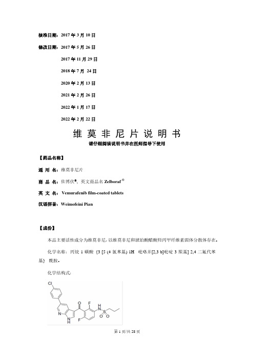
化学名称:丙烷-1-磺酸{3-[5-(4-氯苯基)-1H- 吡咯并[2,3-b]吡啶-3-羰基]-2,4-二氟代苯基}- 酰胺。
化学结构式:分子式:C23H18ClF2N3O3S分子量:489.93【性状】两面凸起、粉白色至橙白色的薄膜衣片。
【适应症】佐博伏®适用于治疗经CFDA批准的检测方法确定的BRAF V600突变阳性的不可切除或转移性黑色素瘤。
【规格】240 mg【用法用量】患者必须经由CFDA批准的检测方法确定的证明肿瘤为BRAF V600突变阳性,才可使用佐博伏®治疗。
佐博伏®不能用于BRAF野生型黑色素瘤患者。
首剂药物应在上午服用,第二剂应在此后约12小时,即晚上服用。
每次服药均可随餐或空腹服用。
用一杯水送服药物,服药时整片吞下佐博伏®片剂。
不应咀嚼或碾碎佐博伏®片剂。
标准剂量佐博伏®的推荐剂量为960 mg(四片240 mg片剂),每日两次。
治疗持续时间建议佐博伏®治疗应持续至疾病进展或发生不可接受的毒性反应(参见表1和表2)。
漏服如果漏服一剂计划的药物,可在下一剂服药4小时以前补服漏服的药物,以维持每日两次的给药方案。
不应同时服用两剂药物。
呕吐如果佐博伏®服药后发生呕吐,患者不应追加剂量,而应按常规剂量继续治疗。
FuGENE HD转染试剂说明书
FuGENE ®HD Transfection Reagent1.What this Product DoesNumber of Transfection ExperimentsIn a typical experiment using HeLa or COS-1 cells, 1 ml of FuGENE ®HD Transfection Reagent can be used to perform up to three hundred transfections in 35-mm tissue-culture dishes, using 3 l of reagent combined with 1 – 2 g DNA per well. This is equivalent to over 6,000wells in a 96-well plate or 1,000 wells in a 24-well plate.L Optimal expression depends upon experimental conditions includ-ing cell type, passage history, confluence, seeding protocol, com-plex incubation time, serum batch, etc. The above amounts of reagents work well with HeLa or COS-1 cells. In other test systems,two- to three-fold higher amounts of reagent yield optimal levels of expression.FormulationFuGENE ® HD Transfection Reagent is a proprietary blend of lipids and other components supplied in 80% ethanol, sterile-filtered, and pack-aged in glass vials. It does not contain any ingredients of human or animal origin.Storage and Stability•FuGENE ® HD Transfection Reagent is shipped at +15 to +25°C.•Store FuGENE ® HD Transfection Reagent at +2 to +8°C, with the lid very tightly closed. The reagent is stable through the expiration date printed on the label when stored under these conditions.L FuGENE ® HD Transfection Reagent remains fully functional evenafter repeatedly opening the vial (at least five times over a two-month period) as long as the vial is tightly recapped and stored at +2 to +8°C between uses.L Do not store FuGENE ® HD Transfection Reagent below 0°C.Components may precipitate and alter results. If you accidently place the reagent at Ϫ20°C, briefly warm it to 37°C to dissolve any precipitate. It should function normally; however, do not return it to the freezer.Special HandlingN Always bring to room temperature and mix FuGENE ® HD Transfec-tion Reagent prior to use (vortex for one second or use inversion).N Do not aliquot FuGENE ® HD Transfection Reagent from the originalglass vials. Chemical residues in plastic vials can significantly decrease the biological activity of the reagent. Minimize the contact of undiluted FuGENE ® HD Transfection Reagent with plastic sur-faces.N Always dilute the reagent by pipetting directlyinto serum-free medium. Do not allow the FuGENE ® HD Transfection Reagent to contact the plastic walls of the tube containing the serum-free medium during the dilution step.N Do not use siliconized pipette tips or tubes.Additional Equipment and Reagents RequiredAdditional reagents and equipment required to perform transfection assays using FuGENE ® HD Transfection Reagent, but not provided,include:General Laboratory Equipment•standard cell culture equipment (e.g., biohazard hoods, incubators,microscope)•standard pipetters and micropipetters •vortex mixerFor Plasmid Preparation•purified plasmid stock (0.1 g/l – 2.0 g/l) in sterile TE (10 mM Tris, 1 mM EDTA, pH 8.0) buffer or sterile water•Genopure Plasmid Midi Kit*, Genopure Plasmid Maxi Kit*, or High Pure Plasmid Isolation Kit* can be used to prepare plasmid.For Verification of Vector Function •assay appropriate for transfected gene•G-418* or Hygromycin B* (optional; for stable transfection experi-ments)For Transfection-Complex Formation•Opti-MEM I Reduced Serum Medium, water, or serum-free medium •24-well plate to serve as test tube rack for FuGENE ® HD Transfec-tion Reagent vial•sterile polystyrene tubes or round-bottom 96-well platesCells Growing in Log Phase•select subconfluent cultures in log phase for preparation of the cell cultures for transfection•method to quantify cell number to reproducibly plate the same number of cellsApplicationFuGENE ® HD Transfection Reagent is a multi-component reagent that forms a complex with DNA, then transports the complex into animal or insect cells. Benefits of FuGENE ® HD Transfection Reagent include:•High transfection efficiency in many common cell types, including HeLa, NIH/3T3, COS-1, COS-7, CHO-K1, Hep G2, HEK-293, MCF7,and some insect cell lines. In addition, you will achieve excellent transfection efficiency in some cell lines (e.g., RAW) that are not transfected well by other reagents. Detailed transfection protocols and sample results are available at .•Demonstrates minimal cytotoxicity or changes in morphology when adequate numbers of cells are transfected, and eliminates the requirement to change media after the addition of transfection complex.•Suitable for transient and stable transfection.•Functions exceptionally well in the presence or absence of serum;eliminates the need to change media.To ensure the quality of cells to be transfected, Roche recommends using freshly-obtained, low-passage cell sines form ATCC ®. For more information please visit and bookmark .For the transient and stable transfection of animal and insect cellsCat. No. 04 709 691 0010.4 ml (up to 120 transfections)Version November 2007Cat. No. 04 709 705 001 1 ml (up to 300 transfections)Store at +2 to +8°CCat. No. 04 709 713 001 1)Mega-pack 5 × 1 ml (up to 1,500 transfections)Cat. No. 05 061 369 00110 ml (up to 3,000 transfections)Cat. No. 04 883 560 001Trial-pack1)The five vials are packaged together in one box with one pack insertFor life science research only. Not for use in diagnostic procedures. FOR IN VITRO USE ONLY.2.How to Use this Product2.1Before you BeginRequired Amount of FuGENE® HD Transfection ReagentFor initial optimization experiments, transfect a monolayer of cells that is 80 – 90% confluent in a six-well culture dish, using 3:2, 4:2, 5:2, 6:2, 7:2, and 8:2 ratios of FuGENE® HD Transfection Reagent (l) to DNA (g), respectively. For most cell types, these FuGENE® HD Transfection Reagent:DNA ratios provide excellent transfection levels.L Subsequent optimization may further increase efficiency in your particular application. In addition to varying the volume of FuGENE® HD Transfection Reagent, other parameters may be evaluated (see section 2.6, Parameters for Optimization, and sec-tion 3, Troubleshooting).Plasmid DNA•It is critical to accurately determine the plasmid DNA concentration using 260-nm absorption (estimates of DNA content based on the intensity of gel bands are not sufficiently accurate). Determine the DNA purity using a 260 nm/280 nm ratio; the optimal ratio is 1.8.•Prepare the plasmid DNA solution in sterile TE (Tris/EDTA) buffer or sterile water at a concentration between 0.1 g/l and 2.0 g/l. Cell Culture ConditionsMinimize both intra- and inter-experimental variance in transfection efficiency by using cells that are regularly passaged, proliferating well (best when in a log-growth phase), and plated at a consistent den-sity. FuGENE® HD Transfection Reagent is different from FuGENE® 6 Transfection Reagent regarding the optimal density of cells required for maximal expression with minimal negative effect; FuGENE®6 Transfection Reagent is formulated to work at low cell densities, whereas FuGENE® HD Transfection Reagent is formulated to work at higher cell densities. Cells must be healthy and free of mycoplasma and other contaminants.L If you have used FuGENE® 6 Transfection Reagent in the past, we suggest that you increase the plating density for initial tests using FuGENE® HD Transfection Reagent. For most cell lines, use the reagent at cell-plating densities at least twice that used with FuGENE® 6 Transfection Reagent to yield maximum protein expression. For most cell lines, cultures should be 80 – 90% con-fluent at the time of transfection. For contact-inhibited cell lines such as NIH/3T3, optimal results are obtained when cells are plated at lower densities.Other Media AdditivesIn some cell types, antimicrobial agents (e.g., antibiotics and fungicides) that are commonly included in cell-culture media may adversely affect the transfection efficiency of FuGENE® HD Transfection Reagent. If possible, exclude additives for initial experi-ments. Once high-efficiency conditions have been established, these components can be added back while monitoring your transfection results. Cell growth and/or transfection efficiency may be affected by variations in sera quality or media formulations.Verification of Vector FunctionOptimize transfection conditions with a known positive-control reporter gene construct prior to transfecting cells with a new vector construct:•Determine transfection efficiency with a reporter gene assay (CAT*,-Gal*, Luciferase*, SEAP*, or hGH*).•Sequence across the flanking vector insert regions to verify the integrity of your new construct.2.2Preparation of Cells for Transfection2.3Overview of Initial Transfection ExperimentAdherent and Suspension Cells in a Six-well Plate or 35-mm Culture DishFor initial optimization, test FuGENE® HD Transfection Reagent:DNA ratios of 3:2, 4:2, 5:2, 6:2, 7:2, and 8:2 (l for FuGENE® HD Transfection Reagent, and g for DNA, respectively). The preparation of the com-plex for a single well of a six-well plate, or a 35-mm culture dish, is described in section 2.4. These ratios will function very well for com-monly used adherent cells and suspension cells. For your particular cell line and culture conditions you may find that ratios of 9:2, 10:2, 11:2, or 12:2 result in even greater expression. Try these ratios if you find the highest expression levels in the 8:2 ratio well.N Prepare the transfection complex in diluent that does not contain serum (e.g., Opti-MEM I Reduced Serum Medium), even if the cells are transfected in the presence of serum. For some cell lines, the complex may be formed in DMEM or sterile water.L For additional optimization tips, see section 2.6 and visit /fugene/hdRatio OverviewPreparation of a transfection complex that is sufficient for a 35-mm culture dish, or one well of a six-well plate, at six different ratios: Tab. 1: Preparation of transfection complex for a 35-mm culture dish.Cell Type ProcedureAdherentcellsOne day before the transfection experiment, trypsinizethe monolayer, adjust cell concentration, and plate thecells in the chosen cell-culture vessel. For most celltypes, plating 3 – 6 × 10⁵ cells in 2 ml of medium in a35-mm culture dish (or six-well plate) overnight willachieve the desired density of Ͼ80% confluency atthe time of transfection. For cell lines with specialcharacteristics, such as contact-inhibited NIH/3T3cells, a lower plating density should be used. If usingculture plates of a different size, adjust the total num-ber of cells, starting volume of FuGENE® HD Transfec-tion Reagent, and the starting mass of DNA inproportion to the relative surface area (Table 2). SuspensioncellsUse freshly passaged cells at a concentration of 5×10⁵/ml to 1 × 10⁶/ml in 2 ml of medium in a 35-mmculture dish (or six-well plate). If using culture platesof a different size, adjust the total number of cells,starting volume of FuGENE® HD TransfectionReagent, and the starting mass of DNA in proportionto the relative volume (Table 2).Tubelabel(ratio)Diluent(l)FuGENE® HDTransfectionReagent (l)DNA(g)Comments3:210032Add the entire volume toa 35-mm culture dish oreach well of a six-wellplate, or 2 – 15 l to eachwell of a 96-well plate.Suggested volumes fordifferent culture vesselsare included in Table 2. 4:2100425:2100526:2100627:2100728:2100822.4Transfection ProcedureNotes:L As with any experiment, include appropriate controls. Preparewells with cells that remain untransfected, cells with transfection reagent alone, and cells with DNA alone.L For stable transfection experiments, the complex-containingmedium should be left unchanged until the cells need to be pas-saged. At that time, include the appropriate selection antibiotics (G 418* or Hygromycin B*).L To prepare transfection complexes for different-sized containers orparallel experiments, proportionally change the quantity of all components according to the total surface area of the cell culture vessel being used (Table 2).L For ease-of-use when transfecting small volumes, as in 96-wellplates containing 0.1 ml culture medium per well, prepare 100 l of transfection complex and add 2 – 15 l to each well depending upon the cell type.L The optimal ratio of transfection reagent:DNA and the optimaltotal amount of complex may vary with cell line, cell density, day of assay, and gene expressed.L After performing the optimization experiment where several ratioswere tested, select a ratio in the middle of the plateau for future experiments.2.5Cotransfection ExperimentsSuggestionsFor cotransfection experiments with FuGENE ® HD Transfection Reagent, maintain the same total reagent:total DNA ratio as that used for a single plasmid in your system. Thus, the total amount of the plas-mid DNA should be equal to the amount of plasmid used in a single plasmid transfection.ᕡAllow FuGENE ® HD Transfection Reagent, DNA, anddiluent to adjust to +15 to +25°C. Vortex for one second or invert the FuGENE ® HD Transfection Reagent vial to mix.ᕢDilute DNA with appropriate diluent, for example, Opti-MEM IReduced Serum Medium, serum-free medium (without anti-biotics or fungicides), or sterile water to a concentration of 2g plasmid DNA/100 l Opti-MEM (0.02g/l).L For insect cells, use sterile water as diluent. For other celllines, try sterile water or serum-free medium as an alterna-tive diluent.ᕣPlace 100 l diluent, containing 2 g DNA into each of six ster-ile tubes labeled 3:2, 4:2, 5:2, 6:2, 7:2, and 8:2.Recommendation : Use sterile polystyrene tubes or round-bottom, 96-well plates to form the transfection complex.L Due to manufacturer variability with release agents for96well plates, we suggest using tissue culture treated 96well plates to reduce variablity.ᕤForm the transfection complex by adding FuGENE ® HDTransfection Reagent to tubes containing diluted DNA :Pipet the FuGENE ® HD Transfection Reagent (3, 4, 5, 6, 7, or 8l) directly into the medium containing the diluted DNA with-out allowing contact with the walls of the plastic tubes.N To avoid adversely affecting transfection efficiency, do notallow undiluted FuGENE ® HD Transfection Reagent to come into contact with plastic surfaces (such as the walls of the tube that contains the serum-free medium) other than pipette tips. Do not use siliconized pipette tips or tubes.ᕥMix and incubate the transfection complex :Vigorously tap the tube or vortex for one to two seconds to mix the contents. If using a 96-well plate, place the plate on a rotat-ing shaker for 5 – 10 seconds. Incubate the transfection reagent:DNA complex for 15 minutes at room temperature.For some ratios and cell types, incubation is not necessary for optimal complex formation, while a longer incubation time is better for other cell types. Determine this for your particular cell line and the ratio you use.ᕦAdd the transfection complex to cell s:Remove culture vessel from the incubator. Removal of growth medium is not necessary. Add the transfection complex to the cells in a drop-wise manner or add below the surface of the medium. Swirl the wells or flasks to ensure distribution over the entire plate surface. Use of a rotating platform shaker for 30 seconds at low speed provides adequate mixing for 96-well plates.Once the FuGENE ® HD Transfection Reagent:DNA complex has been added to the cells, there is no need to remove and replace with fresh medium (as is necessary with some other transfection reagents).L In our experience, the exposure of most common laboratorycell types (COS-1, CHO-K1, HEK-293, HeLa, Hep G2, MCF-7) to the transfection complex until performance of the gene expression assay (24–48 hours later) does not affect the results. If you desire to transfect cells that are in serum-free medium during the transfection process, then replace the medium with serum-containing medium 3 – 8 hours after transfection, unless the cells normally grow in serum-free medium. ᕧIncubate cells and assay the results :Following transfection, incubate the cells for 18 – 72 hours prior to measuring protein expression. The length of incubation depends upon the transfected vector construct, the cell type being transfected, the cell medium, cell density, and the type of protein being expressed. After this incubation period, measure protein expression using an assay that is appropriate for your system.L If you observe low transfection levels or more than10 – 30% cell death, refer to section 3, Troubleshooting and /fugene/hd2.6Parameters for Optimization2.7Transfection of Adherent Cells Adapted for Suspension Growth•In some cases, adherent cells may be adapted for suspension growth, thus enabling the production of transiently transfected cells on a very large scale.•HEK-293 cells grown in suspension in serum-free medium that did not contain heparin or dextran sulfate produced significant amounts of protein following transfection.2.8Guidelines for Preparing FuGENE® HD Transfection Reagent:DNA Complex for Various Culture Vessel SizesThe starting volume and mass to add to the different culture vessels is based upon preparing a 100-l transfection complex as described in sec-tions 2.3 and 2.4. For best results, prepare a 100-l complex at different ratios and add varying amounts of each ratio when optimizing. The amounts below are based on the 100-l complex as prepared in sections 2.3 and 2.4.Suggested seeding density for adherent cells = 30,000 – 70,000 cells per cm2Suggested seeding density for suspension cells = 250,000 – 500,000 cells per mlTab. 2: Refer to the table below when setting up your transfection reac-tions. T hese are suggested seeding densities and are media, passage level, laboratory, and cell-line dependent. It is critical that log phase cultures are selected for subculture for the transfection experiments, and that cultures are seeded at the proper density for the transfection experiment. Observe cultures and plate them so that the monolayer is 80–90% confluent at the time of trans-fection. This must be determined empirically. For some cell lines, 60–80% con-fluency is sufficient. However, a contact-inhibited cell line, such as NIH/3T3, should be plated at lower confluence due to its growth characteristics.1) Scale up total volume for larger vessels.3.TroubleshootingParameter to beoptimizedProcedureFuGENE® HD Transfection Reagent:DNA ratio Form the transfection complex at several ratios: 3:2, 4:2, 5:2, 6:2, 7:2, 8:2, 10:2, and 12:2 (l FuGENE® HD Transfec-tion Reagent: g DNA).In some systems, altering the ratio of FuGENE® HD Transfection Reagent to DNA can increase the level of protein expression.L It has been reported that for some plasmid preparations, a ratio of 2:2 yielded optimal results. This is unusual and may reflect some property of the plasmid preparation rather than a characteristic of the FuGENE® HD Transfec-tion Reagent.Amount of transfectioncomplex addedTry adding 200%, 150%, 75%, 50%, and 25% of the amount of 100-l transfection complex suggested in Table 2.Number of cells plated Plating more cells will overcome negative growth effects of excess transfection complex. For cells with special growth characteristics, such as NIH/3T3 cells, do not use this as the first parameter for optimization.Incubation time for the transfection complex to form Vary the length of incubation time for transfection-complex formation: add the complex to the cells immediately after the components are combined and mixed, and then at several intervals up to 40 minutes (i.e., 0, 15, 25, and 40 min-utes). We have observed that in some cell lines, the transfection-complex incubation time tends to have no effect on results when using higher ratios; however, results using lower-ratio-complexes varied depending on the incubation time for complex formation.Special tips for sensitive cell lines •Reduce the time of exposure to the transfection complex (2–3 hours maximum), then replace the medium.•Use the lower ratios, and allow the complex to form for a longer period of time (determine empirically for your cell line), then add lower amounts of the complex (50% or less of what was originally tested).Culture vessel Surfacearea(cm2)TotalvolumeofmediumSuggested seeding densitySuggested amount ofthe 100-l trans-fection complex toadd to each well (l)Final amount of FuGENE® HDTransfection Reagent (l) ineach well following addition ofsuggested amount of 100-ltransfection complexCells/wellAdherent cellsCells/wellSmall or suspensioncellsUsing the3:2 ratioUsing the8:2 ratio totalvolumevolume forlargerlow high low high96-well plate(1 well)0.30.110,00020,00025,00050,00050.150.424-well plate(1 well)1.90.550,000125,000250,000500,000250.752.012-well plate(1 well)3.8 1.0100,000250,000375,000750,00050 1.54.0 35-mm dish82200,000500,000500,0001,000,000100 3.08.06-well plate(1 well)9.42200,000600,000500,0001,000,000100 3.08.0 60-mm dish215500,0001,400,0001,250,0002,500,000250 1)7.520.0 10-cm dish55101,500,0003,500,0002,500,0005,000,000500 1)15.040.0 T-25 flask256700,0001,700,0001,500,0003,000,000300 1)9.024.0 T-75 flask75202,000,0005,000,0005,000,00010,000,000900 1)27.072.0Low trans-fection efficiency Poor quality orinsufficient quantityof nucleic acidsVerify the amount, purity, and sequence of nucleic acid.Perform a control transfection experiment with a commercially available transfection-grade plasmidpreparation.Chemical contaminants may be in the plasmid preparation. Avoid phosphate buffers until you havetested them in your system.L Endotoxins are reported to be cytotoxic to some very sensitive cell lines.Insufficient numberof cellsUse adherent cells that are at least 80% confluent. Low cell density results in fewer cells available totake up transfection complex, and excess complex may be cytotoxic; in addition, fewer cells yieldless protein.Too many cells orcells post log phaseWhen confluent cultures are subcultured, or cells are plated at too high a density, the cells fail to dividein the culture being transfected. This results in suboptimal expression.Suboptimal FuGENE®HD TransfectionReagent:DNA ratio,complex incubationtime, total amount oftransfection complexadded, or cell densityOptimize the FuGENE® HD Transfection Reagent:DNA ratio, complex incubation time, amount ofcomplex added to cells, and cell density, according to the following procedure:Day before transfection:Prepare two 96-well plates of cells at high and low seeding densities (see Table 2 for suggestions).Day of transfection:•Form 200 l of transfection complex at ratios of 2:2, 3:2, 4:2, 5:2, 6:2. 7:2, and 8:2 (l transfectionreagent:g DNA) following the protocol in this pack insert (sections 2.3, 2.4) and doubling theamounts of all components.•As soon as the complexes are combined and mixed, add 10, 5, or 2.5 l of each complex to one of3columns of cells in each 96-well plate (i.e., columns 2, 3, and 4). Leave all outer wells empty ascontrols.•Continue to incubate the complexes at room temperature. After an additional 10 – 15 minutes, add 10,5, or 2.5 l of each complex to the next 3 columns (5, 6, and 7) of cells in each 96-well plate.•Continue to incubate the complexes at room temperature. After an additional 10 – 15 minutes, add 10,5, or 2.5 l of each complex to the next 3 columns (8, 9, and 10) of cells in each 96-well plate.•Assay the plates 1–2 days later. Select the ratio, amount of complex, and time of transfection-complexincubation that resulted in optimal expression.•If optimal transfection occurs at the higher ratios, repeat this process using ratios of 6:2, 7:2, 8:2, 10:2,12:2, and 14:2. Add 5, 10, and 15 l of complex. We have never successfully transfected cells using theratio of 2:2, but it has been reported that some plasmid preparations transfect at this ratio.See section 2.6, Optimization of FuGENE® HD Transfection Reagent:DNA ratio, for more information andvisit /fugene/hdFuGENE® HD Trans-fection Reagent wasaliquotedCheck that FuGENE® HD Transfection Reagent is stored in the original container. If the reagent wasaliquoted into plastic containers, there is a high chance of inactivation. Make sure the reagent isimmediately mixed with the dilute DNA either by vortexing or pipetting up to 10 – 15 times.FuGENE® HD Trans-fection Reagent cameinto contact withplastic or wasinadequately mixedRepeat transfection, carefully pipetting FuGENE® HD Transfection Reagent directly into the serum-freemedium, being careful not to touch the sides of the container while adding the FuGENE® HD Transfec-tion Reagent to the diluted DNA. If the FuGENE® HD Transfection Reagent is added too gently, it maylayer on top of the medium, thus making contact with the plastic.Transfection complexwas formed in serum-containing mediumCheck original bottle of medium used for complex formation. Repeat experiment using new bottle ofOpti-MEM that does not contain any additives (e.g., serum, antibiotics, growth enhancers, heparin,dextran sulfate, etc.). Try forming the complex in sterile water or plain DMEM.Media and mediacomponentsDifferent media and media components may influence the level of transfection efficiency andsubsequent growth of the transfected cells, as well as expression of the recombinant protein. Some lotsof sera have been reported to interfere with optimal transfection.Quality and/or lot-to-lot differences that affect transfection experiments have been noted in both seraand media. Check that the medium and/or serum is from the same lot that worked previously. Try newlots or a different vendor.Culture may becontaminated withmycoplasmaCultures contaminated with mycoplasma have been shown to have decreased transfection efficacy.Determine if culture is contaminated with mycoplasma; use the Mycoplasma Detection Kit* orMycoplasma PCR ELISA* to assess contamination.Inconsistent results Ratio or amount oftransfection complexis at the edge ofperformance plateauInitial experiments should be completed to determine the ratios, amount of complex to be added, andlength of time for complex formation for optimal performance. In our experience, we have found the pla-teau to be relatively broad. We recommend that future experiments be performed with ratios, incubationtime, and amounts of complex that were in the middle of the plateau. If conditions are selected at theedge of the plateau, very small procedural differences may cause large differences in the resulting pro-tein expression. Increased consistency may be achieved by shifting parameters away from the edge ofthe plateau to the middle of the plateau.Transfection complexformation:timing,amounts, and ratioFormation of the complex involves a multifaceted interaction between the transfection reagent and DNAas well as biological parameters. Differences in any of the components or techniques may result ininconsistencies. If results do not meet your expectations, then repeat the optimization experimentselecting areas near the plateau found in previous experiments. For current experiments, determine ifyou should use a different ratio, length of time, or amount of complex for more consistent transfectionresults.Extensive testing of the FuGENE® HD Transfection Reagent is performed on two cell lines: one easy totransfect and one very difficult to transfect. All reagent lots must pass this rigorous testing before wemake it available to you. However, we cannot test all cell lines, media, sera, and vectors; in your labora-tory, you may find slight differences in the optimal ratio, amount of complex, or time for complex forma-tion for some lots of FuGENE® HD Transfection Reagent.Cells For consistent results, cells must be properly maintained. Cells change with passage level, passage conditions, media, and sera. For some cell lines, these changes have little to no effect on transfectionexperiments, but for other cell lines, these changes have profound effects. Each cell type may have a dif-ferent optimal transfection condition. Optimal values for a single cell type may also change slightly withvector construct and type of protein expressed.Observation Possible cause RecommendationSigns of cytotoxicity Transfected protein iscytotoxic or isproduced at highlevelsReduced viability or slow growth rates may be the result of high levels of protein expression, as the cell’smetabolic resources are directed toward production of the heterologous protein. The expressed proteinmay also be toxic to the cell at the level expressed.To analyze cytotoxicity, prepare experimental controls as described below.Prepare extra control wells containing:ቢ Cells that are not transfectedባ Cells treated with DNA alone (e.g., without FuGENE® HD Transfection Reagent)ቤ Cells treated with FuGENE® HD Transfection Reagent alone (no DNA added)ብ Cells transfected with a non-toxic or secreted protein.Compare experimental transfected cells to cells in the control wells (described above). Considerrepeating the experiment with a secreted reporter gene such as SEAP, hGH, or a standard -gal controlvector. Cells expressing SEAP should show little to no evidence of cytotoxicity.Too much transfec-tion complex fornumber of cellsIncrease the number of cells plated, and/or decrease the total amount of complex added to the cells. Trydifferent ratios and allow the complexes to form for different time intervals. Add different amounts ofcomplex; for example, make the complex as usual but add 75%, 50%, or 25% of the usual amounts toeach well. See Suboptimal FuGENE® HD:DNA Ratio in "Low transfection efficiency" section of this tablefor details or optimization protocol.Culture may becontaminated withmycoplasmaDetermine if culture is contaminated with mycoplasma; use the Mycoplasma Detection Kit* orMycoplasma PCR ELISA* to assess contamination.Cells may not behealthyAssess physiological state of cells and the incubation conditions (e.g., check incubator CO2, humidity,and temperature levels). Observe cells prior to each passage for morphology and absence of contami-nants. Make sure cells do not overgrow. Routinely passage cells prior to reaching confluency. Make surethat culture media and additives are within expiration date and have been stored properly.Diluent is toxic to thecellsDMEM is toxic to some insect cell lines. For these cells, prepare the transfection complex in sterilewater. You may also try forming the complex in the medium in which the cells are growing, providingthat the medium does not contain serum, heparin, or dextran sulfate.Plasmid preparationcontaminated withendotoxinEndotoxin is reported to be cytotoxic to sensitive cell lines.If above tests provenegative, FuGENE®HD TransfectionReagent may not beoptimal for your cells.Try FuGENE® 6 Transfection Reagent*, DOTAP Liposomal Transfection Reagent*, DOSPER LiposomalTransfection Reagent*, or X-tremeGENE Q2 Transfection Reagent*.High protein-expression levelsHigh expression levels of certain intracellular proteins (e.g., Green Fluorescent Protein [GFP]) may becytotoxic to some cell types. Cell proliferation, toxicity, and cell death may be monitored using Apoptosisand Cell Proliferation products from Roche Applied Science (visit /apoptosis for more information).Media and mediacomponentsTest different media and optimize the level of each medium component for these cytotoxic effects.Although it is not usually necessary to remove the transfection complex following the transfection step,it may be necessary to feed your cells with fresh media for extended growth periods. This is particularlyimportant if the transfected cells are allowed to continue to grow for 3 – 7 days to provide maximalprotein expression.。
纽储非妇产科
低浓度的纯次氯酸伤口清洁敷料——纽储非面世。
这是世上首个也是目前唯一一个利用复杂工艺将低浓度的纯次氯酸应用于临床治疗 上的创新产品。
纽储非® :理想的伤口清洁产品
[1] SAM DUKAN,et al. Hypochlorous Acid Stress in Escherichia coli: Resistance, DNA Damage, and Comparison with Hydrogen Peroxide Stress. 1996;178(21):6145-6150
首次证实人体先天免疫中产生次氯酸(HOCl)杀菌。 John E. Harrison [1]在白细胞氧化爆发过程中证明次氯酸存在,并证明次氯酸 有着明显的杀菌作用。
次氯酸的杀菌机理得到进一步揭示。 Thomas EL[2]发现次氯酸(HOCl)通过损坏细菌DNA、蛋白(硫醇、硫醚和氨 基)、酶失活作用,包括切割肽键和氧化细菌成分巯基。
同时人体细胞中存在一种天然的 非必需氨基酸-牛磺酸,作为次 氯酸的清道夫,防止附带损害。
次氯酸盐: 控制1841年维也纳产褥热
塞麦尔韦斯 (Semmel-Weiss,1818~1865),现代医院流行病学之父 在19世纪中叶,时任奥地利维也纳大学附属医学院的产科医师塞麦尔韦斯注意 到,由医师负责的产科病房产褥热的发生率比助产士负责的病房高9倍,前者 的病死率高达10%以上,经过调查,该感染是通过医生的手扩散的,是由于做 过尸体解剖的医师未经洗手消毒,就去处理产科患者造成的。通过实行严格的 漂白粉液洗手措施后,产褥热的传播得到了明显的控制,病死率减少到1.0% 以下。他应用系统的流行病学调查方法,控制了该医院产褥热的流行爆发。
- 1、下载文档前请自行甄别文档内容的完整性,平台不提供额外的编辑、内容补充、找答案等附加服务。
- 2、"仅部分预览"的文档,不可在线预览部分如存在完整性等问题,可反馈申请退款(可完整预览的文档不适用该条件!)。
- 3、如文档侵犯您的权益,请联系客服反馈,我们会尽快为您处理(人工客服工作时间:9:00-18:30)。
CERTIFICATE OF ANALYSISTurboFect™in vitro Transfection Reagent1 ml(for 300-500 in vitro transfections)#R0531Lot: Expiry Date:Store at 4°C.DescriptionTurboFect™ in vitro Transfection Reagent* is a sterile solution of a cationic polymer in water. The polymer forms compact, stable, positively charged complexes with DNA. These complexes protect DNA from degradation and facilitate gene delivery into eukaryotic cells. TurboFect™ is ideal for transfection of a variety of cells, including primary and difficult-to-transfect cells. Transfection can be performed in the presence or absence of serum. TurboFect™ demonstrates superior transfection efficiency and minimal toxicity when compared to lipid-based or other polymer-based transfection reagents.Reagents to be Supplied by the UserSerum-free DMEM, RPMI or other growth medium.The presence of antibiotics in the medium has no effect on transfection efficiency. General ConsiderationsDNA Quality RequirementsDNA quality is critical for successful transfection. An A260/A280 ratio of 1.8 or higher is recommended. Endotoxin-contaminated DNA may result in inefficient transfection and cause unacceptably high cellular toxicity.Cell DensityThe recommended confluency for adherent cells at the day of transfection is 70-90%. Suspension cells should be plated at an optimal density ensuring their logarithmic growth at the time of transfection.Incubation TimeTransient transgene expression takes place within 2-72 hours after transfection. The optimal incubation time depends on the cell type, promoter strength and expression product, and must be determined experimentally.Choice of PromoterHigh transfection efficiency depends both on the transgene promoter and on the cell line used. Cytomegalovirus (CMV) promoter is commonly used for high gene expression in a variety of cell lines. Other promoters, such as those from simian virus (SV40) and from Rous sarcoma virus (RSV) can also be used.Transfection Reagent/DNA RatioThe volume of transfection reagent used depends on the amount of DNA, transgene and cells to be transfected. The ratios presented in the protocols below are starting amounts and can be further optimized for best results. Transfection in the Presence of SerumThe transfection efficiency with TurboFect™in vitro transfection reagent is equally high in the presence of serum. This is not the case with many other transfection reagents. AntibioticsAntibiotics do not interfere with bothDNA/TurboFect™ complex formation and cell transfection.General Protocol for Transfection of Adherent and Suspension Cells in a 24-well Plate Quantities and volumes should be scaled-up according to the number of cells/wells to be transfected (see Table 1 on reverse page).1.In each well, seed ~5x10⁴ adherent cells or~5x10⁵ suspension cells in 1 ml of growth medium 24 hours prior to transfection.Note∙The recommended confluency for adherent cells on the day of transfection is 70-90%.∙Suspension cells should be in logarithmic growth phase at the time of transfection.2.Dilute 1 µg of DNA in 100 µl of serum-free DMEM or other serum-free growth medium.3.Briefly vortex TurboFect™ reagent and add 2 µl of it to the diluted DNA. Mix immediately by pipetting or vortexing.4.Incubate 15-20 min at room temperature. Note∙Prepare immediately prior to transfection.∙We recommend starting with 1 µg of DNA and 2 µl of TurboFect™ per well in a 24-well plate (see scale-up Table on reverse page).∙Subsequent optimization may further increase transfection efficiency depending on the cell line and transgene used.5.Add 100 µl of the TurboFect™/DNA mixture drop-wise to each well. Do not remove the growth medium from the cells.6.Gently rock the plate to achieve even distribution of the complexes.7.Incubate at 37°C in a CO2 incubator.8.Analyze transgene expression 24-48 hours later. For stable transfection, cells should be grown in selective medium for 10-15 days. Protocol for Reverse Transfection of Adherent and Suspension Cells In a 24-well PlateThe protocol is optimized for transfection in 24-well plate format. Quantities and volumes should be scaled-up according to the number of cells/wells to be transfected (see Table 1 on reverse page).1.Dilute 1 µg of DNA in 100 µl of serum-freeDMEM or other serum-free growth medium. 2.Briefly vortex TurboFect™ reagent and add 2 µl of it to the diluted DNA. Mix immediately by pipetting or vortexing.3.Incubate 15-20 minutes at room temperature.4.Evenly distribute 100 µl of the TurboFect™/ DNA mixture at the bottom of the well of a 24-well plate.Note∙Prepare immediately prior to transfection∙We recommend starting with 1 µg of DNA and 2 µl of TurboFect™ per well in a 24-well plate (see scale-up Table on reverse page).∙Subsequent optimization may further increase transfection efficiency depending on the cell line and transgene used.5.Gently layer 1 ml of ~105 adherent cellsor ~106 suspension cells per well on top of the TurboFect™/ DNA mixture.6.Incubate at 37°C in a CO2 incubator.7.Analyze transgene expression 24-48 hours later. NotePlates can be centrifuged for 2-5 min at 200 x g tofacilitate sedimentation of cells to the bottom of theplate.(continued on reverse page)Table 1. Scale-up ratios for transfection of adherent and suspension cells with TurboFect™in vitro Transfection Reagent.Tissue culturevessel Growtharea,cm2/wellVolume ofmedium, mlAdherent (suspension)cells to seed the daybefore transfection*Amount of DNAVolume ofTurboFect™, µl*µg** µl***Recom-mendedRange96-well plate 0.3 0.2 0.5-1.2x10⁴(2.0x10⁴)0.2 20 0.4 0.3-0.648-well plate 0.7 0.5 1.0-3.0x10⁴(5.0x10⁴)0.5 50 1.0 0.5-1.424-well plate 2.0 1.0 2.0-6.0x10⁴(1.0x10⁵)1.0 1002.0 1.0-2.812-well plate 4.0 2.0 0.4-1.2x10⁵(2.0x10⁵)2.0 200 4.0 2.6-6.06-well plate 9.5 4.0 0.8-2.4x10⁵(4.0x10⁵)4.0 400 6.0 4.0-8.060 mm plate20 6.0 2.0-6.3x10⁵(1.0x10⁶)6.0 600 12.0 8.0-16.0*These numbers were determined using HeLa and Jurkat cells. Actual values depend on the cell type. **Amount of DNA and TurboFect™ in vitro Transfection Reagent used may require optimization.*** The volume of DNA should be 1/10 of the volume of the culture medium used for dilution of the DNA. Cells successfully transfected with TurboFect™ include:Permanently growing cell lines Primary cell culturesCos-7 african green monkey kidney cellsHeLa human cervix adenocarcinoma cellsCHO chinese hamster ovary cellsHEK293 human embryonic kidney cellsB50 rat nervous tissue neuronal cellsCalu1 human lung epidermoid carcinoma cells RAW264 mouse leukaemic monocyte-macrophage cells WEHI mouse B cell lymphoma cellsMDCK Madin Darby Canine Kidney cellsRaji human Burkitt’s lymphoma cellsCOLO human colon adenocarcinoma cellsJurkat human leukaemic T cellsSp2/Ag14 mouse myeloma cellsHeLa S3 human cervix carcinoma cellsHep2C human larynx carcinoma cellsL929 mouse connective tissue fibroblastsNIH3T3 mouse embryo fibroblasts Rat fibroblastsMouse bone marrow derived dendritic cells Mouse bone marrow derived macrophages HLF human lung fibroblastsFor cell line updates, see This product or the use of this product is covered by EPO patent application EP2070970A2 and corresponding counterparts owned by Fermentas. The purchase of this product includes a non-transferable license to use this product for the purchaser's internal research. All other commercial uses of this product, including without limitation product use for diagnostic purposes, resale of product in the original or any modified form or product use in providing commercial services require a separate license from Fermentas. For further information on obtaining licenses please contact Fermentas atinfo@.PRODUCT USE LIMITATIONThis product is developed, designed and sold exclusively for research purposes and in vitro use only. The product was not tested for use in diagnostics or for drug development, nor is it suitable for administration to humans.Please refer to for Material Safety Data Sheet of the product.TROUBLESHOOTING Problem Possible Cause and SolutionLow transfection efficiency Suboptimal reagent/DNA ratio.Optimize the amount of transfection reagent added to the fixed amount of DNA. Suboptimal quantity of DNA.Optimize the amount of DNA used for transfection. Keep the amount of transfection reagent constant.Poor DNA quality.Use high quality DNA with an A 260/A280 ratio greater than 1.8.Suboptimal cell confluency.Optimize cell plating conditions. Ensure that adhered cells are 70- 90% confluent at the time of transfection. Ensure that suspension cells are in logarithmic growth phase at the time of transfection.Mycoplasma contamination.Mycoplasma infection in cell culture often results in poor and/or non-reproducible transfection. Regularly check your cells for mycoplasma infection.High cellular toxicity Toxic transgene.Verify if the expressed transgene is toxic.Suboptimal incubation conditions.Reduce incubation time of the polyplexes with the cells. Replace the transfection mixture 3-4 hours later with fresh growth medium.Suboptimal quantity of DNA.Reduce the quantity of DNA used for transfection.Cell density is too low.Increase the plating density of cells used for transfectionQUALITY CONTROLTransfection efficiency was tested on HeLa cells using 1 µg of eGFP expressing plasmid and 2 µl of TurboFect™ per 5x10⁴ cells in 24-well plate. Transfection efficiency, i.e. the percentage of transfected cells, is 90±10% as estimated by flow cytometry.Quality authorized by: Jurgita Zilinskiene。